

Multi-Analytical Characterization of Illuminated Choirbooks from the Royal Audience of Quito

 ,
,
Abstract
1. Introduction
2. Materials and Methods
2.1. Population and Sampling
2.2. Multi-Analytical Methodology
2.2.1. Technical Photography
2.2.2. X-Ray Fluorescence (XRF)
2.2.3. Scanning Electron Microscopy
2.2.4. Infrared Spectrometry (FTIR-ATR)
3. Results and Discussion
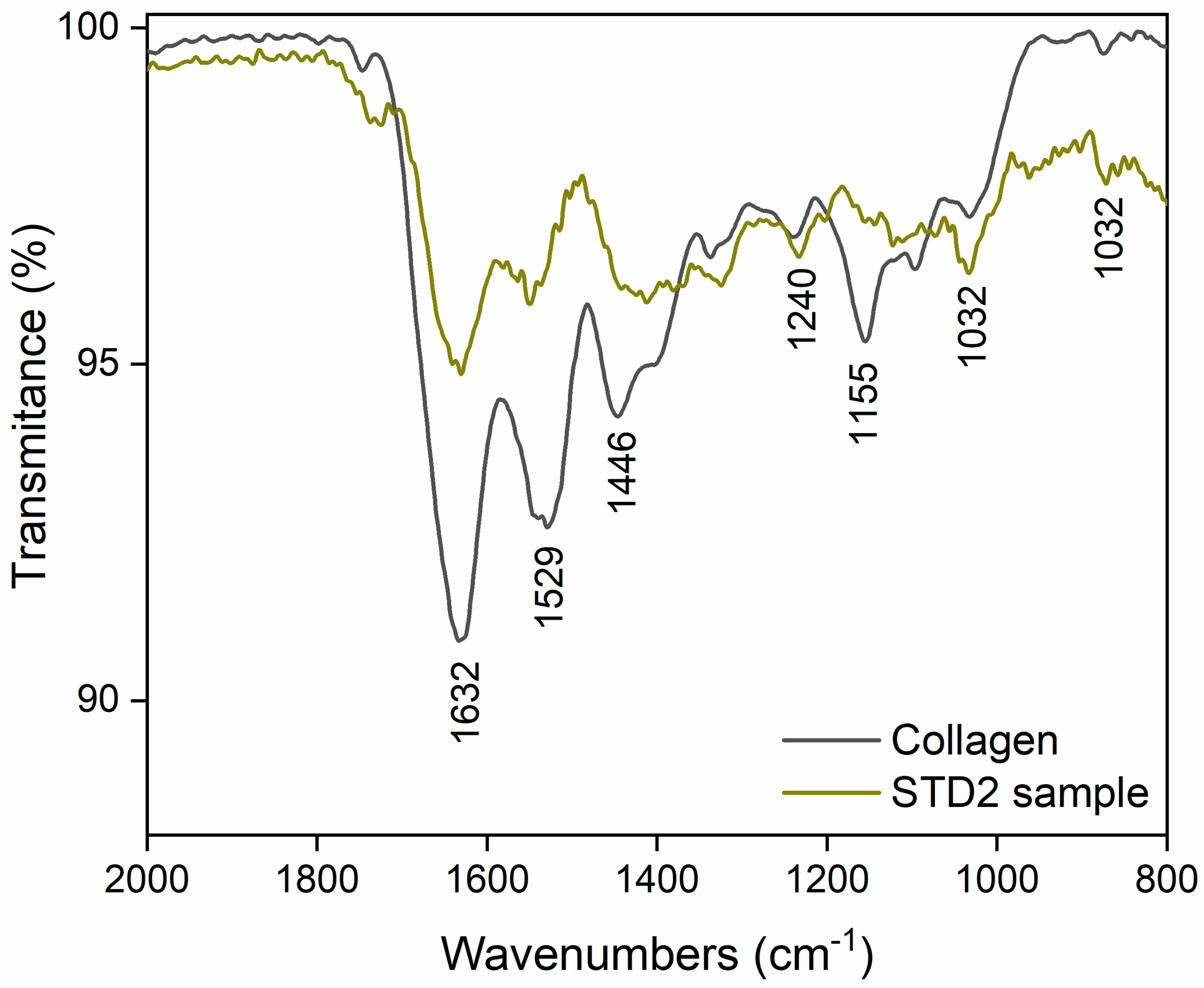
3.1. The Parchment
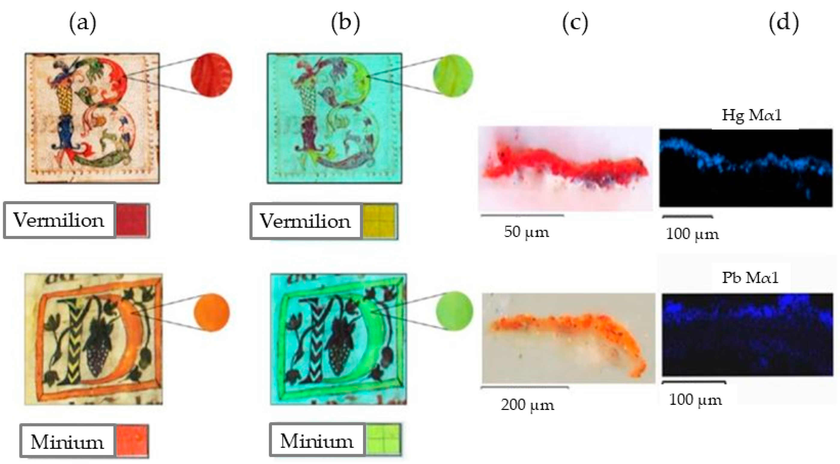
3.2. Red and Orange Illuminations
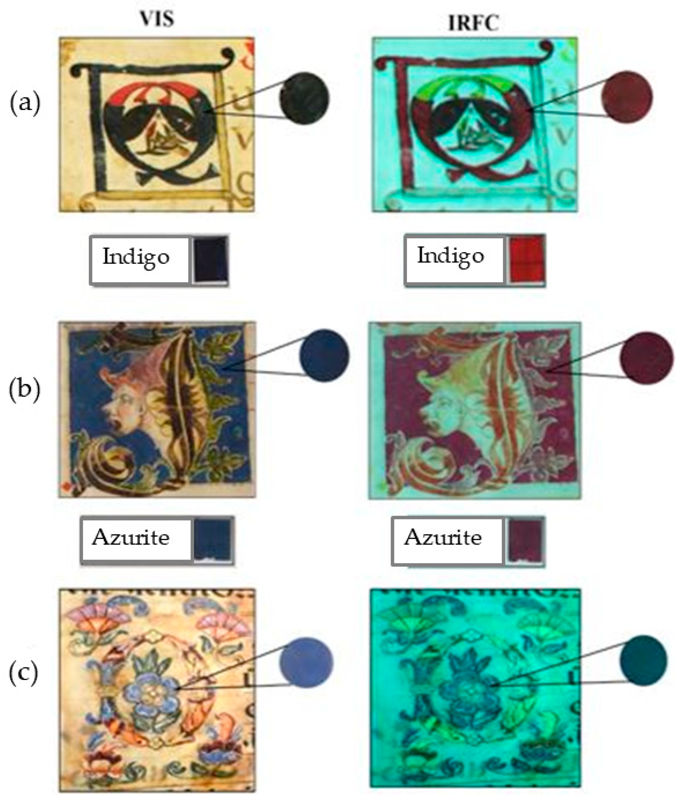
3.3. Blue Illuminations
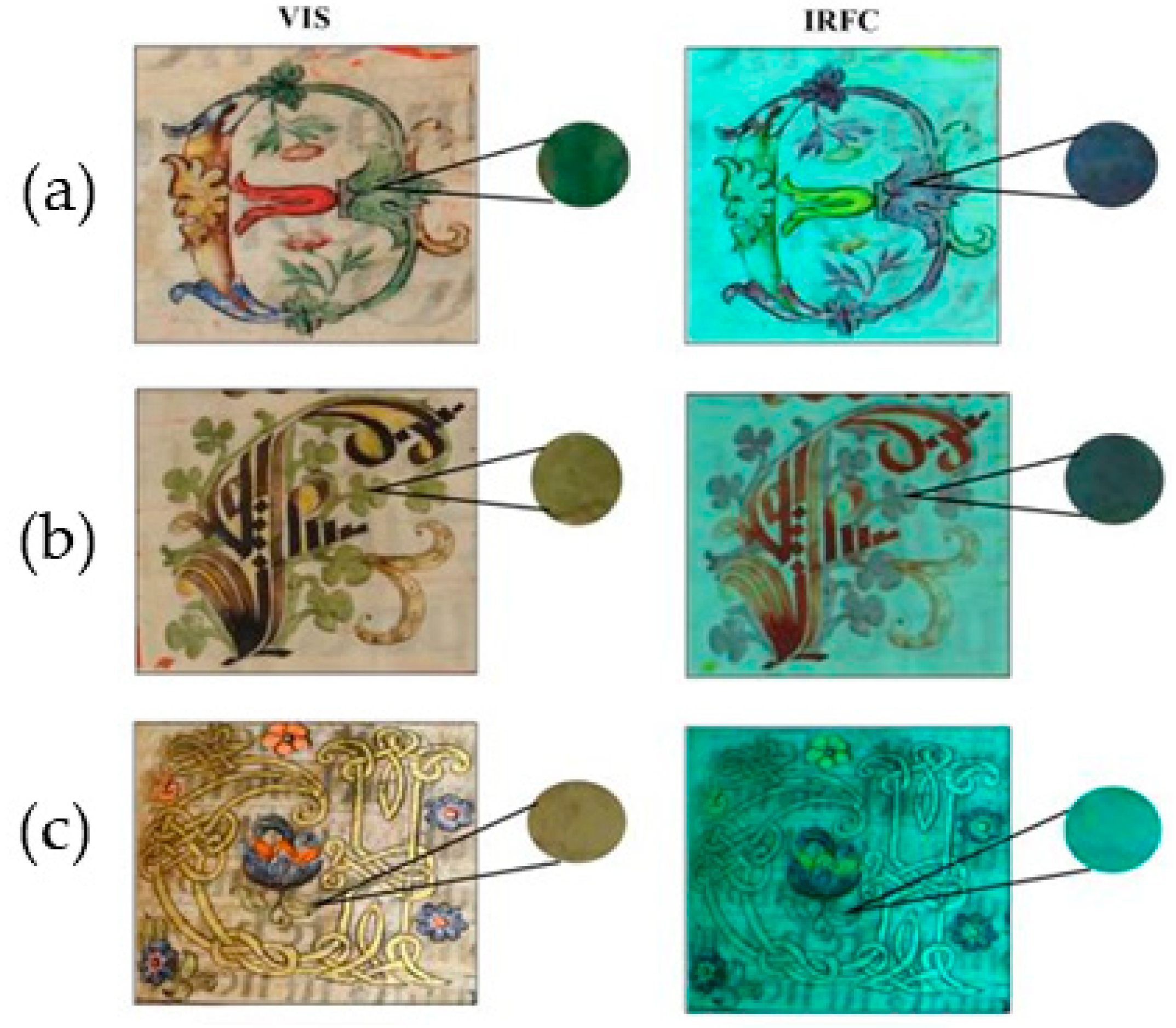
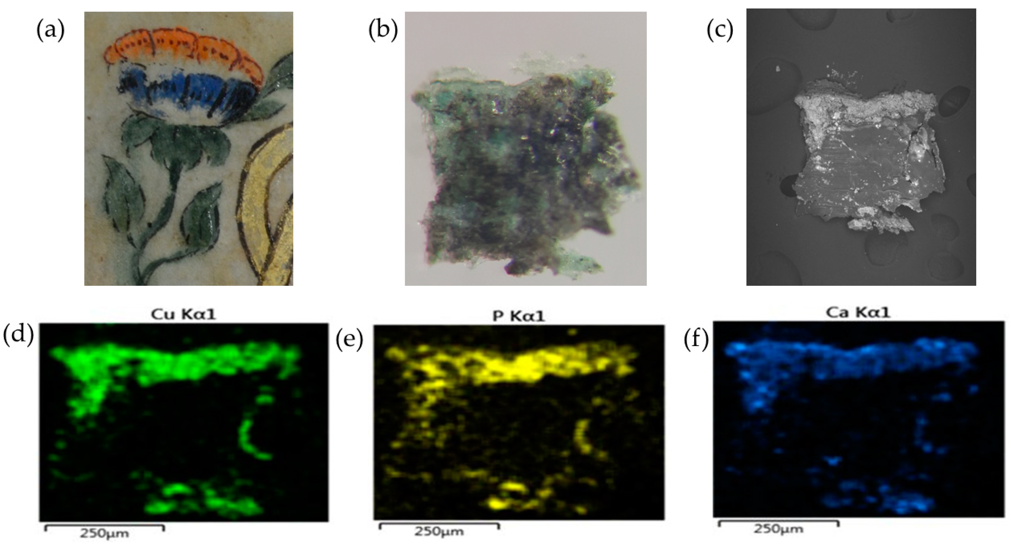
3.4. Green Illuminations
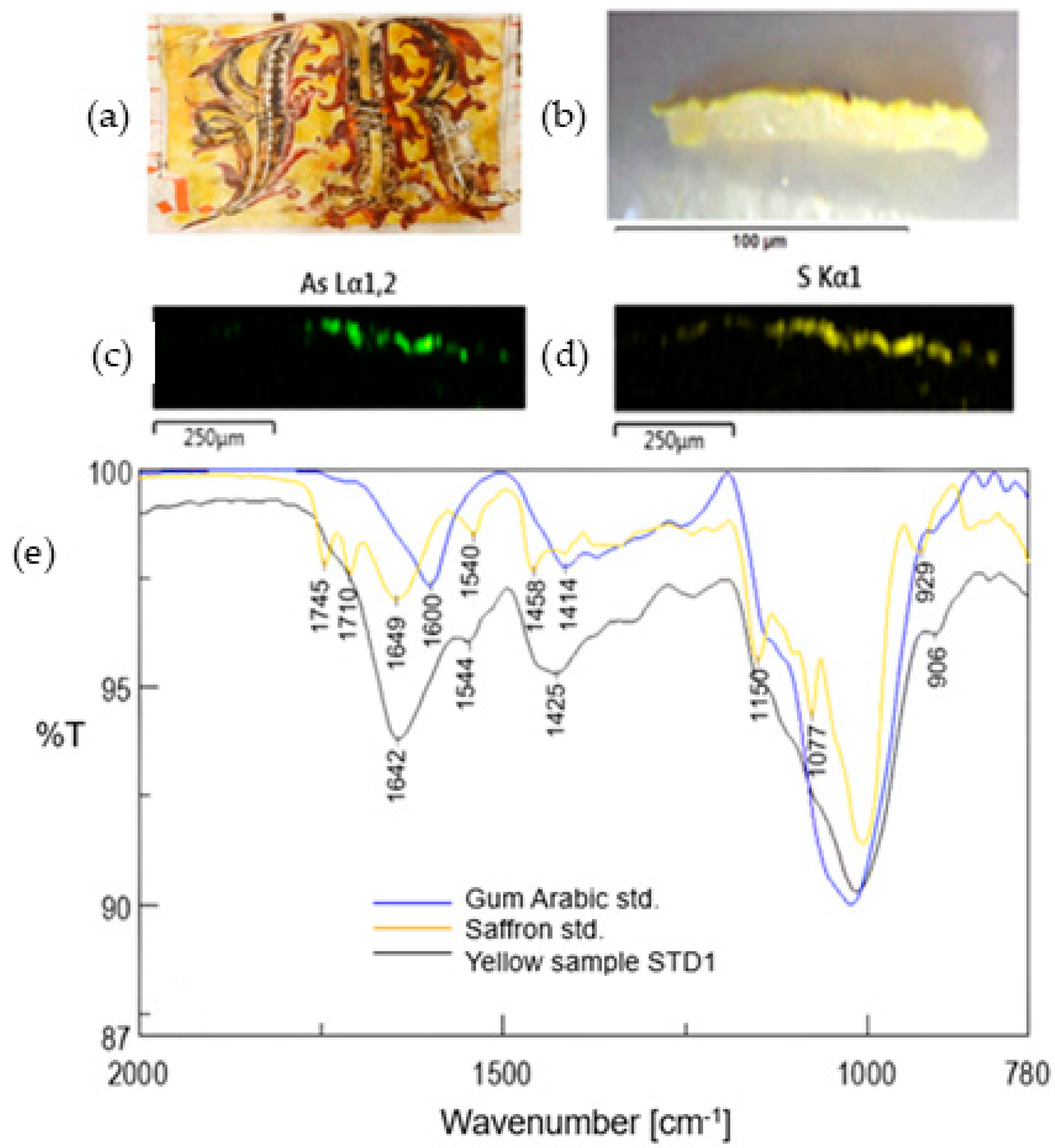
3.5. Yellow Illuminations
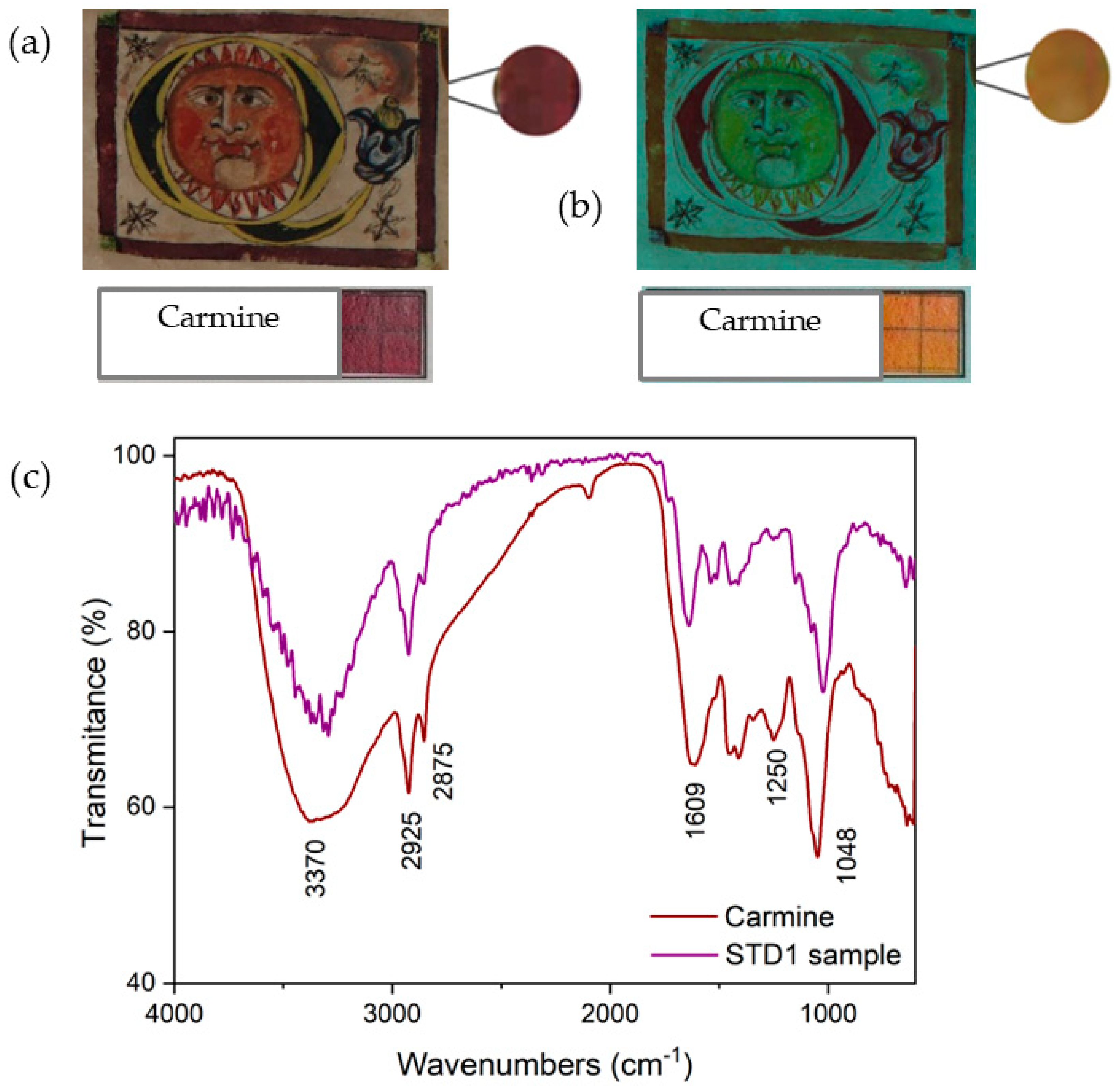
3.6. Violet Illuminations
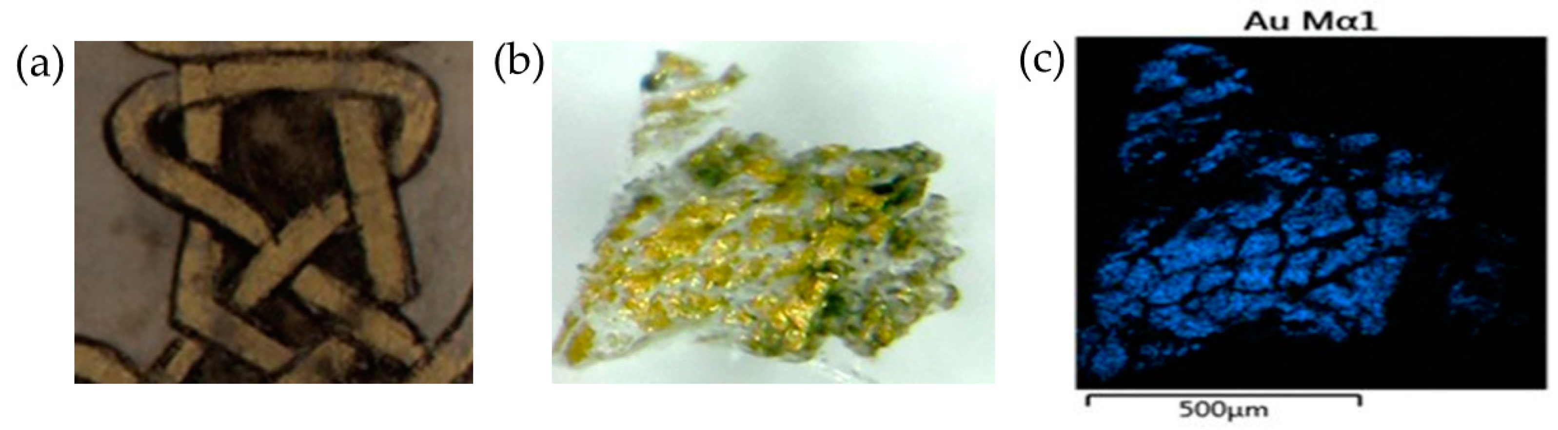
3.7. Gilding Illuminations
3.8. Binders
3.9. Vulnerability and Conservation Criteria
3.9.1. The Parchment
3.9.2. The Binders
3.9.3. Pigments and Dyes
4. Conclusions
Author Contributions
Funding
Data Availability Statement
Acknowledgments
Conflicts of Interest
References
- Bueno-Vargas, J. Deterioro En Encuadernaciones Manuscritas de Gran Formato: Causas Intrínsecas de Alteración En Los Libros de Coro. Cuad. Restaur. 2006, 6, 43–56. [Google Scholar]
- Gento, B. Historia de La Obra Constructiva de San Francisco Desde Su Fundación Hasta Nuestros Días, 1535–1942. 1942. Available online: https://repositorio.bne.gob.ec/xmlui/handle/34000/991 (accessed on 15 June 2024).
- Stevenson, R. Music in Quito: Four Centuries. Hisp. Am. Hist. Rev. 1963, 43, 247–266. [Google Scholar] [CrossRef]
- Webster, V.; Materiales, S. Modelos y Mercado de La Pintura En Quito, 1550–1650. Procesos. Rev. Ecuat. Hist. 2016, 43, 37–64. [Google Scholar] [CrossRef]
- López, G. Archivo y Biblioteca Nacionales de Bolivia. Unidad Documental Simple: 266v-267v—Poder Especial Que Otorga Tomé García. 2022. Available online: https://archivo-abnb.org.bo/index.php/poder-especial-que-otorga-tome-garcia-2 (accessed on 15 June 2024).
- González Suárez, F. Historia General de La República Del Ecuador; Imprenta del Clero: Quito, Ecuador, 1890. [Google Scholar]
- Navarro, J.G. La Pintura En El Ecuador Del XVI Al XIX; Dinediciones: Quito, Ecuador, 1991; pp. 52–59. ISBN 9978954058. [Google Scholar]
- Monagas, J.E. Documentación musical en el Convento de San Francisco deito: Libros litúrgico-musicales, órganos y coristas. Acta Musicol. 2021, 93, 43–66. [Google Scholar]
- Carvalho, I.; Casanova, C.; Araújo, R.; Lemos, A. Colour Identification, Degradation Processes and Findings in a Fifteenth-Century Book of Hours: The Case Study of Cofre n.o 31 from Mafra National Palace. Herit. Sci. 2018, 6, 9. [Google Scholar] [CrossRef]
- Bruni, S.; Caglio, S.; Guglielmi, V.; Poldi, G. The Joined Use of n.i. Spectroscopic Analyses—FTIR, Raman, Visible Reflectance Spectrometry and EDXRF—To Study Drawings and Illuminated Manuscripts. Appl. Phys. A Mater. Sci. Process. 2008, 92, 103–108. [Google Scholar] [CrossRef]
- Manso, M.; Le Gac, A.; Longelin, S.; Pessanha, S.; Frade, J.C.; Guerra, M.; Candeias, A.J.; Carvalho, M.L. Spectroscopic Characterization of a Masterpiece: The Manueline Foral Charter of Sintra. Spectrochim. Acta—Part A Mol. Biomol. Spectrosc. 2013, 105, 288–296. [Google Scholar] [CrossRef]
- Tanevska, V.; Nastova, I.; Minčeva-Šukarova, B.; Grupče, O.; Ozcatal, M.; Kavčić, M.; Jakovlevska-Spirovska, Z. Spectroscopic Analysis of Pigments and Inks in Manuscripts: II. Islamic Illuminated Manuscripts (16th–18th Century). Vib. Spectrosc. 2014, 73, 127–137. [Google Scholar] [CrossRef]
- Clarke, M. The Analysis of Medieval European Manuscripts. Stud. Conserv. 2001, 46, 3–17. [Google Scholar] [CrossRef]
- Poole, J.B.; Reed, R. The Preparation of Leather and Parchment by the Dead Sea Scrolls Community. Technol. Cult. 1962, 3, 1. [Google Scholar] [CrossRef]
- Sterflinger, K.; Pinzari, F. The Revenge of Time: Fungal Deterioration of Cultural Heritage with Particular Reference to Books, Paper and Parchment. Environ. Microbiol. 2012, 14, 559–566. [Google Scholar] [CrossRef] [PubMed]
- Badea, E.; Della Gatta, G.; Budrugeac, P. Characterisation and Evaluation of the Environmental Impact on Historical Parchments by Differential Scanning Calorimetry. J. Therm. Anal. Calorim. 2011, 104, 495–506. [Google Scholar] [CrossRef]
- Axelsson, K.M.; Larsen, R.; Sommer, D.V.P.; Melin, R. Degradation of Collagen in Parchment under the Influence of Heat-Induced Oxidation: Preliminary Study of Changes at Macroscopic, Microscopic, and Molecular Levels. Stud. Conserv. 2016, 61, 46–57. [Google Scholar] [CrossRef]
- Pozo-Antonio, J.S.; Jiménez-Desmond, D.; De Villalobos, L.; Mato, A.; Dionísio, A.; Rivas, T.; Cardell, C. SO2-Induced Aging of Hematite- and Cinnabar-Based Tempera Paint Mock-Ups: Influence of Binder Type/Pigment Size and Composition. Minerals 2023, 13, 289. [Google Scholar] [CrossRef]
- Castillejo, M.; Martín, M.; Oujja, M.; Santamaría, J.; Silva, D.; Torres, R.; Manousaki, A.; Zafiropulos, V.; Van Den Brink, O.F.; Heeren, R.M.A.; et al. Evaluation of the Chemical and Physical Changes Induced by KrF Laser Irradiation of Tempera Paints. J. Cult. Herit. 2003, 4, 257–263. [Google Scholar] [CrossRef]
- Troiano, F.; Polo, A.; Villa, F.; Cappitelli, F. Assessing the Microbiological Risk to Stored Sixteenth Century Parchment Manuscripts: A Holistic Approach Based on Molecular and Environmental Studies. Biofouling 2014, 30, 299–311. [Google Scholar] [CrossRef]
- Koochakzaei, A.; Bidgoli, B.J.; Safapour, S.; Nemati-Babaylo, A. Multispectral Imaging and Hyperspectral Techniques Applied to Dyed Fibers: A Classification Approach. Prog. Color Color. Coat. 2024, 17, 227–238. [Google Scholar] [CrossRef]
- Cosentino, A. Identification of Pigments by Multispectral Imaging; a Flowchart Method. Herit. Sci. 2014, 2, 8. [Google Scholar] [CrossRef]
- Dyer, J.; Verri, G.; Cupitt, J. Multispectral Imaging in Reflectance and Photo-Induced Luminescence Modes: A User Manual. 2013. Available online: https://www.researchgate.net/profile/Giovanni-Verri-2/publication/267266175_Multispectral_Imaging_in_Reflectance_and_Photo-induced_Luminescence_modes_A_User_Manual/links/5448e7560cf2d62c3052d2b7/Multispectral-Imaging-in-Reflectance-and-Photo-induced-Luminescence-modes-A-User-Manual.pdf (accessed on 15 June 2024).
- Lachmann, T.; van der Snickt, G.; Haschke, M.; Mantouvalou, I. Combined 1D, 2D and 3D Micro-XRF Techniques for the Analysis of Illuminated Manuscripts. J. Anal. At. Spectrom. 2016, 31, 1989–1997. [Google Scholar] [CrossRef]
- Hidalgo Brinquis, M. del C. Técnicas Medievales En La Elaboración Del Libro: Aportaciones Hispanas a La Fabricación Del Pergamino y Del Papel y a Los Sistemas de Encuadernación. Anu. Estud. Mediev. 2011, 41, 755–773. [Google Scholar] [CrossRef]
- Moreno González, M. Tratamiento de Los Pergaminos Utilizados En Los Libros de Coro de El Escorial. Signo Rev. Hist. Cult. Escrita 1997, 4, 169–176. [Google Scholar]
- Hagadorn, A. Parchment Making in Eighteenth-Century France: Historical Practices and the Written Record. J. Inst. Conserv. 2012, 35, 165–188. [Google Scholar] [CrossRef]
- Badea, E.; Miu, L.; Budrugeac, P.; Giurginca, M.; Mašić, A.; Badea, N.; Della Gatta, G. Study of Deterioration of Historical Parchments by Various Thermal Analysis Techniques Complemented by SEM, FTIR, UV-Vis-NIR and Unilateral NMR Investigations. J. Therm. Anal. Calorim. 2008, 91, 17–27. [Google Scholar] [CrossRef]
- Odlyha, M.; Theodorakopoulos, C.; Bozec, L. Fourier Transform Infra-Red Spectroscopy (ATR/FTIR) and Scanning Probe Microscopy of Parchment. e-Preserv. Sci. 2009, 6, 138–144. [Google Scholar]
- Boulc’h, F.; Bouillon, N.; Guillon, O.; Vallet, J.M.; Demoulin, A.; Gontero-Lauze, V. Painting Techniques of a 15th Century Illuminated Manuscript Revealed by Combining Multimodal Imaging Methods and X-Ray Fluorescence Spectroscopy. Available online: https://papers.ssrn.com/sol3/papers.cfm?abstract_id=4404515 (accessed on 15 June 2024).
- Biolcati, V.; Wilson, M.; Fiddyment, S.; Unitt, R.; Ryan, C.C.; Hoffmann, A.G.; Gillis, J.; France, F.; Ó Macháin, P.; Iacopino, D. The Book of Uí Mhaine: An Interdisciplinary Analysis of the Materiality of the Gaelic Manuscript Tradition. Heritage 2023, 6, 5393–5409. [Google Scholar] [CrossRef]
- Koochakzaei, A.; Alizadeh Gharetapeh, S.; Jelodarian Bidgoli, B. Identification of Pigments Used in a Qajar Manuscript from Iran by Using Atomic and Molecular Spectroscopy and Technical Photography Methods. Herit. Sci. 2022, 10, 30. [Google Scholar] [CrossRef]
- Castañeda Delgado, M. Caracterización e Identificación Del Índigo Utilizado Como Pigmento En La Pintura de Caballete Novohispana. Bachelor’s Thesis, Escuela de Conservación y Restauración de Occidente, Guadalajara, Mexico, 2017. [Google Scholar]
- Seldes, A.M.; Burucúa, J.E.; Maier, M.S.; Abad, G.; Jáuregui, A.; Siracusano, G. Blue Pigments in South American Painting (1610–1780). J. Am. Inst. Conserv. 1999, 38, 100–123. [Google Scholar] [CrossRef]
- Parrilla Bou, M.A. El Arte de Los Pigmentos Análisis Histórico-Artístico de Su Evolución a Partir de Los Tratados Españoles de Francisco Pacheco y Antonio Palomino; Universitat de València, Servei de Publicacions: València, Spain, 2009; ISBN 9788437074436. [Google Scholar]
- Ordoudi, S.A.; De Los Mozos Pascual, M.; Tsimidou, M.Z. On the Quality Control of Traded Saffron by Means of Transmission Fourier-Transform Mid-Infrared (FT-MIR) Spectroscopy and Chemometrics. Food Chem. 2014, 150, 414–421. [Google Scholar] [CrossRef] [PubMed]
- Celis, F.; Segura, C.; Gómez-Jeria, J.S.; Campos-Vallette, M.; Sanchez-Cortes, S. Analysis of Biomolecules in Cochineal Dyed Archaeological Textiles by Surface-Enhanced Raman Spectroscopy. Sci. Rep. 2021, 11, 6560. [Google Scholar] [CrossRef]
- Kannangara, R.; Siukstaite, L.; Borch-Jensen, J.; Madsen, B.; Kongstad, K.T.; Staerk, D.; Bennedsen, M.; Okkels, F.T.; Rasmussen, S.A.; Larsen, T.O.; et al. Characterization of a Membrane-Bound C-Glucosyltransferase Responsible for Carminic Acid Biosynthesis in Dactylopius Coccus Costa. Nat. Commun. 2017, 8, 1987. [Google Scholar] [CrossRef]
- Kroustallis, S. Binding Media in Medieval Manuscript Illumination: A Source Research. Rev. História da Arte 2011, 1, 112–125. [Google Scholar]
- Valipour, P.; Ekrami, E.; Shams Nateri, A. Colorimetric Properties of Wool Dyed with Cochineal: Effect of Dye-Bath PH. Prog. Color Color. Coat. 2013, 7, 129–138. [Google Scholar] [CrossRef]
- Kroustallis, S. Quomodo Decoretur Pictura Librorum: Materiales y Técnicas de La Iluminación Medieval. Anu. Estud. Mediev. 2011, 41, 775–802. [Google Scholar] [CrossRef]
- Castellanos Rodriguez, D.M. Desarrollo de Metodologías Analíticas Basadas En Espectrometría de Masa y Espectroscopía Infrarroja Para La Identificación de Aglutinantes Orgánicos y Sus Productos de Degradación En Arte Colonial. 2018. Available online: http://hdl.handle.net/11336/83588 (accessed on 18 May 2024).
- Pacheco, F. Arte de La Pintura, Su Antigüedad y Grandezas; Biblioteca Virtual Miguel de Cervantes: Alicante, Spain, 1871. [Google Scholar]
- Bruquetas, R. The Search for the Perfect Color: Pigments, Tints, and Binders in the Scientific Expeditions to the Americas. J. Interdiscip. Hist. 2015, 45, 367–387. [Google Scholar] [CrossRef]
- Larsen, R.; Vest, M.; Kejser, U.B. STEP Leather Project: Evaluation of the Correlation between Natural and Artificial Ageing of Vegetable Tanned Leather and Determination of Parameters for Standardization of an Artificial Ageing Method. 1994. Available online: https://archive.org/details/stepleatherproje0000step/page/n6/mode/1up (accessed on 22 July 2024).
- Sharma, J.; Bohidar, H.B. Gelatin-Glutaraldehyde Supramolecular Structures Studied by Laser Light Scattering. Eur. Polym. J. 2000, 36, 1409–1418. [Google Scholar] [CrossRef]
- Kennedy, C.J.; Hiller, J.; Odlyha, M.; Nielsen, K.; Drakopoulos, M.; Wess, T.J. Degradation in Historical Parchments: Structural, Biochemical, and Thermal Studies TT—Paper Restoration. Pap. Mitteilungen IADA 2002, 3, 23–30. [Google Scholar]
- Hansen, E.F.; Lee, S.N.; Sobel, H. The Effects of Relative Humidity on Some Physical Properties of Modern Vellum: Implications for the Optimum Relative Humidity for the Display and Storage of Parchment. J. Am. Inst. Conserv. 1992, 31, 325–342. [Google Scholar] [CrossRef]
- BS 4971:2017; Conservation and Care of Archive and Library Collections. British Standards Institution: London, UK, 2017. Available online: https://knowledge.bsigroup.com/products/conservation-and-care-of-archive-and-library-collections?version=standard (accessed on 22 July 2024).
- ISO 11799:2024; Information and Documentation—Document Storage Requirements for Archive and Library Materials. International Organization for Standardization: Geneva, Switzerland, 2024. Available online: https://www.iso.org/es/contents/data/standard/08/23/82306.html (accessed on 2 July 2024).
- Ma, Z.; Yang, L.; Wang, L.; Pitthard, V.; Bayerova, T.; Krist, G.; Zhao, X. The Influence of Natural Aging Exerting on the Stability of Some Proteinaceous Binding Media Commonly Used in Painted Artworks. Coatings 2022, 12, 1522. [Google Scholar] [CrossRef]
- Špec, T.; Retko, K.; Ropret, P.; Meden, A.; Bernard, J. The Influence of UV-Vis Radiation, and Oscillations of Temperature and Relative Humidity, on Malachite Alteration in the Presence of Different Organic Binders and Varnishes. J. Raman Spectrosc. 2014, 45, 1068–1075. [Google Scholar] [CrossRef]
- Spring, M.; Grout, R. The Blackening of Vermilion: An Analytical Study of the Process in Paintings. Natl. Gall. Tech. Bull. 2002, 23, 50–61. [Google Scholar]
- Mccormack, J.K. LETTE R The Darkening of Cinnabar in Sunlight. Miner. Depos. 2000, 35, 796–798. [Google Scholar] [CrossRef]
- Da Pieve, F.; Hogan, C.; Lamoen, D.; Verbeeck, J.; Vanmeert, F.; Radepont, M.; Cotte, M.; Janssens, K.; Gonze, X.; Van Tendeloo, G. Casting Light on the Darkening of Colors in Historical Paintings. Phys. Rev. Lett. 2013, 111, 208302. [Google Scholar] [CrossRef] [PubMed]
- Hogan, C.; Da Pieve, F. Colour Degradation of Artworks: An Ab Initio Approach to X-Ray, Electronic and Optical Spectroscopy Analyses of Vermilion Photodarkening. J. Anal. At. Spectrom. 2015, 30, 588–598. [Google Scholar] [CrossRef]
- Anaf, W.; Janssens, K.; De Wael, K. Formation of Metallic Mercury During Photodegradation/Photodarkening of A-HgS: Electrochemical Evidence. Angew. Chem. 2013, 125, 12800–12803. [Google Scholar] [CrossRef]
- Davidson, R.S.; Willsher, C.J. The Light-Induced Blackening of Red Mercury(II) Sulphide. J. Chem. Soc. Dalton Trans. 1981, 3, 833–835. [Google Scholar] [CrossRef]
- Radepont, M.; Coquinot, Y.; Janssens, K.; Ezrati, J.J.; De Nolf, W.; Cotte, M. Thermodynamic and Experimental Study of the Degradation of the Red Pigment Mercury Sulfide. J. Anal. At. Spectrom. 2015, 30, 599–612. [Google Scholar] [CrossRef]
- Elert, K.; Cardell, C. Weathering Behavior of Cinnabar-Based Tempera Paints upon Natural and Accelerated Aging. Spectrochim. Acta—Part A Mol. Biomol. Spectrosc. 2019, 216, 236–248. [Google Scholar] [CrossRef]
- Coccato, A.; Moens, L.; Vandenabeele, P. On the Stability of Mediaeval Inorganic Pigments: A Literature Review of the Effect of Climate, Material Selection, Biological Activity, Analysis and Conservation Treatments. Herit. Sci. 2017, 5, 12. [Google Scholar] [CrossRef]
- Lluveras, A.; Boularand, S.; Andreotti, A.; Vendrell-Saz, M. Degradation of Azurite in Mural Paintings: Distribution of Copper Carbonate, Chlorides and Oxalates by SRFTIR. Appl. Phys. A Mater. Sci. Process. 2010, 99, 363–375. [Google Scholar] [CrossRef]
- van Loon, A.; Noble, P.; de Man, D.; Alfeld, M.; Callewaert, T.; Van der Snickt, G.; Janssens, K.; Dik, J. The Role of Smalt in Complex Pigment Mixtures in Rembrandt’s Homer 1663: Combining MA-XRF Imaging, Microanalysis, Paint Reconstructions and OCT. Herit. Sci. 2020, 8, 90. [Google Scholar] [CrossRef]
- Vickackaite, V.; Romani, A.; Pannacci, D.; Favaro, G. Photochemical and Thermal Degradation of a Naturally Occurring Dye Used in Artistic Painting. A Chromatographic, Spectrophotometric and Fluorimetric Study on Saffron. Int. J. Photoenergy 2004, 6, 175–183. [Google Scholar] [CrossRef]


| Illuminated Manuscripts | Repository | Author | Temporality | Featured Image | |
|---|---|---|---|---|---|
| STD1 | Book of Psalms | Santo Domingo Convent | Anonymous | S. XVII | 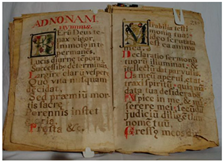 |
| SF1 | Illuminated manuscript of Apostle Commons | San Francisco Convent | Anonymous | S. XVII | 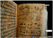 |
| STD2 | Illuminated manuscript SN | Santo Domingo Convent | Attributed to Fray Pedro Bedón | 1613 |  |
| SF2 | Book of psalms | San Francisco Convent | Anonymous | S. XVII | 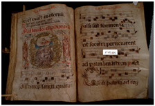 |
| SA1 | St. Augustine’s Festivities Choirbook | San Agustin Convent | Friar Francisco de la Fuente | 1628 | 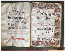 |
| Analyzed Area | Choirbook | IRFC | Chemical Elements (XRF) | FTIR Bands (cm−1) | Interpretation |
|---|---|---|---|---|---|
| Parchment | STD1 SF1 STD2 SF2 SA1 | Ca (33%), S (7%), Al, Si, Cl, K, Fe Ca (38%), S (11%), Al, Si, Cl, K, Fe Ca (45%), S (8%), Al, Si, Cl, K, Fe Ca (50%), S (7%), Al, Si, Cl, K, Fe Ca (42%), S (8%), Al, Si, Cl, K, Fe | 2900–2800, 1000–1250, 1650–1500, 1234 | Use of calcium hydroxide, sodium sulfite, poem stone and salts in the parchment production process Gelatinization processes | |
| Yellow color | SF1, STD1, SA1 | - | As, S, (Si, Al, K, Ca) | Orpiment | |
| SF2 | - | As, Pb, S, Si (Al, K, Fe) | Orpiment, masicot | ||
| STD2 | - | Fe, Si, Al (Na, K, Mg, Ca, S) | 3355, 2923, 2853,1710, 1745, 1643, 1015 | Yellow ochre, saffron | |
| Orange color | STD1, STD2, SA1 | Light yellow | Pb (Al, Si, K, Fe, Ca) | Minium | |
| STD2, SF2, SA1 | Dark yellow | Pb, Hg, S (Al, Si, K, Ca) | Minium, Vermilion | ||
| SA1 | - | Pb (Al, Si, K, Fe, Ca) | - | Minium | |
| Golden color | SF1, STD2, SF2 | - | Au, Cu | - | Gold leaf |
| Blue color | SF1, STD2 | Brown | Cu (Mg, Al, Si, Ca, Fe) | - | Azurite |
| SA1 | Brown | Cu, Pb (Mg, Al, Si, Ca, Fe) | Azurite, lead white | ||
| SF1, SF2 | - | Co, Ni, As, Si, K (Al, Ca, Fe) | - | Smalt | |
| STD1, SF1, STD2 | Bright red | 1670, 1461 | Indigo | ||
| Red color | SF1, STD2, SF2 | Dark yellow | Hg, S (Al, Si, K, Ca) | Vermilion | |
| Green color | STD1, SA1 | Blue | Cu, Si, P (Ca, Al, S) | Verdigris | |
| STD2 | Blue | Cu, Ca, Si, (Al, Si, P) | Verdigris, lime white | ||
| SF1 | Blue | Cu, Si, P (Al, Si, Ca) | Malachite/Verdigris, bone black | ||
| SF2 | Blue | Cu, Si, C (Al, Si,) | Malachite/Verdigris, black with humus | ||
| Violet | STD2, SF2 | Light coffee | Pb (Si, K, Ca) | 1628, 1454, 1418, 1240, 1033 | Carmine, Minium |
| Pigments and Dyes | Choirbooks | ||||
|---|---|---|---|---|---|
| STD1 | SF1 | STD2 | SF2 | SA1 | |
| Blue | Indigo | Indigo Azurite Smalt | Azurite Indigo | Smalt | Azurite + lead white |
| Green | Verdigris | Malachite/Verdigris Verdigris + Bone black | Verdigris + Lime white | Malachite/Verdigris + Carbon black | Verdigris |
| Yellow | Orpiment | Orpiment | Saffron Yellow Ochre | Orpiment Masicot | Orpiment |
| Gilding | Gold leaf | Gold leaf | Gold leaf | ||
| Red | Vermilion | Vermilion | Vermilion | Vermilion | Vermilion |
| Orange | Minium | Minium Vermilion + Minium | Vermilion + Minium | Minium | |
| Violets | Carmine | Carmine | Carmine + Minium | Carmine Carmine + Minium | Carmine |
| Binders | Egg yolk and white + oil | Oil | Arabic gum | Oil | Egg yolk and white + oil, gum arabic |
Disclaimer/Publisher’s Note: The statements, opinions and data contained in all publications are solely those of the individual author(s) and contributor(s) and not of MDPI and/or the editor(s). MDPI and/or the editor(s) disclaim responsibility for any injury to people or property resulting from any ideas, methods, instructions or products referred to in the content. |
© 2024 by the authors. Licensee MDPI, Basel, Switzerland. This article is an open access article distributed under the terms and conditions of the Creative Commons Attribution (CC BY) license (https://creativecommons.org/licenses/by/4.0/).
Share and Cite
Romero-Bastidas, M.; Guacho-Pachacama, K.; Vásquez-Mora, C.; Espinoza-Guerra, F.; Díaz-Benalcázar, R.; Ramírez-Bustamante, J.; Ramos-Guerrero, L. Multi-Analytical Characterization of Illuminated Choirbooks from the Royal Audience of Quito. Heritage 2024, 7, 6592-6613. https://doi.org/10.3390/heritage7120305
Romero-Bastidas M, Guacho-Pachacama K, Vásquez-Mora C, Espinoza-Guerra F, Díaz-Benalcázar R, Ramírez-Bustamante J, Ramos-Guerrero L. Multi-Analytical Characterization of Illuminated Choirbooks from the Royal Audience of Quito. Heritage. 2024; 7(12):6592-6613. https://doi.org/10.3390/heritage7120305
Chicago/Turabian StyleRomero-Bastidas, Martha, Katherine Guacho-Pachacama, Carlos Vásquez-Mora, Fernando Espinoza-Guerra, Rita Díaz-Benalcázar, Johanna Ramírez-Bustamante, and Luis Ramos-Guerrero. 2024. "Multi-Analytical Characterization of Illuminated Choirbooks from the Royal Audience of Quito" Heritage 7, no. 12: 6592-6613. https://doi.org/10.3390/heritage7120305
APA StyleRomero-Bastidas, M., Guacho-Pachacama, K., Vásquez-Mora, C., Espinoza-Guerra, F., Díaz-Benalcázar, R., Ramírez-Bustamante, J., & Ramos-Guerrero, L. (2024). Multi-Analytical Characterization of Illuminated Choirbooks from the Royal Audience of Quito. Heritage, 7(12), 6592-6613. https://doi.org/10.3390/heritage7120305