Article

Rare Earth Elements to Control Bone Diagenesis Processes at Rozafa Castle (Albania)

1
Department of Prehistory, Archaeology and Ancient History, University of Valencia, 46010 Valencia, Spain
2
Institute of Materials Science, University of Valencia, Av. Catedrático José Beltrán, 2, 46980 Paterna, Spain
3
Antiquity of Southeastern Europe Research Centre, University of Warsaw, 32, 00-927 Warsaw, Poland
4
Department of Analytic Chemistry, University of Valencia, 46100 Burjassot, Spain
*
Author to whom correspondence should be addressed.
Heritage 2024, 7(10), 5800-5813; https://doi.org/10.3390/heritage7100273
Submission received: 16 August 2024 / Revised: 3 October 2024 / Accepted: 15 October 2024 / Published: 17 October 2024

Abstract

:
Archaeological bone chemical composition is modified post-mortem by diagenesis processes, and over decades, several authors have proposed different protocols to avoid post-depositional contamination that can carry to misleading interpretations about the lifestyle and origin of ancient populations. In this work, a methodological approach based on rare earth elements analysis was developed to determine diagenetic alterations on femurs, humeri, and skull surfaces, and internal layers from thirteen individuals exhumed during fieldwork in the Fatih Sultan Mehmet Mosque at Rozafa Castle (Shkodër, Albania). Major, minor, and trace elements, including rare earth elements, were measured employing spectrometric techniques, and the obtained data were statistically processed by principal component analysis and one-way ANOVA to select the best preserved bones. The results show that in general, the internal parts of bones, especially skulls, suffered post-depositional chemical contamination. Finally, to show the effectiveness of the proposed approach, a diet reconstruction employing log(Sr/Ca) and Zn/Ca was tested, obtaining results that are in line with the literature describing a diet based on a mixed economy, mostly agricultural products with low protein intakes.

1. Introduction

Skeletal remains are materials commonly found in archaeological contexts, and chemical analyses in bones have been employed to study diet, pathologies, and taphonomy. However, bones undergo degradation induced by different post-depositional processes [1], and the obtention of reliable information has been under discussion. In fact, bones main components, such as the inorganic hydroxyapatite phosphate crystals and organic compounds (collagen proteins, cells, and other macromolecules), are particularly affected by diagenesis mechanisms that include ionic substitutions in the bioapatite, a form of calcium phosphate that is the main component in the bone mineralized part [2]. In this way, it is acting as a body repository of elements, storing useful oligoelements or toxic elements [3]. At the same time, after death, those bone crystals are exposed to the environment. After the hydrolysis of the organic compounds, a wide variety of exogenous alkaline and metal elements are incorporated into the bioapatite crystals or precipitate into bone pores. Therefore, the control of these post-depositional processes has raised the interest of scientists working on archaeological science, forensic anthropology, palaeontology, and environmental sciences.
In the present study, a methodological approach employing rare earth elements (REE) as markers to supervise bone post-depositional processes was tested in twenty-six samples from a total of 13 Late Medieval period skeletons buried in Rozafa Castle (Shkodër, Albania), where a mass grave was found (trench 23) with numerous human remains, from entire skeletons to bone fragments (Figure 1).
The castle is located in the territory of the modern city of Shkodër (ancient Scodra), identified as one of the most representative Mediterranean areas where, since the Bronze Age, human activities were mainly concentrated around fortified hills [4]. Therefore, due to its post-depositional complexity resulting from continued occupational events, Rozafa Castle and especially the identified mass grave become a significant case study for applying our proposed approach.
Archaeological bone chemical element analysis was employed as a proxy to obtain pre-mortem or post-mortem information, and this method was recently used in some very important case studies, such as in Pompeii’s remains [5]. Since half of the XX century, researchers have used chemical elements (i.e., Sr, Ba, and Zn) to assess the paelodiet habits of ancient individuals [6,7,8,9]. Also, Pb, As, or Hg have been used to evaluate heavy metal intoxication for different historical periods [10,11,12], and isotopic studies have been conducted in order to obtain information about human mobility [13,14]. However, the diagenetic processes affecting ancient bones are often just superficially taken into account to guarantee the biogenetic origin of the data collected. In this sense, the use of rare earth elements (REE) has shown to be useful to determine the degree of diagenetic alteration in bone assemblages [15,16,17]. Therefore, use the REE as a methodological tool based on the assumption that the content of REE found in the bone matrix is related to the post-depositional processes driven by the surrounding sediment and groundwater and is not from biological sources.
Diagenetic processes affect buried bones in different ways, and it is of pivotal importance to develop methodological approaches to control these post-depositional aspects. Researchers are employing bones to look at the preserved bone biological markers. Still, today, there is an important discussion about the quality of the data related to elemental, isotopic, organic, or DNA analyses obtained in the last decades. In this work, a proposed methodological approach based on the identification of REE concentrations in different bones has been applied in a real case study. The method is based on REE; these chemical elements are not easily incorporated into the organism with concentrations in living organisms lower than 1 μg/g, and this makes rare earth elements potential markers to evaluate diagenetic impact in bones. Bone samples from femurs, skulls, and humeri from Rozafa Castle remains were sampled and analyzed for the determination of major elements, trace elements, and rare earth elements. The obtained data were processed by multivariate statistics to observe diagenetic processes and cross-referencing the results with bioapatite and paleodiet indexes calculated in order to assess misleading interpretations.

Archaeological Background

Rozafa Castle was built around 1360 by George II Balšić and later was reformed under Venetian and Ottoman rule. Located in ancient Scroda, northern Albania, on the eastern shore of Lake Sköhder (Figure 1A), fieldwork has been carried out in its 3 ha of area since 2011 by archaeologists from the University of Warsaw and the University of Tirana [18,19,20]. The fortress is located on a steep slope, with levels ranging between 130 m.a.s.l. in the citadel and around 95 m.a.s.l. outside the curtain wall. Human remains were excavated during 2016–2017 fieldwork seasons in the Fatih Sultan Mehmet Mosque. The fortress was built during the Venetian Republic occupation in the late 14th century as St. Stephen’s cathedral in the place of an earlier sanctuary, which was turned into a mosque in 1479.
The religious building is located close to the north-western wall of the castle and covers an area of 255 m2, while the small graveyard behind it is 77 m2. In the surroundings, dozens of different burials were disclosed, mostly without human remains. Nevertheless, in the biggest part of the graveyard a mass grave was found (“trench 23”) with numerous human remains, from entire skeletons to bone fragments in the “ossuary” (Figure 1C). Among the findings, two belt buckles were found, which typology allowed researchers to date the mass grave around the 13th–14th c.
The anthropological study carried out during fieldwork identified 39 individuals buried in trench 23 of the mass grave, from which 11 individuals were selected for the present study together with two from grave 21. For this study, sampling, when possible, was carried out in long bones and skulls because, in previous studies, they showed better resistance to diagenetic processes as compared to other skeletal sectors such as ribs [8,15]. The skeletons were exhumed from the burials 3, 4, 5, 6, 7, 9, 17, 19, 21, 23, 23D, 23E, and 25 (Figure 1C) and were sampled for laboratory analysis (Table 1).

2. Materials and Methods

2.1. Samples

Twenty-six samples were analyzed from 13 individuals buried in Rozafa Castle from the Late Medieval period. Three different types of bones were sampled: cranial bones (4 petrous bones named “skulls” throughout the text) and post-cranial bones (diaphysis of 7 femurs and 2 humeri). To study bone diagenesis processes mainly caused by sediments and groundwater, a protocol established by Gallello (2014) [15] and Rasmussen et al. (2019) [21] was implemented.
From each individual, one bone was sampled, obtaining two samples (surface and internal). All anthropological information, as well as kind of bone, sampled type (surface or internal), and sample ID, is summarized in Table 1.
Bone samples were obtained with a mechanical cutter. Two samples were taken for each bone: a surface (S) sample by mechanical abrasion (i.e., 033-S) and another from the internal (I) part (i.e., 033-I), as shown in Table 1. Specifically, the surface samples were taken from the brownish layer of the cortical and trabecular bone that is in direct contact with the sediment and burial environment. On the other side, the internal samples were taken once the surface of cortical and trabecular parts were removed. Furthermore, the elemental composition of modern bones was compared to verify the conservation status of the archaeological bones. To do so, data from Harkness et al. (2019) [22], Zapata et al. (2006) [23], and Hancock et al. (1999) [24] collected from modern populations were taken into consideration (Supplementary Materials Tables S1 and S2).

2.2. Analytical Methodology

As aforementioned, 26 samples were obtained from 13 individuals, which included 2 samples from each individual, from the internal and from the surface through mechanical abrasion with a scalpel. Sample preparation and digestion protocol was a modified version of Gallello et al. (2013) [25]. Samples were homogenized by using an agate mortar, obtaining powdered samples. Bone powder was measured by a portable energy-dispersive X-ray fluorescence. A S1 Titan Bruker device (Bruker, Kennewick, Washington, DC, USA) was used, equipped with a rhodium X-ray emission source and a detector X-Flash® SDD. An internal Geochemtrace setup was used in order to measure Ca, P, Fe, Al, Ti, and Si. Afterwards, powdered samples were prepared to run inductive coupled plasma mass spectrometry (ICP-MS) analyses. In order to do so, 150 mg of each powdered sample was digested in aqua regia (1.35 mL of HCl and 0.45 mL of HNO3) in glass tubes at 100 °C in a water bath for 40 min, adding two blanks. Subsequently, digested solutions were poured into plastic tubes and brought to a final volume of 25 mL with ultra-pure water (18.2 MΩ). The measuring solutions were obtained by diluting 2.5 mL of these 25 mL solutions to 5 mL. To prepare the multi-element standards, two solutions were prepared: the first solution of 2 mg/L of La, Ce, Pr, Nd, Ba, Bi, Cd, Cr, Cd, Co, Cu, Pb, Li, Mn, Mo, Ni, Sr, Tl, Ti, V, Zn, U, and Th, and the second one of 0.4 mg/L of Sm, Eu, Gd, Tb, Dy, Ho, Er, Tm, Yb, Lu, Sc, and Y. For the calibration standards preparation, in a plastic tube the corresponding volume of the multi-element standard solutions, 1 mL of aqua regia was poured and filled up to 5 mL final volume with ultrapure water. All standards were acquired from Sharlab S.L. (Barcelona). Elements were measured using an ICP-MS Perkin Elmer model Elan DRCII (Concord, ON, Canada). Rhodium was employed as an internal standard. Bone ash NIST 1400 was used as standard reference material to evaluate the analytical method. Major, minor, and trace element concentrations, including REE for all samples and referenced fresh bone, are shown in Tables S1 and S2 (Supplementary Materials).

2.3. Rare Earth Elements Data Processing

Rare earth elements are a group of seventeen elements with similar chemical properties, classified by The Union of Pure and Applied Chemistry (IUPAC): lanthanum, fourteen lanthanides series plus scandium and yttrium. They are ubiquitously found together in geological deposits in measurable quantities. During the last 20 years, REE has been recognized as an important mineral source, and demand has recently increased because of their use in new technologies. These elements are not so rare and have an average abundance in the Earth’s crust, comparable to many other elements. There is intense interest in geological research to identify REE sources because of their important use as strategic elements in industry. It is clear that rock-forming processes affect the relative composition of REE. For some years, REE research has focused on how these elements are affected during sedimentation and anthrosol formation processes, as well as during weathering, leaching, adsorption, complexation, and reprecipitation [15].
As aforementioned, due to their chemical characteristics, REE can be employed to monitor bones post-depositional contamination, as their total biological content in bones is less than 1 μg/g. In this work, La, Ce, Pr, Nd, Sm, Eu, Gd, Tb, Dy, Ho, Er, Tm, Yb, Lu plus Sc and Y (promethium, as a radioactive element, is not measured and not included for this study) were analyzed and the obtained data processed. To calculate the total rare earth element contents (represented by ∑REE), the sum of the concentrations of fourteen REE from La to Lu was taken into account (Table S2 of the Supplementary Materials).

2.4. Statistical Analysis

A multifactor analysis of variance (ANOVA) was performed in order to determine the factors that have a significant effect on the element distributions in the data set: A (type of bone); B (bone surface or internal part); and C (the conjunction of both categories) were used as factors. Three hypotheses support this test: normally distributed data in each group, equal variances for all groups, and independent observations [26]. Data were adjusted to ensure a normal distribution, so the ANOVA analysis gave accurate and statistically significant information. A post hoc Tukey test was carried out as a posteriori verifying test. The null hypothesis was verified at the significance level of p ≤ 0.01.
On the other hand, principal component analysis (PCA) was carried out as it is a useful technique to show the elemental relationships that occur in the raw data [11,22,27]. The data were centered around zero, and autoscaling was employed; in this way, the multi-normality and unitvariance, which are the core of a parametric test such as PCA, were ensured [26]. All the analyzed elements (Ca, P, Fe, Al, Ti, Si, Ba, Bi, Cd, Cr, Cd, Co, Cu, Pb, Li, Mn, Mo, Ni, Sr, Tl, Ti, V, Zn, U, Th, La, Ce, Pr, Nd, Sm, Eu, Gd, Tb, Dy, Ho, Er, Tm, Yb, Lu, Sc, and Y) plus Ca/P have been employed as variables to run the PCA analysis. The statistical analyses were carried out employing RStudio (R Core Team (2020) [28]. R: A language and environment for statistical computing. R Foundation for Statistical Computing, Vienna, Austria; “https://www.R-project.org/ accessed on 28 November 2022)”. In particular, for multivariate data analysis, the “factoextra” package was used (Alboukadel Kassambara and Fabian Mundt (2020); factoextra: Extract and Visualize the Results of Multivariate Data Analyses. R package version 1.0.7.). Multifactor analysis was run with package “stats”, part of R, and post hoc Tukey test was carried out with the “multcomp” package (Torsten Hothorn, Frank Bretz, and Peter Westfall, 2008).

2.5. Paleodiet Indexes

Paleodiet studies by archaeological bone analysis [8,22,29,30] were focused on identify food habits, lifestyle, and social conduct.
Chemical elements such as Sr and Zn as biomarkers were based on the assumption that the trophic level or food habit of an ancient individual were identifiable looking at bone elemental concentrations [31,32]).
Following the aforementioned authors’ [31,32] premise, in a biological system, Sr and Zn are non-essential elements in comparison with Ca, one of the two main elements (Ca and P) of the bone inorganic composition. These elements, although not having a recognized vital role in the body, tend to replicate the metabolism of chemically similar bio-essential elements, allowing their incorporation into the body and apatite [3]. Their behavior in the organism is represented as a ratio between its concentration and that of the bio-essential element used as a reference. The ratios Sr/Ca and Zn/Ca decrease during metabolic processes where Ca is involved; this is known as biopurification [6]. It was shown that Sr/Ca ratios decrease from one trophic step to the next constantly [33]. Heinrich and Voorhies pointed out that the Sr/Ca ratio could potentially be used as a tracer of trophic levels in extinct vertebrates if two conditions occurred: on one side, the inorganic source of Sr should be constant, and no significant post-mortem processes should have altered the elemental concentration in bone [6]. These relations with trophic levels rather than food consumption are only feasible if the Sr level in bone is not simply proportional to the amount of Sr in the diet but to the mean Sr/Ca ratio of the diet [34]. The Sr/Ca ratio has therefore been used traditionally as a proxy to identify pastoral or agricultural societies. In this study, reference values [7] were taken into consideration for Sr/Ca, where log(Sr/Ca) < −4 establishes a pastoral economy and log(Sr/Ca) > −3 is associated with an agricultural economy [8]. Values ranging between these limits are interpreted as a mixed economy. The absorption of Zn is also dependent on dietary factors; for instance, the quantity and quality of protein intake and phytate fiber consumption, as well as the amounts of Zn intake in a meal, will alter the absorption of this element [35]. Therefore, Zn is conventionally related in the literature to protein intake habits, rich or poor protein consumption, and not to trophic levels. In this sense, a diet rich in protein has a Zn/Ca ratio >0.5, while values <0.35 are indicative of a poor protein intake [8]. To evaluate the interpretive errors that could take place if diagenetic factors are not taken into account, an attempt has been made to reconstruct the diet using the different groups of samples, including the internal and surface parts of bones.

3. Results

3.1. Elemental Composition and Statistical Analysis

The obtained concentrations of major, minor, and trace elements, including REE for all samples and fresh bone, are shown in Tables S1 and S2 (Supplementary Materials). Mean values of the studied bones, respectively internal and external parts, are shown in Table 2. Calcium (from 359 to 399 mg/g), P (from 160 to 174 mg/g), and other known bio-essential trace elements such as Fe (from 0.48 to 1.05 mg/g), Zn (from 109 to 234 μg/g), and Sr (from 196 to 207 μg/g) show the highest mean values for all samples, followed by V (from 39.8 to 62.9 μg/g), Ba (from 48.6 to 60.9 μg/g), Mn (from 6.28 to 45.6 μg/g), Cu (from 14.7 to 22.9 μg/g), Ni (from 12.9 to 17.3 μg/g), Cr (from 13.9 to 16.4 μg/g), and Mn (6.28 ± 8.08 μg/g), which show the lowest mean values in the internal samples and the highest V (62.9 ± 24.5 μg/g) in surface samples (Table 2). For Mo (from 1.52 to 1.21 μg/g) and Tl (from 0.05 to 0.02 μg/g), concentrations increase in the internal samples with respect to the surface ones, except for Mo in SHK 029 and SHK 026 and Tl in SHK 011. A similar trend is observed in Li and Bi and more evident in skull samples (individuals SHK 033, SHK 032, SHK 031, and SHK 030). Calcium and phosphorous present values for internal samples that fit well with the fresh bone standard concentrations (Supplementary Materials Table S1). Samples SHK 033-I, SHK 031-I, and SHK 019-I show the lowest concentrations with values under 338 mg/g for Ca and 153 mg/g for P. Other known bio-essential elements (Fe, Mn, Co, Ni, Cu, Zn, and Sr) concentrations from the internal part are similar to the fresh bone standard references (Supplementary Materials Table S1). However, sample SHK 030-I (skull) shows higher concentrations of Mn, Co, Ni, and Cu compared with the other internal samples. Other trace elements, such as V and Mo, display concentrations corresponding to two orders of magnitude higher than those reported for fresh bone in all individuals.
REE, Sc, and Y concentrations are shown in Table S2 (Supplementary Materials) for all bone samples. Internal samples present one order of magnitude lower than surface samples.
Internal sample values for La, Ce, Pr, Tb, Dy, Ho, Er, Tm, Yb, and Lu show similar concentration to the standard fresh bone, except for skull samples (SHK 030-I, SHK 031-I, and SHK 032-I). Nevertheless, Sc, Ce, Nd, and Sm concentrations for internal samples are higher than those from the standard fresh bone. Despite this, ∑REE for internal samples in all cases is <1 μg/g, consistent with the standard concentration for ∑REE in vivo bone proposed by Kohn et al., 2013 [36]. The total amount of REE mean concentrations in the Harkness et al., 2019 [22] study are slightly higher than the average of the population (Supplementary Materials Table S2), probable due to the bones sampled from patients who underwent hip replacement surgery contaminated by Gd incorporated from medical imaging contrast agents. In general, surface samples present higher ∑REE values, with values >1 μg/g except for SHK 015-S (femur), SHK 017-S (femur), and SHK 023-S (humerus).
The ANOVA test shows significant differences (p < 0.01) for almost all elemental concentrations, including REE, grouping the variables by factor B (internal or surface bone samples), as can be seen in Table 2. The rest of the aforementioned factors, A (kind of bone) and C (the conjunction of A and B categories, were used as factors), gave no significant differences. Total internal and total surface samples are grouped by the post hoc Tukey test (p < 0.01), where group b corresponds to the highest values and group a corresponds to the lowest. As established by the post hoc Tukey test, elements such as Li, V, Cr, Cu, Sr, Mo, and Cd do not show significant differences between the internal and external parts, while REE and other trace elements show lower values in the internal (a) part compared to the external part (b); the only exception is Tl, which presents the opposite tendency of having the internal (b) part be higher than the external (a) part.
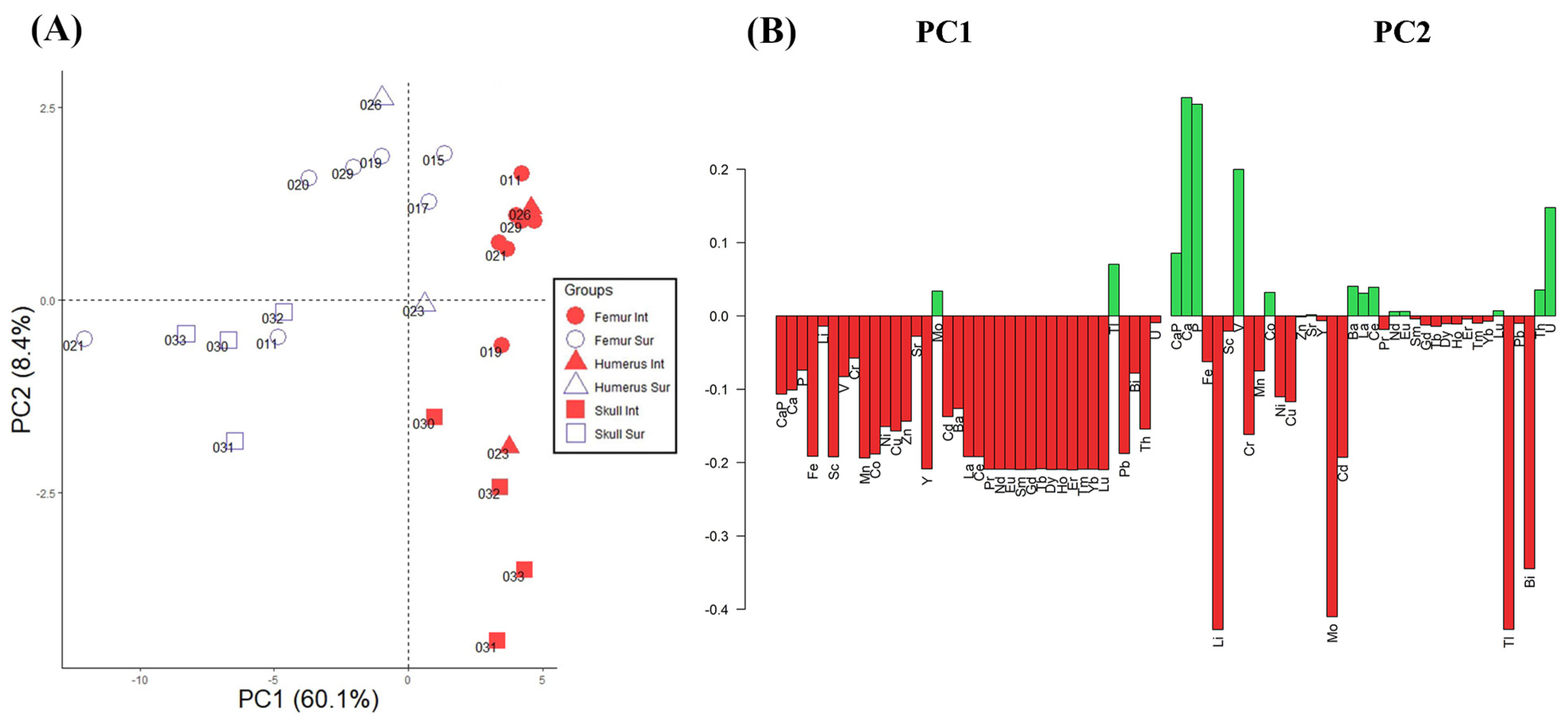
Principal component analysis employing all elements as variables was carried out to observe bone class variability (Figure 2). The first two principal components (PC1 60.1% and PC2 8.4%) contain the main part of the variance of the data with 68.5% total variance. From the score plot shown in Figure 2A, it can be appreciated that surface bone samples are clearly separated from the internal ones. In the PC1 negative direction, where the majority of the surface samples are located, elements such as REE, Fe, Sc, Mn, Co, Y, Ni, Cu, Pb, and Th are the most significant variables (Figure 2B) contributing to the distribution of the scores in the space, with higher concentrations of these elements found in the “Sur” class. However, just Mo and Tl are slightly contributing in the PC1 positive direction, where the “Int” class samples are located. On the other hand, in the PC2 negative direction, where the majority of the skull samples (“Int” and “Suf”) are located together with some femurs (021 Sur, 011 Sur, and 019 Int), metals such as Li, Cr, Mo, Cd, Tl, and Bi are the most important variables contributing to the distribution of the samples in the model. Nevertheless, in the PC2 positive direction, where the majority of the femurs (“Int” and “Suf”) together with the humerus 026 (“Int” and “Sur”) are located, elements such as Ca, P, V, and U are the most significant variables, together with the Ca/P ratio that shows a slightly minor loading value.
Overall, the main information related to diagenesis alteration distinguishing between the internal part and surface samples is shown in the PC1 direction and inside the bone classes. Differences in PC2 show that the majority of the femurs are associated with Ca and P and the structural proxy Ca/P ratio, suggesting a better preservation of these latter compared with other bone sections.
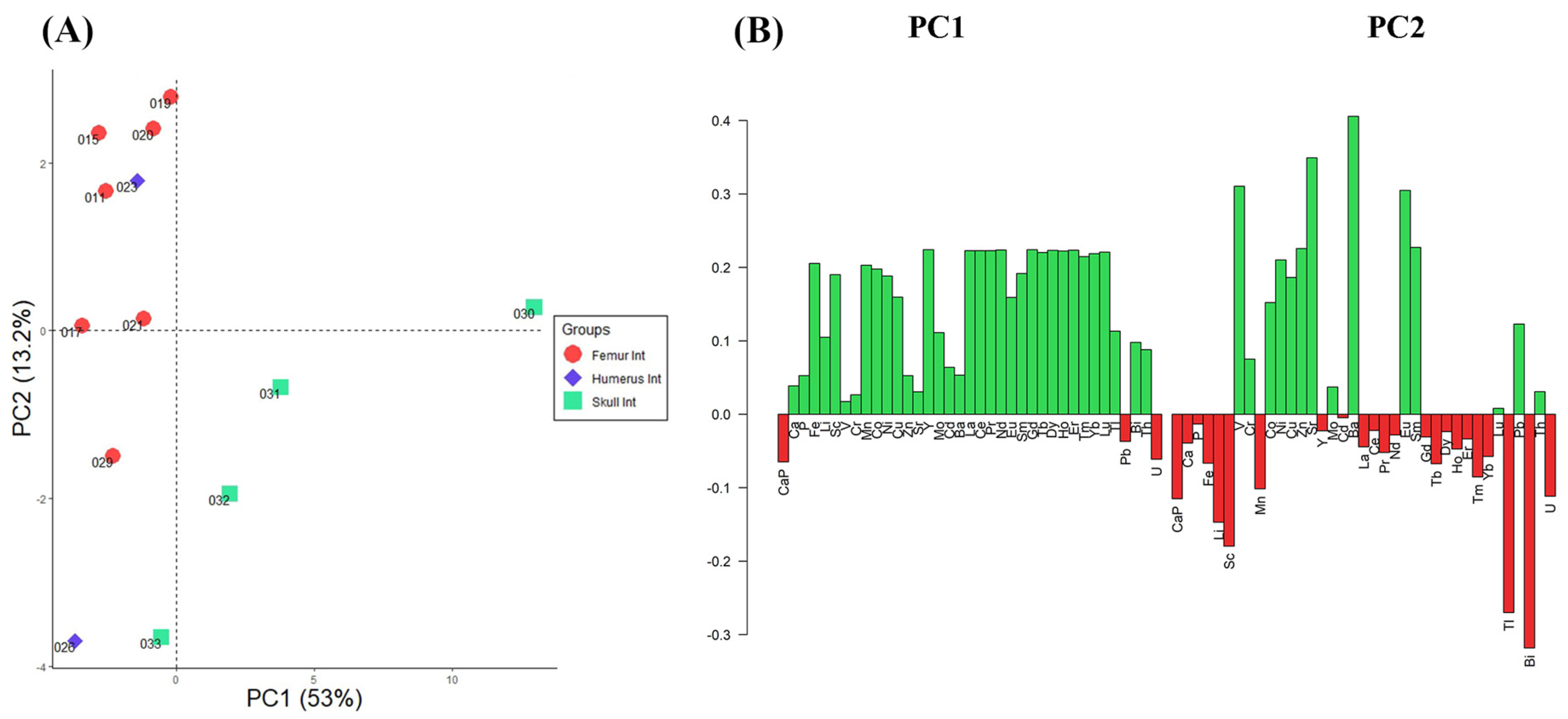
To further understand the bone diagenesis processes and select the best preserved samples, a PCA (Figure 3) was run just employing the internal part of the bones. The PCA model explains 66.2% of the total variance (Figure 3A), including PC1 (53%), and PC2 (13.2%) scores.
From the score plot shown in Figure 3A, it can be seen that the internal femurs and humerus samples are located in the PC1 negative direction, opposite to the skull samples in the PC1 positive direction. The Ca/P ratio and U are the most significant variables contributing in the PC1 negative direction, while elements such as Fe, Li, Sc, Mn, Co, Ni, Cu, Y, Mo, REE, and Tl are the most important in the PC1 positive direction (Figure 3B). In the PC2 negative direction, where the majority of the skull samples are located, elements such as Li, Sc, Tl, Bi, and U are the most important variables, while in the PC2 positive direction, where the majority of the femurs are located, elements such as V, Co, Ni, Cu, Zn, Sr, Ba, Eu, and Sm are the most significant variables.
Lastly, the results of the PCA (Figure 3) just including internal samples from different skeletal parts confirm that REE and heavy metals are especially affecting skulls, while femurs and the humerus mineral structure seem to be better preserved from exogenous chemical elements.

3.2. Testing a Paleodiet Reconstruction

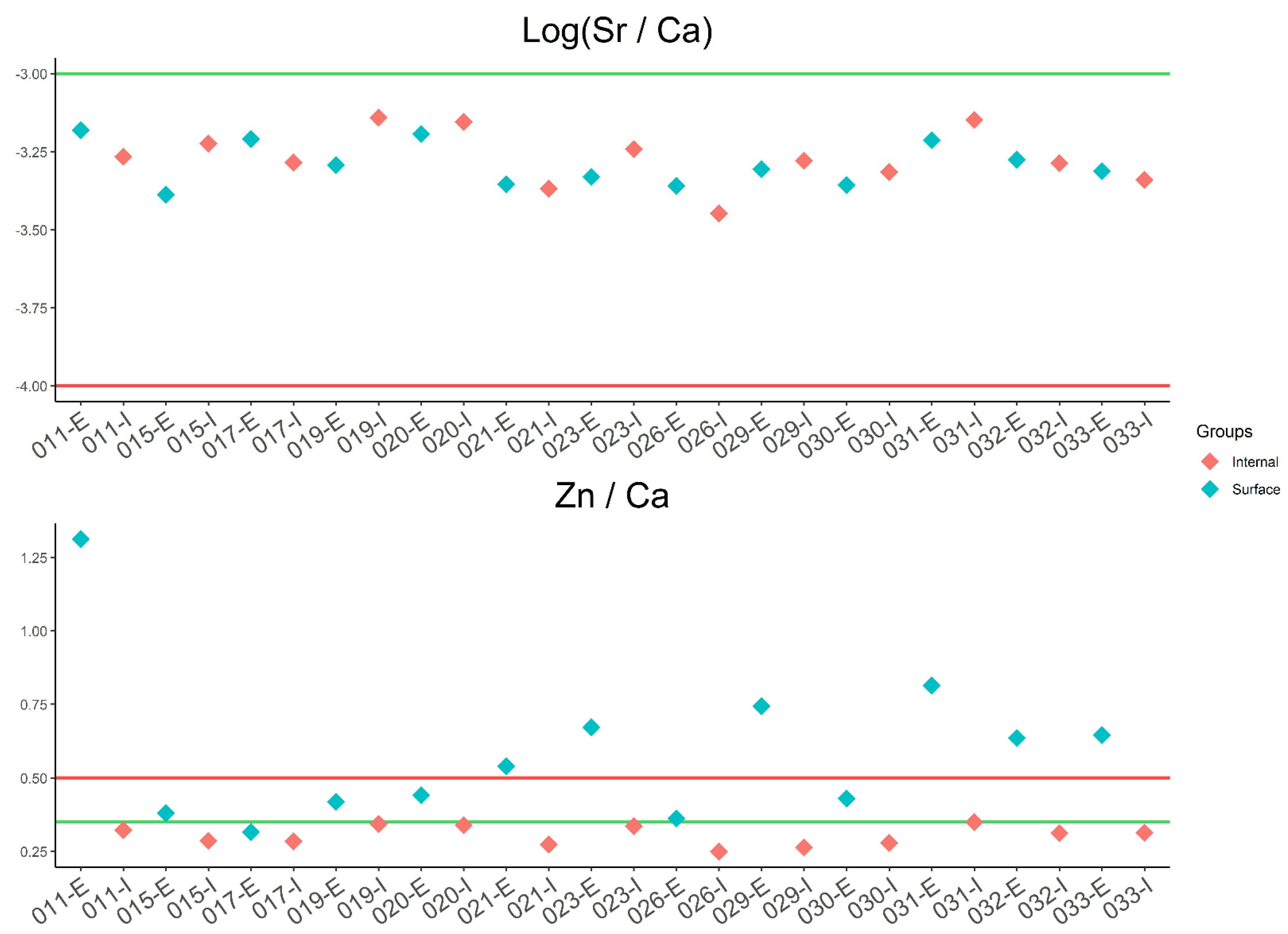
A diet reconstruction test was carried out using all the analyzed bones in order to observe changes related to the part (internal and surface) and type of bone sample employed (Figure 4). In the aforementioned literature, two ratios based on elements such as Ca, Zn, and Sr have been used in order to identify diet habits: the Zn/Ca ratio is used to identify higher or lower protein intake, and Sr/Ca is used to recognize pastoral or agricultural societies. Taking into account the log(Sr/Ca), all samples are classified as part of a mixed economy, with their values within the range −3 to −4. In the case of the Zn/Ca ratio, values for internal samples are characteristic of a poor protein-based diet, and it can be clearly observed that differences in Zn concentration between the surface and internal layers of bones alter the diet interpretation. Therefore, bone diagenetic processes can be identified in Zn/Ca ratio surface samples as incoherently associated with a rich protein diet.

4. Discussion

Many different mechanisms of diagenesis could eventually cause a change in the chemical composition of bone. Either through ionic exchange from the periosteal or endosteal surfaces, or via haversian channels or any kind of chemical or physical disturbance of the outer surface of bone, or even through microbial attack. As established by Rasmussen et al., 2019 [21], the diagenetic alterations start on the surface area of bone; our data are shown in Table 2. ANOVA and post hoc Tukey tests established the significant differences (p < 0.01) existing between the elemental concentrations on the surface and the internal layer of bones. As confirmed by PCA (Figure 2), surface (i.e., SHK 021-E) can be distinguished from internal (i.e., SHK 021-I) samples due to their relative higher concentration in REE and other heavy metals. Furthermore, internal samples present less variance than surface samples, suggesting a more homogeneous elemental concentration, and comparing the data collected from fresh bone with internal samples, very similar elemental values are obtained.
About bioapatite components, the amounts of calcium and phosphate are relatively stable within unchanged limits [37], so the Ca/P ratio can be used to control the integrity of the mineral component of bone. Although Ca/P is a proxy to verify the preservation of the bone, it does not exclude some kind of chemical alteration, as expected in the recrystallization of apatite and/or authigenic precipitation of phases with similar Ca/P ratios [38,39]. Nevertheless, taking into account the fresh bone, as calculated with data from Hancock et al., (1993) [24], which has a Ca/P ratio range of 2.06 to 2.45, all values of studied individuals would be included, as they range from 2.25 ± 0.04 in the inner to 2.29 ± 0.05 in the surface bone samples.
In this study, a methodological approach based on REE employed as a marker has been developed to obtain useful complementary information to evaluate bone diagenesis processes together with the Ca/P ratio. As aforementioned, it must be taken into account that REE are not easily incorporated into the organism by the gastrointestinal tract [33] and have total concentrations in living organisms lower than 1 μg/g [22,36], and this makes REE a useful marker to trace the diagenetic impact in bones. Bone surface results show that their elemental composition is more altered than the internal ones. The total amount of rare earth elements (ΣREE) for internal samples is always smaller than 1 μg/g, with the highest ΣREE concentrations found in skull internal samples SHK 030, SHK 032, and SHK 031. Nevertheless, in surface samples, ΣREE reaches values above 1 μg/g except for SHK 023 (humerus), SHK 017 (femur), and SHK 015 (femur). It can be concluded that, just taking into account internal skull samples, SHK 030, SHK 031, SHK 032, and SHK 033 present more altered chemical composition than femurs and the humerus. However, it was not possible to establish if the different states of conservation depend on external factors (i.e., body positions in burial that create specific microenvironmental conditions).
Post-depositional processes in bones can commonly take action from weathering, dissolution, precipitation, microbial attack, and ionic substitution. Many works carried out during the last decades have consistently investigated how diagenesis is affecting and limiting the reconstruction of past human activities and lifestyles.
Rare earth elements have been shown to be potential markers. Variation in REE composition in bones between and across depositional environments has been shown [40,41,42,43,44]. Our work is pushing forward the potential of REE, as their concentration is significantly different in buried bone. These elements are not common in living tissues and are easily incorporated by digenetic processes into bone post-mortem, maintaining a constant concentration after their initial incorporation into bone. Therefore, all bones exhumed in a common burial environment inherit a common REE signal, but depending on the type of bone, their concentration can be different, assuming that higher REE values in the surface and internal parts of bones are indicating a major environmental impact in the matrix and so more contamination. The REE composition of fossil and archaeological bones could therefore be used to investigate the quality of conservation of the matrix and evaluate its consistency for carrying out further bioarchaeological studies. Previous works have employed REE concentrations for burial associations in scattered bone remains by comparing the chemical composition of the bones [45]. However, preliminary works by Gallello and colleagues [8,27] analyzing bones from some late ancient Roman necropolises employing multivariate statistics showed that REE elements are capable of selecting bones that maintain a biological chemical elemental composition less affected by post-depositional processes. In the aforementioned studies, the bone chemical differences were not related to the position of the graves but to the bone types, and the geochemical profile in both excavation fields was homogeneous. Therefore, differences between femurs and ribs have shown that REE concentrations are higher in the last one. Consequently, the rib layer surfaces have suffered major enrichments of these elements because of the digenetic processes impact. The statistical results have put in evidence that diagenesis on the bone surface, depending on the skeletal sectors, has not had the same impact. These preliminary results have suggested that femurs and other long bones and skulls have suffered a less diagenetic impact than the ribs and bones of children.
The results obtained in this work are pushing forward these early studies and confirm that there is a greater enrichment of REE in the surface and internal part of skulls due to diagenesis compared to femurs or humeri. REE differences between femurs and skull bones could be explained by the variations in mineralogical density; where skulls are more susceptible to diagenetic processes, femurs, due to their denser mineralization, are less influenced by diagenetic changes.
The chemical analysis of REEs and statistical classification of bones exposed to different degrees of diagenesis have been shown to be a good tool, together with the analysis of bones belonging to different skeletal sectors to control for diagenetic factors in order to decide whether a sample is suitable for biological or paleodiet studies.
Furthermore, previous studies [27] testing paleodiet reconstruction showed differences in the diet profile of the same population depending on the class of sample (type of bone) employed. No sexual or social differences have been appreciated between the groups.
This study employing Strontium and Zn to test a diet reconstruction of the studied population confirms the conclusions of the preliminary works. It should be pointed out that Zn displays significant differences between internal and surface samples that may be due to diagenetic alteration, which is reflected in the paleodiet indexes based on the Zn/Ca ratios. Surface samples for the Zn/Ca ratio show higher values than those of internal samples, which would have biased the final diet interpretations. However, in the case of log(Sr/Ca), no diet interpretative differences between internal and surface parts or kinds of bones were identified.
Finally, the paleodiet test in the studied population based on log(Sr/Ca) and Zn/Ca ratios shows a mix of vegetables and low protein intake. This is in accordance with the diet of lower socio-economic classes during Medieval times in the studied region (Adriatic Coast), which was mostly based in carbohydrate intakes (bread consumption represented almost 70% of the diet) with lower intakes of proteins (fresh meat, fish, and dairy products) [46,47]. There is not clear information about the studied Scodra inhabitants’ diet during the XIII or XIV century; however, following the available data, the subsistence strategy should be in line with the population living in a Mediterranean climate, based on agriculture, farming, and fishing. It may be this population was following a diet already marked by the influence of the past Roman Empire with viticulture, cereals, vegetables, some fruit, and olives. Periodical fishing and animal farming may also have been an additional source of protein intake. The nutritional patterns in Scodra should be similar to those of the medieval populations in the Dalmatia region and comparable to previously studied Croatian populations of similar chronologies from coastal communities recognized as commercial centers, including fishing [47].
The results obtained show that the development of a proper methodological approach for diagenesis control can be a reliable tool to consistently interpret dietary habits and, in general, the lifestyle of ancient populations.
Consequently, for biochemical–archaeological studies, trace elements and REE data processing to statistically classify archaeological bones could help to select a better preserved sample. Diagenetic factors caused by the environment may be controlled by analyzing the surface of the bone employing statistical tools. Finally, a protocol for better preserved bone sample selection to prevent misleading archaeological interpretations has been proposed.

5. Conclusions

For the first time, a methodological approach based on REE to control bone diagenesis processes was developed and tested in a selected bone collection from 13 Late Medieval period skeletons buried in Rozafa Castle (Shkodër, Albania). The proposed approach behind this research consisted in obtaining a surface sample by mechanical abrasion and another from the internal parts of femurs, humeri, and skulls, measuring major, minor, and trace elements, including rare earth elements, and statistically processing the data obtained. Then, statistical data analysis allowed to comprehend the relations between the different elemental compositions and verified the efficiency of the sampling and analytical method applied in order to assess the preservation status of the studied set of samples. In general, the internal parts of samples are better preserved than the surface, and femurs are considered less contaminated than skulls. To test the reliability of the proposed approach, a paleodiet reconstruction was carried out using Zn/Ca and log(Sr/Ca) ratios. The data are in line with the literature, as the diet of the studied individuals was low in protein and their society was based on a mixed economy, mainly supported by agricultural activities. The study shows that paleodiet reconstruction based on skeletal remains elemental analyses can be affected by diagenetic alterations. Finally, an approach was proposed to control post-depositional alterations on archaeological bones through a multi-elemental characterization in order to avoid misleading interpretations of the data.

Supplementary Materials

The following supporting information can be downloaded at: https://www.mdpi.com/article/10.3390/heritage7100273/s1, Table S1: Composition values for all major and minor elements analyzed for every individual. Ca, P, and Fe are expressed in mg/g; the other elements are expressed in μg/g, and U and Th are expressed in μg/kg. Modern fresh bone values are compared with the archaeological bones; Table S2: Composition values for all REE, Sc, and Y elements analyzed for every individual. All elements are expressed in μg/kg. Modern fresh bone values are compared with the archaeological bones.

Author Contributions

D.R.N., G.G. and J.R.: conceptualization, methodology, investigation, resources, data curation, formal analysis, supervision, project administration, visualization, writing—original draft preparation, writing—review and editing, funding acquisition. G.P., M.L.C. and A.P.: Investigation, validation, visualization, writing—review and editing. All authors have read and agreed to the published version of the manuscript.

Funding

Project BEAGAL18/00110, “Development of analytical methods applied to archaeology”, funded by the Spanish Ministry of Science and Innovation and the Ministry of Universities.

Data Availability Statement

All the produced data are available in the manuscript and Supplementary Materials.

Acknowledgments

Gianni Gallello acknowledges the financial support of the Beatriz Galindo Fellowship (2018) funded by the Spanish Ministry of Science and Innovation and the Ministry of Universities (BEAGAL18/00110), the Spanish Ministry of Science and Innovation, for funding the project EvolMED, “Evolutionary cultural patterns in the contexts of the neolithization process in the Western Mediterranean” (PID2021-127731NB-C21). The authors are grateful to the reviewers for their suggestions, comments, and feedback that have consistently enhanced the quality of the manuscript.

Conflicts of Interest

On behalf of all authors, Gianni Gallello, as corresponding author, states that there are no conflicts of interest.

References

  1. Bala, Y.; Farlay, D.; Boivin, G. Bone mineralization: From tissue to crystal in normal and pathological contexts. Osteoporos. Int. 2012, 24, 2153–2166. [Google Scholar] [CrossRef]
  2. Keenan, S.W.; Engel, A.S. Early diagénesis and recrystallization of bone. Geochim. Cosmochim. Acta 2017, 196, 209–223. [Google Scholar] [CrossRef]
  3. Vidaud, C.; Bourgeois, D.; Meyer, D. Bone as target organ for metals: The case of f-elements. Chem. Res. Toxicol. 2012, 25, 1161–1175. [Google Scholar] [CrossRef]
  4. Dyczek, P.; Shpuza, S. From Antiquity to Modernity, 1st ed.; University of Warsaw: Warsaw, Poland, 2020; pp. 7–401. [Google Scholar]
  5. Alapont, L.; Gallello, G.; Martinón-Torres, M.; Osanna, M.; Amoretti, V.; Chenery, S.; Ramacciotti, M.; Jiménez, J.L.; Rubio, Á.M.; Cervera, M.L.; et al. The casts of Pompeii: Post-depositional methodological insights. PLoS ONE 2023, 18, e0289378. [Google Scholar] [CrossRef]
  6. Toots, H.; Voorhies, M.R. Strontium in Fossil Bones and the Reconstruction of Food Chains. Science 1965, 149, 854–855. [Google Scholar] [CrossRef] [PubMed]
  7. Sillen, A.; Kavanagh, M. Strontium and paleodietary research: A review. Am. J. Phys. Anthropol. 1982, 25, 67–90. [Google Scholar] [CrossRef]
  8. Gallello, G.; Kuligowski, J.; Pastor, A.; Diez, A.; Bernabeu, J. Chemical element levels as a methodological tool in Forensic Science. J. Forensic Res. 2015, 6, 1. [Google Scholar] [CrossRef]
  9. Winter, N.J.; Snoeck, C.; Claeys, P. Seasonal cyclicity in trace elements and stable isotopes of modern horse enamel. PLoS ONE 2016, 11, e0166678. [Google Scholar] [CrossRef]
  10. Gallello, G.; Cilli, E.; Bartoli, F.; Andretta, M.; Calcagnile, L.; Pastor, A.; de la Guardia, M.; Serventi, P.; Marino, A.; Benazzi, S.; et al. Poisoning histories in the Italian renaissance: The case of Pico Della Mirandola and Angelo Poliziano. J. Forensic Leg. Med. 2018, 56, 83–89. [Google Scholar] [CrossRef]
  11. López-Costas, O.; Lantes-Suarez, O.; Cortizas, M.A. Chemical compositional changes in archaeological human bones due to diagenesis: Type of bone vs soil environment. J. Archaeol. Sci. 2016, 67, 43–51. [Google Scholar] [CrossRef]
  12. Rasmussen, K.L.; Boldsen, J.L.; Kristensen, H.K.; Skytte, L.; Hansen, K.L.; Mølholm, L.; Grootes, P.M.; Nadeau, M.J.; Eriksen, K.M.F. Mercury levels in Danish medieval human bones. J. Archaeol. Sci. 2008, 35, 2295–2306. [Google Scholar] [CrossRef]
  13. Scharlotta, I.; Goriunova, O.I.; Weber, A. Micro-sampling of human bones for mobility studies: Diagenetic impacts and potentials for elemental and isotopic research. J. Archaeol. Sci. 2013, 40, 4509–4527. [Google Scholar] [CrossRef]
  14. Balter, V.; Braga, J.; Télouk, P.; Thackeray, J.F. Evidence for dietary change but not landscape use in South African early hominins. Nature 2012, 489, 558–560. [Google Scholar] [CrossRef]
  15. Gallello, G. Western Mediterranean Archaeology: Chemical Elements Levels in Archaeology Materials as a Methodological Tool. Ph.D. Dissertation, Universidad de Valencia, Valencia, Spain, 2014. [Google Scholar]
  16. Suarez, C.A.; Suarez, M.B.; Terry, D.O.; Grandstaff, J.R.; Grandstaff, D.E. Rare earth element geochemistry and taphonomy of terrestrial vertebrate assemblages. Palaios 2007, 22, 500–512. [Google Scholar] [CrossRef]
  17. Trueman, C.N.; Behrensmeyer, A.K.; Tuross, N.; Weiner, S. Mineralogical and compositional changes in bones exposed on soil surfaces in Amboseli National Park, Kenya: Diagenetic mechanisms and the role of sediment pore fluids. J. Archaeol. Sci. 2004, 31, 721–739. [Google Scholar] [CrossRef]
  18. Karaiskaj, G. Die Spätantiken und Mittelalterlichen Wehranlagen in Albanien; Verlag Dr. Kovac: Hamburg, Germany, 2010; pp. 9–249. [Google Scholar]
  19. Lemke, M. Fieldwork at Scodra 2013. Światowit XI (LII)/A; University of Warsaw: Warsaw, Poland, 2014; pp. 189–196. [Google Scholar]
  20. Dyczek, P. Terra Incognita: Results of Polish Excavations In Albania And Montenegro. Stud. Eur. Gnes. 2017, 16, 351–369. [Google Scholar] [CrossRef]
  21. Rasmussen, K.L.; Milner, G.R.; Skytte, L.; Lynnerup, N.; Lange Thomsen, J.; Lier Boldsen, J. Mapping diagenesis in archaeological human bones. Herit. Sci. 2019, 7, 41. [Google Scholar] [CrossRef]
  22. Harkness, S.H.; Darrah, T.H. From the crust to the cortical: The geochemistry of trace elements in human bone. Geochim. Cosmochim. Acta 2019, 249, 76–94. [Google Scholar] [CrossRef]
  23. Zapata, J.; Pérez-Sirvent, C.; Martínez-Sánchez, M.J.; Tovar, P. Diagenesis, not biogénesis: Two late Roman skeletal examples. Sci. Total Environ. 2006, 369, 357–368. [Google Scholar] [CrossRef]
  24. Hancock, R.G.V.; Grynpas, M.D.; Åkesson, K.; Obrant, J.T.; Kessler, M.J. Baselines and variabilities for major and trace elements in bone. In Prehistoric Human Bone: Archaeology at the Molecular Level; Lambert, J.B., Grupe, G., Eds.; Springer: Berlin/Heidelberg, Germany, 1993; pp. 189–201. [Google Scholar] [CrossRef]
  25. Gallello, G.; Kuligowski, J.; Pastor, A.; Diez, A.; Bernabeu, J. Biological mineral content in Iberian skeletal cremains for control of diagenetic factors employing multivariate statistics. J. Archaeol. Sci. 2013, 40, 2477–2484. [Google Scholar] [CrossRef]
  26. Reymann, C.R.; Filzmoser, P.; Garrett, G.; Dutter, R. Statistical Data Analysis Explained: Applied Environmental Statistics; John Wiley & Sons: Hoboken, NJ, USA, 2008; ISBN 978-0-470-98581-6. [Google Scholar]
  27. Gallello, G.; Scopa, S.; Kuligowski, J.; Bartoli, F.; Mallegni, F.; Pastor, A. Variación química intraesquelética relacionada con la diagenesis en los restos óseos de C/en Gil (Valencia). Saguntum 2015, 47, 175–186. [Google Scholar] [CrossRef]
  28. R Core Team. R: A Language and Environment for Statistical Computing; R Foundation for Statistical Computing: Vienna, Austria, 2020; Available online: https://www.R-project.org/ (accessed on 28 November 2022).
  29. Rasmussen, K.L.; Milner, G.R.; Delbey, T.; Skytte, L.; Lynnerup, N.; Thomsen, J.L.; Schiavone, S.; Torino, M.; Larsen, L.A.; Boldsen, J.L. Trace element distribution in human cortical bone microstructure: The potential for unravelling diet social status in archaeological bones. Herit. Sci. 2020, 8, 111. [Google Scholar] [CrossRef]
  30. Scharlotta, I.; Bazaliiskii, V.I.; Kusaka, S.; Weber, A. Diet or mobility? Multi-isotopic (C, N, and Sr) dietary modeling at Shamanka II, Cis-Baikal, Siberia. Archaeol. Res. Asia 2022, 29, 100340. [Google Scholar] [CrossRef]
  31. Lambert, J.B.; Vlasak, S.M.; Thometz, A.C.; Buikstra, J.E. A comparative study of the chemical analysis of ribs and femurs in Woodland population. Am. J. Phys. Anthropol. 1982, 59, 289–294. [Google Scholar] [CrossRef]
  32. Comar, C.L.; Russell, R.S.; Wasserman, R.H. Strontium-calcium movement from soil to man. Science 1957, 126, 485–492. [Google Scholar] [CrossRef]
  33. Reynard, B.; Balter, V. Trace elements and their isotopes in bones and teeth: Diet, environments, diagenesis, and dating of archaeological and paleontological samples. Palaeogeogr. Palaeoclimatol. Palaeoecol. 2014, 416, 4–16. [Google Scholar] [CrossRef]
  34. Burton, J.; Wright, L. Nonlinearity in the relationship between bone Sr/Ca and diet: Paleodietary implications. Am. J. Phys. Anthropol. 1995, 96, 273–282. [Google Scholar] [CrossRef]
  35. Lönnerdal, B. Dietary factors influencing Zinc absorption. J. Nutr. 2000, 130, 1378–1383. [Google Scholar] [CrossRef]
  36. Kohn, M.J.; Morris, J.; Olin, P. Trace element concentrations in teeth—A modern Idaho baseline with implications for archaeometry, forensics, and palaeontology. J. Archaeol. Sci. 2013, 40, 1689–1699. [Google Scholar] [CrossRef]
  37. Burton, J. Bone chemistry and trace element analysis. In Biological Anthropology of the Human Skeleton, 2nd ed.; Katzenberg, M.A., Shelley, S.R., Eds.; John Wiley & Sons. Inc.: Hoboken, NJ, USA, 2008; Chapter 14. [Google Scholar] [CrossRef]
  38. Hedges, R.E.M.; Millard, A.R.; Pike, A.W.G. Measurements and relationships of diagenetic alteration of bone from three archaeological sites. J. Archaeol. Sci. 1995, 22, 201–209. [Google Scholar] [CrossRef]
  39. Fabig, A.; Herrmann, B. Trace elements in buried human bones: Intra-population variability of Sr/Ca and Ba/Ca ratios—Diet or diagenesis? Naturwissenschaften 2002, 89, 115–119. [Google Scholar] [CrossRef]
  40. Trueman, C.N.; Benton, M.J. A geochemical method to trace the taphonomic history of reworked bones in sedimentary settings. Geology 1997, 25, 263–266. [Google Scholar] [CrossRef]
  41. Trueman, C.N.G.; Behrensmeyer, A.K.; Potts, R.; Tuross, N. High-resolution records of location and stratigraphic provenance from the rare earth element composition of fossil bones. Geochim. Cosmochim. Acta GCA 2006, 70, 4343–4355. [Google Scholar] [CrossRef]
  42. Trueman, C.N.; Palmer, M.R.; Field, J.; Privat, K.; Ludgate, N.; Chavagnac, V.; Eberth, D.A.; Cifelli, R.; Rogers, R.R. Comparing rates of recrystallisation and the potential for preservation of biomolecules from the distribution of trace elements in fossil bones. Comptes Rendus Palevol 2008, 7, 145–158. [Google Scholar] [CrossRef]
  43. Wang, S.; Huang, T.D.; Yan, R.; Chen, H.; Zhang, S.; Li, X.; Kótai, L.; Reisz, R.R. Rare Earth Elements in Dinosaur Bones Across the Embryo-Adult Spectrum. Front. Earth Sci. 2020, 8, 230. [Google Scholar] [CrossRef]
  44. Williams, C.T.; Henderson, P.; Marlow, C.A.; Molleson, T.I. The environment of deposition indicated by the distribution of rare earth elements in fossil bones from Olduvai Gorge, Tanzania. Appl. Geochem. 1997, 12, 537–547. [Google Scholar] [CrossRef]
  45. Trueman, C.N. Forensic geology of bone mineral: Geochemical tracers for post-mortem movement of bone remains. In Forensic Geosciences, Principles, Techniques and Applications; Pye, K., Croft, D.J., Eds.; The Geological Society of London: London, UK, 2004; pp. 249–256. [Google Scholar]
  46. Mazzi, M.S. Consumi Alimentari e Malattie nel Basso Medioevo. In Archeologia Medievale: Cultura Materiale, Insediamenti, Territorio: VIII; All’insegna del Giglio: Firenze, Italy, 1981; Volume 8, pp. 321–336. Available online: http://digital.casalini.it/10.1400/245696 (accessed on 9 October 2023).
  47. Slaus, M.; Bedíc, Z.; Sikanjic, P.R.; Vodanovic, M.; Kunic, A.D. Dental health at the transition from the late antique to the early medieval period on Croatia’s eastern Adriatic coast. Int. J. Osteoarchaeol. 2010, 21, 577–590. [Google Scholar] [CrossRef]
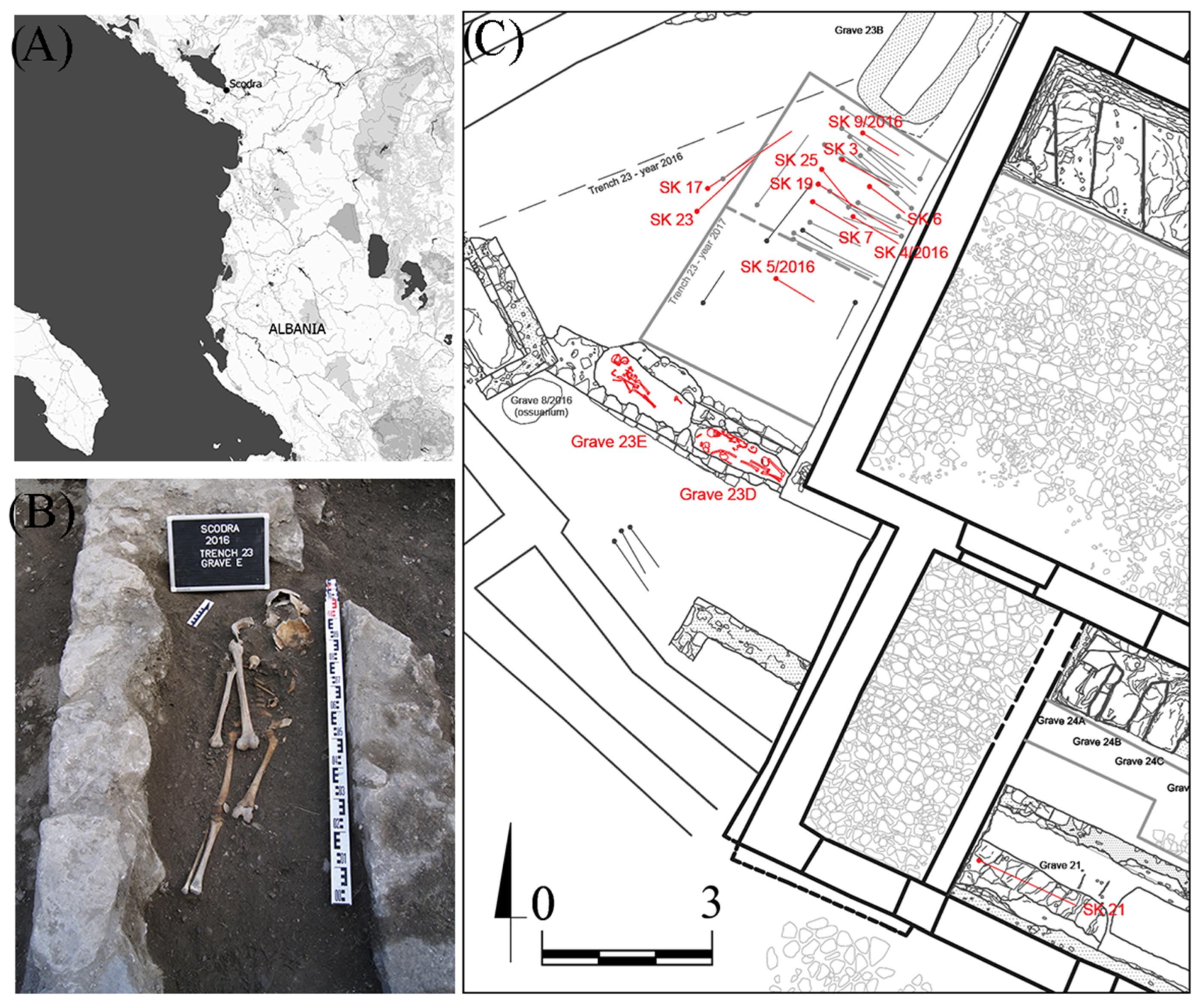
Figure 1. Archaeological site of Rozafa Castle; (A) location of Scodra; (B) image of the “grave 23 E”; (C) mass grave located in trench 23 and grave 21. Studied skeletons in red. (pictures provided by J. Żabko-Potopowicz).
Heritage 07 00273 g001
Figure 2. PCA results of internal (Int) and surface (Sur) bone samples. Scores (A) and loadings (B) for the calculated PCA model representing PCs 1 and 2.
Heritage 07 00273 g002
Figure 3. PCA results of the internal bone samples (Int). Scores (A) and loadings (B) for the calculated PCA model representing PCs 1 and 2.
Heritage 07 00273 g003
Figure 4. Paleodiet ratio results: log(Sr/Ca) and Zn/Ca. The lines represent the limits for the categories. Log(Sr/Ca) ratio values ≥ −3 (green line) is an agricultural economy (vegetable), and values ≤ −4 (red line) is a pastoral economy (milk and meat). A Zn/Ca ratio value ≥ 0.5 (red line) indicates a rich protein diet, and values ≤ 0.35 indicate poor protein intake (green line).
Heritage 07 00273 g004
Table 1. Anthropological information of the studied individuals as well as sample information: laboratory label “Lab ID”; sampled bone “Bone”; sampled part “Type”; and sample ID are reported.
Lab IDGraveSexAgeBoneTypeSample IDBoneTypeSample ID
SHK 033SK4/2016UnknownAdultSkullInternal033-ISkullSurface033-S
SHK 032SK5/2016Female25–29SkullInternal032-ISkullSurface032-S
SHK031SK9/2016Male18–25SkullInternal031-ISkullSurface031-S
SHK 030SK17Unknown18–21SkullInternal030-ISkullSurface030-S
SHK 029SK19Female50+FemurInternal029-IFemurSurface029-S
SHK 026SK21Immature10–12HumerusInternal026-IHumerusSurface026-S
SHK 023SK21UnknownAdultHumerusInternal023-IHumerusSurface023-S
SHK 021SK23Male30–39FemurInternal021-IFemurSurface021-S
SHK 020SK23DMaleAdult (30–39)FemurInternal020-IFemurSurface020-S
SHK 019SK23EUnknownAdultFemurInternal019-IFemurSurface019-S
SHK 017SK3Male20–29FemurInternal017-IFemurSurface017-S
SHK 015SK6Female20–25FemurInternal015-IFemurSurface015-S
SHK 011SK7Male20–29FemurInternal011-IFemurSurface011-S
Table 2. Mean values (±st.dv) of major, trace elements, and REE for all studied samples. Ca, P, and Fe are expressed in mg/g, trace elements, and REE in μg/g. The letters (a, b) indicate the homogeneous groups resulted from post hoc Tukey test (p < 0.01) applied over one-way ANOVA results: (a) minor values and (b) higher values. Values without letters were not significantly distinguishable from groups a or b.
REEInternalSurfaceMajors and TraceInternalSurfaceMajors and TraceInternalSurface
La26.6 ± 32.9 a442 ± 289 bCa/P2.25 ± 0.04 a2.29 ± 0.05 bBi0.02 ± 0.010.02 ± 0.01
Ce49.4 ± 54.4 a607 ± 355 bCa359 ± 26.5 a399 ± 16.9 bCd0.22 ± 0.140.54 ± 0.43
Pr6.09 ± 7.51 a79.5 ± 48.2 bP160 ± 11.9 a174 ± 9.04 bBa48.6 ± 11.2 a60.9 ± 11.4 b
Nd30.2 ± 37.9 a404 ± 222 bFe0.48 ± 0.10 a1.05 ± 0.43 bTl0.05 ± 0.03 b0.02 ± 0.01 a
Eu8.46 ± 2.52 a29.9 ± 12.8 bLi1.24 ± 0.531.21 ± 0.32Pb3.42 ± 2.07 a15.0 ± 9.30 b
Sm29.3 ± 9.14 a107± 48.5 bTh0.09 ± 0.02 a 0.16 ± 0.05 b
Gd7.60 ± 8.30 a94.8 ± 55.8 bV39.8 ± 5.66 a62.9 ± 24.5 b
Tb0.92 ± 1.18 a13.5 ± 8.16 bCr13.9 ± 3.53 16.4 ± 4.17
Dy5.45 ± 6.83 a78.4 ± 45.4 bMn6.28 ± 8.08 a45.6 ± 33.7 b
Ho1.04 ± 1.30 a15.8 ± 9.27 bCo0.90 ± 0.17 a1.39 ± 0.28 b
Er2.81 ± 3.24 a43.1 ± 24.9 bNi12.9 ± 3.76 a17.3 ± 3.60 b
Tm0.50 ± 0.45 a5.86 ± 3.45 bCu14.7 ± 8.88 22.9 ± 10.8
Yb2.81 ± 2.32 a34.9 ± 20.2 bZn109 ± 9.53 a234 ± 92.9 b
Lu0.49 ± 0.35 a5.43 ± 2.93 bSr196 ± 32.2207 ± 31.6
Sc409 ± 36.2 a557 ± 115 bU0.73 ± 0.650.82 ± 0.61
Y46.3 ± 43.3 a670 ± 401 bMo1.52 ± 0.691.21 ± 0.51
Disclaimer/Publisher’s Note: The statements, opinions and data contained in all publications are solely those of the individual author(s) and contributor(s) and not of MDPI and/or the editor(s). MDPI and/or the editor(s) disclaim responsibility for any injury to people or property resulting from any ideas, methods, instructions or products referred to in the content.