Abstract
In this work, a painted gypsum bas-relief from the facades of the inner courtyard of the St. Petersburg Academy of Arts building was examined using UV and visible light photography and optical and electron scanning microscopy, which showed the heterogeneous layers of white painting on the surface of the bas-relief that covered the historical ones. These undesirable layers should be removed during the restoration work, but it was found that the traditional method of removing surface layers of painting with the help of chemical solvents and mechanical cleaning does not solve the problem to the full extent. A cross-section of all the painting layers was prepared to investigate the stratigraphy of the paint layers. These studies were conducted using optical and electron scanning microscopy in order to determine the structure of the paint layers more properly and study the chemical composition of every layer. After this study, a complex cleaning technique was developed. This technique combines chemical and laser cleaning, making it possible to effectively remove the upper dense layers of paint without damaging the historical paint layers.
1. Introduction
The construction of the main building of the St. Petersburg Academy of Arts was completed in 1788. A distinctive feature of this architectural ensemble is the central circular courtyard. According to a legend, by decree of Empress Catherine II, it was requested to be made in the same size as the dome of St. Peter’s Basilica in Rome. Designed in the classical style, the so-called “compass” is decorated with 24 metopes (bas-reliefs) located above the windows of the second floor.
In the process of the most recent rehabilitation of the courtyard facades, some of the original bas-reliefs were dismantled and replaced with copies. One critically dilapidated fragment of a removed metope was admitted for study and conservation (Figure 1).
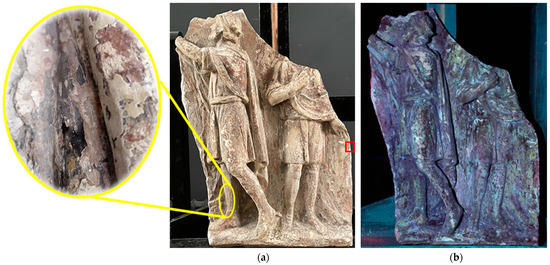
Figure 1.
General view of the bas-relief: (a) in visible light; (b) UV photography. Red square marks the area where the probe for cross-section was taken. Yellow sign shows the detailed picture of the marked area.
There were multiple losses of the gypsum and the paint layer was heterogeneous, with accumulated dirt (shown in a detailed picture, Figure 1). The bonding of the lower paint layer with the surface was weak. The upper covering layers formed a dense, hard crust. Over the course of its existence, the bas-relief has been repainted multiple times; however, specifying the exact number of over-painted layers and dating them simply through visual observation proved to be difficult. It was decided that samples of paint layers would be taken for detailed analysis and stratigraphy studies. A series of optoelectronic tests would also be performed, after which the elemental composition of the layers would be determined, followed by the removal of late over-painting from the surface of the bas-relief.
To remove dense surface layers, a traditional restoration cleaning technique with chemically active solvents in compress was first applied to this art object. However, it proved to be ineffective. The chemical solution, as a result of the long exposure time, not only softened the hard surface layers but could also penetrate into the gypsum base in some places in the bas-relief surface. In this case, the complete loss of red paint layers occurred. This observation prompted the research of another restoration method, such as laser cleaning.
Nowadays, laser cleaning is applied widely enough in the restoration of cultural heritages [1]. It is used on a routine basis in many museums around the world for the removal of contaminants of natural and anthropogenic origin from the surfaces of cultural heritage objects made of various materials (stone, metal, ceramics, fabric, paper, etc.) [1,2]. It was used in the restoration of a number of world-famous monuments, including the friezes of the ancient Greek temple of the Parthenon and monuments from the Acropolis of Athens in Greece [3] as well as a masterpiece of the Renaissance—the Golden Gate of the Baptistery of the Cathedral of Cathedral of Saint Mary of the Flower in Florence (Italy) [4]. At the moment, the state of the art of dissemination of laser cleaning in the conservation of cultural heritages is continuously growing in Europe, which is demonstrated by many recent publications [5,6,7,8,9,10]. Laser cleaning is a contactless, chemically pure, and selective method which offers many advantages compared to traditional cleaning techniques—for instance, in cases when chemically resistant layers must be removed from fragile surfaces.
Presently, a pulsed solid-state Nd:YAG laser with a wavelength of 1064 nm is the most commonly applied one for the task of cultural heritage conservation [1,11]. It is normally used to clean stone surfaces and metal objects. However, there are cases in which it has been used for the restoration of wall paintings and stone surfaces with decorative paint layers [12], which makes this type of laser potentially useful for our task. Moreover, black gypsum crusts, which develop on statues and buildings in open air [13], have high absorption of Nd:YAG laser radiation; this also makes this laser a universally applicable tool for cleaning carbonate rock artifacts from surface soiling.
The bas-relief studied in this work is a complex, multi-layered object whose surface includes not only contaminations but also dense heterogeneous paint layers resistant to chemical solvents as well as, presumably, gypsum crusts. The aim of this article was to conduct a complex structure investigation of this object and study the chemical composition of the painting layers for developing a restoration technique including modern methods of diagnostics, such as optical and electron scanning microscopy, EDX analysis, and UV and visible light photography, combined with cleaning methodology using laser and chemical treatment in combination with the mechanical cleaning.
2. Materials and Methods
The gypsum bas-relief was examined using infrared (980–1100 nm), ultraviolet (365 nm), and visible spectrum photofixation methods using optical filters and a Canon EOS 600D camera (sensor type: 22.3 × 14.9 mm CMOS, 18.7 megapixels).
A painting cross-section sample (perpendicular cut of all paint layers without a gypsum base) was prepared and polished by the drop method [14]. Since the surface of the painted bas-relief had many losses and the paint layers were not uniform, it was decided to take a sample in an area located close to the edges of the bas-relief, retaining the maximum number of layers that were easiest to distinguish during UV shooting (Figure 1b). The cross-section was studied using a DigiMicro Prof 300× digital USB microscope (DigiMicro, Germany/China) in direct non-polarized light of a wide spectrum and in ultraviolet radiation with a wavelength of 365 nm.
The cross-section was also analyzed on a Vega3 SBH scanning electron microscope (TESCAN, Brno, Czech Republic) (resolution 3 nm, accelerating voltage up to 30 kV, magnification from 4.5 to 1,000,000) to determine the elemental composition of each layer. The data on the chemical composition of each layer were obtained, and a map of element distribution was compiled.
Black crusts from the bas-relief surface were analyzed with a LOMO METAM LV-41 metallographic microscope (LOMO, St. Petersburg, Russia) at 50–1500× magnification with an LED backlight.
The cleaning of the bas-relief from late white layers using a traditional restoration technique for cleaning gypsum was carried out using a polar 10% solvent of dimethyl sulfoxide and dimethylformamide in a compress. The exposure time was from 10 to 60 min. Laser cleaning was performed using a pulsed Nd:YAG laser with a pulse duration of 100 µs, a wavelength of 1064 nm, and a maximum radiation energy of 2 J. The energy density varied in the range from 4 to 15 J/cm2, and the pulse repetition rate was 1 Hz.
3. Results
3.1. Photofixation and Stratigraphic Studies
Photofixation of the bas-relief was carried out in several wavelength ranges. Photography in the IR range did not bring appreciable results. When photographing a fragment of the metope in long-wave ultraviolet radiation (365 nm), it was possible to register the visible luminescence of separate paint layers (Figure 1b).
Photofixation in UV light showed the presence of heterogeneous paint layers on the surface of the bas-relief. A cross-section from the surface of the bas-relief (marked by the red area in Figure 1a) was taken and examined using optical microscopy (Figure 2).
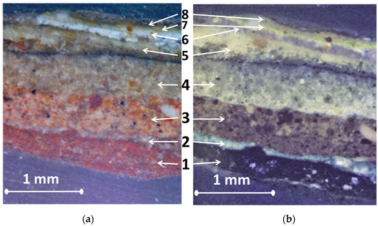
Figure 2.
Cross-section of the metope fragment: (a) in visible light; (b) in UV light.
Optical microscopy of the prepared cross-section sample showed the presence of eight layers. Layers 1 (about 500 µm thickness) and 3 (about 400 µm thickness) are red-colored materials. These layers are supposed to be historical ones that should be saved. Figure 2b shows light color luminescence in the area between these two layers. It means that this thin layer (about 100 µm thickness) is different from the chemical composition of layers 1 and 3. Layers 4 (about 500 µm thickness) and 5 (about 300 µm thickness) have similar luminescence (Figure 2b) but different particle sizes. Layer 4 contains bigger impurities than layer 5. Between these layers, one can notice a thin black line, which is supposed to be a black crust.
Moreover, the presence of three layers (layers 6, 7, and 8 (about 200 µm thickness)) of white paint on the surface of the bas-relief can be noticed (Figure 2a). The UV image (Figure 2b) also showed different luminescence in the layers of white paint, which provided the prerequisite for studying the elemental composition of the cross-section, which was carried out using a scanning electron microscope.
Data obtained from the scanning electron microscope are presented in Figure 3 and Figure 4 below. Information about the elemental distribution on the surface of the cross-section was obtained in high vacuum mode (<1 × 10⁻² Pa) with an accelerating voltage of 20 kV.
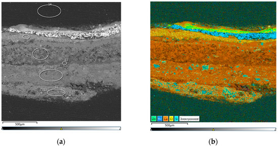
Figure 3.
General view of cross-section: (a) electronic image in BSE mode; (b) mapping of the cross-section surface.
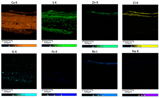
Figure 4.
Distribution of elements in cross-section.
The mapping of the sample showed the presence of layers of white shading containing a large amount of barium and zinc. The two layers of red paint contained large inclusions of silicon and iron (Figure 3 and Figure 4).
The elemental distribution in each layer clearly shows the presence of sulfur and chlorine impurities on the surface layers, as well as an abundance of calcium on almost the entire surface of the cross-section (Figure 4).
After scanning, it became possible to correlate the obtained data on the nature of the materials with historical research and suggest in what period each layer of paint was applied. The results of the analysis are presented in Table 1, where all the layers are described in order (Figure 2a,b), starting from layer 1 and ending with the layers of white paint. A description for every layer’s appearance is also presented in Table 1.

Table 1.
Stratigraphy of the bas-relief.
3.2. Selection of Laser Exposure Modes
In the case of the studied bas-relief, it was necessary to clean not only surface contamination but also unwanted near-surface layers of paint. In addition, due to the multiple losses of the white cover paint, the dark crusts were visible to the naked eye and covered a large area of the bas-relief. However, it should be noted that it is rather difficult to unambiguously judge the nature of black crusts since this issue requires detailed study. Typically, black gypsum crusts on the surface of marble and limestone are the result of the transformation of calcite into calcium sulfate (CaSO4*H2O) because of a chemical reaction, which becomes possible in urban atmospheres containing S and C from industry heating processes and CaSO4 aerosols [19]. For porous materials that are able to retain rain moisture inside themselves, the formation of black gypsum crusts is especially characteristic. The bas-relief studied in this work has a gypsum base and the presence of gypsum layers or lime in its structure, and rains are typical for the northwest region of Russia.
Since the bas-relief was in the open air and Ca and S were found on its surface as part of its colorful layers, it can be assumed that in this case, just as in the case of stone monuments, we may deal with crusts, which are named in the scientific literature as a patina enriched with gypsum [20].
We took a probe from two areas on the surface of the bas-relief, which visually had a black color (Figure 5, red marks), and carried out a study on a metallographic microscope at a magnification of 50×.
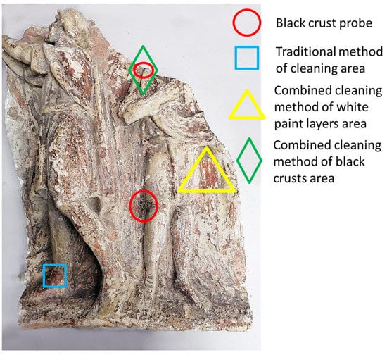
Figure 5.
General view of the bas-relief with marked areas involved in the experiment.
We compared the obtained photos from the metallographic microscope with the results from another investigation of different gypsum crusts (Figure 6).
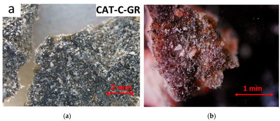
Figure 6.
View of the black crust: (a) under the stereomicroscope from the paper J.S. Pozo-Antonio etc. [21] …; (b) under the metallographic microscope from our experiment.
One can notice a similar surface structure and white crystals’ formation on the surfaces of the black crusts shown in Figure 6. The authors of the reference work (Figure 6a) claimed that such black crusts form as a result of the sulfation process of calcite in the open air, when gypsum is formed and continues to react with the environmental S and C. Thus, we may conclude that in our case, we are also dealing with gypsum crusts. This is important from the point of view of the laser source choice, since the Nd:YAG laser with a wavelength of 1064 nm removes black gypsum crusts easily [1,13,22,23].
The traditional cleaning method using chemical solvents showed that areas with white paint and gypsum crusts are resistant to chemical effects (Figure 5, blue mark), although gypsum in its pure form reacts well with the used solvents dimethyl sulfoxide and dimethylformamide [24]. Traditional cleaning began with the mildest, most gentle reagents at their minimum concentrations. In the final phase of trial clearings, solutions such as polar 10% dimethyl sulfoxide and dimethylformamide in a compress were used. In each case, the compress exposure was gradually increased up to 60 min. This prolonged exposure softened the gypsum layer and the lower layers of paint, while the upper white layers of paint and gypsum crusts remained resistant to the solvents. The inefficiency of the tested methods led to the search for a different solution to the problem and to the development of a combined cleaning method using laser technologies. It included laser removal of the gypsum crust, destruction of the near-surface layer of white paint using laser heating, and the use of the chemical solvent dimethyl sulfoxide followed by mechanical removal of unwanted layers.
Laser action on the near-surface layer of white paint in this technique does not remove it but destroys the surface structure [25], which contributes to faster penetration of the solvent into the white layers and subsequent mechanical removal. Laser cleaning of the gypsum crust allows the solvent to react directly with the gypsum layer covering the historical layer. The gypsum layer thus softens and is very easy to remove mechanically. It is noteworthy that in this case, the historical layer of red paint is not removed along with the softened gypsum—that is, the gypsum layer, due to its optical and chemical properties, serves to protect the historical patina from direct exposure to both laser radiation and the solvent, which makes the developed complex technique effective and safe.
In the case of studying the laser impact on the gypsum crust and the near-surface layer of white shading, two methods of cleaning were tested: dry and wet. In the wet method, the surface of the bas-relief was treated with dimethyl sulfoxide before laser impact (exposure for 5 min), after which it was treated by laser and then again exposed to dimethyl sulfoxide and cleaned mechanically. The results showed that both the dry and wet cleaning methods were equally effective in removing the gypsum crusts and surface layers of white pigments, so it was decided to use the dry cleaning method in order to use less solvent and not expose the bas-relief areas to a longer chemical attack. It was found that when exposed to the gypsum crust, the energy density parameter of laser radiation did not play a key role. The crust was removed both at low values of energy density (about 4 J/cm2) and at high values (about 15 J/cm2), after which no visible changes in the color or structure of the gypsum layer occurred that were observed by optical microscopy with the portable USB device described in the “Materials and Methods” section, at a magnification of 50×.
For the near-surface layer of white paint, on the contrary, the energy density turned out to be important, despite the weak absorption at a wavelength of 1064 nm by white pigments that included Ba and Zn in their chemical composition [25]. Moreover, gypsum itself also strongly reflects near-IR radiation [26], which also prevents one-stage cleaning of the bas-relief using laser radiation since the gypsum layer is comparable in thickness to the white layers (Figure 2). Thermal exposure at a high radiation power of about 13–15 J/cm2 caused darkening of the white layers and a change in their structure, which led to an uncontrolled cleaning process of unevenly distributed layers on the surface of the bas-relief in which the historical layer could be damaged in some places. Experimentally, the optimal mode of laser action on the white painting layers was obtained. The energy density was about 11 J/cm2, the pulse repetition rate was 1 Hz, the laser beam diameter was 2.5 mm, and the number of scans was four. This mode was used during all further experiments, including the removal of gypsum crusts (one scan was sufficient).
3.3. Combined Cleaning Results
After the optimal energy density values were found, the method of complex cleaning of the area with a gypsum crust which is not amenable to the action of a chemical solvent was tested (Figure 7).
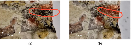
Figure 7.
Photos of the area with a gypsum crust cleared by the combined method: (a) area before the cleaning process; (b) after combined cleaning.
Figure 5 shows the area where this method was applied (green mark). With the help of laser radiation, the black crust was removed, after which a layer of gypsum remained and was exposed to dimethyl sulfoxide (exposure for 10 min). Then, using a scalpel, the softened layer of gypsum was removed mechanically. The cleaning result is shown in Figure 7b.
Then, an area of 3 × 4 cm (Figure 5, yellow mark) on the surface of bas-relief, which was covered with layers of white paint, was cleaned (Figure 8).
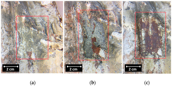
Figure 8.
Photos of the area cleaned by the combined method from white paint: (a) before the cleaning process; (b) dimethyl sulfoxide treatment; (c) cleaning result.
After laser exposure and application of dimethyl sulfoxide (exposure for 10 min), part of the white layers almost immediately began to peel off (Figure 8b), and the remaining area was cleaned with a scalpel and additional treatment with dimethyl sulfoxide (depending on the thickness of the layer) during mechanical action. As a result, the site was uncovered with no visible damage to the historic red paint layer (Figure 8c).
Using a DigiMicro Prof 300× digital USB microscope, images of the surface of the bas-relief were obtained (Figure 9).
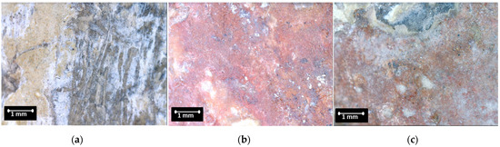
Figure 9.
Photos of the bas-relief surface: (a) the original surface with white paint; (b) surface cleaned by the combined method; (c) surface cleaned by the traditional method.
The photographs show that after the combined cleaning method (Figure 9b), the surface had a more pronounced red color compared to the surface cleaned with the traditional chemical method. Whitish traces (Figure 9c) may indicate that a gypsum layer remains, distorting the color of the surface after traditional cleaning.
4. Discussion
Table 1 provides a detailed description of the elemental compositions and the estimated times when the layers of white paint present on the surface of the bas-relief were applied. It is difficult to estimate the application time of the iron-containing layers of reddish and orange color. In the drawings from the Academy of Arts Museum collection made in 1794 by S.F. Ivanov [27], there is no visible contrast which could make it possible to suggest that the metopes were a different color from the facade. Since there are no data on the original color solution for the courtyard metopes, we offer a hypothesis for the reasons the gypsum was painted.
The decoration style of the courtyard’s facades that was envisioned by the architect proposed a terracotta-imitating paint, which conformed to the knowledge of the 18th century classics on the Greek style. In this case, a later orange layer might have appeared in the 1820s–1830s, over the course of the construction work supervised by K.A. Ton. Alternatively, it might have emerged somewhere between 1859 and 1864, in the 8-year-long period of major building rehabilitation, which was recorded in the memoir of V.P. Lvov [15].
The moving architectural model of the Academy of Arts building displayed in the permanent exhibition of the museum is an intricate miniature of the prospective building executed in 1764 from the designs of the architectural faculty professors A.F. Kokorinov and J.-B. M. Vallin de la Mothe. It is possible that many elements of the building were originally supposed to be decorated with gold leaf, as was done in the model interior. The commenced construction of the building halted in the early 1770s due to the cessation of funding. For the sake of reducing the costs, the architects might have abandoned the gilding plans, although only after the metopes had been prepared for oil varnish and painted terracotta. The model does not offer a definitive answer to the question of the architects’ intention, since it, too, has been repainted.
The bas-relief studied here is a complex, multi-layered object. Optical microscopy and EDX analysis allowed for accurately determining the positioning of the layers and their chemical composition, which are important both for provenance research and for developing a method of further restoration.
In Figure 9a, there are clearly visible smooth depressions on the surface, which could have emerged after mechanical cleaning of the bas-relief performed before another repair or restoration procedure. This explains the heterogeneity of the distribution of the white over-paint layers as well as the absence of gypsum crusts in some places. A surface such as this is difficult to clean with universal solvents, as the first attempts in cleaning confirmed; however, in combination with laser exposure, dimethyl sulfoxide proved to be an effective solvent. The effectiveness of the combined cleaning was also affected by the positioning of the protective gypsum layer on top of the terracotta paint. A layer arrangement of this type may be characteristic of individual decorative elements, historical buildings, or painted monuments, to which this combined cleaning technique could also be applied.
5. Conclusions
Stratigraphic analysis, optical microscopy, and photography techniques as well as electron scanning microscopy allowed us to speculate which layers were applied at later stages and how the monument was changed. After that, it was possible to clean it by developing a complex technique of removing unwanted paint layers with a chemical solvent and a laser. This technique has demonstrated its effectiveness and safety in comparison with the traditional cleaning methods, as evidenced by microscopy analysis of the cleaned surface.
This work is an example of a comprehensive study of an art object through modern optoelectronic methods that permits to solve complex problems of cultural heritage conservation and restoration, which cannot be fully accomplished via traditional methods only. This confirms the relevance of adjusting modern restorative techniques for complex, multi-layered art objects (e.g., fine art and murals), which contain low-solubility layers of synthetic coating varnishes, drying oil, soot, and various surface oxides.
Author Contributions
Stratigraphic research and analysis and photofixation, A.K.; laser cleaning experiments and method development, A.V. and G.Z.; scanning electron microscopy experiments, D.D.; resources and project administration, V.P. All authors have read and agreed to the published version of the manuscript.
Funding
This research received no external funding.
Institutional Review Board Statement
Not applicable.
Informed Consent Statement
Not applicable.
Data Availability Statement
Not applicable.
Acknowledgments
The authors express their gratitude to Yu. G. Bobrov, Department of Easel Painting Restoration of the St. Petersburg Academy of Fine Arts (St. Petersburg, Russian Federation), for fruitful discussions and valuable advice during the research.
Conflicts of Interest
The authors declare no conflict of interest.
References
- Cooper, M. Laser Cleaning in Conservation: An Introduction; Butterworth-Heinemann: Oxford, UK, 1998; p. 120. [Google Scholar]
- Fotakis, C.; Anglos, D.; Zafiropulos, V.; Georgiou, S.; Tornari, V. Lasers in the Preservation of Cultural Heritage. In Principles and Applications; CRC Press: Boca Rayton, FL, USA,; Taylor & Francis Group: Abingdon, UK, 2007; p. 336. [Google Scholar]
- Pouli, P.; Papakonstantinou, E.; Frantzikinaki, K.; Panou, A.; Frantzi, G.; Vasiliadis, C.; Fotakis, C. The two-wavelength laser cleaning methodology; theoretical background and examples from its application on CH objects and monuments with emphasis to the Athens Acropolis sculptures. Herit. Sci. 2016, 4, 9. [Google Scholar] [CrossRef]
- Matteini, M.; Lalli, C.; Tosini, I.; Giusti, A.; Siano, S. Laser and chemical cleaning tests for the conservation of the Porta del Paradiso by Lorenzo Ghiberti. J. Cult. Herit. 2003, 4, 147–151. [Google Scholar] [CrossRef]
- Bertasa, M.; Korenberg, C. Successes and challenges in laser cleaning metal artefacts: A review. J. Cult. Herit. 2022, 53, 100–117. [Google Scholar] [CrossRef]
- Basso, E.; Pozzi, F.; Reiley, M.C. The Samuel FB Morse statue in Central Park: Scientific study and laser cleaning of a 19th-century American outdoor bronze monument. Herit. Sci. 2020, 8, 81. [Google Scholar] [CrossRef]
- Melita, L.N.; Węgłowska, K.; Tamburini, D.; Korenberg, C. Investigating the Potential of the Er:YAG Laser for the Removal of Cemented Dust from Limestone and Painted Plaster. Coatings 2020, 10, 1099. [Google Scholar] [CrossRef]
- Petiti, C.; Toniolo, L.; Gulotta, D.; Mariani, B.; Goidanich, S. Effects of cleaning procedures on the long-term corrosion behavior of bronze artifacts of the cultural heritage in outdoor environment. Environ. Sci. Pollut. Res. 2020, 27, 13081–13094. [Google Scholar] [CrossRef] [PubMed]
- Berthonneau, J.; Parent, P.; Grauby, O.; Ferry, D.; Laffon, C.; Colombini, A.; Courtois, B.; Bromblet, P. Yellowing of laser-cleaned artworks: Formation of residual hydrocarbon compounds after Nd:YAG laser cleaning of gypsum plates covered by lamp black. J. Cult. Herit. 2019, 39, 57–65. [Google Scholar] [CrossRef]
- Parfenov, V.; Galushkin, A.; Tkachenko, T.; Aseev, V. Laser Cleaning as Novel Approach to Preservation of Historical Books and Documents on a Paper Basis. Quantum Beam Sci. 2022, 6, 23. [Google Scholar] [CrossRef]
- Siano, S.; Agresti, J.; Cacciari, I.; Ciofini, D.; Mascalchi, M.; Osticioli, I.; Mencaglia, A.A. Laser cleaning in conservation of stone, metal, and painted artifacts: State of the art and new insights on the use of the Nd:YAG lasers. Appl. Phys. A 2012, 106, 419–446. [Google Scholar] [CrossRef]
- Science and Art. The Painted Suiface; Sgamellotti, A., Brunetti, B.G., Miliani, C., Eds.; The Royal Society of Chemistry: Cambridge/London, UK, 2014; p. 499. [Google Scholar]
- Grammatikakis, G.; Demadis, K.D.; Melessanaki, K.; Pouli, P. Laser-assisted removal of dark cement crusts from mineral gypsum (selenite) architectural elements of peripheral monuments at Knossos. Stud. Conserv. 2015, 60 (Suppl. S1), S3–S11. [Google Scholar] [CrossRef]
- Malinovsky, N.V. Thin Cross-Sections: Research Features; Terra artis. Art and design, ed. T.V. Gorbunova. SPb.; Publishing House of SPSAPA named after A.L. Stieglitz: St. Petersburg, Russian, 2021; p. 93. (In Russian) [Google Scholar]
- Litovchenko, E.N.; Polyakova, L.S. Academy of Arts. The History of Everyday Life in the Memoirs and Images of Contemporaries. 19th–Early 20th Centuries; Premium Press: St. Petersburg, Russian, 2014; pp. 111–113. (In Russian) [Google Scholar]
- Academy of Arts: History in Photographs. 1850s–1950s; Album: St. Petersburg, Russian, 2004. (In Russian)
- Shilov, V.A. Behind the Facades of the Familiar Buildings of St. Petersburg; Stroyizdat: St. Petersburg, Russian, 2003; p. 316. (In Russian) [Google Scholar]
- Nikitenko, G.Y.; Sobol, V.D. Houses and People of Vasilyevsky Island; Tsentrpoligraf: Moscow, Russian, 2007; p. 735. (In Russian) [Google Scholar]
- Camuffo, D.; Del Monte, M.; Sabbioni, C. Origin and growth mechanisms of the sulfated crusts on urban limestone. Water Air Soil Pollut. 1983, 19, 351–359. [Google Scholar] [CrossRef]
- Vlasova, D.Y.; Rytikova, V.V.; Frank-Kamenetskaya, O.V. (Eds.) The Effect of the Environment on Saint Petersburg’s Cultural Heritage: Results of Monitoring the Historical Necropolis Monuments (Geoheritage, Geoparks and Geotourism); Springer Nature Switzerland AG: Cham, Switzerland, 2019; p. 188. [Google Scholar] [CrossRef]
- Pozo-Antonio, J.S.; Cardell, C.; Comite, V.; Fermo, P. Characterization of black crusts developed on historic stones with diverse mineralogy under different air quality environments. Environ. Sci. Pollut. Res. 2022, 29, 29438–29454. [Google Scholar] [CrossRef] [PubMed]
- Parfenov, V.A. Use of laser technologies for restoration, documentation and replication of sculptural monuments in St. Petersburg. Insight Non-Destr. Test. Cond. Monit. 2020, 62, 129–133. [Google Scholar] [CrossRef]
- Siano, S.; Giamello, M.; Bartoli, L.; Mencaglia, A.; Parfenov, V.; Salimbeni, R. Laser cleaning of stone by different laser pulse duration and wavelength. Laser Phys. 2008, 18, 27–36. [Google Scholar] [CrossRef]
- Yu, B.; Wang, J.; Miao, S.; Ren, Y. Solubility and thermodynamic modeling of calcium sulfate dihydrate in three organic aqueous solutions (ethylene glycol, acetic acid, dimethyl sulfoxide) from 303.15 K to 348.15 K. J. Chem. Thermodyn. 2022, 166, 106684. [Google Scholar] [CrossRef]
- Schnell, A.; Goretzki, L.; Kaps, C. IR-Laser Effects on Pigments and Paint Layers. In Lasers in the Conservation of Artworks; Springer: Berlin, Germany, 2005; pp. 291–296. [Google Scholar] [CrossRef]
- Long, L.L.; Querry, M.R.; Bell, R.J.; Alexander, R.W. Optical properties of calcite and gypsum in crystalline and powdered form in the infrared and far-infrared. Infrared Phys. 1993, 34, 191–201. [Google Scholar] [CrossRef]
- Ivanov, S.F. The Building of the Academy of Arts. Section along the Central Axis. Academy of Arts: Architectural Materials [Online Collections]. 1794. Available online: https://collection.artsacademymuseum.org/entity/OBJECT (accessed on 6 March 2022).
Disclaimer/Publisher’s Note: The statements, opinions and data contained in all publications are solely those of the individual author(s) and contributor(s) and not of MDPI and/or the editor(s). MDPI and/or the editor(s) disclaim responsibility for any injury to people or property resulting from any ideas, methods, instructions or products referred to in the content. |
© 2023 by the authors. Licensee MDPI, Basel, Switzerland. This article is an open access article distributed under the terms and conditions of the Creative Commons Attribution (CC BY) license (https://creativecommons.org/licenses/by/4.0/).