Article

Mapping with Macro X-ray Fluorescence Scanning of Raffaello’s Portrait of Leo X

1
Department of Physics and Astronomy, University of Florence, 50019 Sesto Fiorentino, Italy
2
Florence Division, INFN/CHNet, 50019 Sesto Fiorentino, Italy
*
Author to whom correspondence should be addressed.
Heritage 2022, 5(4), 3993-4005; https://doi.org/10.3390/heritage5040205
Submission received: 24 November 2022 / Revised: 29 November 2022 / Accepted: 1 December 2022 / Published: 6 December 2022

Abstract

:
Raffaello is renowned as one of the Old Renaissance Masters and his paintings and painting technique are famous for the details and naturality of the characters. Raffaello is famous in particular for the then-new technique of oil painting, which he mastered and perfected. On the occasion of the 500th anniversary of the death of Raffaello (2020), there was a large exhibition at the Scuderie del Quirinale in Rome, where many paintings and drawings by the Old Master were on show. One of these paintings was the portrait of Leo X with two cardinals belonging to the collection of the Uffizi galleries in Florence. Before going to Rome, the painting underwent conservation treatments at the Opificio delle Pietre Dure, where a comprehensive diagnostic campaign was carried out with the aim of understanding the painting materials and technique of the Old Master. In this paper, the results of macro X-ray fluorescence (MA-XRF) analysis, carried out exploiting the instrument developed by INFN-CHNet, are shown. Among the results, “bismuth black” and the likely use of glass powders in lakes are discussed.

1. Introduction

The analysis of the painting materials employed by an artist to produce a work of art is considered essential for a deep comprehension of his work, style and technique. Moreover, knowledge of the materials is crucial for conservation treatments. It is, however, true that material analysis should not be limited to knowledge of a single painting or artwork but should be contextualised within the production of the artist and other contemporary masters. This multidisciplinary approach increases the knowledge of the history of materials (e.g., trade routes), artists and techniques [1,2,3,4,5], which may also be useful for authentication and forensic science [6,7].
Analytical methods aiding the purpose of material characterisation are now numerous and many instruments allow for in situ non-invasive measurements with high performances [8,9,10]. In recent years, X-ray fluorescence (XRF), one of the principal techniques employed in this field, has been developed from an elemental compositional analysis giving spectra into a mapping method giving elemental distribution maps and thus images. This technique is now called macro-XRF (MA-XRF) [11,12] and its contribution to heritage science is undisputed.
The MA-XRF instrument employed in this work is a MA-XRF scanner developed by the Cultural Heritage Network of the Italian National Institute of Nuclear Physics (INFN-CHNet), which works in the development of novel instruments to aid heritage science [13,14].
The study presented here deals with MA-XRF analysis of the Portrait of Leo X by Raffaello Sanzio. This painting belongs to the collection of the Uffizi Galleries and has been under conservation interventions for a couple of years at the Opificio delle Pietre Dure in Florence for the occasion of the exhibition at the Scuderie del Quirinale for the 500th anniversary of the death of the Old Master (2020). The aim of the MA-XRF campaign was to study in detail the painting palette and technique employed by Raffaello, contextualizing the results with other works by the artist.

2. Materials and Methods

2.1. The INFN-CHNet MA-XRF Scanner

The INFN-CHNet instrument, thoroughly described in [15], is a light highly transportable device specifically designed for in situ heritage science applications [16]. Briefly, the measuring head has an X-ray tube (Moxtek, 40 kV maximum voltage, 0.1 mA maximum anode current), a silicon drift detector (Amptek XR100 SDD, 25 mm2 effective active surface, 500 μm thickness) and a telemeter (Keyence IA-100) for the continuous control and adjustment of the sample–instrument distance. All these elements are installed on three linear motor stages by Physik Instrumente, 200 mm travel range in the x and y directions for this version, plus a 50 mm stage along the z perpendicular direction. Motor controllers, signal digitizer (CAEN DT5780) and other auxiliary elements are contained in carbon-fibre box on which the motors are installed. Instrument control and data analysis are carried out with in-house developed software. The instrument has been successfully employed in several heritage science applications over the years [17,18,19].
The operating conditions of the X-ray tube for all measurements discussed here were 30 kV anode voltage, 90 μA filament current, Mo anode with an 800 μm diameter collimator. Scanning velocity was 10 mm/s and the equivalent pixel size 1 mm. Several scans were performed for the almost full mapping of the painting. Due to the dimensions and the weight of the painting itself—which made its handling laborious—and the encumbrance of the easel, some areas could not be reached with the MA-XRF scanner.

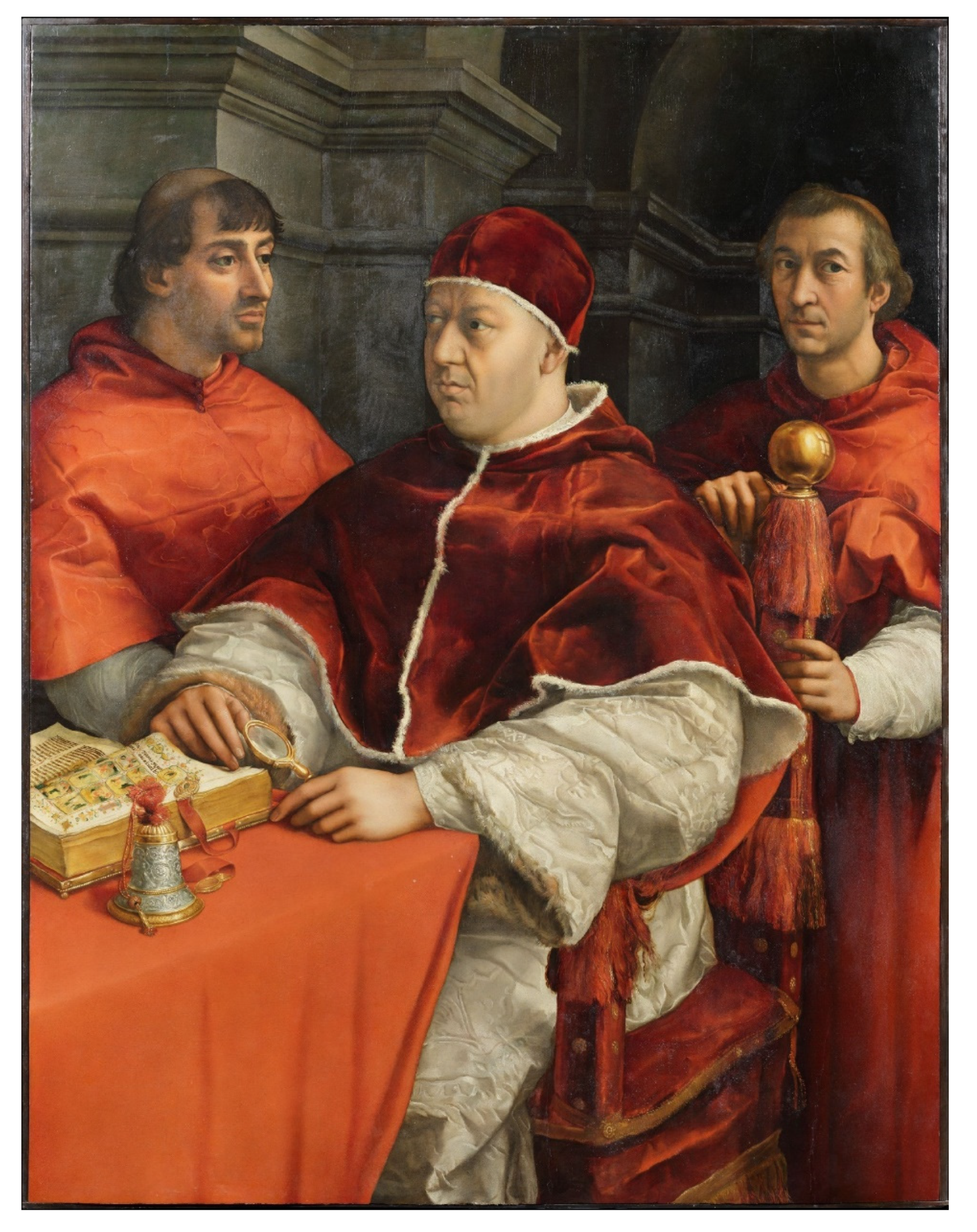
2.2. The Portrait of Leo X by Raffaello Sanzio

The painting analysed here and shown in Figure 1 depicts Pope Leo X (1475–1521), born Giovanni di Lorenzo de’ Medici and son of Lorenzo il Magnifico, portrayed in a three-quarter pose with two cardinals, Luigi de’ Rossi and Giulio de’ Medici, both members of the Medici family and cousins of the pope. The three are represented in a grey architectural background.
The painting was executed in Rome and most likely commissioned by the Medicis on THE occasion of the wedding of Leo X’s nephew Lorenzo de’ Medici, Duke of Urbino, with Madeleine de la Tour d’Auvergne, a ceremony that was held in Florence in September 1518. According to a report at the time, the portrait was placed on the middle of the bridal banquet table to allow the papal and cardinal relatives to be symbolically present at the festivities [20].
Red fabrics are dominant and masterfully painted by the artist; these are the dark red velvet of the mozzetta (the elbow-length cape with hood of the pope) and of the camauro (his felt cap), edged with ermine, the similar fabric of the chair and armrest, the heavy tablecloth, the gros de tours silk cardinal cassocks.
The pope is vested with a light grey, fur-inner-lined brocade cassock decorated with a pomegranate and ivy shoots motif. He is holding a magnifying glass with his left hand and turning the page of precious illuminated Bible, identified by scholars as the Hamilton Bible, with his right [21]. In foreground there is a finely carved silver handbell decorated with a sheaf of red and gold thread, which was mentioned by Vasari as “a little bell of wrought silver, which is more beautiful than words can tell” [22].
An interesting detail is that at the top of the back post of the chair, where on the golden ball there is the reflection of the room, there are included the shoulder of the pope and a window, which is the source of light in the painting, although not visible.

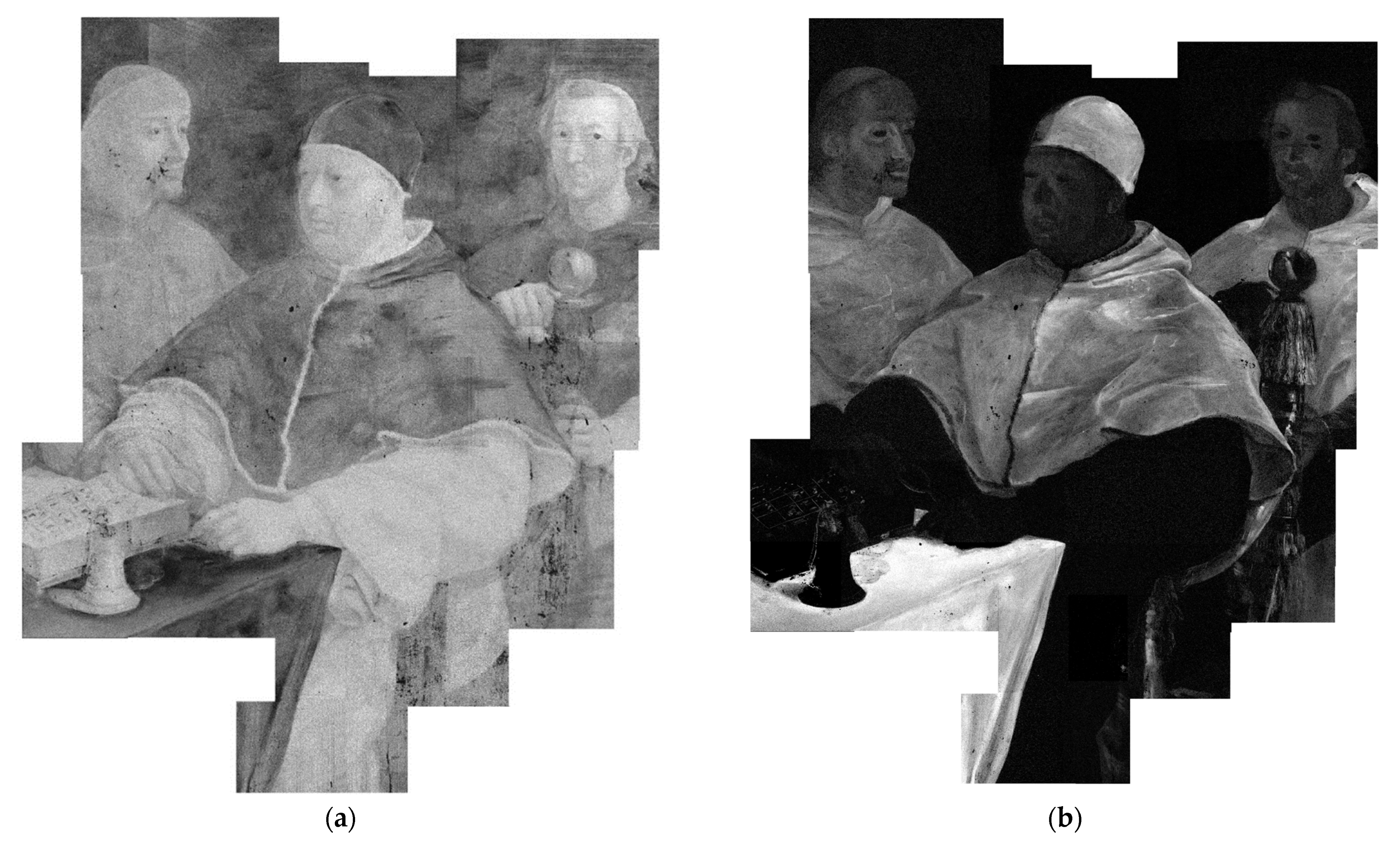
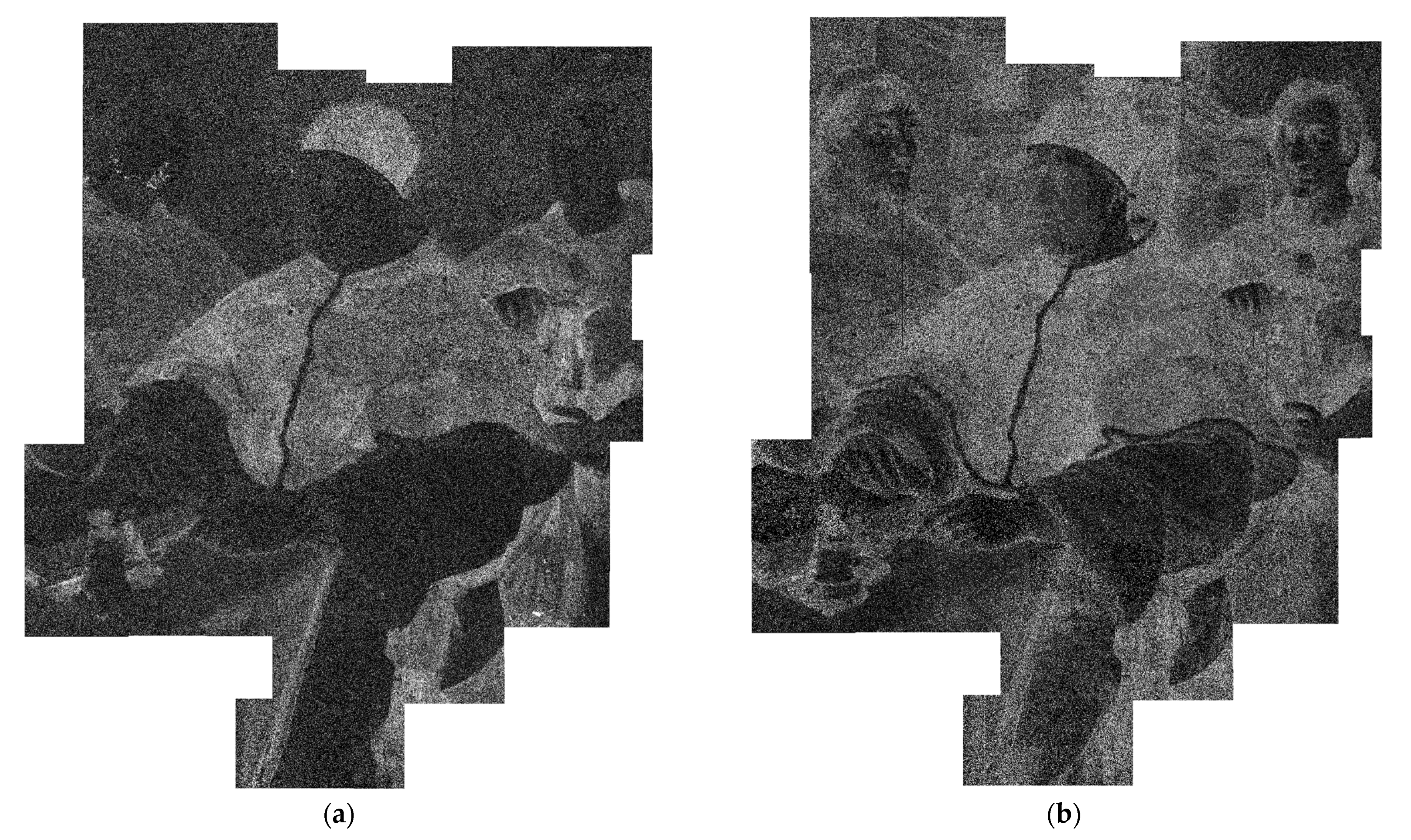
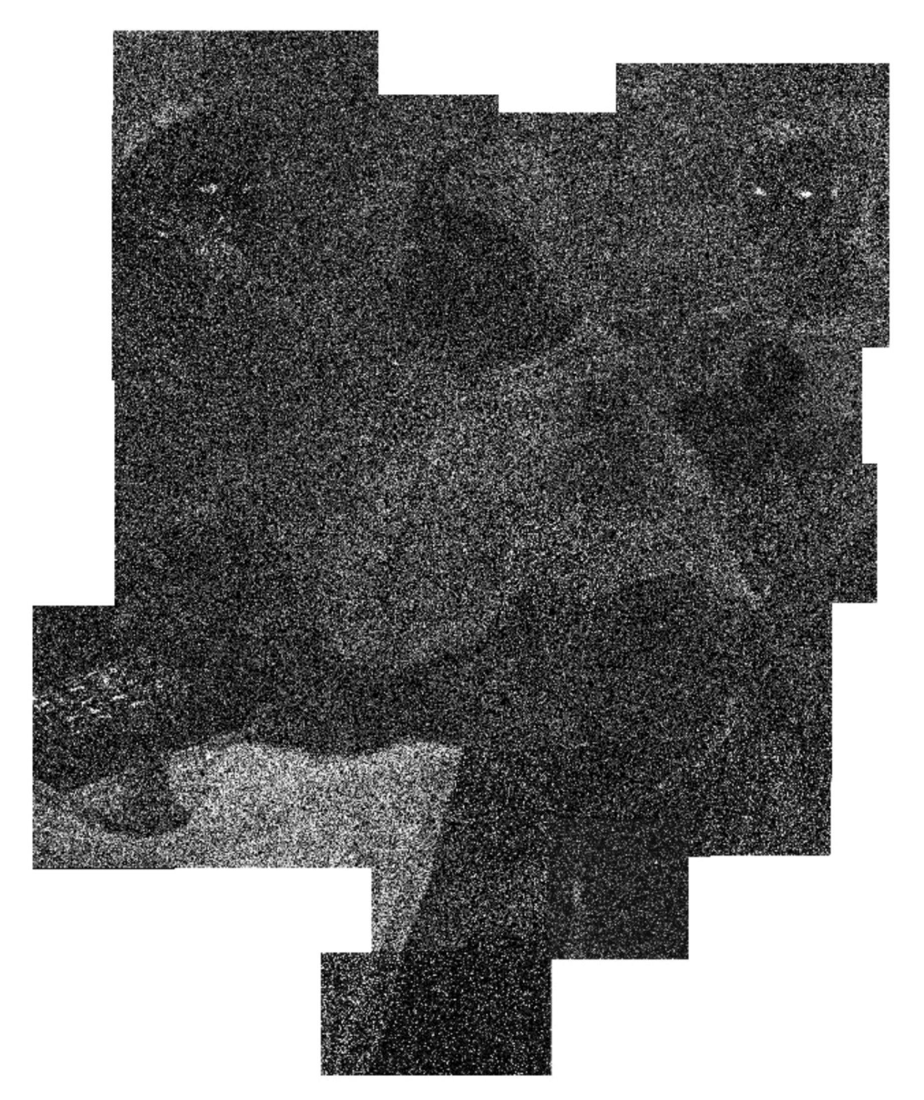
3. Results and Discussion

The painting palette hypothesised by MA-XRF analysis is broadly consistent with other paintings by Raffaello Sanzio and with the materials available to artists during the Renaissance period. Here, the hypothesis of pigments and materials employed based on the detected elements is discussed.
Pb is ubiquitously present in the painting (Figure 2a), a fact that suggests the use of a traditional lead-white-based imprimitura layer. The heterogeneous distribution of this element is most probably due to its additional use as lead white as a proper pigment in the painting layer, both in white areas and/or in mixture with other pigments/materials such as in cassocks, furs and flesh tones.
The painting’s main colour is red, which is found in several different red fabrics, such as the velvet camauro (hat) of the pope and velvet or silk mozzettas (the short elbow-length mantles), the tablecloth and the trimmings of the chair. These are all characterised by the presence of Hg (Figure 2b), indicating the use of vermilion/cinnabar. The modelling of red garments was achieved with Cu-based pigments (Figure 2c). Giulio de’ Medici’s (the cardinal at the pope’s proper right) mozzetta contains a smaller amount of copper but more lead than the other two and is indeed lighter in tone. The velvet appearance of the pope’s mozzetta and camauro was then obtained with the addition of small quantities of Fe-based pigments (Figure 2d), such as iron oxides/hydroxides contained in earth and ochres.
In addition, Mn traces were detected in most of the red areas (Figure 3a). This element is a typical trace of pigments such as earth and ochres, particularly of umber [23], but is also considered a marker of the use of glass powders employed as a siccative of the oil medium [24]. Mn is thought to be present in glass, particularly in that of Italian production, acting as a decolouriser of the light green tone given by the presence of iron traces, usually present in the production materials of glass [25]. Interestingly, glass powders were detected in red lakes in other paintings by Raffaello [26,27,28] and other artists [29,30,31]. Indeed, in correspondence of the Mn-rich areas, K and Ca were also detected, elements which have been found in glass powders in paintings by Raffaello [25]. The presence of K may also attest the use of alum as is typical of the lakes [32]. The use of glass powder in this painting was indeed confirmed by cross sections in red areas [33]. Mn and K were also detected in shadows on red fabrics as seen on the reading desk, the handbell and the hand of the pope. It is realistic to hypothesise the use of umber (a type of Mn-rich earth [34]) in these areas, but the use (also) of red lakes cannot be excluded. The red velvet of the chair was instead most likely painted with red lakes only (with the addition of glass powder) and not with vermilion. K traces were also found in other areas of the painting not matching the distribution of Mn; in these cases, their presence was not fully understood, but it has to be taken into account that K is present in several artistic compounds (e.g., green earth, lakes and varnish).
The pope’s white damask cassock is mainly characterised by the presence of lead (Figure 2a), indicating, as previously discussed, the use of lead white. As similarly happens in red garments, modelling, shading and also the inner fur of this robe are painted with Cu- and Fe-based compounds (Figure 2c,d). In addition, the distribution of Ca likely also matches with the pomegranate and ivy shoots motif (Figure 4a). Ca may be due to the gypsum-based preparation layer, but its elemental distribution may suggest its use in the pictorial layer to execute shadows and darker areas. The use of bone black (identified in some cases in this painting [35] and also in other paintings by Raffaello [36]) cannot be excluded, although XRF may not discriminate between other Ca-containing compounds and organic materials [37].
The cold-grey tone of the cassock was then obtained with a rare Bi-based pigment (Figure 4b). Known as “bismuth black”, it exists in the metallic and in the trisulphide (bismuthinite mineral) forms, It is not properly a black but rather a grey pigment, and it is documented in paintings in a rather short timeframe from the end of the 15th century, almost during the discovery of the mines in Germany (around 1460), to the first decades of the 16th century [38]. The use of this pigment is reported in other works by Raffaello, such as the Madonna dell’Impannata (1511–1515) of the Palatine Gallery in Florence [39], the Ansidei Madonna (1505), likely the Procession to Calvary (1504–1505) and the Madonna of the Pinks (1506–1507) of the National Gallery in London [25]. This pigment was first documented in Fra Bartolomeo’s works (Pitti Panel, 1512) [40,41,42]. Its use is not limited to paintings but is also seen in illuminated manuscripts, such as those by Jean Bourdichon [43,44], as well as in inks to give a silvery appearance [45] and as metal foil to imitate silver [46].
The yellow areas, such as the magnifying glass, the reading desk and the handbell, were painted with complex mixtures to imitate the metallic appearance of these objects. Yellow, brown, blue/green and red pigments (Cu- and Fe-based compounds, lead–tin yellow and vermilion traces) were skilfully employed, differentiating lights and shadows in order to realistically render the tridimensionality of the objects (Figure 5). “Bismuth black” was employed to give them their metallic appearance.
Details of the miniatures in the book were realized with vermilion, a Cu-based material and lead–tin yellow. The glass of the magnifying glass was painted with a Cu-based pigment and “bismuth black” traces (possibly coming from the cassock behind). On the golden ball at the top of the papal throne, the reflection of the pope’s mozzetta was painted with vermilion.
Flesh tones were painted with lead white, vermilion and Fe- and Cu-based compounds (the last also employed in the shaved beards and tonsures). The irises of the eyes were painted with a Cu-based pigment, possibly employed as a drier of the black pigment [47]. Hair was painted with Fe-based compounds with a non-negligible amount of Ca, which is found in most of the shadows. The hair of Luigi de’ Rossi (the cardinal at the pope’s proper left) is slightly darker compared to the others due to the use of Cu-based compounds.
The architectural background is characterised by a rather uniform distribution of Cu (Figure 2c)—most likely indicating its use as a greenish preparation layer. The Cu-containing material in this case was identified as copper resinate with cross sections [33], a pigment which has been found in a number of paintings from this time [47,48]. Prospective lines visible in the Cu map are present as engravings. It is worth noting that the homogeneity of the Cu map likely undermines the hypothesis that Raffaello initially painted a green curtain instead of the architectural background, as is present in the portrait of Julius II [49]. Similarly, there is no evidence that the painting was initially meant to be a solitary portrait of the pope, as hypothesised by other scholars [50]: in that case, in place of the cardinals, Raffaello would have painted the architectural background with a copper resinate base layer which is instead not detected. It cannot be ruled out, however, that an eventual change of mind happened during the drawing phase of the portrait. Ca-, Fe- and Bi-based materials are also detected in lower quantities than Cu, most likely painted over the green copper resinate background [33] as a way to reach the appearance of the pietra serena. These elements are present in the background and are shaded following the volumes, rendering the tridimensionality of the architecture with a light coming from the left side of the painting (i.e., from the window visible only in the reflection of the ball on the chair). This possibly suggests the use of these materials in more superficial layers of the painting, as determined by cross sections [33].
Zn traces are typical in a number of artistic materials, such as vermilion and malachite, and also in iron-gall inks where they are present in white vitriol (ZnSO4) [51]. Although in this painting Zn (Figure 6) is detected in association with Hg- and some Cu-based areas (such as blue/green details of the miniatures of the Bible), the Zn map draws attention to the eyes of the three characters. Indeed, all three pairs of eyes are painted with Cu-based materials, but it is interesting to note that only the eyes of the two cardinals evidently contain Zn. IR reflectography actually highlighted two different techniques of underdrawings: a pouncing technique was likely used for the cardinals (although hardly visible) and freehand drawing with black chalk for the pope [52]. Recently, works exploiting MA-XRF on paintings have, interestingly, found that the distribution of Zn detected in paintings is most likely related to iron-gall-ink underdrawings [53,54]. Unfortunately, the dwell time of the measurements carried out here does not give a sufficient resolution for a proper identification based on the visualization of characteristic features of underdrawings such as hatching, but the use—not yet documented in Raffaello’s painting—of iron-gall ink as underdrawing remains an intriguing possibility.
A summary of the results is presented in Table 1.

4. Conclusions

The MA-XRF analysis carried out on the Portrait of Leo X by Raffaello gave information on the materials employed. Traditional materials, such as lead white, vermilion, earth/ochres, Cu-based compounds (copper resinate at least in the background) and lead–tin yellow were masterfully used by the artist to paint different fabrics and flesh tones. Glass powder was most probably employed in red lakes, as documented in the Mn map. The metallic appearance of objects such as the silver handbell, but also the grey tone of the cassocks, was painted using a rare “bismuth black”, also attested in other paintings by Raffaello.
The architectural background was painted with a copper resinate base layer with volume and tridimensionality achieved using “bismuth black”, a Ca-based pigment (possibly bone black) and earths/ochres. Interestingly, the distribution of the Cu map attests that the painting was meant from the beginning as a triple portrait and not with the pope represented individually as hypothesised by scholars (in that case, a homogeneous distribution of Cu would have also been detected below the cardinals). In addition, the homogeneity of the Cu map undermines the hypothesis of a green curtain painted in the first phase of painting similar to the one in the portrait of Julius II. Another interesting hypothesis is the possible use of iron-gall ink in the underdrawing—especially in the eyes of the two cardinals—as hypothesised by the Zn map.
As is also demonstrated in this study, MA-XRF allows the non-invasive mapping of the elemental distribution of the materials, making the technique a powerful tool for a preliminary non-invasive and non-destructive analytical method. It can also be considered a good guide for a subsequent, more accurate scientific analysis.

Author Contributions

Conceptualization, A.M., C.R. and L.C.; methodology, F.T., L.G. and P.A.M.; software, F.T. and L.C; validation, P.A.M.; formal analysis, A.M. and C.R.; investigation, A.M. and C.R.; resources, F.T., L.G. and P.A.M.; data curation, F.T. and L.C.; writing—original draft preparation, A.M.; writing—review and editing, A.M., C.R. and L.G.; visualization, A.M., C.R. and L.C.; supervision, F.T. and P.A.M.; project administration, F.T. All authors have read and agreed to the published version of the manuscript.

Funding

This research received no external funding.

Institutional Review Board Statement

Not applicable.

Informed Consent Statement

Not applicable.

Data Availability Statement

Not applicable.

Acknowledgments

The authors are grateful to the Opificio delle Pietre Dure and the Gallerie degli Uffizi for allowing this study. In particular, Cecilia Frosinini, Oriana Sartiani, Roberto Bellucci (OPD), Eike D. Schmidt and Carolina Forasassi (Uffizi) are gratefully acknowledged. The authors warmly thank Lara Palla and Caroline Czelusniak for their contributions to the MA-XRF software. Thanks are also due to Marco Manetti for his invaluable technical support and to Leandro Sottili for his helpful suggestions.

Conflicts of Interest

The authors declare no conflict of interest.

References

  1. Kirby, J.; Nash, S.; Cannon, J. Trade in Artists’ Materials Markets and Commerce in Europe to 1700; Archetype Publications: London, UK, 2010. [Google Scholar]
  2. Berrie, B.H. Rethinking the history of artists’ pigments through chemical analysis. Annu. Rev. Anal. Chem. 2012, 5, 441–459. [Google Scholar] [CrossRef] [PubMed]
  3. Ricciardi, P.; Dooley, K.A.; MacLennan, D.; Bertolotti, G.; Gabrieli, F.; Patterson, C.S.; Delaney, J.K. Use of standard analytical tools to detect small amounts of smalt in the presence of ultramarine as observed in 15th-century Venetian illuminated manuscripts. Herit. Sci. 2022, 10, 38. [Google Scholar] [CrossRef]
  4. Fiorillo, F.; Burgio, L.; Kimbriel, C.S.; Ricciardi, P. Non-Invasive Technical Investigation of English Portrait Miniatures Attributed to Nicholas Hilliard and Isaac Oliver. Heritage 2021, 4, 1165–1181. [Google Scholar] [CrossRef]
  5. Radpour, R.; Gates, G.A.; Kakoulli, I.; Delaney, J.K. Identification and mapping of ancient pigments in a Roman Egyptian funerary portrait by application of reflectance and luminescence imaging spectroscopy. Herit. Sci. 2022, 10, 8. [Google Scholar] [CrossRef]
  6. Calligaro, T.; Banas, A.; Banas, K.; Bogdanović Radović, I.; Brajković, M.; Chiari, M.; Forss, A.; Hajdas, I.; Krmpotić, M.; Mazzinghi, A.; et al. Emerging nuclear methods for historical painting authentication: AMS-14C dating, MeV-SIMS and O-PTIR imaging, global IBA, differential-PIXE and full-field PIXE mapping. Forensic. Sci. Int. 2022, 336, 111327. [Google Scholar] [CrossRef]
  7. Fiorillo, F.; Hendriks, L.; Hajdas, I.; Vandini, M.; Huysecom, E. The Rediscovery of Jan Ruyscher and Its Consequence. J. Am. Inst. Conserv. 2022, 61, 55–63. [Google Scholar] [CrossRef]
  8. Miliani, C.; Rosi, F.; Brunetti, B.G.; Sgamellotti, A. In Situ Noninvasive Study of Artworks: The MOLAB Multitechnique Approach. Acc. Chem. Res. 2010, 43, 728–738. [Google Scholar] [CrossRef]
  9. Legrand, S.; Ricciardi, P.; Nodari, L.; Janssens, K. Non-invasive analysis of a 15th century illuminated manuscript fragment: Point-based vs imaging spectroscopy. Microchem. J. 2018, 138, 162–172. [Google Scholar] [CrossRef]
  10. Karydas, A.G. Application of a Portable XRF Spectrometer for the Non-Invasive analysis of Museum Metal Artefacts. Ann. Chim. 2007, 97, 419–432. [Google Scholar] [CrossRef]
  11. Alfeld, M.; Vaz Pedroso, J.; van Eikema Hommes, M.; van der Snickt, G.; Tauber, G.; Blaas, J.; Haschke, M.; Erler, K.; Dik, J.; Janssens, K. A mobile instrument for in situ scanning macro-XRF investigation of historical paintings. J. Anal. At. Spectrom. 2013, 28, 760. [Google Scholar] [CrossRef]
  12. Romano, F.P.; Caliri, C.; Nicotra, P.; Di Martino, S.; Pappalardo, L.; Rizzo, F.; Santos, H.C.J. Real-time elemental imaging of large dimension paintings with a novel mobile macro X-ray fluorescence (MA-XRF) scanning technique. Anal. At. Spectrom. 2017, 32, 773. [Google Scholar] [CrossRef]
  13. Giuntini, L.; Castelli, L.; Massi, M.; Fedi, M.; Czelusniak, C.; Gelli, N.; Liccioli, L.; Giambi, F.; Ruberto, C.; Mazzinghi, A.; et al. Detectors and Cultural Heritage: The INFN-CHNet Experience. Appl. Sci. 2021, 11, 3462. [Google Scholar] [CrossRef]
  14. Sottili, L.; Giuntini, L.; Mazzinghi, A.; Massi, M.; Carraresi, L.; Castelli, L.; Czelusniak, C.; Giambi, F.; Mandò, P.A.; Manetti, M.; et al. The Role of PIXE and XRF in Heritage Science: The INFN-CHNet LABEC Experience. Appl. Sci. 2022, 12, 6585. [Google Scholar] [CrossRef]
  15. Taccetti, F.; Castelli, L.; Czelusniak, C.; Gelli, N.; Mazzinghi, A.; Palla, L.; Ruberto, C.; Censori, C.; Lo Giudice, A.; Re, A.; et al. A multipurpose X-ray fluorescence scanner developed for in situ analysis. Rend. Lincei. Sci. Fis. Nat. 2019, 30, 307–322. [Google Scholar] [CrossRef]
  16. Mazzinghi, A.; Ruberto, C.; Castelli, L.; Ricciardi, P.; Czelusniak, C.; Giuntini, L.; Mandò, P.A.; Manetti, M.; Palla, L.; Taccetti, F. The importance of being little: MA-XRF on manuscripts on a Venetian island. X-ray Spectrom. 2021, 50, 272–278. [Google Scholar] [CrossRef]
  17. Romani, M.; Pronti, L.; Ruberto, C.; Severini, L.; Mazzuca, C.; Viviani, G.; Mazzinghi, A.; Chiari, M.; Castelli, L.; Taccetti, F.; et al. Toward an assessment of cleaning treatments onto nineteenth–twentieth-century phographs by using a multi-analytic approach. Eur. Phys. J. Plus 2022, 137, 757. [Google Scholar] [CrossRef]
  18. Sottili, L.; Guidorzi, L.; Mazzinghi, A.; Ruberto, C.; Castelli, L.; Czelusniak, C.; Giuntini, L.; Massi, M.; Taccetti, F.; Nervo, M.; et al. The Importance of Being Versatile: INFN-CHNet MA-XRF Scanner on Furniture at the CCR “La Venaria Reale”. Appl. Sci. 2021, 11, 1197. [Google Scholar] [CrossRef]
  19. Mangani, S.M.E.; Mazzinghi, A.; Mandò, P.A.; Legnaioli, S.; Chiari, M. Characterisation of decoration and glazing materials of late 19th-early 20th century French porcelain and fine earthenware enamels: A preliminary non-invasive study. Eur. Phys. J. Plus 2021, 136, 1079. [Google Scholar] [CrossRef]
  20. Minnich, N.H. Raphael’s Portrait “Leo X with Cardinals Giulio de’ Medici and Luigi de’ Rossi”: A Religious Interpretation, Renaiss. Q. 2003, 56, 1005–1052. [Google Scholar] [CrossRef]
  21. Lollini, F. Con gli occhi di Giovanni. In Raffaello e il Ritorno del Papa Medici: Restauri e Scoperte sul Ritratto di Leone X con i DUE CARdinali; Ciatti, M., Schmidt, E.D., Eds.; Edifir—Edizioni Firenze: Firenze, Italy, 2020; pp. 61–83. [Google Scholar]
  22. Vasari, G. Le Vite de’ più Eccellenti Pittori, Scultori e Architettori Nelle Redazioni del 1550 a 1568; Bettarini, R., Barocchi, P., Eds.; Edizioni Giuntina: Firenze, Italy, 1976; Volume 4, p. 188. [Google Scholar]
  23. Helwig, K. Iron Oxide Pigments: Natural and Synthetic. In Artists’ Pigments. A Handbook of Their History and Characteristics; Berrie, B.H., Ed.; National Gallery of Art: Washington, DC, USA, 2007; Volume 4, pp. 39–96. [Google Scholar]
  24. Spring, M. Colourless Powdered Glass as an Additive in Fifteenth- and Sixteenth-Century European Paintings; Technical Bulletin Volume 33; National Gallery: London, UK, 2012; pp. 4–26. [Google Scholar]
  25. Spring, M. Raphael’ s Materials: Some New Discoveries and their Context within Early Sixteenth-Century Painting. Nat. Gal. Tech. Bull. 2005, 77–86. [Google Scholar]
  26. Roy, A.; Spring, M.; Plazzotta, C. Raphael’s Early Work in the National Gallery: Paintings before Rome; Technical Bulletin Volume 25; National Gallery: London, UK, 2004; pp. 4–35. [Google Scholar]
  27. Borgia, I.; Cauzzi, D.; Radicati, B.; Seccaroni, C. Raphael’s Saint Cecilia in Bologna: New Data about its Genesis and Materials. In Raphael’s Painting Technique: Working Practices before Rome; Proceedings of the Eu-ARTECH Workshop; Nardini Editore: Firenze, Italy, 2007; pp. 93–99. [Google Scholar]
  28. Berrie, B.H.; Walmsley, E. Raphael’s Alba Madonna. In Raphael’s Painting Technique: Working Practices before Rome; Proceedings of the Eu-ARTECH Workshop; Nardini Editore: Firenze, Italy, 2007; pp. 101–108. [Google Scholar]
  29. Ricci, C.; Borgia, I.; Brunetti, B.G.; Miliani, C.; Sgamellotti, A.; Seccaroni, C.; Passalacqua, P. The Perugino’s palette: Integration of an extended in situ XRF study by Raman spectroscopy. J. Raman Spectrosc. 2004, 35, 616–621. [Google Scholar] [CrossRef]
  30. Lutzenberger, K.; Stege, H.; Tilenschi, C. A note on glass and silica in oil paintings from the 15th to the 17th century. J. Cult. Herit. 2010, 11, 365–372. [Google Scholar] [CrossRef]
  31. Mazzinghi, A.; Ruberto, C.; Castelli, L.; Czelusniak, C.; Giuntini, L.; Mandò, P.A.; Taccetti, F. MA-XRF for the Characterisation of the Painting Materials and Technique of the Entombment of Christ by Rogier van der Weyden. Appl. Sci. 2021, 11, 6151. [Google Scholar] [CrossRef]
  32. Kirby, J.; Spring, M.; Higgitt, C. The Technology of Red Lake Pigment Manufacture: Study of the Dyestuff Substrate. Nat. Gallery Tech. Bull. 2005, 26, 71–87. [Google Scholar]
  33. Lalli, C.G.; Innocenti, F. Analisi stratigrafiche eseguite sul Ritratto di papa Leone X con i cugini cardinali. In Raffaello e il Ritorno del Papa Medici: Restauri e Scoperte sul Ritratto di Leone X con i Due Cardinali; Ciatti, M., Schmidt, E.D., Eds.; EdiFir Edizioni Firenze: Firenze, Italy, 2020; pp. 211–218. [Google Scholar]
  34. Hradil, D.; Grygar, T.; Hradilová, J.; Bezdička, P. Clay and iron oxide pigments in the history of painting. Appl. Clay Sci. 2003, 22, 223–236. [Google Scholar] [CrossRef]
  35. Lanterna, G. Analisi non invasive e considerazioni sul Ritratto di papa Leone X con i cugini cardinali di Raffello. In Raffaello e il Ritorno del Papa Medici: Restauri e Scoperte sul Ritratto di Leone X con i Due Cardinali; Ciatti, M., Schmidt, E.D., Eds.; EdiFir Edizioni Firenze: Firenze, Italy, 2020; pp. 201–210. [Google Scholar]
  36. Ruberto, C.; Mazzinghi, A.; Massi, M.; Castelli, L.; Czelusniak, C.; Palla, L.; Gelli, N.; Betuzzi, M.; Impallaria, A.; Brancaccio, B.; et al. Imaging study of Raffaello’s “La Muta” by a portable XRF spectrometer. Microchem. J. 2016, 126, 63–69. [Google Scholar] [CrossRef]
  37. Perry, J.J.; Brown, L.; Jurneczko, E.; Ludkin, E.; Singer, B.W. Identifying the plant origin of artists’ yellow lake pigments by electrospray mass spectrometry. Archaeometry 2011, 53, 164–177. [Google Scholar] [CrossRef]
  38. Spring, M.; Grout, R.; White, R. ‘Black Earths’: A Study of Unusual Black and Dark Grey Pigments used by Artists in the Sixteenth Century. Nat. Gal. Tech. Bull. 2003, 24, 103–105. [Google Scholar]
  39. Ciatti, M.; Castelli, C.; Frosinini, C.; Gusmeroli, L.; Cartechini, L.; Rosi, F.; Seccaroni, C.; Miliani, C.; Moioli, P.; Lalli, C.G.; et al. Il restauro della Madonna dell’Impannata di Raffaello delle Gallerie degli Uffizi, Galleria Palatina di Palazzo Pitti. OPD Restauro 2017, 29, 48–90. [Google Scholar]
  40. Moioli, P.; Scafè, R.; Seccaroni, C. Appendice I, Analisi di fluorescenza X su sei dipinti di Fra’ Bartolomeo. In L’età di Savonarola. Fra Bartolomeo e la Scuola di San Marco; Padovani, S., Scudieri, M., Damiani, G., Eds.; Marisilio: Venezia, Italy, 1996; pp. 314–315. [Google Scholar]
  41. Lanterna, G.; Matteini, M. Appendice II, Analisi SEM/EDS di un campione di film pittorico (sez. n. 5312—s. 744.3—Fra’ Bartolomeo, pala Pitti). In L’età di Savonarola. Fra Bartolomeo e la Scuola di San Marco; Padovani, S., Scudieri, M., Damiani, G., Eds.; Marisilio: Venezia, Italy, 1996; pp. 316–318. [Google Scholar]
  42. Buzzegoli, E.; Kunzelman, D.; Giovannini, C.; Lanterna, G.; Petrone, F.; Ramat, A.; Sartiani, O.; Moioli, P.; Seccaroni, C. The use of dark pigments in Fra’ Bartolomeo’s paintings. In Art et Chimie, la Couleur: Actes du Congrés; CNRS Editions; Urbis Library Network: Paris, France, 2000; pp. 203–208. [Google Scholar]
  43. Burgio, L.; Clark, R.J.H.; Hark, R.R.; Rumsey, M.S.; Zannini, C. Spectroscopic Investigations of Bourdichon Miniatures: Masterpieces of Light and Color. Appl. Spectrosc. 2009, 63–66, 611–620. [Google Scholar] [CrossRef]
  44. Trentelman, K.; Turner, N. Investigation of the painting materials and techniques of the late-15th century manuscript illuminator Jean Bourdichon. J. Raman Spectrosc. 2009, 40, 577–584. [Google Scholar] [CrossRef]
  45. Ricciardi, P.; Mazzinghi, A.; Legnaioli, S.; Ruberto, C.; Castelli, L. The Choir Books of San Giorgio Maggiore in Venice: Results of in Depth Non-Invasive Analyses. Heritage 2019, 2, 1684–1701. [Google Scholar] [CrossRef] [Green Version]
  46. Gold, R. Reconstruction and Analysis of Bismuth Painting. In Painted Wood: History and Conservation, Proceedings of a Symposium Organized by the Wooden Artifacts Group of the American Institute for Conservation of Historic and Artistic Works; Dorge, V., Howlett, F.C., Eds.; Getty Conservation Institute: Los Angeles, CA, USA, 1998; pp. 166–178. [Google Scholar]
  47. Kuhn, H. Verdigris and Copper Resinate in Artists’Pigments. In A Handbook of Their History and Characteristics; Oxford University Press: New York, NY, USA, 2008. [Google Scholar]
  48. Colombini, M.P.; Lanterna, G.; Mairani, A.; Matteini, M.; Modugno, F.; Rizzi, M. Copper resinate: Preparation, characterisation and study of degradation. Ann. Chim. 2001, 91, 749–757. [Google Scholar]
  49. Del Serra, A. (Ed.) Nota di Restauro. In Raffaello e il Ritratto di Papa Leone: Per il Restauro del Leone X con Due Cardinali Nella Galleria Degli Uffizi; Silvana Editore: Milano, Italy, 1996; pp. 67–88. [Google Scholar]
  50. Natali, A. Leone come Giulio? Tracce per un’indagine sull’invenzione del ritratto di Leone X con i due cardinali. In Raffaello e il Ritratto di Papa Leone: Per il Restauro del Leone X con Due Cardinali Nella Galleria Degli Uffizi; Del Serra, A., Ed.; Silvana Editore: Milano, Italy, 1996; pp. 51–66. [Google Scholar]
  51. Krekel, C. The chemistry of historical iron gall inks: Understanding the chemistry of writing inks used to prepare historical documents. Int. J. Forensic. Doc. Exam. 1999, 5, 54–58. [Google Scholar]
  52. Bellucci, R.; Frosinini, C. Un caso complesso di pianificazione dell’immagine in Raffaello: Il triplice ritratto di Leone X, Giulio de’ Medici e Luigi de’ Rossi. In Raffaello e il Ritorno del Papa Medici: Restauri e Scoperte sul Ritratto di Leone X con i Due Cardinali; Ciatti, M., Schmidt, E.D., Eds.; EdiFir Edizioni Firenze: Firenze, Italy, 2020; pp. 119–142. [Google Scholar]
  53. Gerken, M.; Sander, J.; Krekel, C. Visualising iron gall ink underdrawings in sixteenth century paintings in-situ by micro-XRF scanning (MA-XRF) and LED-excited IRR (LEDE-IRR). Herit. Sci. 2022, 10, 78. [Google Scholar] [CrossRef]
  54. Yan, S.; Huang, J.; Daly, N.; Higgitt, C.; Dragotti, P.L. Revealing Hidden Drawings in Leonardo’s ‘the Virgin of the Rocks’ from Macro X-Ray Fluorescence Scanning Data through Element Line Localisation. In Proceedings of the ICASSP 2020–2020 IEEE International Conference on Acoustics, Speech and Signal Processing (ICASSP), Barcelona, Spain, 4–8 May 2020; pp. 1444–1448. [Google Scholar] [CrossRef]
Figure 1. Portrait of Leo X with two cardinals by Raffaello Sanzio (oil on panel, 155 cm × 119 cm). (Courtesy of the Uffizi Galleries).
Heritage 05 00205 g001
Figure 2. Elemental distribution of (a) Pb (Lα line), (b) Hg (Lα line), (c) Cu (Kα line) and (d) Fe (Kα line). White is associated with maximum counts and black with minimum.
Heritage 05 00205 g002aHeritage 05 00205 g002b
Figure 3. Elemental distribution of (a) Mn (Kα line) and (b) K (K lines). White is associated with maximum counts and black with minimum.
Heritage 05 00205 g003
Figure 4. Elemental distribution of (a) Ca (Kα line) and (b) Bi (Lα line). White is associated with maximum counts and black with minimum.
Heritage 05 00205 g004
Figure 5. Detail of the handbell, book and magnifying glass. Elemental distribution of (a) detail in visible light, (b) Cu (Kα line), (c) Hg (Lα line), (d) Fe (Kα line), (e) Sn (Lα line), (f) Bi (Lα line). White is associated with maximum counts and black with minimum.
Heritage 05 00205 g005
Figure 6. Elemental distribution of Zn (Kα line). White is associated with maximum counts and black with minimum.
Heritage 05 00205 g006
Table 1. Summary of the results. Elements reported in brackets are intended in traces. Pb was detected in all areas and is not commented on.
ColourElements DetectedHypothesised
Pigments/Colourants
whitePbLead white
redHg
Cu, (Fe), (Mn), K
Vermilion/Cinnabar
Copper-based compound and
Fe oxides/hydroxides (for modelling); also umber?
Mn traces possibly indicating glass powder in red lakes
K likely from red lakes
grey
(garments)
Cu, Bi
(Fe)
Ca
Copper-based compound
“Bismuth black”
Fe oxides/hydroxides (for shading)
Bone black (or other Ca-based material)
yellowFe, Sn
Bi
Fe oxides/hydroxides
Lead–tin yellow
“Bismuth black” (in case of metallic objects)
flesh toneHg, Fe, Cu
(Ca)
Vermilion/Cinnabar
Fe oxides/hydroxides (likely with Ca traces)
Copper-based compound
backgroundCu, Ca, Fe, BiCopper resinate [46]
Bone black (or other Ca-based material)
Fe oxides/hydroxides
“Bismuth black”
Publisher’s Note: MDPI stays neutral with regard to jurisdictional claims in published maps and institutional affiliations.

