Article

Preliminary Studies of the Effects of Nanoconsolidants on Mural Paint Layers with a Lack of Cohesion

1
HERCULES Laboratory, University of Évora, Palácio do Vimioso, Largo Marquês de Marialva, 8, 7000-809 Évora, Portugal
2
Chemistry Department of School of Sciences and Technology, University of Évora, Rua Romão Ramalho 59, 7000-671 Évora, Portugal
*
Author to whom correspondence should be addressed.
Heritage 2021, 4(4), 3288-3306; https://doi.org/10.3390/heritage4040183
Submission received: 30 August 2021 / Revised: 24 September 2021 / Accepted: 6 October 2021 / Published: 12 October 2021

Abstract

:
This paper reports the preliminary results of a comparative analysis of the effects of three consolidants on the color appearance of fresco paint layers affected by lack of cohesion. In vitro assays were performed with a laboratory-synthesized nanolime, a commercial nanolime (CaLoSiL® IP25), and a commercial acrylic resin (PrimalTM SF-016 ER®) applied by nebulization over two sets of replicas of buon and lime fresco painted with red and yellow ochres and smalt pigments. The paint layers were surveyed before, one week, and one month after treatment with technical photography in the visible range (Vis) and ultraviolet-induced fluorescence in the visible range (UVF), as well as optical microscopy (OM-Vis), colorimetry, spectrophotometry, and scanning electron microscopy coupled with energy dispersive x-ray spectroscopy (SEM-EDS). Experimental work also comprised the synthesis of nanolime and its characterization by X-ray diffraction (XRD), scanning electron microscopy (SEM), Fourier-transform infrared spectroscopy (FTIR), and thermogravimetry analysis (TGA-DTG). The results show no alteration on pigments’ spectral curves and elemental composition. The increase in the CIEL* coordinate and ∆E color variation noticed after the treatment with the nanolimes is associated with a white haze formation on the paint surfaces. The impact on color appearance is higher on the darker tones.

Graphical Abstract

1. Introduction

According to the International Council on Monuments and Sites (ICOMOS), mural paintings form an integral part of the monuments and heritage sites that should be preserved in situ whenever possible [1]. Frescoes are one of the main types of mural paintings, used since antiquity till the twentieth century. What makes this technique so unique is that the pigments are laid down on damp lime-based mortar in a certain way that the pigments become fixed by the carbonatation of the calcium hydroxide contained in the underneath ground [2,3].
Fresco and secco mural paintings, being part of an architectural surface, are often exposed to weathering phenomena, which over time may severely affect their structure and composition [4]. The resulting degradation can vary from structural damage, aesthetic modifications, chemical and physical alterations of the paint layers, and mortar constituents [5]. Among the deterioration features commonly found in these cases is the lack of cohesion of paint layers. Lack of cohesion occurs when the pigment particles in a paint layer become exposed and gradually loose. When it manifests, the need for a consolidation treatment emerges to strengthen its crystalline network and to improve the mechanical resistances within its structure.
Consolidation of a porous material is an irreversible process. Any intervention should respect the integrity, nature, elaboration technique, and aesthetic of the decayed masterpiece being treated [6,7]. Consolidation does not solely depend on the composition of the art object but on different factors concerning the consolidant, such as type, concentration, solvent, ambient conditions, and application technique [8,9]. An effective consolidant must improve the physicochemical properties of the fresco while preserving its aesthetic properties and be efficient, durable, and environmentally friendly without producing harmful by-products [1,6].
For the past sixty years, different types of products have been successfully applied but have also produced some drawbacks (e.g., vinyl and acrylic polymers, organosilicates, baryta water, and lime water) [10,11,12,13]. Acrylic and vinyl resins, for example, have a low rate of penetration that may produce a hydrophobic film that obstructs water permeability [12]. Lime water (Ca(OH)2(aq)) is harmless on mortars, but over paint layers can form a carbonatation veil over the surface [10]. Baryte water (Ba(OH)2(aq)) is best when applied to mural paintings affected by calcium sulfate damage. However, Ba(OH)2(aq) is not fully compatible with the lime-based mural painting matrix, leading to the deposition of small amounts of barium carbonate or sulfate to the calcium carbonate matrix [11]. To overcome some of these issues, the development of nanoconsolidants as an alternative to traditional consolidants has occurred over the past few decades [14,15,16]. Nanoconsolidants, having the advantage of nanomaterials, are expected to present improved properties and open new research topics [14,15]. Inorganic nanoconsolidants and their composites, in particular, alkali earth metals hydroxides and oxides (e.g., Ca(OH)2, Mg(OH)2, MgO), have become of interest for consolidation and conservation of paper [16,17,18], lime mortar and stone [15,16,18,19], wood [16,18,20], and earthen constructions [21]. Some of their beneficial characteristics are enhanced stability, good penetration capacity inside decayed substrates, high potential for long-term durability, and efficiency [15]. The effectiveness of nanoconsolidant dispersions particularly depends on the dimension of the particles and is undesirable and harmful for application particles that tend to agglomerate and form clusters. Particles dimensions influence the lattice symmetry, surface free energy, electronic structure, carbonatation mechanism, and together with the preparation method (for example, the use of templating agent), could influence the particles agglomeration rate.
Nanoparticles (NPs) of calcium hydroxide (Ca(OH)2) dispersed in short-chain alcohols (generically named nanolime) have become a material of interest for the consolidation of frescoes due to their high compatibility, as both are lime-based materials [18]. If applied to frescoes, limestones, and lime mortars, nanolime can enhance the strength and the cohesion in the porous decayed material [10,18,22,23]. Synthesis methods of preparation of nanolime and nanolime consolidation effectiveness have already been a subject of a preview study carried out in our research group [23]. The main advantages of nanolime are the high surface area available and higher reactivity, assuring a faster carbonatation process. As a result, consistent consolidation can be expected within a time frame of seven days to one month after treatment [24]. However, several authors have concluded that nanolime’s main limitation is white haze formation, markedly when applied in frescoes [25,26,27,28]. Moreover, the application of nanolime has many variables that may limit its effectiveness, such as dispersing solvent, dispersion concentration, application procedure, and environmental conditions [17,24,26,28,29,30,31,32].
Despite the research already undertaken in previous studies with commercial and laboratory-synthesized nanolime, there is still little research on the assessment of nanolime treatments in mural paint surfaces. To the best of our knowledge, the most recent was made by Becherini et al. [27] and Bourguignon et al. [28] in 2018 and Normand et al. [13] in 2020, which was a comparative analysis of their efficacy and compatibility and showed that nanolime dispersions (5 g/L in ethanol (applied till saturation); 20 g/L in 2-propanol) induced notable color variation seen as white haze formation in the painted surfaces. They suggest reducing white haze by applying lower concentrations of the product. They also observed that many variabilities affect the treatment, such as the painting technique and water and salt content. According to Bourguignon et al. [28], surface hardness after treatment is dependent on the painting technique but not on the pigment. Normand et al. [13] found that water absorption of the substrate incremented after application.
In this context, the current research intends to widen the understanding of the effect of two nanolimes (laboratory prepared and commercial) and commercial acrylic resin produced on a set of frescoes paint replicas with a lack of cohesion. The main purpose was to verify the impact on color appearance suffered by the frescoes paint layers after the products were applied, using a multi-analytical setup, in an attempt to answer doubts raised by conservator-restorers. Herein we report our first preliminary results.

2. Materials and Methods

2.1. Nanolime Synthesis

Ca(OH)2 NPs were synthesized by precipitation method [29,33] of equal volumes of aqueous solutions of 0.4 M calcium chloride (CaCl2, 99.9% pure, Sigma-Aldrich) and 0.8 M sodium hydroxide (NaOH, 99.9% pure, Sigma-Aldrich) at T = 90 °C with the addition of Triton X-100 (5 g, t-Oct-C6H4-(OCH2CH2)xOH) [23,34], according to Equation (1).
CaCl 2 ( aq ) + 2 NaOH ( aq )   90   ° C ,   H 2 O ,   Triton   X 100 Ca ( OH ) 2 ( s ) + 2 NaCl ( aq )
The reaction took place for one hour after the dropwise addition of hot NaOH solution was completed. The obtained white precipitate of Ca(OH)2 NPs and supernatant were cooled down to room temperature under normal atmosphere and washed with Milli Q water with consecutive cycles of agitation (vortex)/sonication (US bath)/centrifugation/decantation until the by-product of the reaction, sodium chloride (NaCl), was removed. Silver nitrate test (0.1 M AgNO3) confirmed the presence or absence of chloride anions. Once the white precipitate of silver chloride (AgCl) was not observable, Milli Q water was substituted by ethanol for the last cycle wash, followed by a drying process at a temperature of 70 °C.

2.2. Fresco Replicas Preparation

Sixty replicas of paint layers with lack of cohesion (dimensions 5 × 5 × 5 cm) were made in buon fresco and lime fresco painting techniques with red and yellow ochres and smalt pigments from Kremer pigmente (Aichstette, Germany). Ochres are among the most common pigments used worldwide in both painting techniques. Smalt pigment was of particular interest for future conservation purposes as it was present in 16–17 c. Portuguese frescoes, show a lack of cohesion and different signs of physical and chemical deterioration [35].
The paint replicas were made in vitro at ambient conditions (T = 22 °C and RH = 59%). The rendering of the fresco replicas consisted of two layers of lime-based mortars. The inner layer of arriccio was of coarser siliceous-based aggregates, while the superficial layer, called intonaco, was of finer calcium-based aggregates. The composition of the two mortar layers and the ratio of lime/aggregates by volume is presented in Table 1.
The preparation of the paint layers for the buon fresco (pigments + water) and the lime fresco (pigments + lime milk) is in Table 2. Two sets of twelve replicas for each pictorial technique were made, except for smalt, which was only prepared in lime fresco. To assure the deterioration by lack of cohesion, pigment content was oversaturated. Later, the paint mixtures were laid over the fresh intonaco with three thick brush strokes.
After laying down the paint layers, the fresco sets remained to dry in the laboratory under ambient conditions (T = 22 ± 2 °C, RH = 57 ± 9%) for a month.

2.3. Consolidants Application

The consolidants were applied as described in Table 3. They were applied to the replica surfaces five consecutive times by nebulization at a distance circa 5 cm under ambient conditions: T = 23 °C, RH = 50% (laboratory-synthesized nanolime), and T = 21 °C, RH = 46% (CaLoSiL® IP25 and Primal®). Consolidant application via nebulization was preferred over brush in order to prevent possible dragging of parts of the paint layer of fresco with loss of cohesion due to brush intervention. In addition, this preferred technique could also allow us to limit the amount of dispersion applied to the substrate and ensure better control of the uniformity of the consolidant product over the surface.
The total number of treated samples was forty-five: three replicas for each pigment/painting technique/consolidant. Upon completion, the paint replicas were kept under ambient conditions (T = 24 ± 3 °C, RH = 50 ± 4%).

2.4. Instrumentation

2.4.1. Nanolime Characterization

Commercial and laboratory-synthesized nanolime were characterized by X-Ray Diffraction (XRD), Scanning Electron Microscopy (SEM), Fourier-transform infrared spectroscopy (FTIR), and Thermogravimetry Analysis (TGA-DTG).
The identification of crystalline phases of the dried NPs was performed with a BRUKER AXS D8 Discovery XRD with monochromatized Cu Kα radiation k = 1.5406 Å operating at 40 kV and 40 mA in the 2θ range 3–75° with a step size of 0.05° (2θ) and 1 s per step (increment: 0.05°, time 1000 s, steps = 1438). The DIFRAC.SUITE.EVA software identified the mineral phases with Powder Diffraction Files of the International Center for Diffraction Data-2.
SEM analyses were performed on a variable pressure scanning electron microscope HITACHI S-3700N coupled with an energy dispersive X-ray spectrometer BRUKER XFlash 5010 SDD EDS. For SEM analysis, diluted nanolime dispersions in 2-propanol were deposited over a glass plate and coated with a metallic conductive layer of gold/palladium with Quantum Q5150RES/sputter coater SC 7620 Polaron.
FTIR spectroscopy of KBr pellets (FTIR grade) was recorded on a BRUKER Hyperion 3000 spectrophotometer with OPUS 7.2 software in the range of 400–4000 cm−1 with a resolution of 4 cm−1.
TGA-DTG analyses were performed with a Simultaneous Thermal Analyzer STA NETZSCH 449 F3 Jupiter under an inert atmosphere of Nitrogen (Air Liquid Alpha gas compressed N2) with a flow rate of 70 mL/min, in a temperature range of 10 °C–1000 °C and a heating rate of 10 °C/min.

2.4.2. Treatment Evaluation

The paint layers were documented before and after treatment by:
(a)
Technical photography with visible (Vis) and ultraviolet-induced visible fluorescence (UVF) captured with a Canon EOS 800D 24 Mpx. Vis was shot in daylight; for UVF, a XeLED-Ni3UV-R4-365 (120-250VAC) input was used;
(b)
Optical microscopy (OM-Vis) performed with a stereozoom microscope Leica M205C at 7.8× magnification coupled with a Nikon DS-Fil digital camera;
(c)
Colorimetry and spectrophotometry measured with a Data Color Check Plus II, in SCE and Standard Illuminant/Observer D65/10°, and aperture size USAV (⌀ 25 mm). Three replicas of the same pigment/paint technique were measured for each product before, one week and one month after consolidation. The results obtained in CIEL*a*b* chromatic space are the average of three measurements (three light flashes) taken on each of the replicas paint layers in an area of 2.5 cm in diameter (Figure 1). Total color variations (∆E*) and absolute color difference (ΔH) were calculated according to Equations (2) and (3), respectively [36].
E ab * = ( ( L * ) 2 + ( a * ) 2 + ( b * ) 2 ) 1 / 2
L*, Δa*, and Δb* correspond to variations in the color values in the dark–light, red–green, and yellow–blue axes.
H ab * = [ ( E ab * ) 2 ( L ab * ) 2 ( C ) 2 ] 1 / 2
where chroma difference ∆C* values were taken by the colorimetry software.
(d)
Painted surfaces were analyzed before and after treatment (one week and one month) by SEM-EDS. SEM imaging was completed in backscattered electron (BSE) mode. EDS analysis and quantification were acquired with Esprit 1.9 software from Brüker corporation.
Two-way ANOVA followed by Bonferroni’s multiple comparisons test statistical analysis was performed with the ΔE* parameter using the software GraphPadPrism 7.00. Differences with a p-value ≥ 0.05 were considered statistically significant.

3. Results and Discussion

3.1. Characterization of the Laboratory-Synthesized Nanolime (LAB Nanolime)

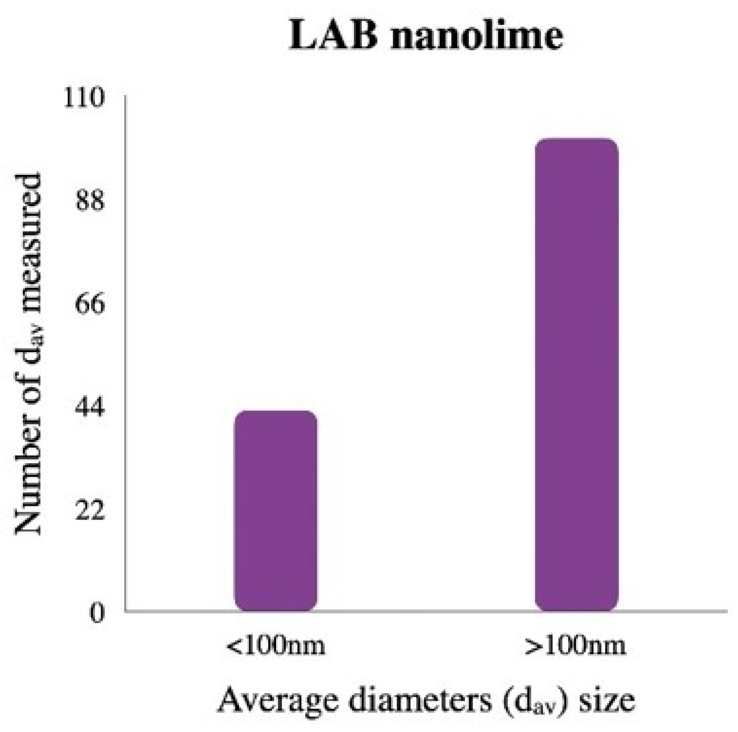
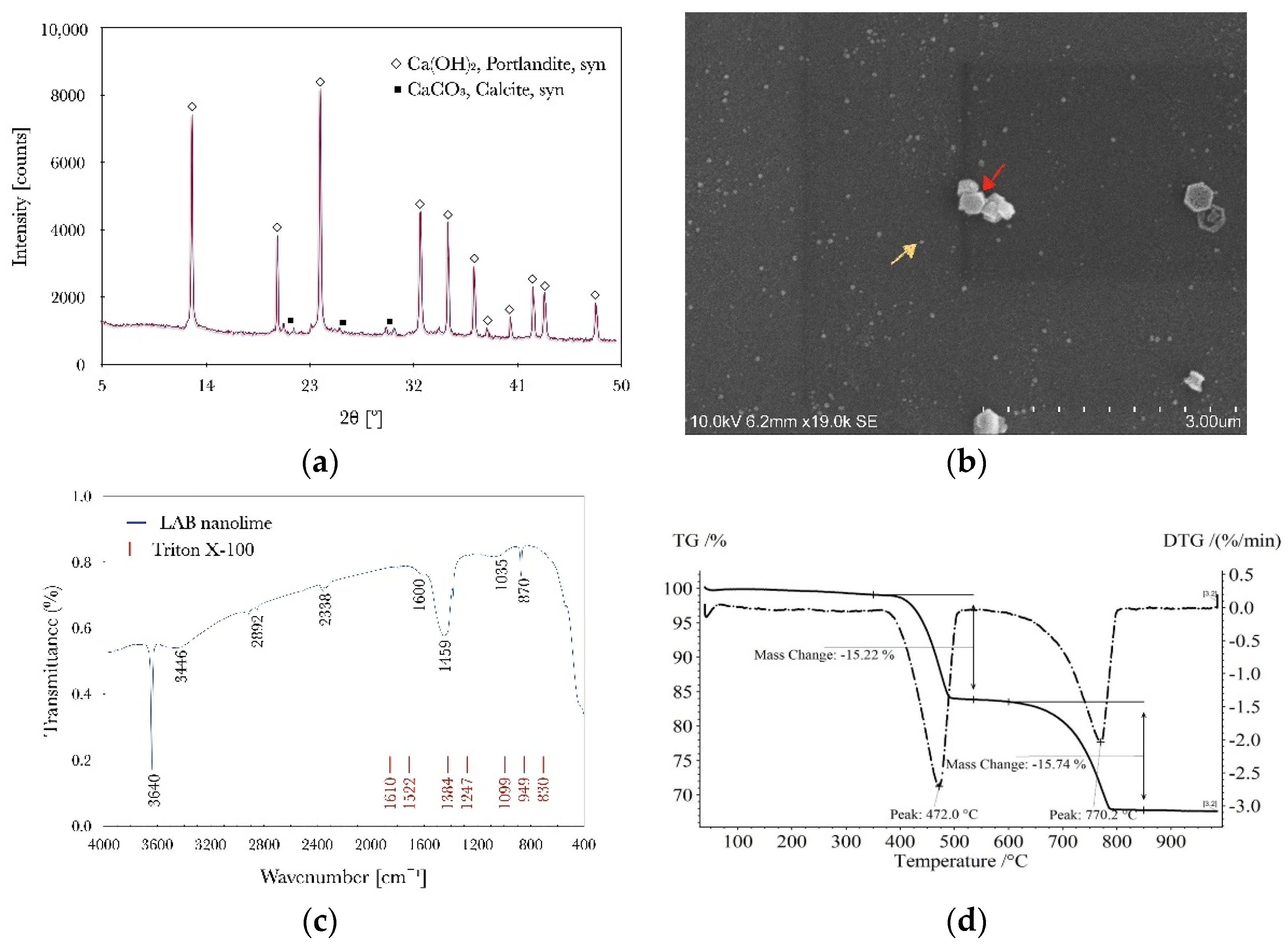
The powder XRD pattern of LAB nanolime (Figure 2a) shows the characteristic peaks of the main crystalline phase of Ca(OH)2 (calcium hydroxide) and the presence of low-intensity peaks of the crystalline phase of CaCO3 (calcium carbonate). The presence of CaCO3 indicates a partial carbonatation of LAB nanolime by atmospheric carbon dioxide (CO2) and humidity when exposed to air during the drying process [37] and analysis. The characteristic hexagonal shape of the Ca(OH)2 was shown by SEM analysis (Figure 2b and Figure A1, Appendix A). Two groups of distinct particles dimensions were observed: a large number of NPs of diameter <100 nm and clusters of larger particles of diameter >100 nm (Figure A2, Appendix A). The former NPs display a mean diameter of 47 nm (observed dmin of 18 nm and dmax of 83), and the later particles show a mean diameter of 300 nm (observed dmin of 165 nm and dmax of 592 nm) and less uniform orientation compared to the smallest NPs.
The infrared spectra (Figure 2c) of the synthesized Ca(OH)2 NPs confirms the presence of Ca(OH)2. The presence of the bands at wavelengths 3640 cm−1 (s), 3446 cm−1 (w), and 1600 cm−1 (w) correspond to the stretching of O-H and H-O-H [38], while the bands present at 2892 cm−1 (w), 2338 cm−1 (w), 1459 cm−1 (m), and 870 cm−1 (w) suggest the formation of CaCO3. Surfactant characteristic bands [39] were not found.
The TGA-DTG curve (Figure 2d) presents two temperature ranges: 350–550 °C and 600–850 °C, where significant weight loss occurred, determining the decomposition of Ca(OH)2 and CaCO3, respectively [40,41]. The average percentage of calcite was 33%, signifying the amount of nanolime that carbonated at any stage: synthesis, drying, and storage. TGA-DTG analysis confirms the absence of surfactant.

3.2. Evaluation of Treatment

The next sections’ discussions on the effects of the consolidants on the color appearance in the paint layers of the replicas have been based on the preliminary data obtained by colorimetry and optical and electron microscopy.

3.2.1. Optical Microscopy and Colorimetry Analysis

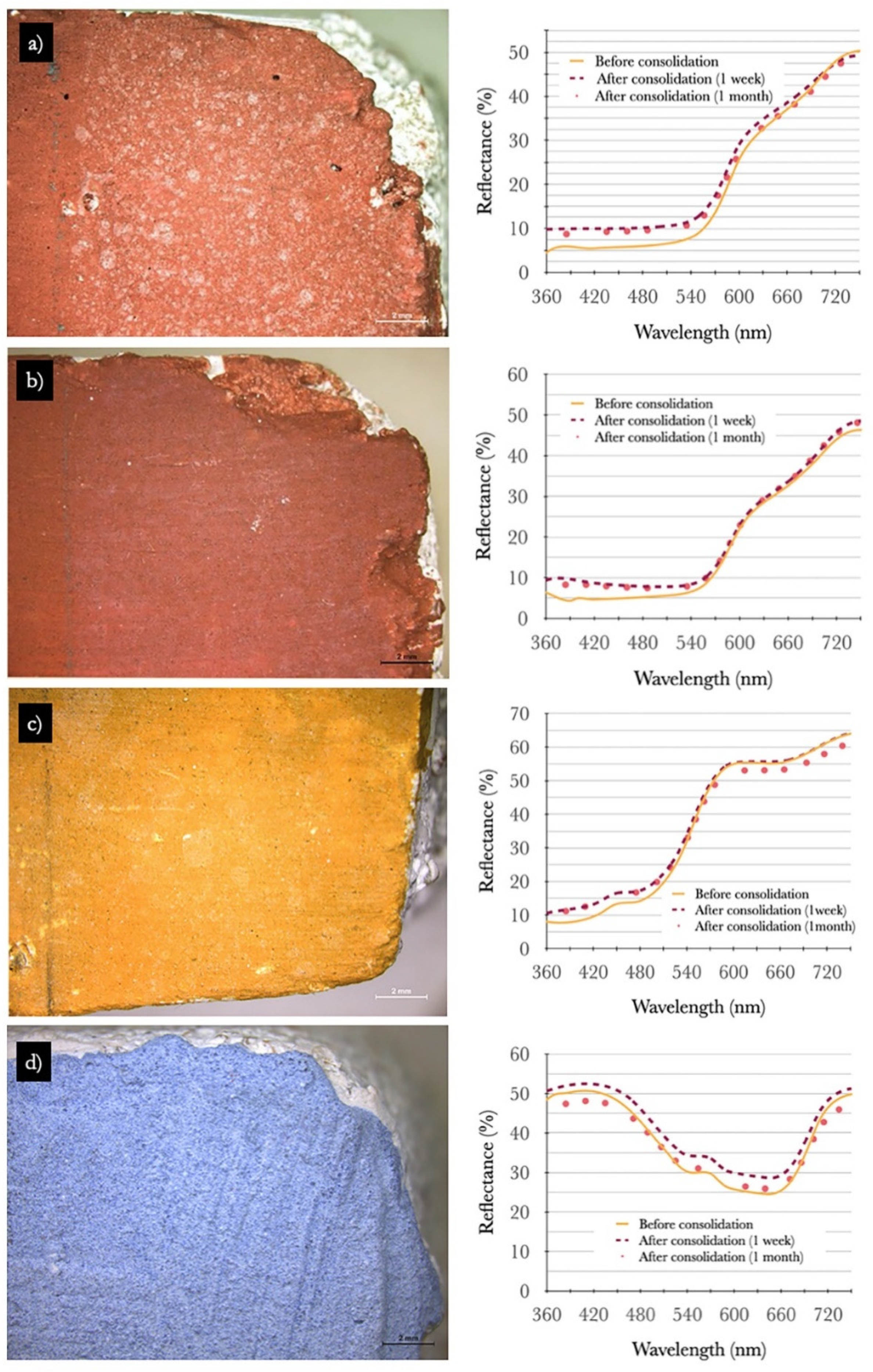
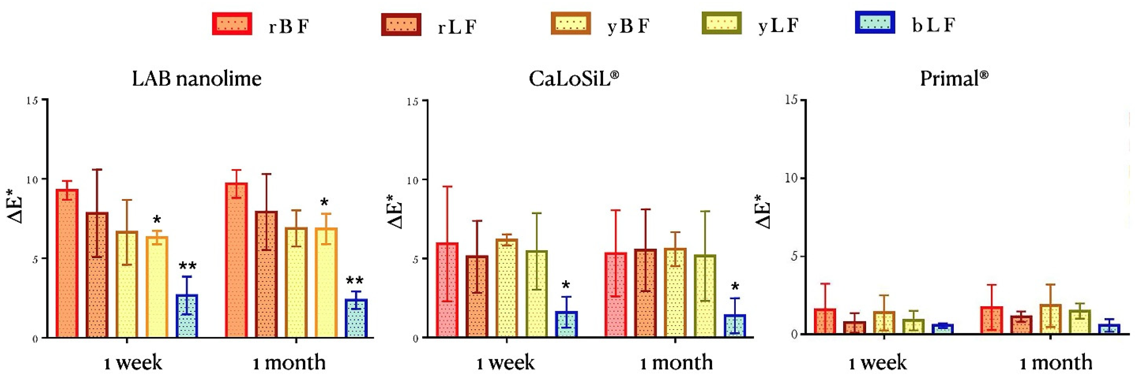
Figure 3 shows details of OM-Vis and the spectral curves of four paint layers made with buon and lime fresco after one week of treatment with two nanolimes. Figure 4 displays the average overall color difference ΔE* for all the replicas after one week and one month.
From Figure 3a–c, a white haze is observable over the paint layers after consolidation with the nanolimes. The visual impact is more noticeable in the darker tones of red (Figure 3). With the LAB nanolime, the white haze appears as spots spread unevenly with different sizes and shapes, while CaLoSiL® produced a more translucid pinkish veil over the surface (Figure 3a,b).
Color changes in consolidation treatments of paint layers are of particular importance, as they can alter the entire visual perception of a painting [42,43]. In the field of conservation, ∆E* ≥ 5 is an acceptable threshold for the impact on the color change that a consolidant may produce on the substrate color [27,28,42,43]. Red paint layers treated with the nanolimes LAB nanolime and CaLoSiL® showed ΔE* ≥ 5 even one month after treatment (Figure 4 and Table A1 in Appendix B). However, the shape of the spectral curves of the red ochres is not affected. Only a slight increase in the R% is noticed (Figure 3a,b).
By OM in the yellow paint layers of Figure 3c, a white haze is also perceived as translucid white spots, but the lighter hues of the pigment mask the effect reflected in lower values of ΔE*.
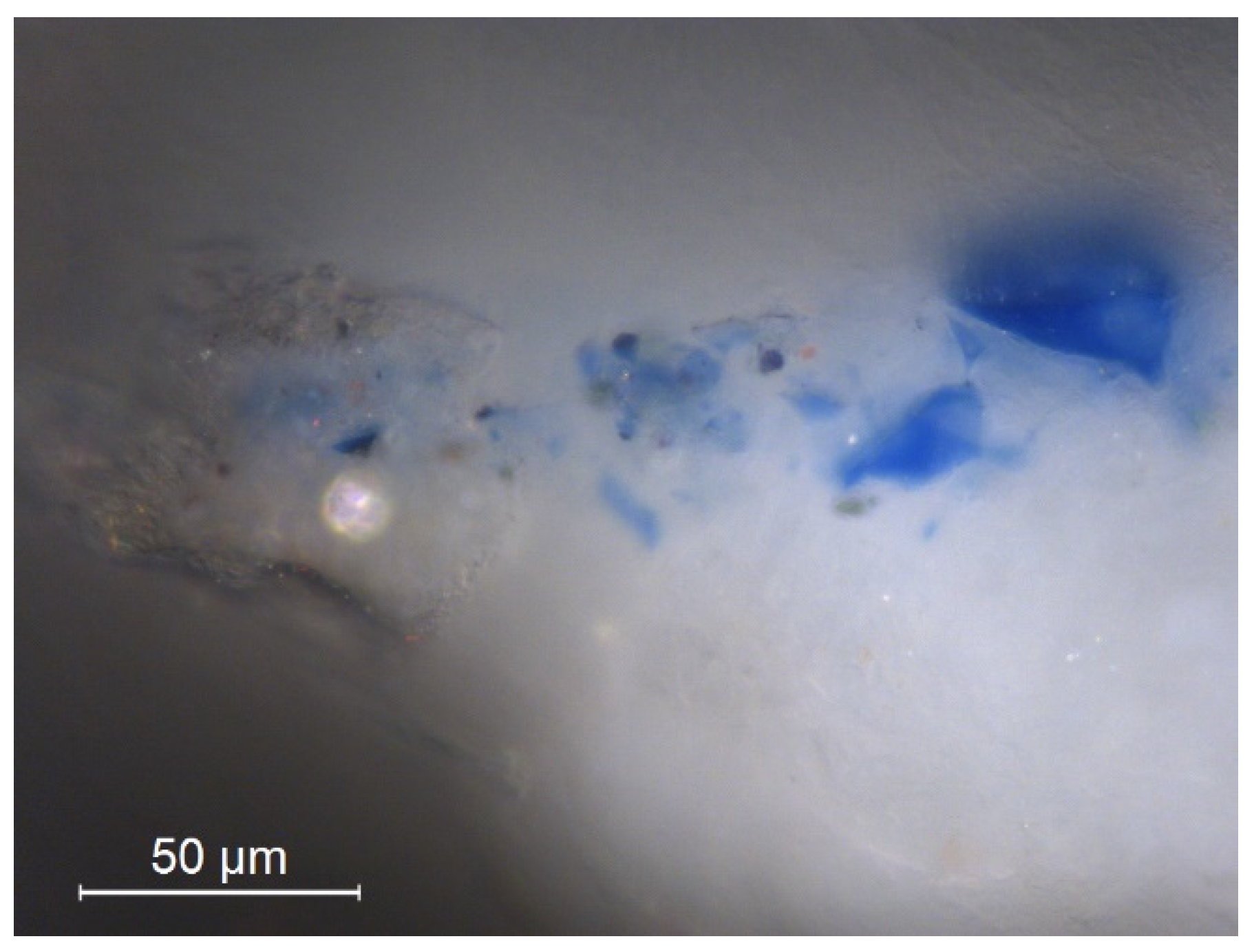
In turn, in the blue paint layers, the consolidation with the two nanolimes did not induce changes in the visual color perception, which could be explained by the low hiding power of smalt coarse particles in a white calcium matrix (Figure 3d and Figure 5). As Figure 4 shows, ΔE < 3 is significantly lower than the ΔE* obtained for the red and yellow ochre paint layers made with buon and lime fresco (Figure 3d and Figure 4).
Values of ΔH (shown in Table A1 in Appendix B) hint at the slight color variation for almost all paint layers, except for the red ochre paint layers, in particular treated with the commercial nanolime (applied in high concentration), where the values ΔH were higher than 2.5.
The finer particles of the ochre paint layers are intimately related to the increased hiding power and tinting strength of the red and yellow paint layers. This explains why these paint layers are more prone to color differences after treatment, in agreement with previous studies by Becherini et al. [27].
With nanolime treatments, ∆E* followed the next order: ∆E* rBF ≥ ∆E* rLF ≥ ∆E* yLF ≥ ∆E* yBF > ∆E* bLF (Figure 4). Besides particle sizes and pigment reflective index, the painting techniques also influence the end outcomes. With the buon fresco technique, the pigments are only mixed with water, while on lime fresco, the pigments are previously combined with lime milk before being laid down on the surface of fresh mortar. The thickness of the dry paint layers is significantly higher in the second case by the amount of calcium carbonate in the composition. The pigments are more distributed in the calcium matrices and tend to enlighten, justifying right from the start the increase of L* and decrease of color coordinates +a* and +b* (Figure 5).
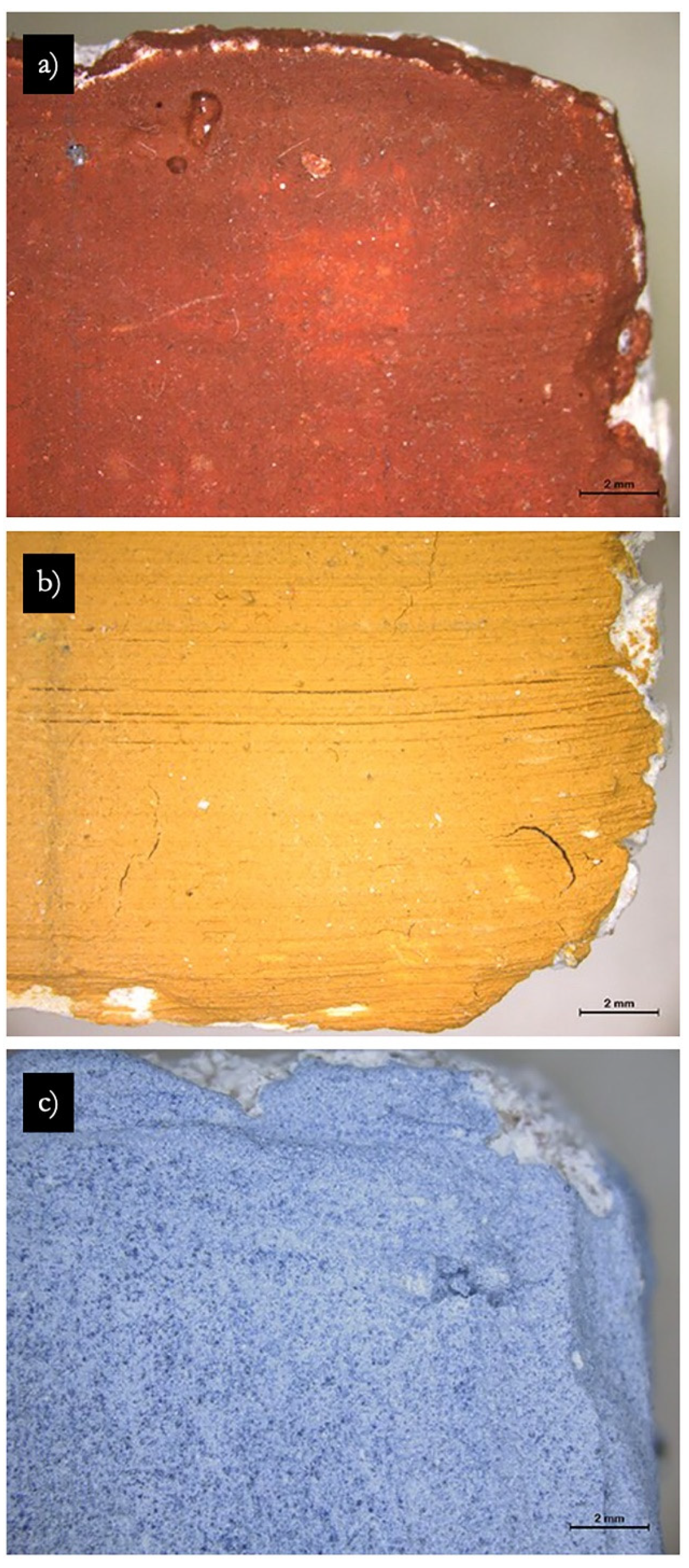
The mural set consolidated with the commercial acrylic resin PrimalTM SF-016 ER®, an improved version of Primal AC33 commonly used in the past as a consolidant/adhesive on mural paint layers, shows ∆E* < 3 for all the paint layers one week and one month after consolidation (Figure 4), where no visual color differences were noticeable on the paint surfaces (Figure 6). Based on the SEM-EDS analysis—discussed next in Section 3.2.2—the explanation relies on the chemical and morphological differences between the products and how they act as consolidants.

3.2.2. Scanning Electron Microscopy Analysis

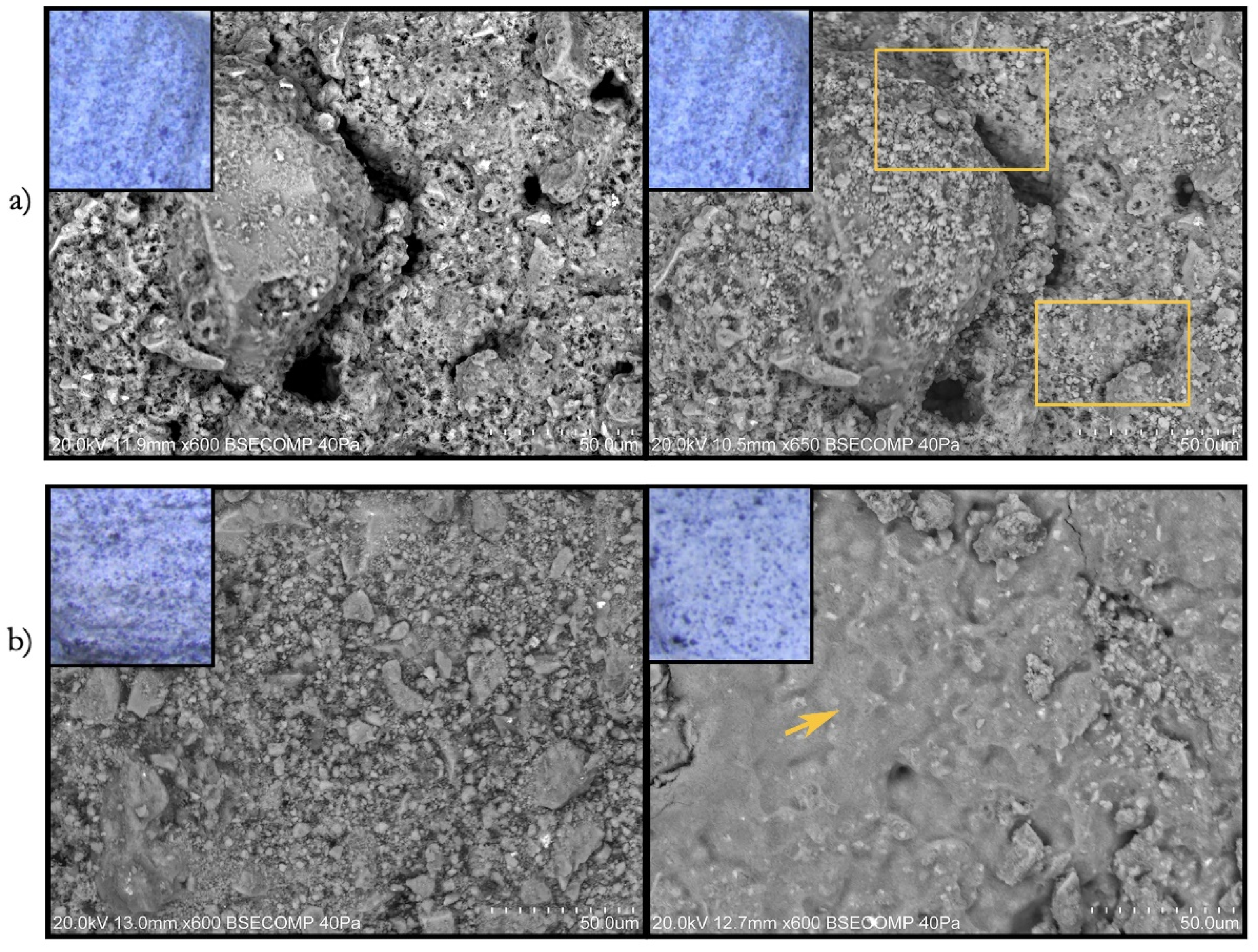
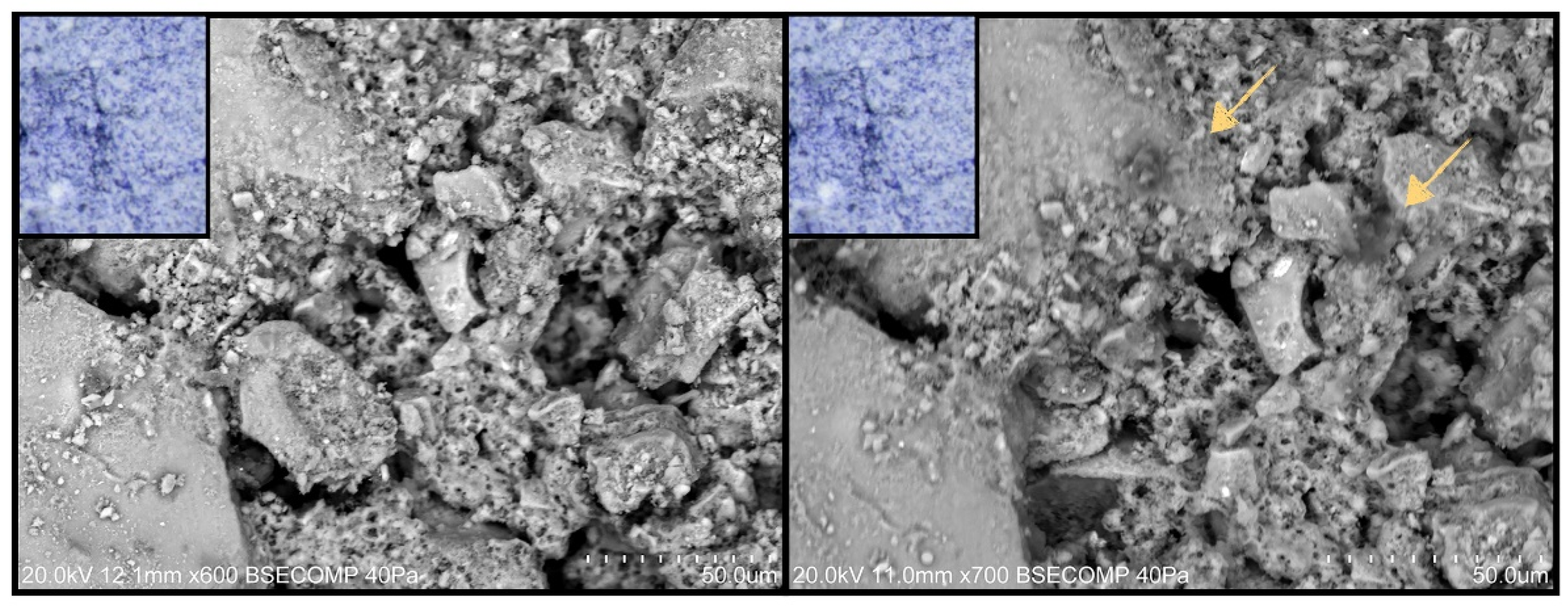
Figure 7 shows SEM BSE images of a blue paint layer before and one week after treatment with two nanolimes.
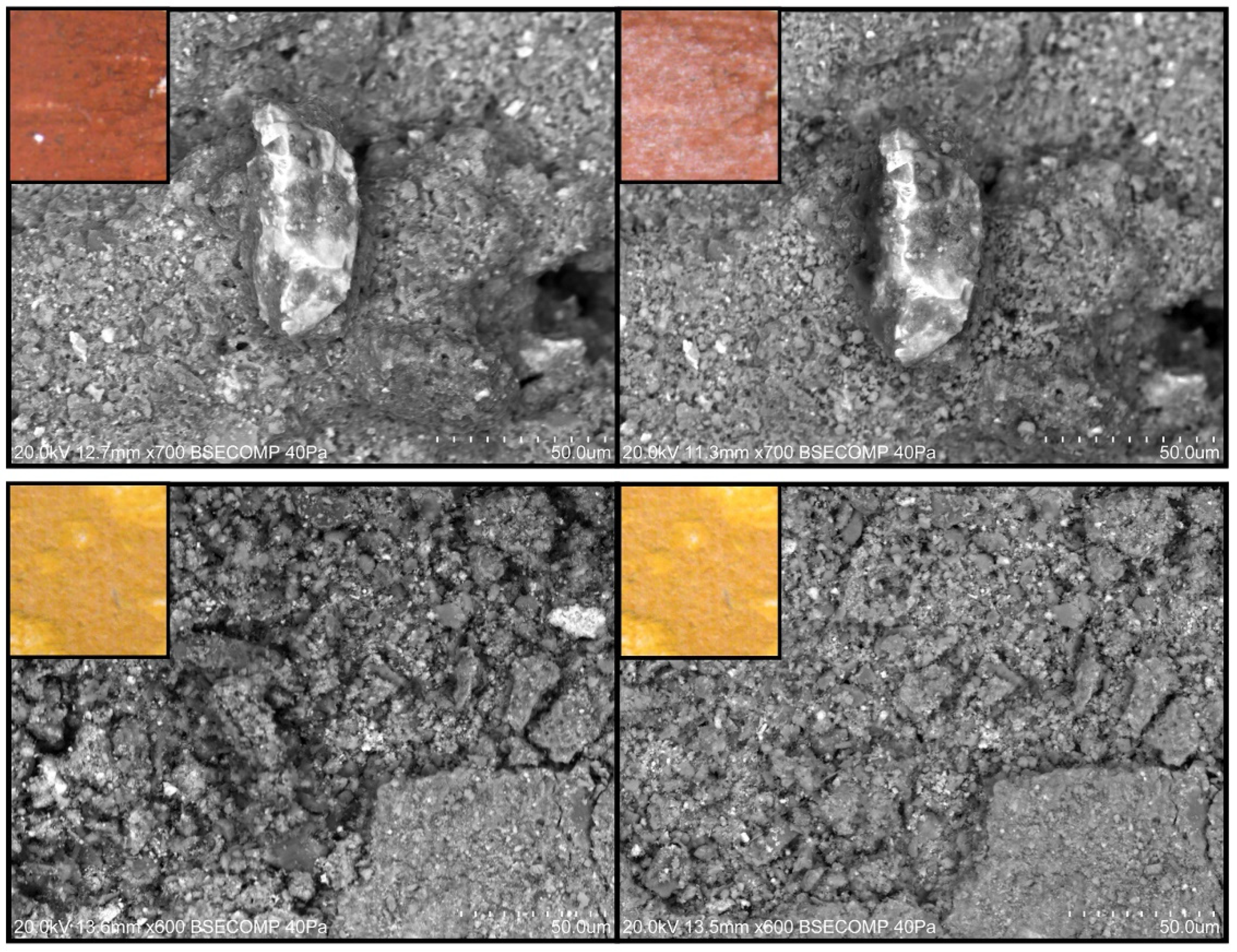
The surface of the replicas treated with LAB nanolime showed hexagonal-shaped particles of sizes ranging from 1.67 to 3.2 μm spread over the paint layers after one week of treatment (Figure 7a), with the same shape but bigger size than those of the synthesized nanolime (Figure 2b). Instead of penetrating in the lime mortar replica network or forming a layer, they appear heterogeneously accumulated on the paint layers, isolated and in clusters (Figure 7a), similarly to other studies on different lime-based substrates, such as carbonated stone buildings [43], limestone [44], and earthen walls [45]. A similar distribution of LAB nanolime was observable in the red and yellow paint layers (Figure A3, Appendix B). The distribution of the NPs in the painted surface in clusters appears to be a probable reason for the spotted appearance of the white haze in the red and yellow paint layers treated with LAB nanolime (Figure 3a,c).
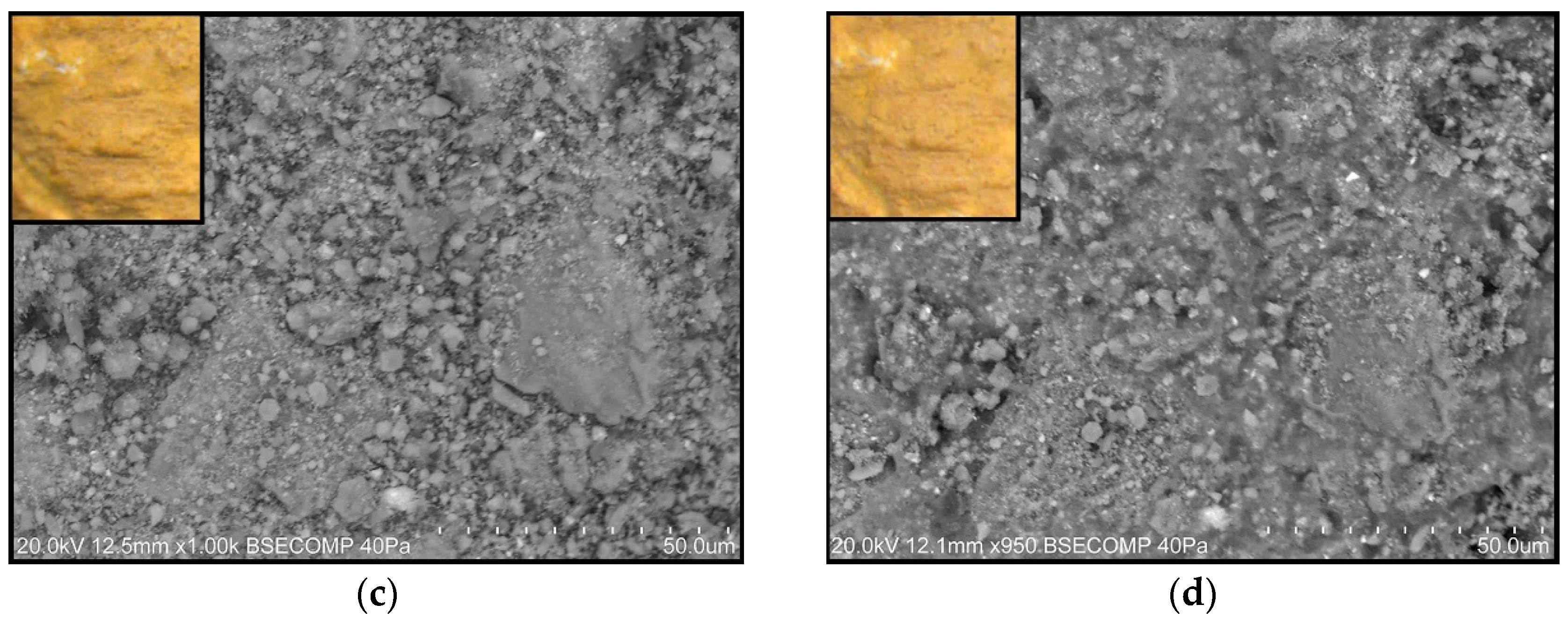
Alternately, treatment with CaLoSiL® has led to the formation of a layer with a homogenous appearance on the red, yellow, and blue painted surfaces after one week of treatment (Figure 7b and Figure 8), and suggests that CaLoSiL® or at least part of it accumulated on the surface, creating a coating layer. According to Ševčík et al. [46], this homogeneous layer indicates a more compacted microstructure and that nanolime has carbonatated. A similar homogeneous layer was observed by Gherardi et al. [47] after applying CaLoSiL® on limestone. At the same time, CaLoSiL® application changes the texture of the painted surfaces as is shown in Figure 7b and Figure 8; Dei and Salvadori [48] observed the same behavior, describing it as an indication of the strengthening and re-cohesion of the paint layers.
In Figure 7b, the blue-painted surface treated with CaLoSiL® appears completely covered after one week of treatment, whereas, in the ochre paint layers, some cracks and pores are incompletely covered (Figure 8). These could be because the coarser pigment particles and higher porosity of the blue paint layers permitted a higher deposition of the Ca(OH)2 NPs within the substrate matrix. However, not all NPs penetrated the matrix, as the covering layer observed in the surface indicates that particles at the surface carbonatated.
Although SEM analysis showed morphological change for the two nanolimes, it is hard to conclude that it is white haze, as changes also appeared on the blue-painted surface, where the effect was not observed by OM and spectrophotometry (Figure 3d, Figure 4, and Figure 7).
The two nanolimes appear to have the potential to recuperate the physicochemical network of the fresco replicas. However, these changes seem to be faster in the case of the commercial nanolime, which has carbonatated quicker than the synthesized nanolime. The difference between the nanolimes characteristics (particle size, degree of particles agglomeration, and dispersing solvent) influenced their reactivity, penetration degrees, and carbonatation mechanisms. When applied, the smaller-sized nanolime, CaLoSiL® (Table 3), carbonated more quickly. Consequently, the nanolimes had a different effect on the morphological of the paint layers. Therefore, the mechanism of the white haze formation caused by the two nanolimes might be different for each case: the accumulation of Ca(OH)2 NPs on the surface (LAB nanolime) and the superficial carbonatation of the product (CaLoSiL®), as proposed by Normand et al. [13]. Control over the CaCO3 polymorphs during carbonatation of NPs [46] and the speed of carbonatation through relative humidity [24] could be the key to limit the white haze formation [46]. The use of diluted dispersions (0.05 to 5 g/L) [29,30,31,48] or using mixed dispersing solvents [9] can also control white haze appearance.
Another route to tackle white haze formation could be by studying the application method [26,49] or mechanical cleaning [49]. During research on the restriction of white haze formation during and after application of nanolime, it is possible to reduce the formed white haze by wetting the surface with water after application; however, do not eliminate it [26]. As an alternative, the removal of the excess nanoconsolidant after each application was the best course of action to avoid, to a large extent, the appearance of the white haze without a decrease in the consolidation effectiveness [49].
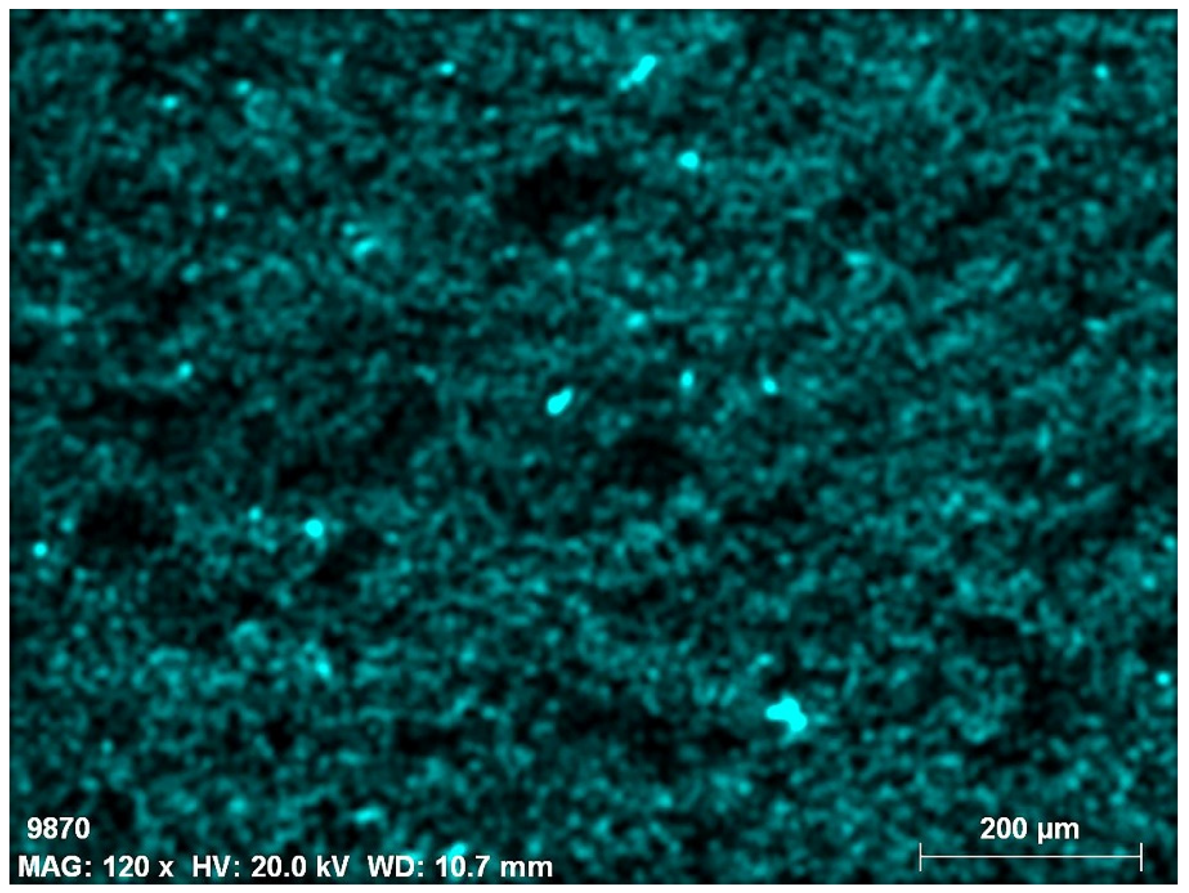
Treatment with PrimalTM SF-016 ER® produced few changes in the appearance of the blue paint layers. OM images and colorimetry results confirmed results. After one week of treatment, a thin film formed in some cracks surrounding the pigment particles (Figure 9). The UVF photographic documentation and the elemental map of carbon (C) shown in Figure 10 and Figure 11 support that the treatment with PrimalTM SF-016 ER® did not form any organic coating layer on the surface.
Figure 7 and Figure 9 show that the treatment with nanolime compounds caused more noticeable changes in the paint layers than PrimalTM SF-016 ER®. It is relevant to note that the concentration of the Primal® (2% of v/v of a commercial solution that contains 50–51% solid content) is lower than the concentration of LAB nanolime and CaLoSiL®, which was 25 g/L.
Yet, higher concentrations of PrimalTM SF-016 ER® can lead to the formation of a film over the surface, which can cause the loss of the readability and integrity of the artwork in the long term, especially when exposed to aggressive environmental conditions [12,27]. Consequently, the usage of low concentrations of acrylic resins is preferred, as it can promote the penetration of the acrylic resin within the substrate and control the hydrophobic film formation.

4. Conclusions

The evaluation of three consolidant revealed that the color changes followed the order: LAB nanolime > CaLoSiL® IP25 > PrimalTM SF-016 ER®.
The increase of the CIEL* coordinate and ∆E color noticed with the two nanolimes after treatment is associated with a white haze formation. The impact on color appearance is higher in the darker tones. It was also dependent on the pigment. Ochre paint layers were more prone to differences in color perception after treatment with nanolime.
The obtained preliminary data open up perspectives for further studies on understating the effect of different nanoconsolidants on deteriorated frescoes, in particular, the effect of nanolime dispersions on the white haze formation over the paint layer and for a larger time scale. From a materials science point of view, there is a set of several factors to be explored, such as synthesis approaches to tailor particle morphology, and to avoid high agglomeration, uniform particle orientation in addition to particle kinetic stability, nature of dispersing media and dispersions concertation, and numbers of application. Assessment of the cohesion and consolidation characteristics of the treated surfaces, carbonatation mechanism, and interrelation with the nanoconsolidant nature is another further challenge. Conservation science demands consistent and various studies of the origin of white haze formation, such as, for instance, the possible relation between the distinct textures (smooth or coarse) of the paint layer surface, nanoparticles arrangement over the treated surface, and veil formation. We anticipate that this preliminary research could be extended for synthetic pigments, with potential for modern wall painting conservation.

Author Contributions

Conceptualization, M.G., C.G., A.C. and P.I.G.; methodology, B.B., M.G., C.G. and P.I.G.; validation, B.B., M.G., C.G. and P.I.G.; formal analysis, B.B., M.G., C.G. and P.I.G.; investigation, B.B., M.G., C.G. and P.I.G.; resources, M.G., C.G., A.C. and P.I.G.; writing—original draft preparation, B.B., M.G., C.G. and P.I.G.; writing—review and editing, B.B., M.G., C.G., A.C. and P.I.G.; supervision, M.G., C.G., and P.I.G.; project administration, M.G., C.G., and P.I.G.; funding acquisition, M.G., C.G., A.C. and P.I.G. All authors have read and agreed to the published version of the manuscript.

Funding

Fundação para a Ciência e a Technologia FCT, Portugal (the Portuguese national funding agency for science, research and technology): UIDB/04449/2020; UIDP/04449/2020; DL 57/2016/CP1372/CT0013; DL/57/2016/CP1338. EMJMD ARCHMAT, co-founded by Erasmus+ program under GA N. 2018-1468/001-001.

Institutional Review Board Statement

Not applicable.

Informed Consent Statement

Not applicable.

Data Availability Statement

The data presented in this study are available in main content and Appendix A and Appendix B.

Acknowledgments

The authors thank FCT for the contract programs of Ref. DL 57/2016/CP1372/CT0013 (PG) and DL/57/2016/CP1338. Furthermore, the authors acknowledge FCT for the funding support through UIDB/04449/2020 and UIDP/04449/2020, and to EMJMD ARCHMAT, co-founded by Erasmus+ program under GA N. 2018-1468/001-001. The authors also acknowledge. Luís Dias for the support with the SEM.

Conflicts of Interest

The authors declare no conflict of interest.

Appendix A. Laboratory-Synthesized Nanolime Characterization
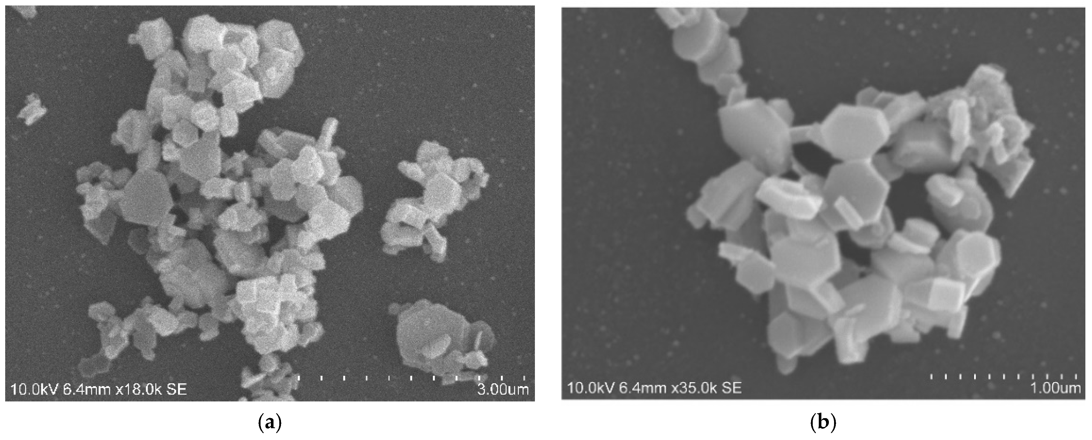
Figure A1. Characterization of LAB nanoparticles: SEM images. (a) Magnification 18.0 k. (b) Magnification 35.0 k.
Heritage 04 00183 g0a1
Figure A2. Particles size diagram for lab nanolime; particles dimensions taken from SEM images. Presentation is rather schematic because, while the shape of the particles is uniform, particle dimensions vary.
Heritage 04 00183 g0a2

Appendix B. Treatment Evaluation

Table A1. Color differences using CIE*L*a*b* coordinates for the paint layers treated with the two kinds of nanolime-Lab nanolime and commercial CaLoSiL® IP25 (measurements taken one week and one month after replicas treatment). ∆L* corresponds to variations in the color values in the dark–light axis; ∆E*—total color variations and ΔH—absolute color differences were calculated according to Equations (2) and (3), respectively [37].
ConsolidantSample Ref.ColorPainting TechniqueΔL*ΔE*ΔH*ΔL*ΔE*ΔH*
One week afterOne month after
LAB nanolime3rBFRedBuon Fresco4.489.912.55.059.102.21
7rBFRedBuon Fresco3.768.731.765.2810.772.47
11rBFRedBuon Fresco4.999.211.843.499.282.26
3rLFRedLime Fresco2.4910.482.82.1710.172.89
7rLFRedLime Fresco3.648.532.063.148.442.10
11rLFRedLime Fresco3.325.001.153.095.311.10
4yBFYellowBuon Fresco0.668.991.551.528.142.14
7yBFYellowBuon Fresco0.515.361.921.205.911.87
11yBFYellowBuon Fresco−0.755.580.61−0.946.620.39
1yLFYellowLime Fresco0.335.970.91−0.155.810.89
7yLFYellowLime Fresco1.116.201.15−0.067.691.19
11yLFYellowLime Fresco0.726.791.15−0.487.061.10
3bLFBlueLime Fresco−0.311.750.091.291.830.07
7bLFBlueLime Fresco2.042.250.011.732.420.24
12bLFBlueLime Fresco3.024.000.260.442.890.32
CaLoSiL® IP2512rBFRedBuon Fresco−0.012.191.260.883.101.40
5rBFRedBuon Fresco2.629.455.642.248.364.97
9rBFRedBuon Fresco3.756.174.052.564.553.34
12rLFRedLime Fresco1.85.312.983.838.032.42
5rLFRedLime Fresco1.687.294.121.155.703.00
9rLFRedLime Fresco0.82.741.480.792.871.34
1yBFYellowBuon Fresco−1.346.121.700.516.631.89
5yBFYellowBuon Fresco−1.655.851.860.354.472.01
9yBFYellowBuon Fresco−0.756.551.960.085.682.11
3yLFYellowLime Fresco0.853.401.060.582.780.90
5yLFYellowLime Fresco0.324.851.150.054.391.38
9yLFYellowLime Fresco0.698.122.510.698.292.57
1bLFBlueLime Fresco2.382.690.472.442.630.51
5bLFBlueLime Fresco1.321.340.010.931.030.15
9bLFBlueLime Fresco0.680.790.22−0.390.510.19
PrimalTM SF-016 ER®2rBFRedBuon Fresco−0.410.440.16−0.350.990.18
6rBFRedBuon Fresco−0.151.480.18−0.931.530.40
10rBFRedBuon Fresco0.140.340.020.150.930.43
2rLFRedLime Fresco0.411.120.18−0.242.110.57
6rLFRedLime Fresco0.361.000.720.071.080.23
10rLFRedLime Fresco0.180.360.05−0.030.760.21
2yBFYellowBuon Fresco−0.931.070.08−1.031.120.003
6yBFYellowBuon Fresco−0.372.640.40−0.883.420.49
10yBFYellowBuon Fresco0.450.460.10−0.141.010.90
2yLFYellowLime Fresco−0.090.210.11−1.071.280.30
6yLFYellowLime Fresco0.551.110.03−0.71.640.11
10yLFYellowLime Fresco−0.711.400.41−2.633.440.43
2bLFBlueLime Fresco0.450.470.14−0.210.220.06
6bLFBlueLime Fresco0.520.520.641.011.010.03
10bLFBlueLime Fresco−0.680.750.14−0.510.530.09
Figure A3. SEM images of red and yellow paint layers treated with LAB nanolime; before and one week after treatment: Upper image: rLF; Lower image: yBF. Detail images obtained by OM-Vis of the studied areas are in the top left corner.
Heritage 04 00183 g0a3

References

  1. ICOMOS. Principios Para la Preservación, Conservación y Restauración de Pinturas Murales. In Proceedings of the 14ª Asamblea General del ICOMOS, Victoria Falls, Zimbabwe, 27–31 October 2003. [Google Scholar]
  2. Mora, P.; Mora, L.; Philipott, P. Conservation of Wall Paintings; Butterwoths: Oxford, UK, 1984. [Google Scholar]
  3. Fichner-Rathus, L. Understanding Art, 10th ed.; Wadsworth: Boston, MA, USA, 2013; ISBN 13-978-1-111-83695-5. [Google Scholar]
  4. Mora, P. Causes of Deterioration of Mural Paintings; International Centre for the Study of the Preservation and Restoration of Cultural Property (ICCROM): Rome, Italy, 2018. [Google Scholar]
  5. Spencer, B.N.; Rosenthal, A.; Podany, J.; Larson, J.H.; Zaccari, F. Art Conservation and Restoration, Encyclopaedia Britannica. Available online: https://www.britannica.com/art/art-conservation-and-restoration (accessed on 1 November 2020).
  6. Gil, M. Conservação de Pintura Mural, Estudo e Consolidação de Argamassas de Cal Aérea e Areia com Falta de Coesão; Laboratório Nacional de Engenharia Civil: Lisboa, Portugal, 2002. [Google Scholar]
  7. Magaloni, K.D. La pintura mural y su conservación. Arqueol. Mex. 2011, 108, 33–37. [Google Scholar]
  8. Rodrigues, J.D. Consolidation of decayed stones. A delicate problem with few practical solutions. In Proceedings of the 3rd International Seminar, Guimarães, Portugal, 7–9 November 2001; pp. 3–14. [Google Scholar]
  9. Zhu, J.; Zhang, P.; Ding, J.; Dong, Y.; Cao, Y.; Dong, W.; Zhao, X.; Li, X.; Camaiti, M. Nano Ca(OH)2: A review on synthesis, properties and applications. J. Cult. Herit. 2021, 50, 25–42. [Google Scholar] [CrossRef]
  10. Brajer, I.; Kalsbeek, N. Limewater Absorption and Calcite Crystal Formation on a Limewater-Impregnated Secco Wall Painting. Stud. Conserv. 1999, 44, 145–156. [Google Scholar] [CrossRef]
  11. Baglioni, P.; Chelazzi, D.; Giorgi, R. Nanotechnologies in the Conservation of Cultural Heritage, a Compendium of Materials and Techniques; Springer: Heidelberg, Germany, 2015. [Google Scholar] [CrossRef]
  12. Kaszowska, Z.; Kot, M.; Białek-Kostecka, D.; Forczek-Sajdak, A. Application of micro-indentation tests to assess the consolidation procedure of historic wall paintings. J. Cult. Herit. 2018, 36, 286–296. [Google Scholar] [CrossRef]
  13. Normand, L.; Duchêne, S.; Vergès-Belmin, V.; Dandrel, C.; Giovannacci, D.; Nowik, W. Comparative in Situ Study of Nanolime, Ethyl Silicate and Acrylic Resin for Consolidation of Wall Paintings with High Water and Salt Contents at the Chapter Hall of Chartres Cathedral. Int. J. Arch. Herit. 2020, 14, 1120–1133. [Google Scholar] [CrossRef]
  14. Baglioni, P.; Carretti, E.; Dei, L.; Giorgi, R. Nanotechnology in wall painting conservation. In Self-Assambly; Robinson, B.H., Ed.; IOS-Press: Amsterdam, The Netherlands, 2013; pp. 32–41. ISBN 1-58603-382-4. [Google Scholar]
  15. Girginova, P.I.; Galacho, C.; Veiga, R.; Silva, A.S.; Candeias, A. Inorganic Nanomaterials for Restoration of Cultural Heritage: Synthesis Approaches towards Nanoconsolidants for Stone and Wall Paintings. ChemSusChem 2018, 11, 4168–4182. [Google Scholar] [CrossRef]
  16. David, M.E.; Ion, R.-M.; Grigorescu, R.M.; Iancu, L.; Andrei, E.R. Nanomaterials Used in Conservation and Restoration of Cultural Heritage: An Up-to-Date Overview. Materials 2020, 13, 2064. [Google Scholar] [CrossRef]
  17. Lisuzzo, L.; Cavallaro, G.; Milioto, S.; Lazzara, G. Halloysite nanotubes filled with MgO for paper reinforcement and deacidification. Appl. Clay Sci. 2021, 213, 106231. [Google Scholar] [CrossRef]
  18. Rodriguez-Navarro, C.; Ruiz-Agudo, E. Nanolimes: From synthesis to application. Pure Appl. Chem. 2017, 90, 523–550. [Google Scholar] [CrossRef]
  19. Becerra, J.; Zaderenko, A.P.; Ortiz, P. Basic Protocol for on-site testing consoli-dant nanoparticles on stone cultural heritage. Heritage 2019, 2, 2712–2724. [Google Scholar] [CrossRef] [Green Version]
  20. Cavallaro, G.; Milioto, S.; Parisi, F.; Lazzara, G. Halloysite Nanotubes Loaded with Calcium Hydroxide: Alkaline Fillers for the Deacidification of Waterlogged Archeological Woods. ACS Appl. Mater. Interfaces 2018, 10, 27355–27364. [Google Scholar] [CrossRef]
  21. Camerini, R.; Chelazzi, D.; Giorgi, R.; Baglioni, P. Hybrid nano-composites for the consolidation of earthen masonry. J. Colloid Interface Sci. 2018, 539, 504–515. [Google Scholar] [CrossRef]
  22. Gómez-Villalba, L.S.; López-Arce, P.; Fort, R.; Álvarez, M. La aportación de la nanociencia a la conservación de bienes del patrimonio cultural. Patrim. Cult. Esp. 2010, 4, 43–56. [Google Scholar]
  23. Girginova, P.I.; Galacho, C.; Veiga, R.; Silva, A.S.; Candeias, A. Study of mechanical properties of alkaline earth hydroxide nanoconsolidants for lime mortars. Constr. Build. Mater. 2019, 236, 117520. [Google Scholar] [CrossRef]
  24. López-Arce, P.; Gómez-Villalba, L.; Martinez-Ramirez, S.; de Buergo, M.; Fort, R. Influence of relative humidity on the carbonation of calcium hydroxide nanoparticles and the formation of calcium carbonate polymorphs. Powder Technol. 2011, 205, 263–269. [Google Scholar] [CrossRef] [Green Version]
  25. Álvarez, A.E.; Nadal, L.F. Evaluación del proceso de carbonatación de nanocales aplicadas a pinturas murales prehispánicas de origen maya. Interv. Rev. Int. Conserv. Restauración Museol. 2010, 1, 31–41. [Google Scholar] [CrossRef] [Green Version]
  26. Vojtěchovský, J. Surface consolidation of wall paintings using lime nano-suspensions. Acta Polytech. 2017, 57, 139–148. [Google Scholar] [CrossRef] [Green Version]
  27. Becherini, F.; Durante, C.; Bourguignon, E.; Vigni, M.L.; Detalle, V.; Bernardi, A.; Tomasin, P. Aesthetic compatibility assessment of consolidants for wall paintings by means of multivariate analysis of colorimetric data. Chem. Cent. J. 2018, 12, 98. [Google Scholar] [CrossRef] [Green Version]
  28. Bourguignon, E.; Tomasin, P.; Detalle, V.; Vallet, J.-M.; Labouré, M.; Olteanu, I.; Favaro, M.; Chiurato, M.A.; Bernardi, A.; Becherini, F. Calcium alkoxides as alternative consolidants for wall paintings: Evaluation of their performance in laboratory and on site, on model and original samples, in comparison to conventional products. J. Cult. Herit. 2018, 29, 54–66. [Google Scholar] [CrossRef]
  29. Ambrosi, M.; Dei, L.; Giorgi, R.; Neto, C.; Baglioni, P. Colloidal Particles of Ca(OH)2: Properties and Applications to Restoration of Frescoes. Langmuir 2001, 17, 4251–4255. [Google Scholar] [CrossRef]
  30. Daehne, A.; Herm, C. Calcium hydroxide nanosols for the consolidation of porous building materials-results from EU-STONECORE. Herit. Sci. 2013, 1, 11. [Google Scholar] [CrossRef] [Green Version]
  31. Baglioni, P.; Chelazzi, D.; Giorgi, R.; Carretti, E.; Toccafondi, N.; Jaidar, Y. Commercial Ca(OH)2 nanoparticles for the consolidation of immovable works of art. Appl. Phys. A 2013, 114, 723–732. [Google Scholar] [CrossRef]
  32. Natali, I.; Saladino, M.L.; Andriulo, F.; Martino, D.C.; Caponetti, E.; Carretti, E.; Dei, L. Consolidation and protection by nanolime: Recent advances for the conservation of the graffiti, Carceri dello Steri Palermo and of the 18th century lunettes, SS. Giuda e Simone Cloister, Corniola (Empoli). J. Cult. Herit. 2014, 15, 151–158. [Google Scholar] [CrossRef]
  33. Giorgi, R.; Bozzi, C.; Dei, L.; Gabbiani, C.; Ninham, A.B.W.; Baglioni, P. Nanoparticles of Mg(OH)2: Synthesis and Application to Paper Conservation. Langmuir 2005, 21, 8495–8501. [Google Scholar] [CrossRef]
  34. Girginova, P.I.; Galacho, C.; Mirão, J.; Veiga, R.; Silva, A.S.; Candeias, A. Preliminary studies of consolidation of wall paintings: Synthesis and characterisation of nanolime. Conserv. Património 2016, 23, 103–107. [Google Scholar] [CrossRef] [Green Version]
  35. Gil, M.; Rosado, T.; Ribeiro, I.; Pestana, J.A.; Caldeira, A.T.; Carvalho, M.L.; Dias, L.; Mirão, J.; Candeias, A. Are theyfrescopaintings? Technical and material study ofCasas Pintadasof Vasco da Gama house in Évora (Southern Portugal). X-Ray Spectrom. 2015, 44, 154–162. [Google Scholar] [CrossRef]
  36. Johnston-Feller, R. Color science in the examination of museum objects: Nondestructive procedures. Color Res. Appl. 2002, 27, 456–457. [Google Scholar] [CrossRef]
  37. Michalopoulou, A.; Maravelaki, P.-N.; Kilikoglou, V.; Karatasios, I. Morphological characterization of water-based nanolime dispersions. J. Cult. Herit. 2020, 46, 11–20. [Google Scholar] [CrossRef]
  38. Nasrazadani, S.; Eureste, E. Application of FTIR for Quantitive Lime Analysis. Available online: https://library.ctr.utexas.edu/digitized/texasarchive/phase2/9028-01-1.pdf (accessed on 1 December 2000).
  39. Kimura, N.; Umemura, J.; Hayashi, S. Polarized FT-IR Spectra of Water in the Middle Phase of Triton X100-Water System. J. Colloid Interface Sci. 1996, 182, 356–364. [Google Scholar] [CrossRef]
  40. Rajisha, K.R.; Deepa, B.; Pothan, L.A.; Thomas, S. Thermochemical and spectroscopic characterization of natural fibre composites. In Interface Engineering of Natural Fibre Composites for Maximum Performance; Zafeiropoulos, N.E., Ed.; Woodhead Publishing: Cambridge, UK, 2011; pp. 241–274. [Google Scholar]
  41. Rodriguez-Navarro, C.; Suzuki, M.A.; Ruiz-Agudo, E. Alcohol Dispersions of Calcium Hydroxide Nanoparticles for Stone Conservation. Langmuir 2013, 29, 11457–11470. [Google Scholar] [CrossRef] [PubMed]
  42. Delgado, J.R.; Grossi, A. Indicators and ratings for the compatibility assessment of conservation actions. J. Cult. Herit. 2007, 8, 32–43. [Google Scholar] [CrossRef]
  43. Becerra, J.; Zaderenko, A.; Ortiz, R.; Karapanagiotis, I.; Ortiz, P. Comparison of the performance of a novel nanolime doped with ZnO quantum dots with common consolidants for historical carbonate stone buildings. Appl. Clay Sci. 2020, 195, 105732. [Google Scholar] [CrossRef]
  44. Borsoi, G.; Lubelli, B.; van Hees, R.; Veiga, R.; Silva, A.S.; Colla, L.; Fedele, L.; Tomasin, P. Effect of solvent on nanolime transport within limestone: How to improve in-depth deposition. Colloids Surf. A Physicochem. Eng. Asp. 2016, 497, 171–181. [Google Scholar] [CrossRef]
  45. Lanzón, M.; De Stefano, V.; Gaitán, J.C.M.; Cardiel, I.B.; Gutiérrez-Carrillo, M.L. Characterisation of earthen walls in the Generalife (Alhambra): Microstructural and physical changes induced by deposition of Ca(OH)2 nanoparticles in original and reconstructed samples. Constr. Build. Mater. 2019, 232, 117202. [Google Scholar] [CrossRef]
  46. Ševčík, R.; Mácová, P.; Estébanez, M.P.; Viani, A. Influence of additions of synthetic anhydrous calcium carbonate polymorphs on nanolime carbonation. Constr. Build. Mater. 2019, 228, 116802. [Google Scholar] [CrossRef]
  47. Gherardi, F.; Otero, J.; Blakeley, R.; Colston, B. Application of Nanolimes for the Consolidation of Limestone from the Medieval Bishop’s Palace, Lincoln, UK. Stud. Conserv. 2020, 65, P90–P97. [Google Scholar] [CrossRef]
  48. Dei, L.; Salvadori, B. Nanotechnology in cultural heritage conservation: Nanometric slaked lime saves architectonic and artistic surfaces from decay. J. Cult. Herit. 2006, 7, 110–115. [Google Scholar] [CrossRef]
  49. López-Martínez, T.; Otero, J. Preventing the Undesired Surface Veiling after Nanolime Treatments on Wall Paintings: Preliminary Investigations. Coatings 2021, 11, 1083. [Google Scholar] [CrossRef]
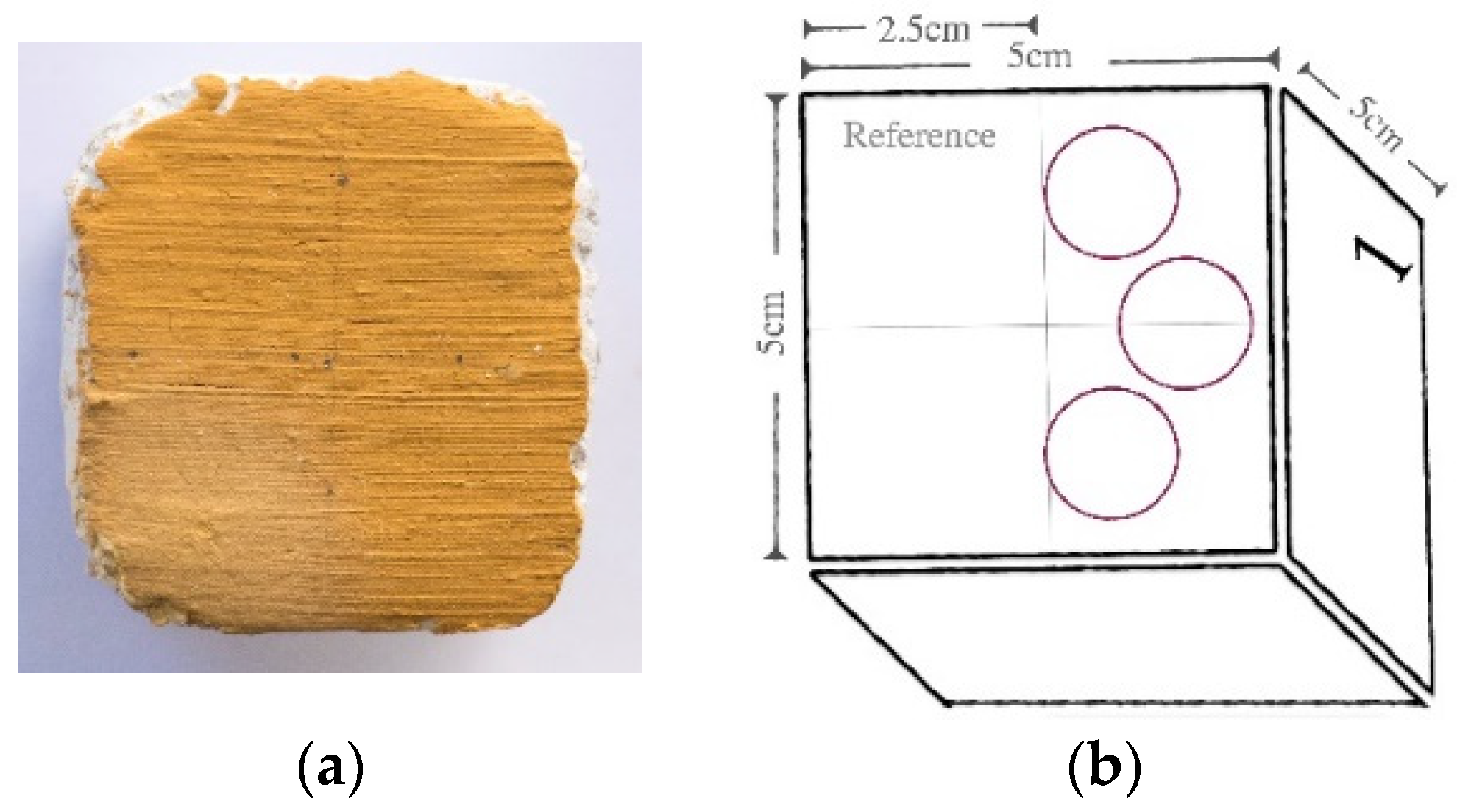
Figure 1. Colorimetry and spectrophotometry: analyzed areas. The results obtained in CIEL*a*b* chromatic space are the average of three measurements (three points and three light flashes) taken on each of the replica’s paint layers in an area of 2.5 cm in diameter. (a) Real sample surface; (b) Schematic representation of the analyzed surfaces.
Heritage 04 00183 g001
Figure 2. Characterization of LAB nanoparticles: (a) XRD pattern; (b) SEM image (yellow arrow—NPs < 100 nm; red arrow—NPs > 100 nm); (c) FT-IR spectra (red: characteristic peaks of Triton X-100 [39]); (d) TGA-DTG curves.
Heritage 04 00183 g002
Figure 3. OM images (at 7.8× magnification) and reflectance spectral curve of paint layers consolidated with nanolimes: (a) rBF, LAB nanolime; (b) rBF, CaLoSiL®; (c) yBF, LAB nanolime; (d) bLF, LAB nanolime.
Heritage 04 00183 g003
Figure 4. Average overall color difference for all the replicas (pigment/technique) at one week and one month after treatment, analyzed by ANOVA followed by Bonferronis multiple comparisons test. In LAB nanolime: * p < 0.05 compared to the rBF replica; ** p < 0.001 compared to ochre pigments. In CaLoSiL® * p < 0.001 compared to the ochre pigments. The means (average) and standard deviations of the ∆E* values are shown. Values of ΔL*, ΔE*, and ΔH* are shown in Table A1, Appendix B.
Heritage 04 00183 g004
Figure 5. OM-Vis of cross-section of a blue paint layer made with lime fresco technique at 500x magnification.
Heritage 04 00183 g005
Figure 6. OM images (at 7.8× magnification) of paint layers consolidated with PrimalTM SF-016 ER®: (a) rBF; (b) yLF; (c) bLF.
Heritage 04 00183 g006
Figure 7. SEM images of LF blue paint layer before (left) and one week after consolidation (right) with (a) LAB nanolime; (b) CaLoSiL®. Detailed images obtained by OM-Vis of the studied areas are in the top left corner. Yellow rectangles enclose areas where the pores and cracks have been partially filled; yellow arrow shows the observed layer.
Heritage 04 00183 g007
Figure 8. SEM images of red and yellow paint layers treated with CaLoSiL® before, and one week after treatment. (a) Red paint layer rBF before treatment; (b) Red paint layer rBF one week after treatment; (c) Yellow paint layer yLF before treatment; (d) Yellow paint layer yLF one week after treatment. Detailed images obtained by OM-Vis of the studied areas are in the top left corner. ((a,b)—the same area before and after treatment; (c,d)—the same area before and after treatment).
Heritage 04 00183 g008aHeritage 04 00183 g008b
Figure 9. SEM image of an LF blue paint layer before (left) and one week (right) after consolidation with PrimalTM SF-016 ER®. Yellow arrow shows the observed thin film.
Heritage 04 00183 g009
Figure 10. UV photographic documentation of the blue replica studied.
Heritage 04 00183 g010
Figure 11. SEM-EDS elemental map of C of the surface area studied under SEM.
Heritage 04 00183 g011
Table 1. Composition of the mortar layers of the replicas (parts by volume).
AriccioIntonaco
1 part hydrated lime putty;1 part hydrated lime putty;
2 parts siliceous sand;2 part cream of marble;
Table 2. Composition of the paint layers of buon and lime fresco replicas (BF—buon fresco; LF—lime fresco; r—red; y—yellow; b—blue).
Buon fresco paint layer
rBFyBF
4 g red ochre pigment (Kremer French Ochre RTFLES-40020®, Kremer pigmente, Aichstetten, Germany);
10 mL tap water;
4 g yellow ochre pigment (Kremer French Ochre JCLES-40040®, Kremer pigmente, Aichstetten, Germany);
10 mL tap water;
Lime fresco paint layer
rLFyLFbLF
5 g red ochre pigment (Kremer French Ochre RTFLES-40020®)
4 pp of milk lime
5 g yellow ochre pigment (Kremer French Ochre JCLES -40040®)
4 pp of milk lime
5 g of smalt pigment (Kremer Smalt-10010®, Kremer pigmente, Aichstetten, Germany)
4 pp of milk lime
Table 3. Consolidants tested on the fresco replicas.
NameChemical Composition/Manufacturer
LAB nanolimeDispersion of Ca(OH)2 NPs, 25 g/L in Acetone:Ethanol (1:10).
Size (SEM): between 18–713 nm
CaLoSiL® IP25 Dispersion of Ca(OH)2 NPs, 25 g/L in iso-propanol (IBZ Salzchemie GmbH & Co. KG, Halsbruecke, Germany) Applied as acquired.
Size: between 50–250 nm (Technical Datasheet)
PrimalTM SF-016 ER® Acrylic polymer dispersion (Dow Coating Materials, United States of America): 50–51% solid content. Applied diluted at 2% in tap water, which is a common concentration used in past conservation works [12].
Publisher’s Note: MDPI stays neutral with regard to jurisdictional claims in published maps and institutional affiliations.