
Things Always Come in Three: Non-Invasive Investigations of Alexander and Roxane’s Wedding Room in Villa Farnesina


Abstract
:1. Introduction
2. Materials and Methods
2.1. X-ray Fluorescence Spectrometer (XRF)
2.2. Portable Raman Spectrometer
2.3. Visible Reflectance
2.4. Reflection Infrared Spectroscopy
3. Results
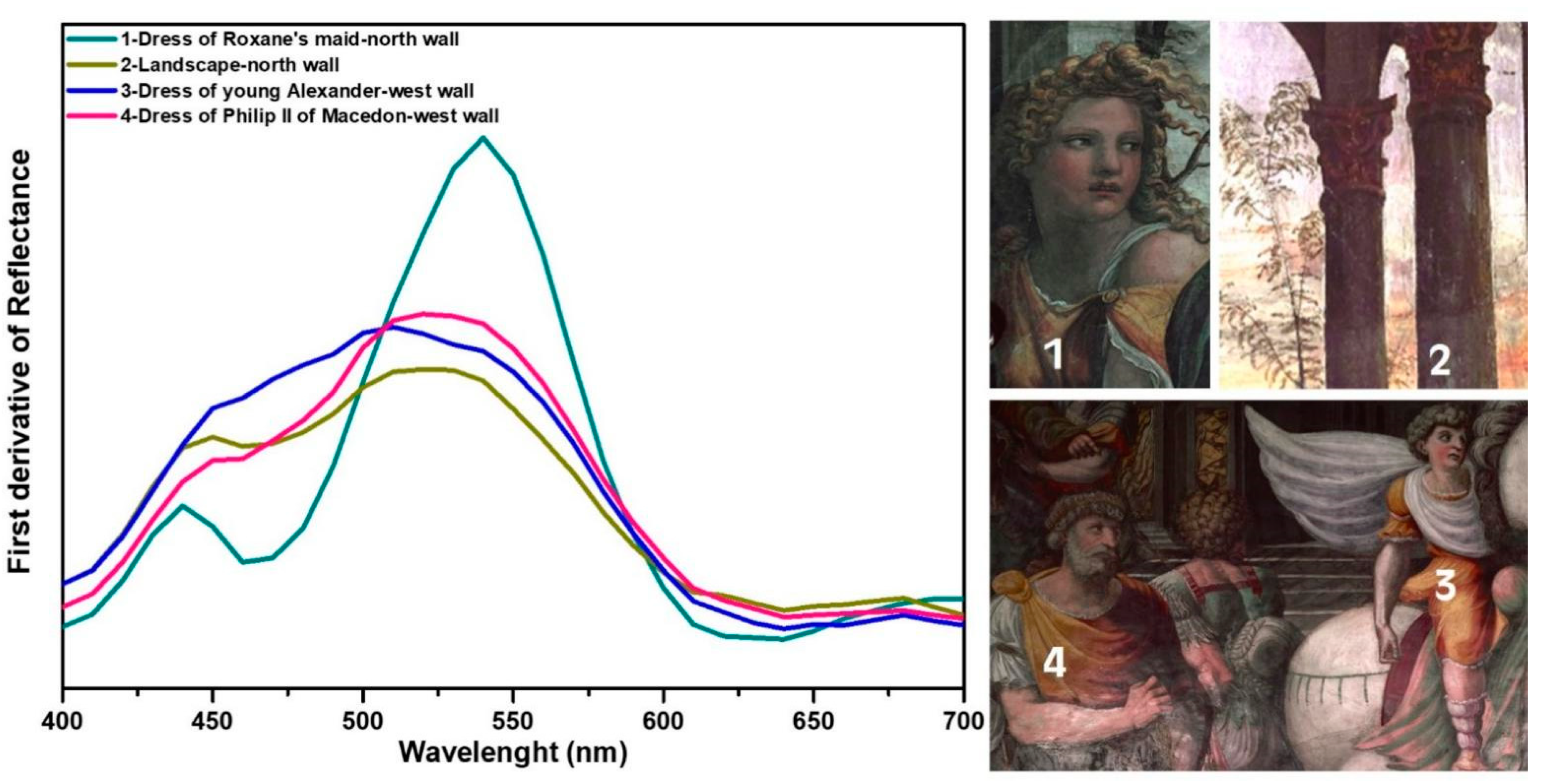
3.1. Red
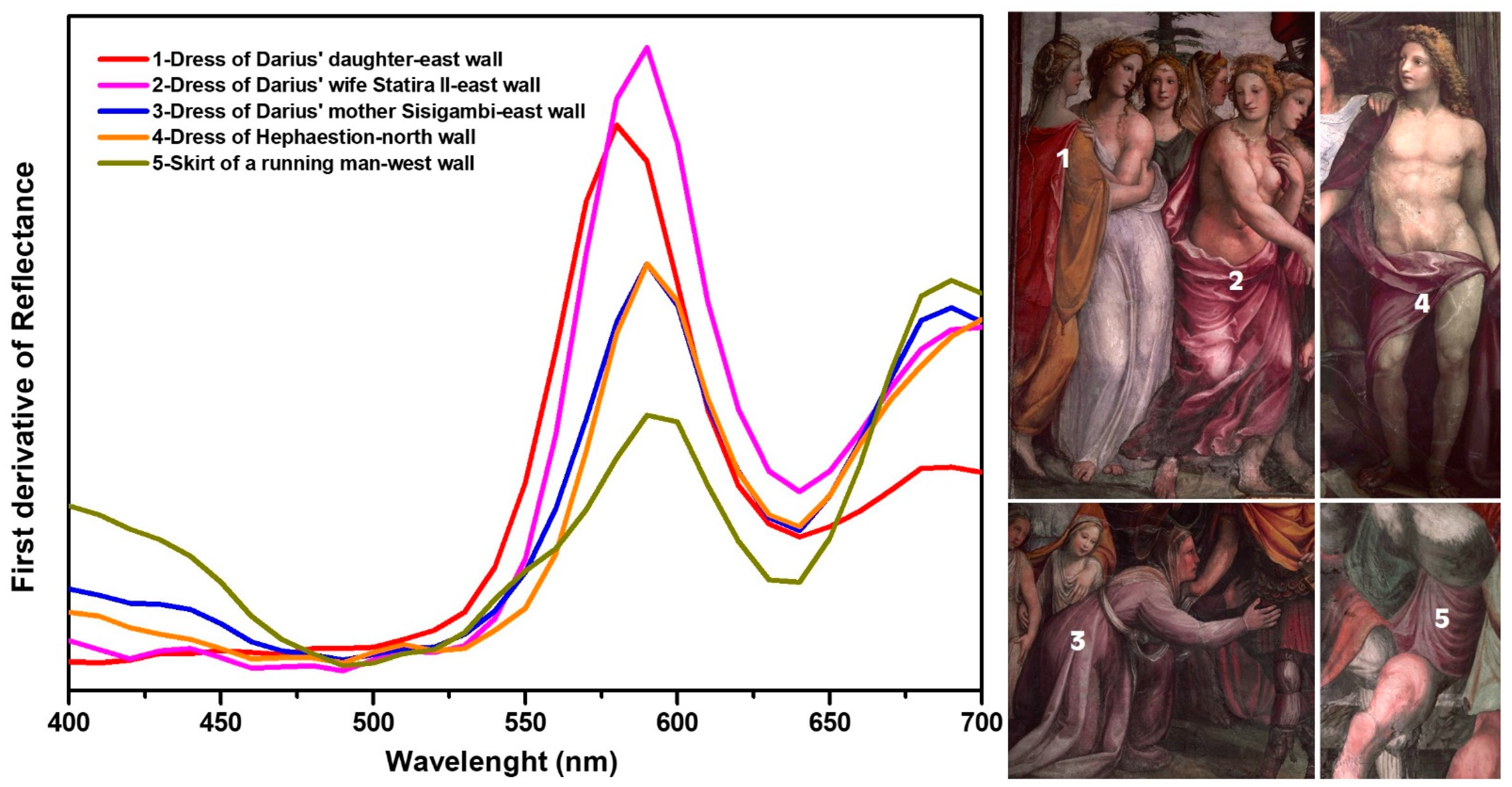
3.2. Purple
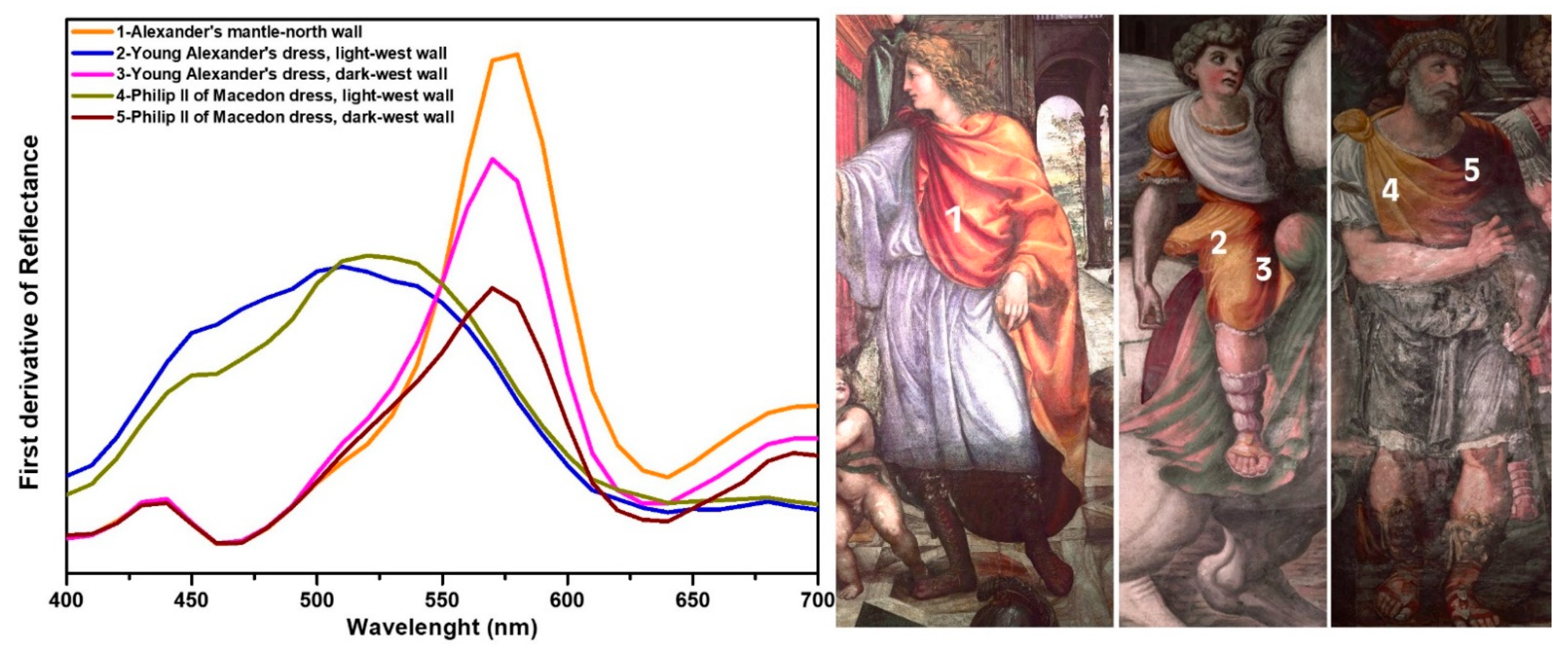
3.3. Yellow
3.4. Orange
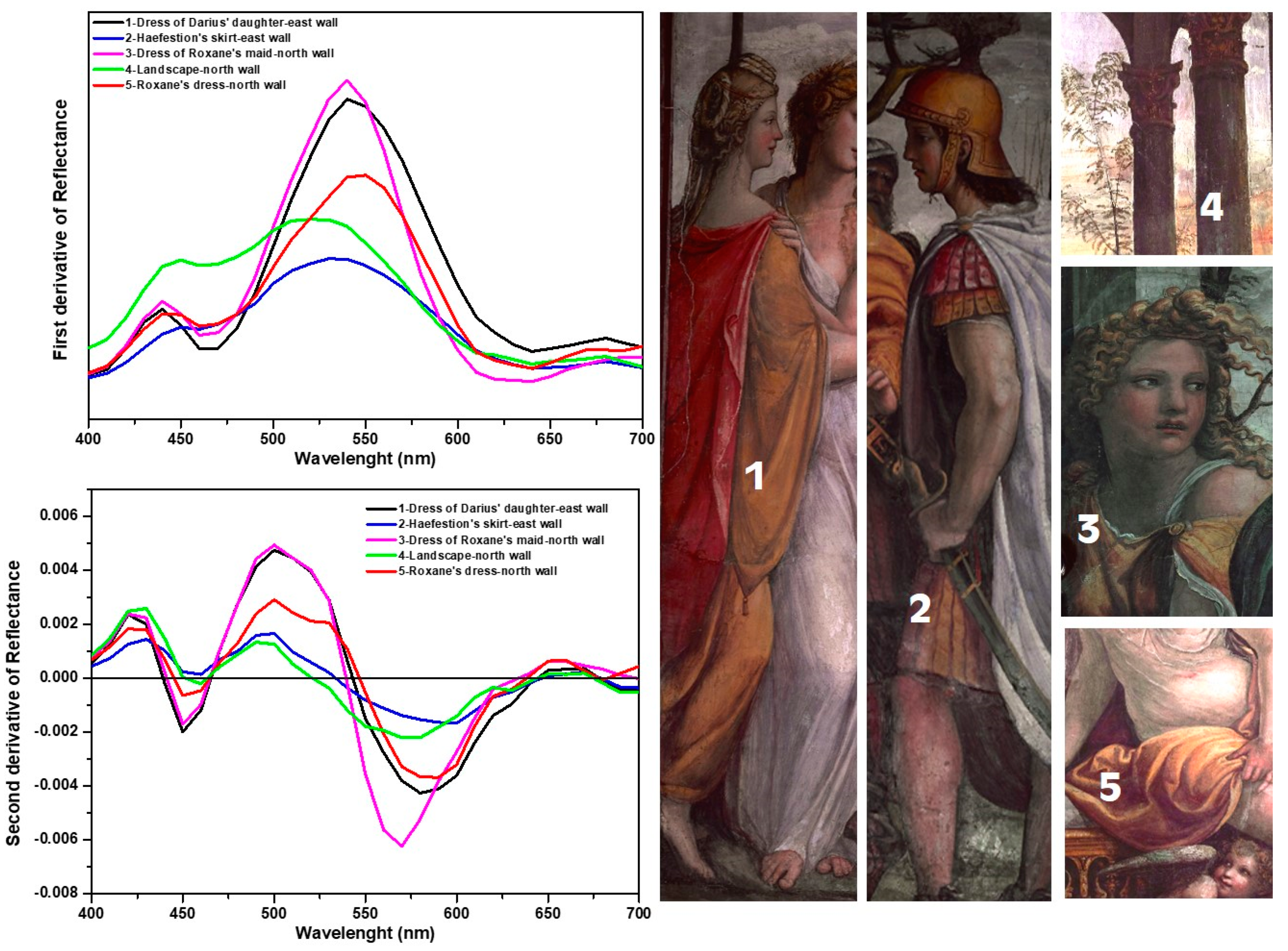
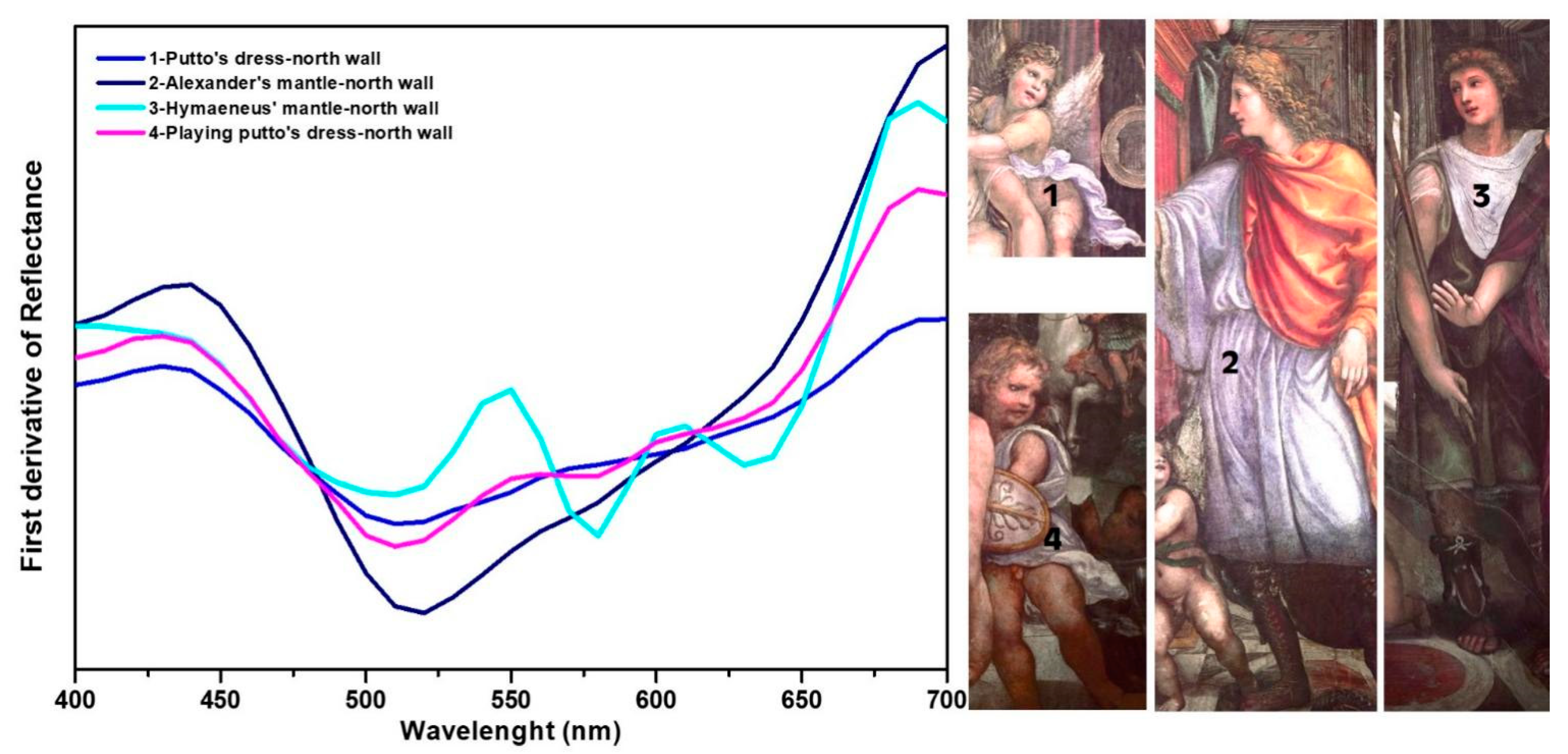
3.5. Blue
- (1)
- The use of lapis lazuli, only on the north wall, for the blue highlights on white draperies, such as on the white dress of the putto that helps Roxane to get ready (Figure 12, line 1).
- (2)
- The use of smalt. It is found as the only blue for highlighting the white dress of Hymenaeus (Figure 12, area 3): from XRF, the characterizing elements Co, Bi, and As emerged, and the reflectance spectrum corresponds to that of smalt (Figure 12, line 3). It is also found in those areas currently perceived as grey, such as in the helmet in the foreground, on the north wall. XRF indicates the presence of Co, Bi, and As, throughout the whole helmet.
- (3)
- The use of smalt and lapis lazuli in single superimposed layers, with lapis lazuli on the surface, distinguishable only with the joint use of XRF and reflectance spectroscopy, because the latter individuates only pigments on the surface. This is the case of the upper part of Alexander’s dress, where the characteristic elements of the blue enamel emerged from the XRF, but its reflectance is almost entirely attributable to that of lapis lazuli, whose elements cannot be revealed by XRF (Figure 12, line 2).
- (4)
- The use of smalt and lapis lazuli mixed together, which is evident from the reflectance spectra of the points analyzed, where the characteristics of both pigments are found. This is the case of the lower part of Alexander’s dress and that of the putto on the right, intent on playing with Alexander’s shield (Figure 12, line 4).
4. Discussion
5. Conclusions
Author Contributions
Funding
Institutional Review Board Statement
Informed Consent Statement
Acknowledgments
Conflicts of Interest
References
- Lapenta, V. Il Duca di Ripalta (Salvador Bermúdez de Castro) in Villa Farnesina. In La Saletta Pompeiana e l’Ottocento in Villa Farnesina; Lapenta, V., Sgamellotti, A., Eds.; Bardi Edizioni: Rome, Italy, 2020. [Google Scholar]
- Bigot, C. Raphaël et la Farnésine. In Gazette des Beaux-Arts; Paris, France. 1884, p. 10. Available online: http://www.icr.beniculturali.it/pagina.cfm?usz=5&uid=68&rid=93 (accessed on 15 July 2021).
- Invernizzi, C.; Daveri, A.; Rovetta, T.; Vagnini, M.; Licchelli, M.; Cacciatori, F.; Malagodi, M. A multi-analytical non-invasive approach to violin materials: The case of Antonio Stradivari “Hellier” (1679). Microchem. J. 2016, 124, 743–750. [Google Scholar] [CrossRef]
- Vagnini, M.; Gabrieli, F.; Daveri, A.; Sali, D. Handheld new technology Raman and portable FT-IR spectrometers as complementary tools for the in situ identification of organic materials in modern art. Spectrochim. Acta Part A Mol. Biomol. Spectrosc. 2017, 176, 174–182. [Google Scholar] [CrossRef]
- Cooper, J.B.; Abdelkader, M.; Wise, K.L. Sequentially Shifted Excitation Raman Spectroscopy: Novel Algorithm and In-strumentation for Fluorescence-Free Raman Spectroscopy in Spectral Space. Appl. Spectrosc. 2013, 67, 973–984. [Google Scholar] [CrossRef] [PubMed]
- Cooper, J.B.; Marshall, S.; Jones, R.; Abdelkader, M.; Wise, K.L. Spatially compressed dual-wavelength excitation Raman spectrometer. Appl. Opt. 2014, 53, 3333–3340. [Google Scholar] [CrossRef] [PubMed]
- DeCarlo, T.M. Characterizing coral skeleton mineralogy with Raman spectroscopy. Nat. Commun. 2018, 9, 5325–5327. [Google Scholar] [CrossRef]
- Oltrogge, D. The Use of VIS Spectroscopy in Non Destructive Paint Analysis Research Project Painting Techniques of Impressionism and Postimpressionism. Köln. 2008. Available online: www.museenkoeln.de/impressionismus (accessed on 15 July 2021).
- Jubb, A.M.; Allen, H.C. Vibrational Spectroscopic Characterization of Hematite, Maghemite, and Magnetite Thin Films Produced by Vapor Deposition. Appl. Mater. Interfaces 2010, 2, 2804–2812. [Google Scholar] [CrossRef]
- Froment, F.; Tournié, A.; Colomban, P. Raman identification of natural red to yellow pigments: Ochre and iron-containing ores. J. Raman Spectrosc. 2008, 39, 560–568. [Google Scholar] [CrossRef]
- Toffoletto, E. Analisi non Invasive per la Caratterizzazione di Dipinti ad Olio su tela, Conservati nel Museo Ca’ Rezzonico di Venezia. Master’s Thesis, Università Ca’ Foscari, Venezia, Italy, 2015. [Google Scholar]
- Huang, Y.; Jiang, Z.; Schwieger, W. Vibrational Spectroscopic Studies of Layered Silicates. Chem. Mater. 1999, 11, 1210–1217. [Google Scholar]
- Manfredi, M.; Barberis, E.; Rava, A.; Robotti, E.; Gosettia, F.; Marengo, E. Portable diffuse reflectance infrared Fourier transform (DRIFT) technique for the non-invasive identification of canvas ground: IR spectra reference collection. Anal. Methods 2015, 7, 2313–2322. [Google Scholar] [CrossRef]
- Castagnotto, E.; Locardi, F.; Slimani, S.; Peddis, D.; Gaggero, L.; Ferretti, M. Characterization of the Caput Mortuum purple hematite pigment and synthesis of a modern analogue. Dyes Pigment. 2021, 185, 108881–108889. [Google Scholar] [CrossRef]
- Poldi, G. L’individuazione del blu di smalto sano e decolorato in dipinti mediante ED-XRF e spettrometria in riflettanza, conference paper. In Proceedings of the ATIV Conference, Parma, Italy, 9–10 July 2009. [Google Scholar]
- Moon, D.-H.; Lee, N.-R.; Lee, E.-W. Ancient Pigments in Afrasiab Murals: Characterization by XRD, SEM, and Raman Spectroscopy. Minerals 2021, 11, 939. [Google Scholar] [CrossRef]
- Rosi, F.; Manuali, V.; Miliani, C.; Brunetti, B.G.; Sgamellotti, A.; Grygar, T.; Hradil, D. Raman scattering features of lead pyroantimonate compounds. Part I: XRD and Raman characterization of Pb2Sb2O7 doped with tin and zinc. J. Raman Spectrosc. 2008, 40, 107–111. [Google Scholar] [CrossRef]
- Simsek, G.; Geckinli, A.E. An assessment study of tiles from Topkapı Palace Museum with energy-dispersive X-ray and Raman spectrometers. J. Raman Spectrosc. 2012, 43, 917–927. [Google Scholar] [CrossRef]
- Smith, G.D.; Klinshaw, R.J. The presence of trapped carbon dioxide in lapis lazuli and its potential use in geo-sourcing natural ultramarine pigment. J. Cult. Herit. 2009, 10, 415–421. [Google Scholar] [CrossRef]
- Roy, A. Artists’ Pigments: A Handbook of Their History and Characteristics; Oxford University Press: Oxford, UK, 1993; Volume 1. [Google Scholar]
- Seccaroni, C. Giallorino. Storia dei Pigmenti Gialli di Natura Sintetica; De Luca Editori d’arte: Rome, Italy, 2005. [Google Scholar]
- Seccaroni, C.; Aresi, N.; Frizzi, T.; Anselmi, C.; Sgamellotti, A. Raphael’s workshop at Villa Farnesina in Rome: The frescoed vault of Cupid and Psyche investigated by macro-X-ray fluorescence scanning. Rend. Fis. Acc. Lincei 2018, 29, 499–510. [Google Scholar] [CrossRef]
- Haldi, J.-P.; Seccaroni, C. Cobalto, Zaffera, Smalto Dall’Antichità al XVIII Secolo; ENEA: Rome, Italy, 2016. [Google Scholar]
- Colomban, P.; Kırmızı, B.; Franci, G.S. Cobalt and Associated Impurities in Blue (and Green) Glass, Glaze and Enamel: Relationships between Raw Materials, Processing, Composition, Phases and International Trade. Minerals 2021, 11, 633. [Google Scholar] [CrossRef]
- Bartalini, R. On the Room of Alexander and Roxane at the Farnesina and on the activity of Sodoma in Rome (with reference to Girolamo Genga and his relations with the Chigi family). Prospettiva 2014, 153, 39–73. [Google Scholar]


| Wall | Colored Area | X-ray Fluorescence (XRF) Main Elements | Vis-Reflectance (Vis-R) | Raman | Medium Infrared (MIR) | Comments |
|---|---|---|---|---|---|---|
| The Darius family before Alexander (east wall) | Red | Ca, Fe, Sr, K (bright red) | Ip:580,640 nm | CaCO3 [8] | Generic Fe-based red from Vis-R [9] | |
| Fe, Ca, K, Sr (dark red) | Ip:580,640 nm | Hematite [10] | Generic Fe-based red from Vis-R [9] | |||
| Yellow | Ca, Fe, Pb, Sr, Sb (bright yellow) | Ip:440,540,630 nm | Yellow ochre [11] | From Vis-R yellow ochre [12] + Naples yellow [12] | ||
| Ca, Fe, Pb, Sr (dark yellow) | Ip:451,533,640 nm | Mostly Pb-yellow [12] +yellow ochre from Vis-R | ||||
| Purple | Ca, Fe, K, Sr | Ip:589,640,695 nm | CaCO3 [8], hematite [10] | CaCO3 [8], silicates [13] and acrylic resin [14] | Hematite-based purple, possible caput mortuum [15] | |
| Purple/greyish | Ca, Fe, Co, K, Si, As, Bi, Sr | Ip:589,640,690 nm | CaCO3 [8], hematite [10] | Hematite-based purple, possible caput mortuum [15]. Smalt is not visible in Vis-R | ||
| Blue | Ca, Co, Fe, Pb, As, K, Bi, Sr, Si | Max:468,561,622 nm; Min:530,590,650 nm; Ip:687 nm | CaCO3 [8] | Smalt [16] | ||
| Ca, Co, Fe, Pb, As, Ti, K, Bi, Si | Max:468,561,622 nm; Min:530,590,650 nm; Ip:687 nm | Smalt [16] and traces of restoration materials (Ti) |
| Wall | Colored Area | X-ray Fluorescence (XRF) Main Elements | Vis-Reflectance (Vis-R) | Raman | Medium Infrared (MIR) | Comments |
|---|---|---|---|---|---|---|
| Alexander meeting his new bride-to-be Roxane (north wall) | Red | Fe, Ca, K, S, Sr, Pb, Hg | Ip:580, 640 nm | Cinnabar [17] | Generic Fe-based red from Vis-R [9] Cinnabar from XRF and Raman, but not visible by Vis-R. | |
| Ca, Fe, Sr, K, S, Pb | Ip:583, 640 nm | Generic Fe-based red from Vis-R [9] | ||||
| Orange | Fe, Ca, K, Sr, Pb | Ip:434,510,579,636 nm | CaCO3 [8] + yellow ochre [11] | . | Mixture of red and yellow ochre. Yellow ochre visible from Raman and Vis-R. Pb-yellow not to be excluded due to the inflection at 510 nm from Vis-R [12] | |
| Yellow | Ca, Fe, K, Sr, Pb | Ip: 440, 544, 640 nm | Only yellow ochre from Vis-R | |||
| Fe, Ca, Pb, K, Sr, Sb | Ip: 436,541,631 nm | Pb-antimonate [18,19] | Yellow ochre+Naples yellow from Vis-R | |||
| Ca, Pb, Fe | Ip:450, 519,631 nm | Mostly Pb-yellow from Vis-R [12] | ||||
| Pb, Sb, Fe (K) | Ip:443,508,546,640 nm | Pb-antimonate [18,19] | Naples yellow+Pb-yellow +yellow ochre from Vis-R [12] | |||
| Purple | Fe, Ca, K, Sr, S, | Ip:589,640,695 nm | Hematite [10] | Hematite-based purple, possible caput mortuum [15] | ||
| Blue | Ca, Fe, K, Sr, S | Max:477 nm, Min:603 nm, Ip:683 nm | CaCO3 [8] | CaCO3 [8], weak signal of lapislazuli [20], acrylic resin [14] | Lapislazuli from Vis-R [12] and traces of restoration materials | |
| Ca, Fe, Co, As, K, Si, Sr, Bi | Max:480 nm, Min:595 nm, Ip:680 nm | Lapislazuli and smalt [16] in different superimposed layers. From Vis-R only Lapislazuli [12] | ||||
| Ca, Fe, Co, As, K, Sr, Bi | Max:481 nm, Min:541,590,637 nm, Ip:680 nm | Lapislazuli [12] and smalt [16] mixed together; Vis-R spectrumhas both the features of these pigments | ||||
| Ca, As, Co, Fe, K, Bi, Sr, Pb, Si | Max:470,574,623 nm, Min:524, 593,652 nm, Ip:685 nm | Smalt [16] | ||||
| Ca, Fe, As, Co, K, Sr, Bi | Max:478 nm, Min:576 nm, Ip:688 nm | Lapislazuli-like spectrum in Vis-R [12]; smalt visible only in first derivative of Vis-R |
| Wall | Colored Area | X-ray Fluorescence (XRF) Main Elements | Vis-Reflectance (Vis-R) | Raman | Comments |
|---|---|---|---|---|---|
| The Taming of Bucephalus (west wall) | Red | Ca, Fe, K Sr | Ip:582,640 nm | CaCO3 [8] | Generic Fe-based red from Vis-R [9] |
| Fe, Ca, Sr, K, S (dark red) | Ip:580,640 nm | Hematite [10] | Generic Fe-based red from Vis-R [9] | ||
| Orange | Fe, Ca, Sr, Pb, K | Ip:435,510,570,635 nm | CaCO3 [8] | Mixture of red and yellow ochre; Pb-yellow not to be excluded due to the inflection at 510 nm [12] | |
| Yellow | Pb, Fe, Sb (K) | Ip:450,521,627 nm | Pb-antimonate [18,19] | Pb-yellow and few yellow ochre from Vis-R [12]. Naples yellow from XRF and Raman | |
| Pb, Fe, Sb | Ip:449,506,627 nm | Pb-antimonate [18,19] | Mostly Pb-yellow from Vis-R [12] | ||
| Purple | Fe, Ca, Sr, K | Ip:590,639,688 nm | CaCO3 [8], hematite | Hematite-based purple, possible caput mortuum [15] | |
| Blue | Ca, Co, Fe, Bi, As, K, Si, Pb | Max:440,549,608 nm; Min:500,580,637 nm; Ip:692 nm | CaCO3 [8] | Smalt [16] |
Publisher’s Note: MDPI stays neutral with regard to jurisdictional claims in published maps and institutional affiliations. |
© 2021 by the authors. Licensee MDPI, Basel, Switzerland. This article is an open access article distributed under the terms and conditions of the Creative Commons Attribution (CC BY) license (https://creativecommons.org/licenses/by/4.0/).
Share and Cite
Vagnini, M.; Anselmi, C.; Azzarelli, M.; Sgamellotti, A. Things Always Come in Three: Non-Invasive Investigations of Alexander and Roxane’s Wedding Room in Villa Farnesina. Heritage 2021, 4, 2792-2809. https://doi.org/10.3390/heritage4040157
Vagnini M, Anselmi C, Azzarelli M, Sgamellotti A. Things Always Come in Three: Non-Invasive Investigations of Alexander and Roxane’s Wedding Room in Villa Farnesina. Heritage. 2021; 4(4):2792-2809. https://doi.org/10.3390/heritage4040157
Chicago/Turabian StyleVagnini, Manuela, Chiara Anselmi, Michela Azzarelli, and Antonio Sgamellotti. 2021. "Things Always Come in Three: Non-Invasive Investigations of Alexander and Roxane’s Wedding Room in Villa Farnesina" Heritage 4, no. 4: 2792-2809. https://doi.org/10.3390/heritage4040157