Article

Identification of Coatings on Persian Lacquer Papier Mache Penboxes by Fourier Transform Infrared Spectroscopy and Luminescence Imaging

by
Alireza Koochakzaei
*,
Ali Nemati Babaylou
and
Behrooz Jelodarian Bidgoli
Faculty of Cultural Materials Conservation, Tabriz Islamic Art University, Tabriz 5164736931, Iran
*
Author to whom correspondence should be addressed.
Heritage 2021, 4(3), 1962-1969; https://doi.org/10.3390/heritage4030111
Submission received: 30 July 2021 / Revised: 21 August 2021 / Accepted: 23 August 2021 / Published: 24 August 2021
(This article belongs to the Special Issue Organic Materials in Heritage Science)

Abstract

:
In this study, Fourier transform infrared spectroscopy (FTIR) and luminescence imaging were used to identify the coatings of seven Persian lacquer papier mache penboxes, of which two were contemporary, one was from the Pahlavi era, and four belonged to the Qajar era. First, FTIR was used to identify the nature of the coating. Then, UV-induced visible luminescence imaging at the spectral ranges of 420–680 nm (UVL), 425–495 nm (UVIBL), and 615–645 nm (UVIRL) was performed for further examination. The FTIR results showed that the coatings were made of alkyd resin, oil-resin varnish (Kaman oil), and shellac. In UV-induced visible luminescence images, synthetic alkyd resin showed no fluorescence, which made it distinguishable from the natural organic coatings. While it is slightly challenging to differentiate Kaman oil from shellac based on FTIR results, these two coatings can be easily distinguished by their fluorescence in UVL and UVIBL images. The results suggest that the combined use of spectroscopy and spectral imaging methods can provide substantial information about the organic coatings of historical objects.

1. Introduction

Persian lacquer objects, especially penboxes (Qalmadan in Persian), are among valuable Persian historical and artistic objects that have survived in significant numbers from multiple historical periods. Despite their global renown as lacquer art, as some researchers have pointed out, these lacquerwares are essentially painted surfaces that are coated with a protective oil or resin [1,2]. In addition to its protective role, this coating often contributes to the aesthetic value of the coated object. However, research into lacquerwares has been generally focused on their historical significance, fabrication technology, and illustrations. This is why the identification of organic coatings is also an important part of the study of these historically and culturally significant objects [3,4,5,6,7,8].
In the case of penboxes, these coatings used to comprise a thin layer of oil-resin veneer. According to historical records, this veneer, generally referred to as “Kaman oil”, used to be prepared by mixing a terpenoid resin, typically sandarac, with linseed oil at boiling temperature. The use of this coating in Persian artworks dates back to at least the 11th century. From the 18th century onwards, Persian artists started to use shellac as well as Kaman oil [9], until the twentieth century, when synthetic resins, especially alkyd, became common. While the process of making papier mache penboxes has not changed much over these centuries, the coatings applied to these objects have changed depending on circumstances [10].
Given the limitations in taking samples from such objects, how often they are recoated or restored, and how often they may have lost a significant part of their original coating, the identification of these original coatings is one of the main challenges for researchers. There are a variety of methods for identifying historical resins and coatings, including techniques based on chromatography and mass spectrometry, which are relatively expensive [11,12,13,14,15,16]. However, more affordable spectroscopic methods such as Raman spectroscopy and especially Fourier transform infrared spectroscopy also have a good track record in identifying the molecular structure of complex organic compounds like those of coatings used on historical objects [17,18,19,20,21,22,23,24]. With these methods, it is possible to study a wide range of synthetic and natural organic oils, resins and coatings very quickly and at reasonably low cost [25].
This identification can also be done with the help of ultraviolet fluorescence examination. The use of ultraviolet light sources to examine visible fluorescence emissions from the outermost layer of painted artworks dates back to the 1920s [26,27,28,29,30]. Despite recent advances in instrumental analysis methods, this method is still one of the fundamental techniques for evaluating the quality of coatings based on their fluorescence in the visible region [31]. However, the results of this method depend on a variety of factors including the experimental process, the post-processing method, and the data usage procedure [32,33]. While ultraviolet imaging allows us to collect data from an object without physical contact, it cannot provide definite results for complex coatings. Therefore, recent years have seen more interest in the compilation of ultraviolet fluorescence databases for artwork materials [34,35].
In this study, we used Fourier transform infrared spectroscopy (FTIR) and spectral imaging to identify the coatings of multiple historical and contemporary lacquer papier mache penboxes in order to determine whether these methods are suitable for the systematic study of such historical and artistic objects. The results of this study can pave the way for the wider use of non-destructive imaging methods in the technical study of historical lacquer objects.

2. Materials and Methods

2.1. Samples

The study was performed on seven Persian lacquer papier mache penboxes (Figure 1). The length of these penboxes varied from about 20 to 25 cm. In terms of age, the penboxes labeled LP1 and LP2 were contemporary, LP3, LP4, LP5, and LP6 belonged to the Qajar period (late 18th to early 20th century), and LP7 belonged to the first Pahlavi period (2nd to the 4th decade of the 20th century). Despite some conservation actions done on these penboxes, they still had their original coatings, making it possible to determine whether such coatings can be identified by FTIR spectroscopy and spectral imaging methods.

2.2. Fourier Transform Infrared Spectroscopy

Fourier Transform Infrared Spectroscopy (FTIR) was used for the structural study of the coating applied to the papier mache base. Before sampling, the surfaces of the penboxes were cleaned with ethanol. Sampling was performed with a scalpel. The samples were analyzed using the KBr pellet method with an FT/IR-680Plus spectrometer (Jasco, Tokyo, Japan). The spectra were recorded in 32 scans with a resolution of 4 cm−1 in the range of 400–4000 cm−1. The spectra of Indian shellac, alkyd resin, and a historical sample of Safavid Kaman oil were also recorded to be used as the reference.

2.3. Luminescence Imaging

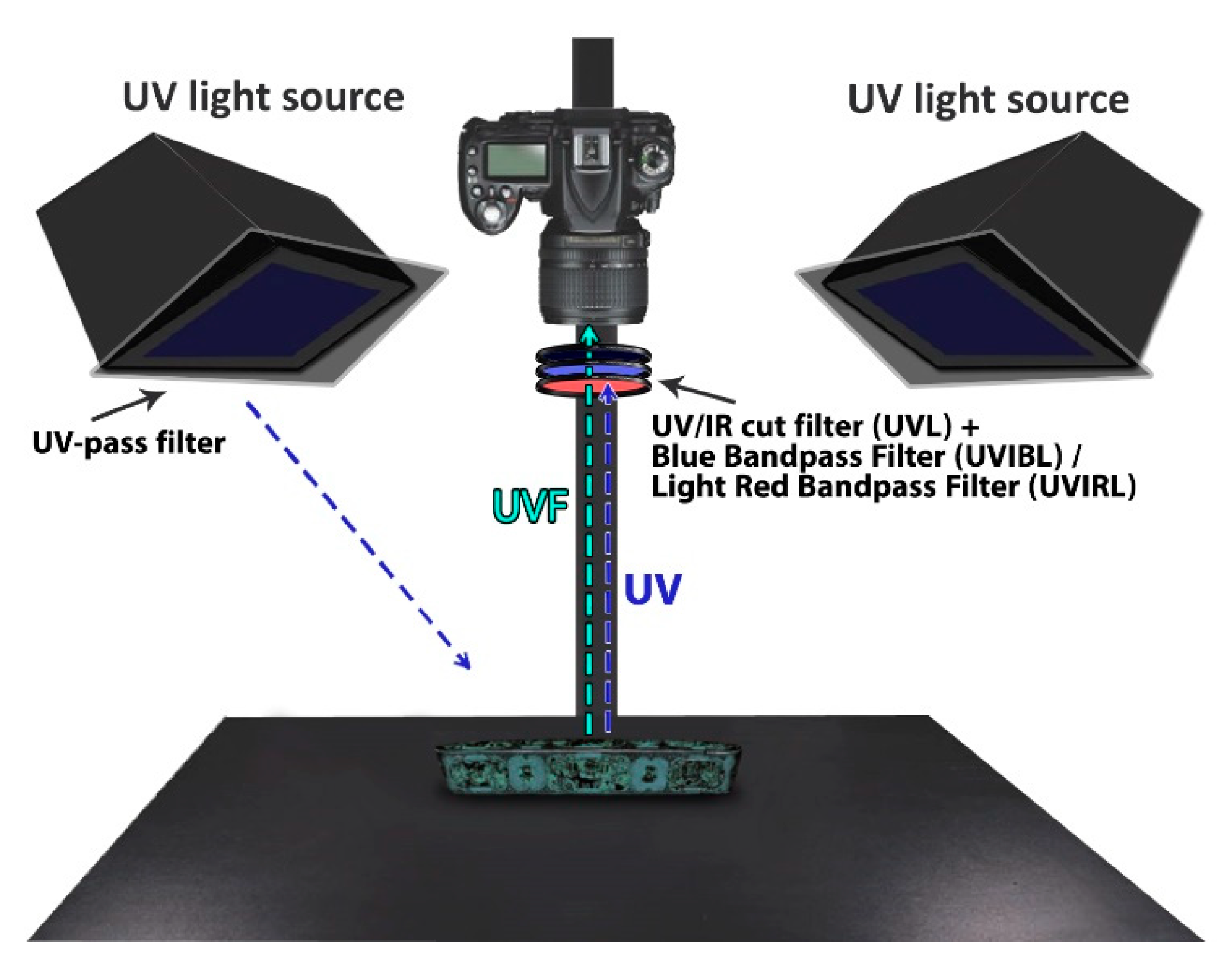
All images were taken with a modified D750 camera (Nikon, Tokyo, Japan) equipped with an AF Nikkor 50 mm f/1.8D lens. To take full advantage of the CMOS sensor (350–1100 nm), the internal UV-IR block filter was removed. Following the instructions of Verri [36], two electronic xenon flashes equipped with a U360 UV beam filter (Hoya, Tokyo, Japan) fixed at a 45-degree angle to the subject were used as the light source (Figure 2). Visible reflected images (VIS) and visible luminescence images were recorded in RAW format at the highest possible resolution (24 megapixels: 6016 × 4016) using the filters described in Table 1 and then converted to 16-bit TIF format in Adobe Photoshop (Adobe, San Jose, CA, USA). Post-processing and calibration procedures were performed using the Kushel method [37] and as instructed by Cosentino [38]. In the UVL1 method, PTFE was used as the white balance reference.

3. Results and Discussion

3.1. FTIR Spectroscopy

Until the 18th century, the coating used in the majority of Persian lacquer works was Kaman oil [39,40]. However, starting from the Safavid era, the use of shellac as an alternative coating also became common. In the 20th century, an increasing number of Persian artists began to use alkyd resins in their work. While acrylic resins have been occasionally used in the restoration of other coatings, in such cases, the structural differences are quite easily distinguishable in FTIR, but it is fairly difficult to distinguish Kaman oil from shellac, alkyd, and triterpenoid varnishes that some artists have used as its substitute. However, given the differences in the FTIR spectra characteristics of these materials, several researchers have suggested using these spectra to distinguish them from another.
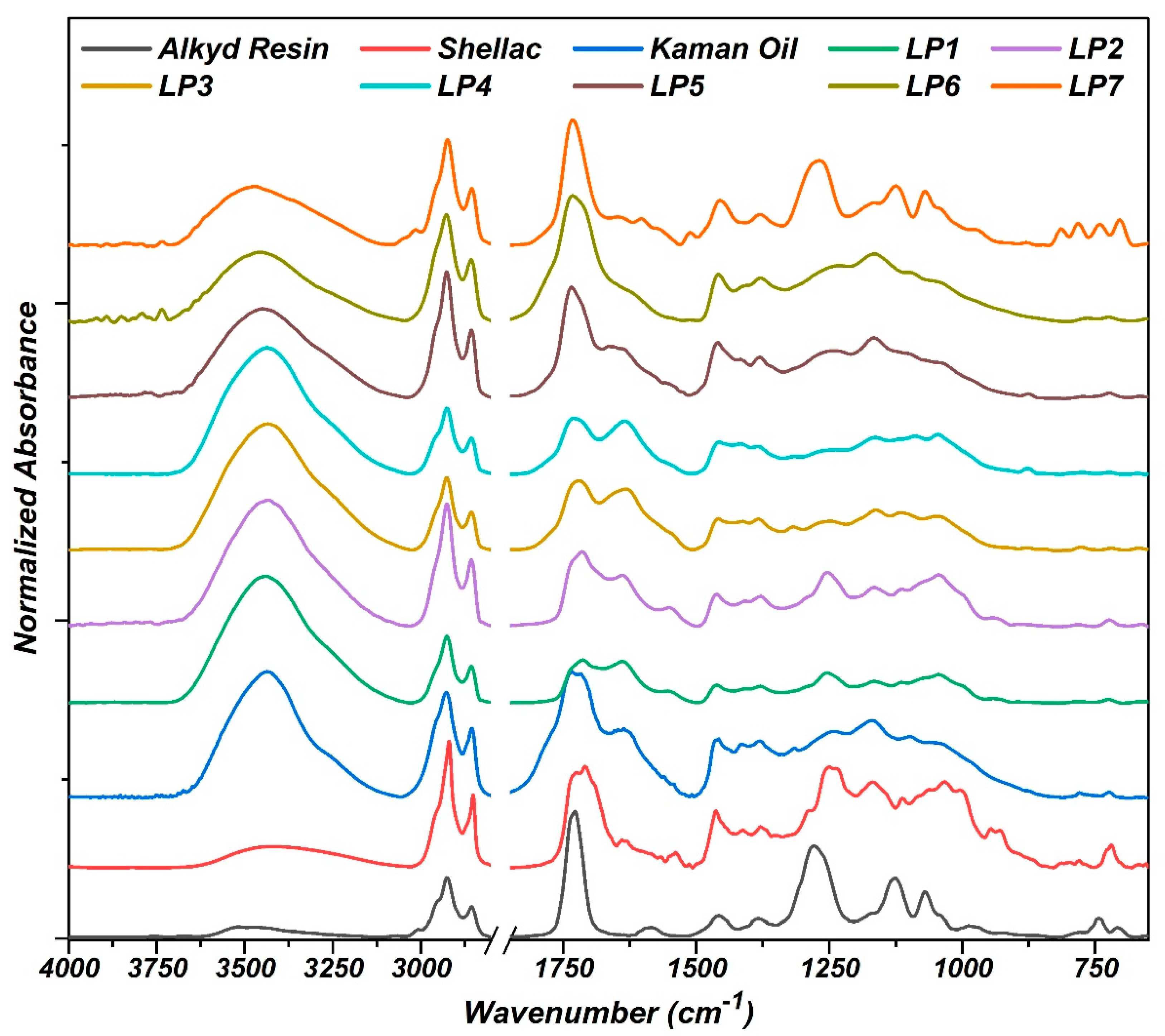
The FTIR spectra of the studied samples are illustrated in Figure 3. Initial examinations showed a clear difference between LP7 and other samples. In the spectrum of LP7, the multiple peaks belonging to OH groups at 3470 cm−1, symmetric and asymmetric stretching vibrations of CH2 at 2925 and 2855 cm−1, bending vibrations of CH2 at 1457 cm−1, and the strong peak related to the stretching vibration of C=O at about 1733 cm−1 are among the characteristics of alkyds. This spectrum also shows the bending vibrations of C-H and the stretching vibrations of C-O and C-C at 1124 cm−1 and especially a strong peak at 1268 cm−1 (probably related to the vibrations of C-O-C esters). The absorption bands 1603, 1071, 704 and 742 cm−1 have also been attributed to the stretching and bending vibrations of the aromatic ring and aromatic CH in alkyds [41,42,43,44]. Comparison of this spectrum with the reference alkyd spectrum also showed the remarkable similarity of the two, further indicating that the coating applied on LP7 is alkyd. Furthermore, a weak absorption band at about 3020 cm−1 was only observed in this spectrum and the reference alkyd spectrum.
For LP1 and LP2, the spectra showed a weak twin absorption band related to C-H vibrations at 929 and 946 cm−1, which is similar to the one in the reference shellac spectrum. These spectra also showed C-O vibrations at 1045 and 1165 cm−1 and a pronounced peak around 1250 cm−1, which is a feature distinguishing shellac from Kaman oil. In the reference Kaman oil spectrum, the peak of this region is positioned around 1165 cm−1 and there is a clearly visible shoulder peak around 1245 cm−1, which can also be seen in the spectra of LP3, LP4, LP5, and LP6. Like shellac, LP1 and LP2 showed the stretching vibrations of carbonyl at around 1714 cm−1. It should be noted that in fresh shellac, C=O vibration appears as a twin peak around 1715 and 1735 cm−1 [44,45,46,47]. In shellac, vinyl (C=C) vibrations were also observed around 1640 cm−1 [47]. Unlike Kaman oil, shellac had a clearly visible absorption band at 1540 cm−1, which was also observed in LP1 and LP2. These results suggested that LP3, LP4, LP5, and LP6 have an oil-resin coating and LP1 and LP2 have a shellac coating. However, the high similarity of the FTIR spectra of these two coatings makes them difficult to distinguish.

3.2. Spectral Imaging

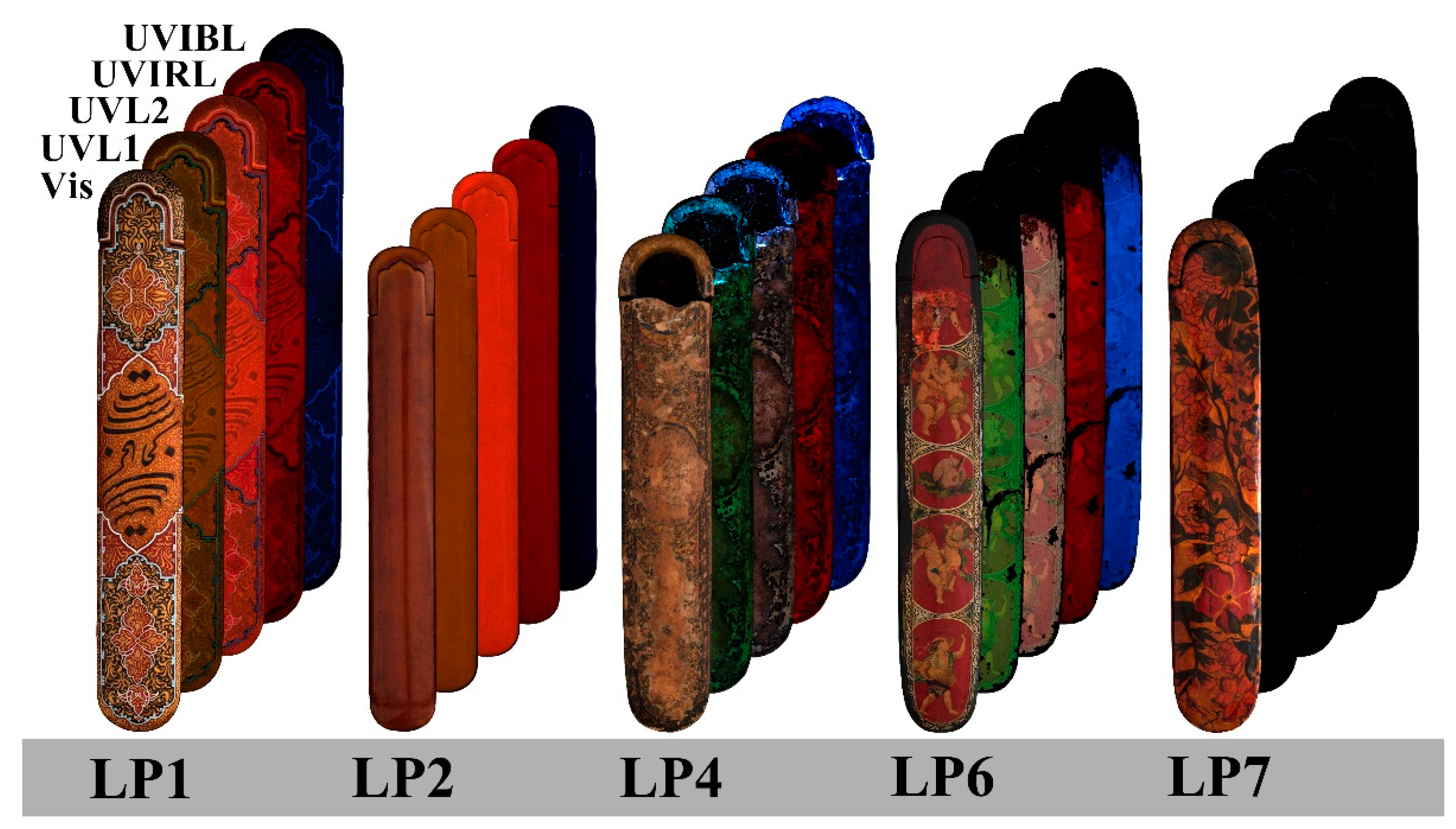
Spectral imaging, in general, and UV-induced visible luminescence imaging, in particular, are very suitable for studying organic coatings and coloring agents on historical objects. The scalability of this method and the fact that it does not require any sampling make it a good complement to spectroscopy-based methods in cultural heritage studies. The results of spectral imaging of the samples are presented in Figure 4 and a summary of the observations is provided in Table 2. While FTIR conducted in the previous step managed to identify the organic coatings, here, an obvious difference was detected between the synthetic coating and natural ones. More specifically, the alkyd resin in LP7 did not show any fluorescence in any of the imaging methods. Therefore, this resin can be easily distinguished from Kaman oil and shellac by its lack of fluorescence.
While the similarity of the FTIR absorption bands of shellac (LP1 and LP2) and Kaman oil (LP3, LP4, LP5, and LP6) make them slightly difficult to distinguish by FTIR, imaging methods make it easy to differentiate them from another. As Figure 4 illustrates, the only image in which shellac and Kaman oil had a similar reddish appearance was UVIRL. The shellac coating had a brown appearance in UVL1 and an orange appearance in UVL2. But Kaman oil had a green color and a milky color in UVL1 and UVL2 respectively. Previous studies have also reported an orange fluorescence for shellac [48,49,50]. In UVIBL, shellac and Kaman oil appeared in dark blue and light blue colors, respectively. These results suggested that shellac and Kaman oil coating can be properly distinguished through spectral imaging.

4. Conclusions

In this study, FTIR spectroscopy and spectral imaging were used to identify the surface coatings of seven contemporary and historical Persian papier mache penboxes. The results of FTIR spectroscopy showed the use of alkyd resins, Kaman oil, and shellac in the coatings. It was found that alkyd resin can be distinguished from shellac and Kaman oil by the absorption bands 1124, 1268, and 3020 cm−1 as well as the absorption bands related to the vibrations of aromatic structures around 1603, 1071, 742, and 704 cm−1. However, it is slightly more difficult to differentiate Kaman oil from shellac coating based on FTIR results. The twin peaks appearing at 929 and 946 cm−1 and the absorption bands 1250, 1540 and 1714 cm−1 can be regarded as the features distinguishing shellac from Kaman oil. Shellac was only detected in the two contemporary samples. Oil-resin base coatings typically showed a peak at 1165 cm−1 with a shoulder at 1245 cm−1. Comparison of the absorption bands of the samples with the reference Kaman oil spectrum indicated the use of this coating in all four Qajar-era penboxes.
Spectral imaging also showed good performance in differentiating the coatings of penboxes. This method was found to be especially effective in distinguishing synthetic coatings from those made of natural oils or resins. While the similarity of the FTIR absorption bands of shellac and Kaman oil in historical objects make them difficult to differentiate, UV-induced visible luminescence imaging made it possible to quickly and easily distinguish them without needing to take any sample. Overall, the results clearly demonstrated the utility of luminescence imaging in the fast and inexpensive identification of organic coatings on historical objects, making it particularly useful for screening large numbers of these artifacts. However, to determine the exact type of coating with this method, a comprehensive reference database will be needed covering all types of coating or must combine this technique with other methods.

Author Contributions

A.K. designed the work process and analyzed and interpreted the data. B.J.B. and A.N.B. participated in experiments. All authors discussed the results and contributed to the final manuscript. All authors have read and agreed to the published version of the manuscript.

Funding

This research received no external funding.

Institutional Review Board Statement

Not applicable.

Informed Consent Statement

Not applicable.

Data Availability Statement

The data presented in this study are available on request from the corresponding author.

Acknowledgments

The authors would like to thank Hassanzad Fanid for providing contemporary penboxes and Sarabi, Shojaeifar and Khanmohammadi for providing Historical penboxes.

Conflicts of Interest

The authors declare no conflict of interest.

References

  1. Decq, L.; Jones, Y.; Steyaert, D.; Cattersel, V.; Indekeu, C.; Van Binnebeke, E.; Fremout, W.; Lynen, F.; Saverwyns, S. Black Lacquered Papier-mâché and Turned Wooden Furniture: Unravelling the Art History, Technology and Chemistry of the 19th-Century Japanning Industry. Stud. Conserv. 2019, 64 (Suppl. S1), S31–S44. [Google Scholar] [CrossRef] [Green Version]
  2. Robinson, B.W. Some thoughts on Qajar Lacquer. In Lacquerwork in Asia and Beyond; Watson, W., Ed.; University of London, Percival David Foundation of Chinese Art, School of Oriental and African Studies: London, UK, 1982; pp. 67–77. [Google Scholar]
  3. Robinson, B.W. Persian Lacquer in the Bern Historical Museum. Iran 1970, 8, 47. [Google Scholar] [CrossRef]
  4. von der Osten-Woldenburg, H. Radartomographie der römischen Villa in Epfendorf TT—Radar tomography of the Roman villa in Epfendorf. Fundber. Baden-Württemberg 1999, 23, 243–252. [Google Scholar]
  5. Diba, L.S. Lacquerwork of Safavid Persia and Its Relationship to Persian Painting; The Institute of Fine Arts, New York University: New York, NY, USA, 1994. [Google Scholar]
  6. Fehervari, G. Near Eastern Lacquerwork: History and Early Guidance. In Lacquerwork in Asia and Beyond; Watson, W., Ed.; University of London, Percival David Foundation of Chinese Art, School of Oriental and African Studies: London, UK, 1982; pp. 225–231. [Google Scholar]
  7. Kiani, M.Y. Early Lacquer in Iran. In Lacquerwork in Asia and Beyond; Watson, W., Ed.; University of London, Percival David Foundation of Chinese Art, School of Oriental and African Studies: London, UK, 1982; pp. 211–224. [Google Scholar]
  8. Robinson, B. Qajar Lacquer. Muqarnas 1989, 6, 131–146. [Google Scholar] [CrossRef]
  9. Porter, Y.; Butani, S. Painters, Paintings and Books: An Essay on Indo-Persian Technical Literature, 12–19th Centuries; Routledge: Abingdon, UK, 2020. [Google Scholar]
  10. Khalili, N.D. Persian Lacquer Painting in the 18th and 19th Centuries; SOAS University of London: London, UK, 1988. [Google Scholar]
  11. Echard, J.; Benoit, C.; Peris-Vicente, J.; Malecki, V.; Gimeno-Adelantado, J.; Vaiedelich, S. Gas chromatography/mass spectrometry characterization of historical varnishes of ancient Italian lutes and violin. Anal. Chim. Acta 2007, 584, 172–180. [Google Scholar] [CrossRef] [PubMed]
  12. Modugno, F.; Ribechini, E. GC/MS in the Characterisation of Resinous Materials. In Organic Mass Spectrometry in Art and Archaeology; John Wiley & Sons: Hoboken, NJ, USA, 2009; pp. 215–235. [Google Scholar]
  13. Scalarone, D.; Chiantore, O. Py-GC/MS of natural and synthetic resins. In Organic Mass Spectrometry in Art and Archaeology; Colombini, M.P., Modugno, F., Eds.; John Wiley & Sons: Hoboken, NJ, USA, 2009; pp. 327–361. [Google Scholar]
  14. Ribechini, E. Direct mass spectrometric techniques: Versatile tools to characterise resinous materials. In Organic Mass Spectrometry in Art and Archaeology; Colombini, M.P., Modugno, F., Eds.; John Wiley & Sons: Hoboken, NJ, USA, 2009; pp. 77–95. [Google Scholar]
  15. Chiavari, G.; Fabbri, D.; Prati, S. Characterisation of natural resins by pyrolysis—Silylation. Chromatographia 2002, 55, 611–616. [Google Scholar] [CrossRef]
  16. Dietemann, P.; Herm, C. GALDI-MS applied to characterise natural varnishes and binders. In Organic Mass Spectrometry in Art and Archaeology; Colombini, M.P., Modugno, F., Eds.; John Wiley & Sons: Hoboken, NJ, USA, 2009; pp. 131–163. [Google Scholar]
  17. Daher, C.; Bellot-Gurlet, L. Non-destructive characterization of archaeological resins: Seeking alteration criteria through vibrational signatures. Anal. Methods 2013, 5, 6583. [Google Scholar] [CrossRef] [Green Version]
  18. Daher, C.; Bellot-Gurlet, L.; Le Hô, A.-S.; Paris, C.; Regert, M. Advanced discriminating criteria for natural organic substances of Cultural Heritage interest: Spectral decomposition and multivariate analyses of FT-Raman and FT-IR signatures. Talanta 2013, 115, 540–547. [Google Scholar] [CrossRef] [Green Version]
  19. Daher, C.; Paris, C.; Le Hô, A.-S.; Bellot-Gurlet, L.; Échard, J.-P. A joint use of Raman and infrared spectroscopies for the identification of natural organic media used in ancient varnishes. J. Raman Spectrosc. 2010, 41, 1494–1499. [Google Scholar] [CrossRef] [Green Version]
  20. Daher, C.; Pimenta, V.; Bellot-Gurlet, L. Towards a non-invasive quantitative analysis of the organic components in museum objects varnishes by vibrational spectroscopies: Methodological approach. Talanta 2014, 129, 336–345. [Google Scholar] [CrossRef] [PubMed] [Green Version]
  21. Ciofini, D.; Striova, J.; Camaiti, M.; Siano, S. Photo-oxidative kinetics of solvent and oil-based terpenoid varnishes. Polym. Degrad. Stab. 2016, 123, 47–61. [Google Scholar] [CrossRef] [Green Version]
  22. Winkler, W.; Kirchner, E.C.; Asenbaum, A.; Musso, M. A Raman spectroscopic approach to the maturation process of fossil resins. J. Raman Spectrosc. 2001, 32, 59–63. [Google Scholar] [CrossRef]
  23. Beltran, V.; Salvador, B.; Butí, S.; Pradell, T. Ageing of resin from Pinus species assessed by infrared spectroscopy. Anal. Bioanal. Chem. 2016, 408, 4073–4082. [Google Scholar] [CrossRef] [Green Version]
  24. Azémard, C.; Vieillescazes, C.; Ménager, M. Effect of photodegradation on the identification of natural varnishes by FT-IR spectroscopy. Microchem. J. 2014, 112, 137–149. [Google Scholar] [CrossRef]
  25. Salzer, R. Infrared and Raman Spectroscopy. In Conservation Science for the Cultural Heritage: Applications of Instrumental Analysis; Varella, E.A., Ed.; Springer Science & Business Media: Berlin/Heidelberg, Germany, 2012; Volume 79, pp. 65–79. [Google Scholar]
  26. de la Rie, E.R. Fluorescence of paint and varnish layers (Part I). Stud. Conserv. 1982, 27, 1–7. [Google Scholar]
  27. de la Rie, E.R. Fluorescence of paint and varnish layers (Part II). Stud. Conserv. 1982, 27, 65–69. [Google Scholar]
  28. de la Rie, E.R. Fluorescence of paint and varnish layers (part III). Stud. Conserv. 1982, 27, 102–108. [Google Scholar]
  29. Goulinat, J.-G. L’apport des procédés scientifiques dans la restoration des peintures TT—The contribution of scientific procedures to the restoration of paintings. Mous. Bull. l’Office Int. Musées 1931, 15, 47–53. [Google Scholar]
  30. Rorimer, J.J. Ultra-Violet Rays and Their Use in the Examination of Works of Art; Metropolitan Museum of Art: New York, NY, USA, 1931. [Google Scholar]
  31. Hickey-Friedman, L. A Review of Ultra-Violet Light and Examination Techniques. In Objects Specialty Group Postprints; The American Institute for Conservation of Historic & Artistic Works: Washington, DC, USA, 2002; Volume 9, pp. 161–168. [Google Scholar]
  32. Dyer, J.; Verri, G.; Cupitt, J. Multispectral Imaging in Reflectance and Photo-Induced Luminescence Modes: A User Manual. European CHARISMA Project, 2013. Available online: https://www.researchgate.net/profile/Giovanni-Verri-2/publication/267266175_Multispectral_Imaging_in_Reflectance_and_Photo-induced_Luminescence_modes_A_User_Manual/links/5448e7560cf2d62c3052d2b7/Multispectral-Imaging-in-Reflectance-and-Photo-induced-Luminescence-modes-A-User-Manual.pdf (accessed on 8 July 2021).
  33. McGlinchey Sexton, J.; Messier, P.; Chen, J.J. Development and testing of a fluorescence standard for documenting ultraviolet induced visible fluorescence. In Proceedings of the 42nd Annual Meeting of the American Institute for Conservation, San Francisco, CA, USA, 28–31 May 2014. [Google Scholar]
  34. CAMEO. CAMEO Materials Database. Conservation & Art Materials Encyclopedia Online. 2018. Available online: http://cameo.mfa.org/wiki/Category:Materials_database (accessed on 8 July 2021).
  35. Measday, D. A Summary of Ultra-Violet Fluorescent Materials Relevant to Conservation. AICCM Natl. Newsl. 2017, 137. Available online: https://b2n.ir/p15183 (accessed on 8 July 2021).
  36. Verri, G.; Saunders, D. Xenon flash for reflectance and luminescence (multispectral) imaging in cultural heritage applications. Br. Mus. Tech. Res. Bull. 2014, 8, 83–92. [Google Scholar]
  37. Kushel, D. Photographic Techniques. In The AIC Guide to Digital Photography and Conservation Documentation; Frey, F.S., Ed.; American Institute for Conservation of Historic and Artistic Works: Washington, DC, USA, 2017. [Google Scholar]
  38. Cosentino, A. Practical notes on ultraviolet technical photography for art examination. Conserv. Patrim. 2015, 21, 53–62. [Google Scholar] [CrossRef] [Green Version]
  39. Khalili, N.D.; Robinson, B.W.; Stanley, T. Lacquer of the Islamic Lands; Nour Foundation: New York, NY, USA, 1996. [Google Scholar]
  40. Babaylou, A.N.; Boyaghchi, M.A.; Najafi, F.; Achachlouei, M.M.; Tabriz Islamic Art University; Art University of Isfahan, Institute for Color Science and Technology. Review on Identification of Diterpenoid Resins in Artworks Varnishes by FTIR. J. Res. Archaeom. 2016, 2, 67–80. [Google Scholar] [CrossRef] [Green Version]
  41. Ploeger, R.; Scalarone, D.; Chiantore, O. The characterization of commercial artists’ alkyd paints. J. Cult. Herit. 2008, 9, 412–419. [Google Scholar] [CrossRef]
  42. Hayes, P.A.; Vahur, S.; Leito, I. ATR-FTIR spectroscopy and quantitative multivariate analysis of paints and coating materials. Spectrochim. Acta Part A Mol. Biomol. Spectrosc. 2014, 133, 207–213. [Google Scholar] [CrossRef]
  43. Anghelone, M.; Jembrih-Simbürger, D.; Schreiner, M. Influence of phthalocyanine pigments on the photo-degradation of alkyd artists’ paints under different conditions of artificial solar radiation. Polym. Degrad. Stab. 2016, 134, 157–168. [Google Scholar] [CrossRef]
  44. Derrick, M.R.; Stulik, D.; Landry, J.M. Infrared Spectroscopy in Conservation Science; Getty Conservation Institute: Los Angeles, CA, USA, 2000. [Google Scholar]
  45. Invernizzi, C.; Rovetta, T.; Licchelli, M.; Malagodi, M. Mid and Near-Infrared Reflection Spectral Database of Natural Organic Materials in the Cultural Heritage Field. Int. J. Anal. Chem. 2018, 2018, 1–16. [Google Scholar] [CrossRef]
  46. Invernizzi, C.; Daveri, A.; Vagnini, M.; Malagodi, M. Non-invasive identification of organic materials in historical stringed musical instruments by reflection infrared spectroscopy: A methodological approach. Anal. Bioanal. Chem. 2017, 409, 3281–3288. [Google Scholar] [CrossRef] [PubMed]
  47. Brajnicov, S.; Bercea, A.; Marascu, V.; Matei, A.; Mitu, B. Shellac Thin Films Obtained by Matrix-Assisted Pulsed Laser Evaporation (MAPLE). Coatings 2018, 8, 275. [Google Scholar] [CrossRef] [Green Version]
  48. Kirsh, A.; Levenson, R.S. Seeing Through Paintings: Physical Examination in Art Historical Studies; Yale University Press: New Haven, CT, USA, 2000. [Google Scholar]
  49. Giachet, M.T.; Schröter, J.; Brambilla, L. Characterization and Identification of Varnishes on Copper Alloys by Means of UV Imaging and FTIR. Coatings 2021, 11, 298. [Google Scholar] [CrossRef]
  50. Sutherland, K. Bleached shellac picture varnishes: Characterization and case studies. J. Inst. Conserv. 2010, 33, 129–145. [Google Scholar] [CrossRef]
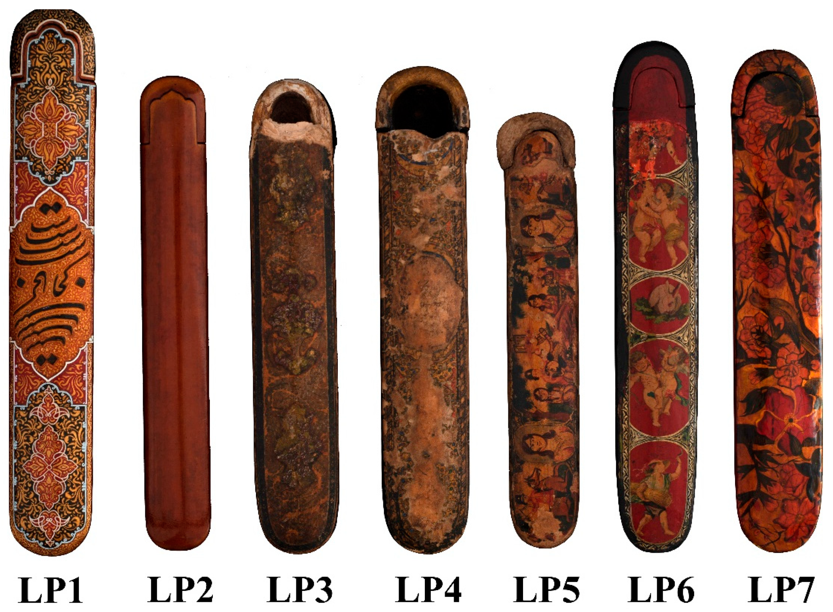
Figure 1. The studied lacquer papier mache penboxes and their codes.
Heritage 04 00111 g001
Figure 2. Arrangement of the camera, light sources, penboxes, and filters used in UV fluorescence imaging.
Heritage 04 00111 g002
Figure 3. FTIR spectra of the studied samples and references in the range of 650–4000 cm−1 after baseline correction.
Heritage 04 00111 g003
Figure 4. UV-induced visible luminescence imaging of the samples with two processing methods (UVL1 and UVL2) and with image capture in the bright red range (UVIRL) and the blue range (UVIBL).
Heritage 04 00111 g004
Table 1. Radiation sources and filters used in each imaging method.
Imaging TechniqueFilter(s) in Front of Radiation SourcesFilter(s) in Front of CameraRange Investigated
Visible-reflected Imaging (VIS)2 × Youngenu NY660 Xenon flashlight (without filter)Baader UV/IR Cut420–680 nm
UV-induced visible luminescence imaging (UVL)+2 × Hoya U-360Baader UV/IR Cut420–680 nm
UV-induced Blue luminescence imaging (UVIBL)+2 × Hoya U-360Baader UV/IR Cut + MidOpt BP470425–495 nm
UV-induced Red luminescence imaging (UVIRL)+2 × Hoya U-360Baader UV/IR Cut + MidOpt BP635615–645 nm
Table 2. Summary of the results of luminance imaging of the coatings identified by FTIR.
SamplesCoating Type *Imaging Methods
UVL1UVL2UVIRLUVIBL
LP1ShellacBrownOrangeRedDark blue
LP2
LP3Kaman oilGreenMilkyRedLight blue
LP4
LP5
LP6
LP7Alkyd resinBlackBlackBlackBlack
* Based on FTIR spectroscopy results.
Publisher’s Note: MDPI stays neutral with regard to jurisdictional claims in published maps and institutional affiliations.