Non-Invasive Investigation of Pigments of Wall Painting in S. Maria Delle Palate di Tusa (Messina, Italy)

 , and
, and 
Abstract
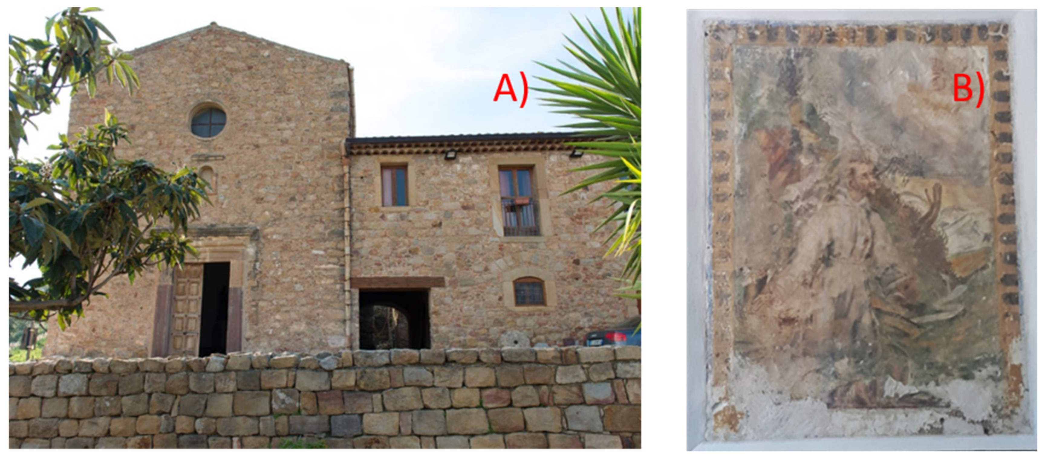
1. Introduction
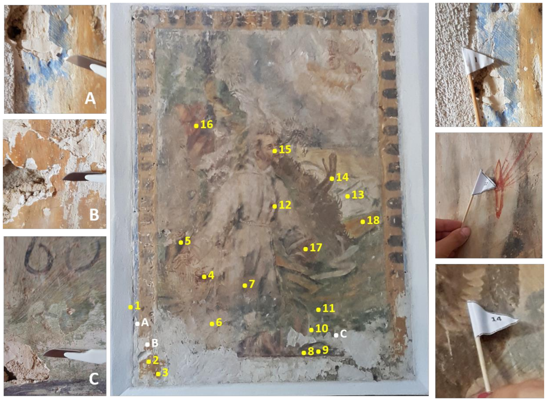
2. Instrumentation and Methods
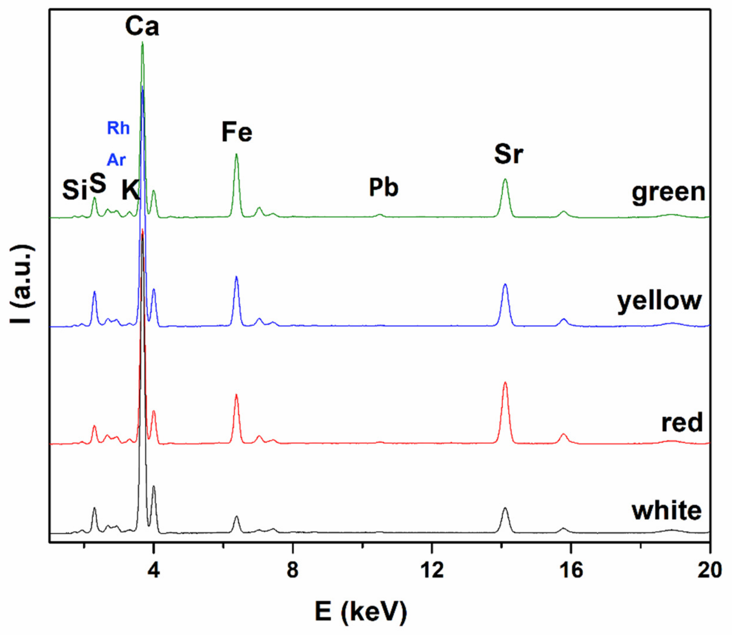
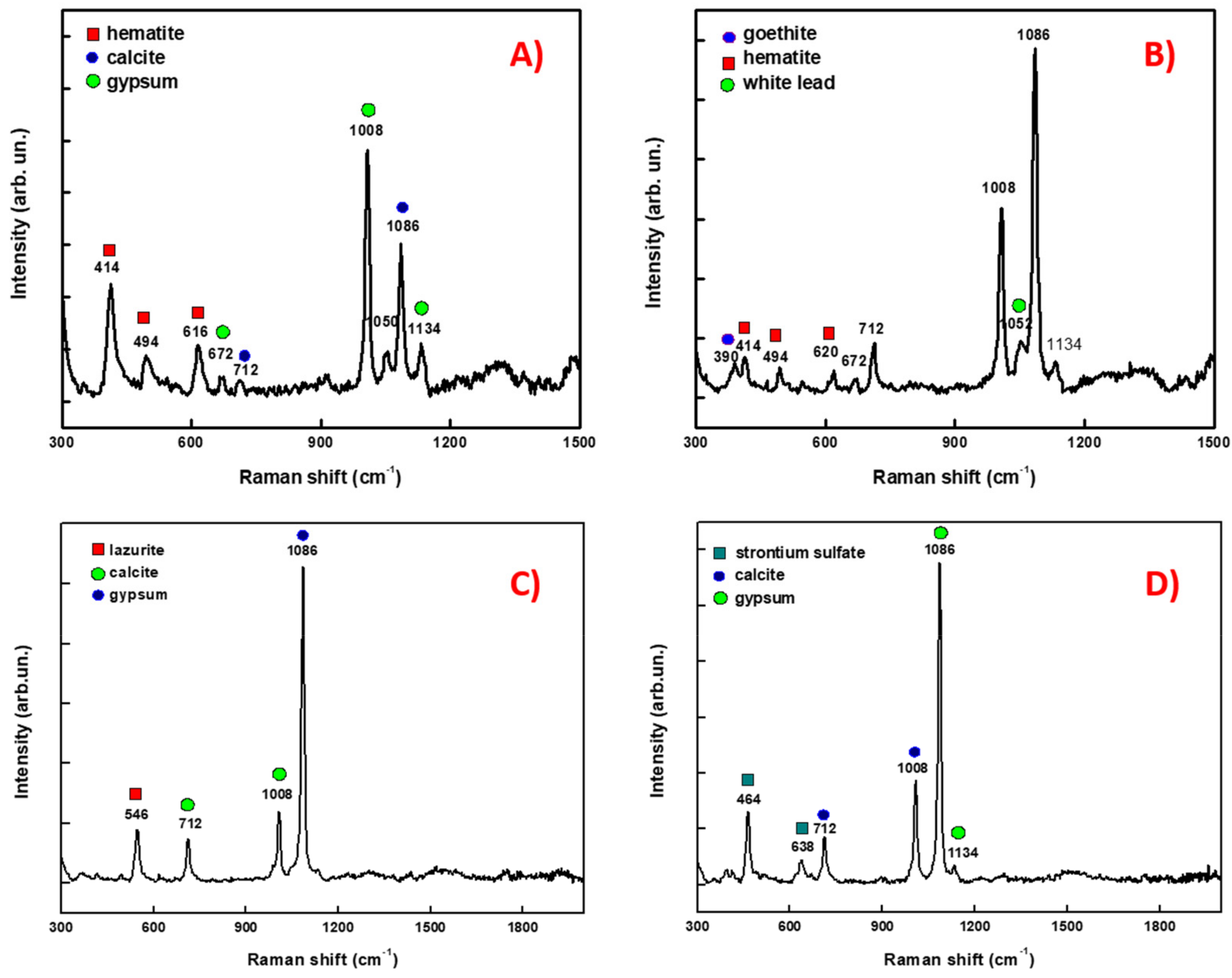
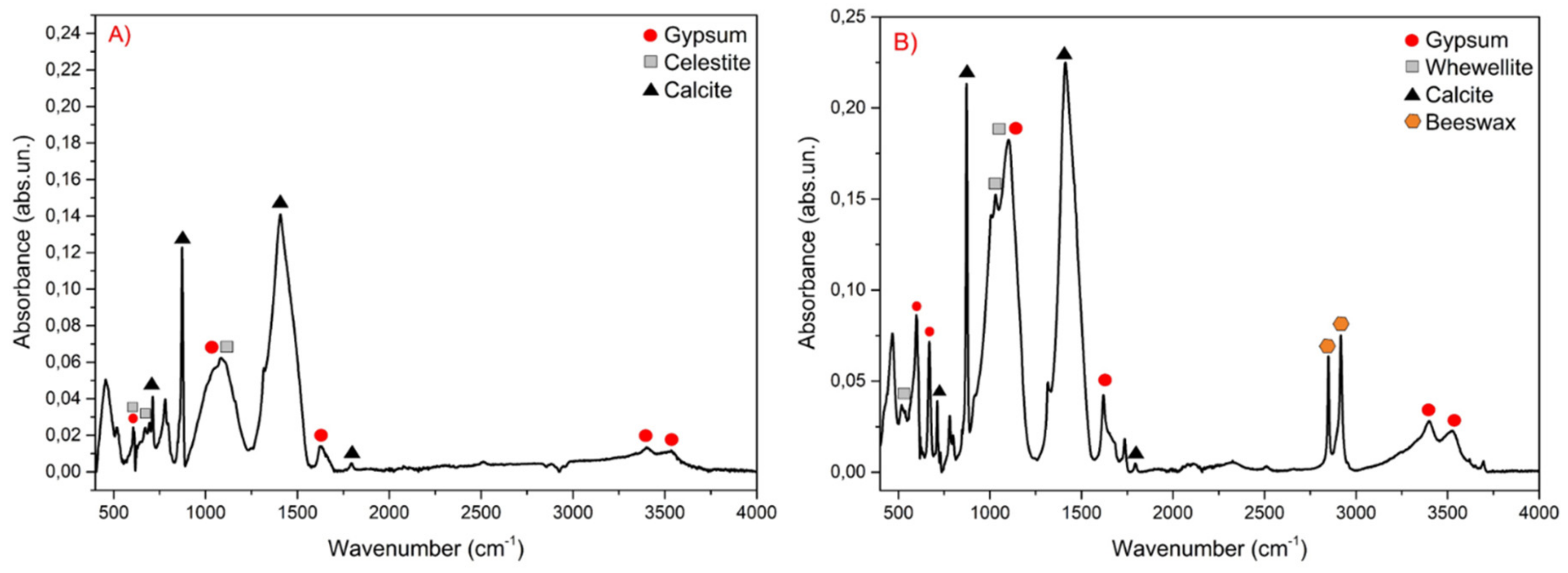
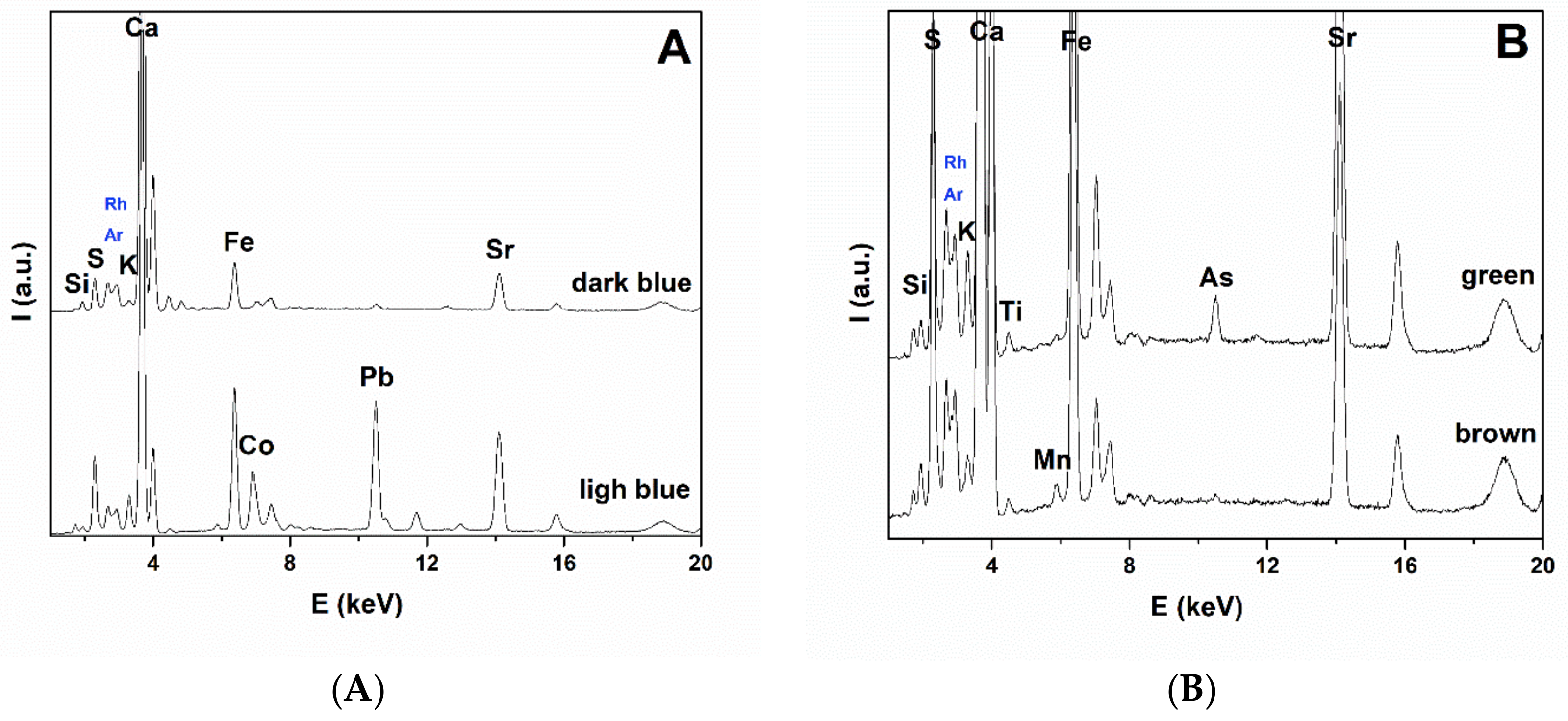
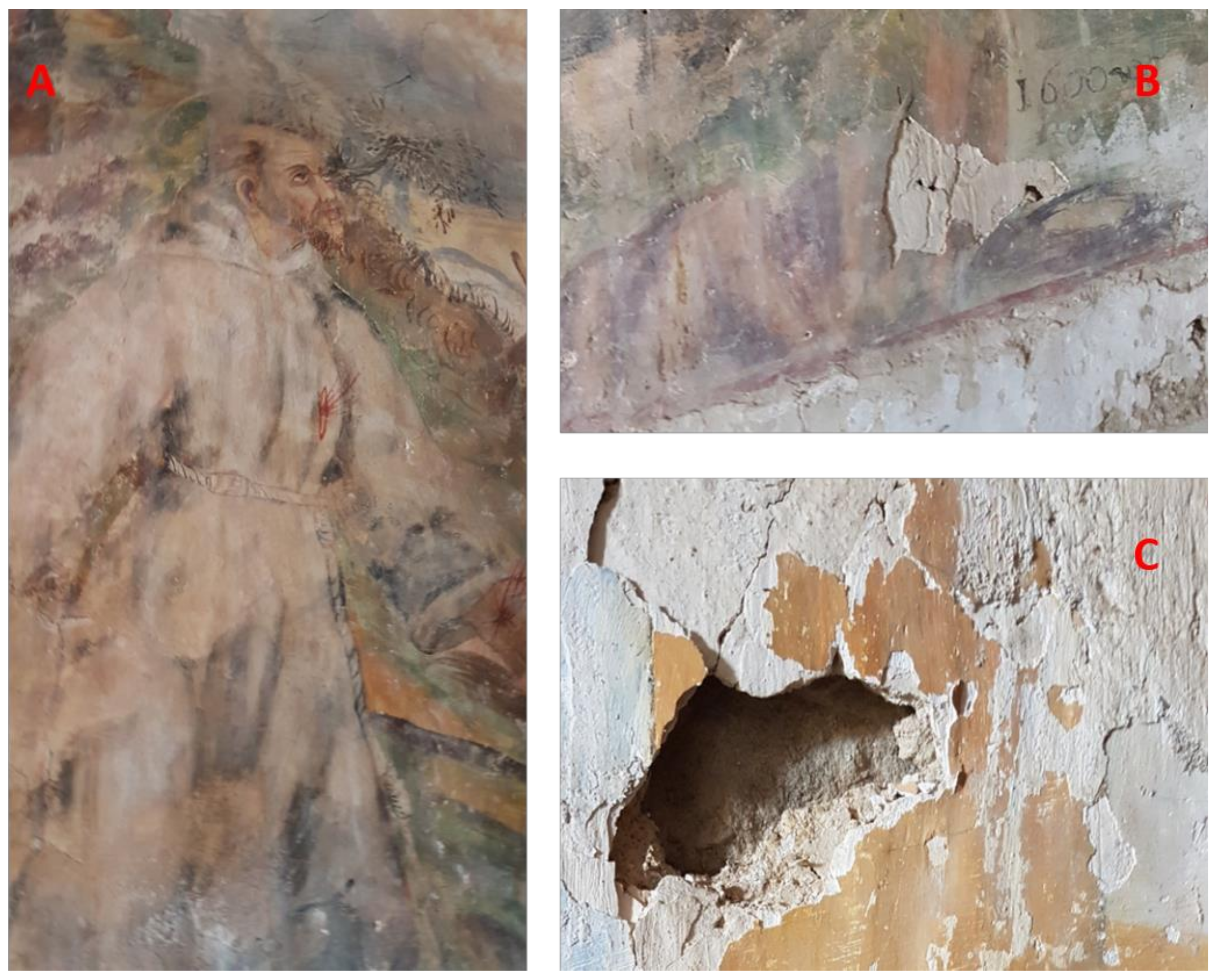
3. Results and Discussion
4. Conclusions
Author Contributions
Funding
Conflicts of Interest
References
- Scibona, G. Alesa Archonidea: La Chiesa e il Complesso Edilizio Postmedievale; Regione Siciliana, Assessorato dei Beni Culturali e Ambientali e della Pubblica Istruzione, Dipartimento per i Beni Culturali e Ambientali ed Educazione Permanente: Palermo, Italy, 2008; ISBN 9788861640283. [Google Scholar]
- Scibona, G. Alesa Archonidea: Dalla Riscoperta Tardo Rinascimentale Agli Scavi del 20; Regione Siciliana, Assessorato dei Beni Culturali e Ambientali e della Pubblica Istruzione, Dipartimento per i Beni Culturali e Ambientali ed Educazione Permanente: Palermo, Italy, 2008; ISBN 9788861640290. [Google Scholar]
- Barbera, G.; Gullotta, S. The Halaesa landscape (III b.c.) as ancient example of the complex and bio-diverse traditional Mediterranean polycultural landscape. Landsc. Hist. 2014, 35, 53–66. [Google Scholar] [CrossRef]
- Crisà, A. GL Castelli, principe di Torremuzza. LANX 2009, 2, 116–149. [Google Scholar]
- Marcaida, I.; Maguregui, M.; Morillas, H.; Taboada, N.P.; de Vallejuelo, S.F.-O.; Veneranda, M.; Madariaga, J.M.; Martellone, A.; de Nigris, B.; Osanna, M. In situ non-invasive characterization of the composition of Pompeian pigments preserved in their original bowls. Microchem. J. 2018, 139, 458–466. [Google Scholar] [CrossRef]
- Giustetto, R.; Gonella, D.; Diana, E. Decay of red pigments on a wall painting adorning the church of ‘San Francesco Dei Capuccini’ in Racconigi (Italy): Archaeometric survey and restoration intervention. Mediterr. Archaeol. Archaeom. 2018, 5, 65–80. [Google Scholar]
- Saladino, M.L.; Ridolfi, S.; Carocci, I.; Chirco, G.; Caramanna, S.; Caponetti, E. A multi-disciplinary investigation of the “Tavolette fuori posto” of the main hall wooden ceiling of the “Steri” (Palermo, Italy). Microchem. J. 2016, 126, 132–137. [Google Scholar] [CrossRef]
- Saladino, M.L.; Ridolfi, S.; Carocci, I.; Martino, D.C.; Lombardo, R.; Spinella, A.; Traina, G.; Caponetti, E. A multi-analytical non-invasive and micro-invasive approach to canvas oil paintings. General considerations from a specific case. Microchem. J. 2017, 133, 607–613. [Google Scholar] [CrossRef]
- Lauwers, D.; Hutado, A.G.; Tanevska, V.; Moens, L.; Bersani, D.; Vandenabeele, P. Characterisation of a portable Raman spectrometer for in situ analysis of art objects. Spectrochim. Acta Part A 2014, 118, 294–301. [Google Scholar] [CrossRef] [PubMed]
- Armetta, F.; Caponetti, E.; Brusca, L.; Martino, D.C.; Saladino, M.L.; Ridolfi, S.; Chirco, G.; Berrettoni, M.; Conti, P.; Nicolò, B.; et al. A multivariate approach to the study of orichalcum ingots from the underwater Gela’s archaeological site. Microchem. J. 2017, 135, 163–170. [Google Scholar]
- Vandenabeele, P.; Edwards, H.G.M.; Jehlička, J. The role of mobile instrumentation in novel applications of Raman spectroscopy: Archaeometry, geosciences, and forensics. Chem. Soc. Rev. 2016, 43, 2628–2649. [Google Scholar] [CrossRef]
- Vandenabeele, P.; Donais, M.K. Mobile spectroscopic instrumentation in archaeometry research. Appl. Spectrosc. 2016, 70, 27–41. [Google Scholar] [CrossRef]
- Mollica Nardo, V.; Aliotta, F.; Mastelloni, M.A.; Ponterio, R.C.; Saija, F.; Trusso, S.; Vasi, C.S. A spectroscopic approach to the study of organic pigments in the field of Cultural Heritage. Atti della Accademia Peloritana dei Pericolanti 2017, 95, A5. [Google Scholar]
- Marcaida, I.; Maguregui, M.; Morillas, H.; García-Florentino, C.; Knuutinen, U.; Carrero, J.A.; de Vallejuelo, S.F.-O.; Martı, A.P.; Castro, K.; Madariaga, J.M. Multispectroscopic and Isotopic Ratio Analysis to Characterize the Inorganic Binder Used on Pompeian Pink and Purple Lake Pigments. Anal. Chem. 2016, 88, 6395–6402. [Google Scholar] [CrossRef]
- Angeli, L.; Legnaioli, S.; Fabbri, C.; Grifoni, E.; Lorenzetti, G.; Guilaine, J.; Palleschi, V.; Radi, G. Analysis of Serra d’Alto figuline pottery (Matera, Italy): Characterization of the dark decorations using XRF. Microchem. J. 2018, 137, 174–180. [Google Scholar] [CrossRef]
- Conti, C.; Botteon, A.; Bertasa, M.; Colombo, C.; Realini, M.; Sali, D. Portable Sequentially Shifted Excitation Raman spectroscopy as an innovative tool for in situ chemical interrogation of painted surfaces. Analyst 2016, 141, 4599–4607. [Google Scholar] [CrossRef]
- Pozzi, F.; Basso, E.; Rizzo, A.; Cesaratto, A.; Tague, T.J. Evaluation and optimization of the potential of a handheld Raman spectrometer: In situ, noninvasive materials characterization in artworks. J. Raman Spectr. 2019, 50, 861–872. [Google Scholar] [CrossRef]
- Renda, V.; Saladino, M.L.; Caramanna, S.; Chirco, G.; Castello, A.F.; Conti, C.; Marco, A.; Sali, D.; Caponetti, E. Investigation on a low environmental impact solvent mixture applied to a wooden painted slab. IJCS 2016, SP2, 789–796. [Google Scholar]
- Burgio, L.; Clark, R.H. Library of FT-Raman spectra of pigments, minerals, pigment media and varnishes, and supplement to existing library of Raman spectra of pigments with visible excitation. Spectrochim. Acta Part A 2001, 57, 1491–1521. [Google Scholar] [CrossRef]
- Marcaida, I.; Maguregui, M.; Morillas, H.; Taboada, N.P.; Veneranda, M.; de Vallejuelo, S.F.O.; Martellone, A.; de Nigris, B.; Osanna, M.; Madariaga, J.M. In situ non-invasive multianalytical methodology to characterize mosaic tesserae from the House of Gilded Cupids, Pompeii. Herit. Sci. 2019, 7, 3. [Google Scholar] [CrossRef]
- Mollica Nardo, V.; Renda, V.; Anastasio, G.; Caponetti, E.; Saladino, M.L.; Vasi, C.S.; Ponterio, R.C. A combination of portable non-invasive techniques to study on reverse glass paintings at Mistretta museum. Microchem. J. 2019, 146, 640–644. [Google Scholar] [CrossRef]
- Salvadori, B.; Errico, V.; Mauro, M.; Melnik, E.; Dei, L. Evaluation of Gypsum and Calcium Oxalates in Deteriorated Mural Paintings by Quantitative FTIR Spectroscopy. Spectrosc. Lett. 2003, 36, 501–513. [Google Scholar] [CrossRef]
- Baglioni, M.; Poggi, G.; Ciolli, G.; Fratini, E.; Giorgi, R.; Baglioni, P. A Triton X-100-Based Microemulsion for the Removal of Hydrophobic Materials from Works of Art: SAXS Characterization and Application. Materials 2018, 11, 1144. [Google Scholar] [CrossRef]
- Buzgar, N.; Buzatu, A.; Sanislav, I.V. The Raman study on certain sulfates. Analele Stiintifice ale Universitatii Al. I. Cuza 2009, 55, 5–23. [Google Scholar]
- Ferroni, E.; Malaguzzi Valerj, V.; Rovida, G. Experimental Study by Diffraction of Heterogeneous Systems as a Preliminary to the Proposal of a Technique for the Restoration of Gypsum Polluted Murals. In Proceedings of the ICOM Conference, Amsterdam, The Netherlands, September 1969. [Google Scholar]


© 2019 by the authors. Licensee MDPI, Basel, Switzerland. This article is an open access article distributed under the terms and conditions of the Creative Commons Attribution (CC BY) license (http://creativecommons.org/licenses/by/4.0/).
Share and Cite
Mollica Nardo, V.; Renda, V.; Bonanno, S.; Parrotta, F.; Anastasio, G.; Caponetti, E.; Saladino, M.L.; Vasi, C.S.; Ponterio, R.C. Non-Invasive Investigation of Pigments of Wall Painting in S. Maria Delle Palate di Tusa (Messina, Italy). Heritage 2019, 2, 2398-2407. https://doi.org/10.3390/heritage2030147
Mollica Nardo V, Renda V, Bonanno S, Parrotta F, Anastasio G, Caponetti E, Saladino ML, Vasi CS, Ponterio RC. Non-Invasive Investigation of Pigments of Wall Painting in S. Maria Delle Palate di Tusa (Messina, Italy). Heritage. 2019; 2(3):2398-2407. https://doi.org/10.3390/heritage2030147
Chicago/Turabian StyleMollica Nardo, Viviana, Vincenzo Renda, Sara Bonanno, Francesco Parrotta, Gianfranco Anastasio, Eugenio Caponetti, Maria Luisa Saladino, Cirino Salvatore Vasi, and Rosina Celeste Ponterio. 2019. "Non-Invasive Investigation of Pigments of Wall Painting in S. Maria Delle Palate di Tusa (Messina, Italy)" Heritage 2, no. 3: 2398-2407. https://doi.org/10.3390/heritage2030147
APA StyleMollica Nardo, V., Renda, V., Bonanno, S., Parrotta, F., Anastasio, G., Caponetti, E., Saladino, M. L., Vasi, C. S., & Ponterio, R. C. (2019). Non-Invasive Investigation of Pigments of Wall Painting in S. Maria Delle Palate di Tusa (Messina, Italy). Heritage, 2(3), 2398-2407. https://doi.org/10.3390/heritage2030147