Article

Rutin Attenuates Microglial Inflammatory Responses by Promoting M2-like Polarization via GDNF and SHH/GLI-1 Signaling and NLRP3 Inflammasome Inhibition


by
Érica Novaes Soares
1,
Julita Maria Pereira Borges
2,
Luciana dos Santos Freitas
3,
Monique Reis de Santana
1,
Alexandre Moraes Pinheiro
3,
Maria de Fátima Dias Costa
1,
Silvia Lima Costa
1,* and
Victor Diogenes Amaral da Silva
1,*
1
Laboratory of Neurochemistry and Cell Biology, Department of Biochemistry and Biophysics, Institute of Health Sciences, Federal University of Bahia (UFBA), Salvador 40110-902, Bahia State, Brazil
2
Laboratory of Pathology and Cell Culture, Department of Health of Science, University State of Southwest of Bahia (UESB), Vitória da Conquista 45083-900, Bahia State, Brazil
3
Laboratory of Veterinary Biochemistry and Immunology, Agricultural Science Centre, Federal University of Recôncavo of Bahia (UFRB), Cruz das Almas 44380-000, Bahia State, Brazil
*
Authors to whom correspondence should be addressed.
Neuroglia 2026, 7(2), 15; https://doi.org/10.3390/neuroglia7020015
Submission received: 11 February 2026 / Revised: 5 May 2026 / Accepted: 8 May 2026 / Published: 17 May 2026

Abstract

Introduction: Rutin is a heterocyclic flavonol glycoside found in plants like apples, citrus fruits and buckwheat, with demonstrated anti-inflammatory properties. However, the molecular mechanisms underlying rutin’s direct effects on microglia, the main immune effector cells in the central nervous system, are not fully understood. The SHH/GLI-1 pathway is a neuronal repair pathway that modulates microglial activity and cell proliferation. Objective: For better compression of the rutin anti-inflammatory effects, this work evaluated the action of rutin on SHH/GLI-1 regulation. Methodology: For this, primary cultures of microglia from postnatal P0–2 days Wistar rats were stimulated with LPS (1 µg/mL) and/or treated with rutin (0.5–1 µM). Microglia morphology was characterized by immunofluorescence for Iba1. Gene expression of cytokines, inflammasome, glial-derived neurotrophic factors (GDNFs), and Sonic Hedgehog and family zinc finger-1 (SHH/GLI) were evaluated by real-time qPCR. Result: The results demonstrated that rutin inhibited the LPS-induced inflammatory response in microglia regulating negatively TNF-alpha, IL-6, and NLR family pyrin domain-containing 3 (NLRP3) mRNA expression. In addition, rutin increased GDNF and SHH-GLI-1 mRNA expression. Furthermore, conditioned medium from rutin-treated microglia showed a protective effect on PC-12 cells against LPS-induced cytotoxicity, reducing cell death as measured by the propidium iodide test and preserving cell morphology. Conclusions: This is the first evidence of the effect of rutin in SHH-GLI-1 signaling, contributing to the understanding of its pharmacological mechanisms and potentially revealing new molecular targets for treatment of neuroinflammatory diseases.

1. Introduction

Neuroinflammation is orchestrated by glial cells, especially microglia and astrocytes, which constitute the first line of defense of the central nervous system (CNS) against different types of damage, and is a central event in the pathophysiology of neurodegenerative diseases (NDDs), such as Parkinson’s disease (PD) [1] and Alzheimer’s disease (AD) [2]. In the context of inflammatory stimuli, microglia can exhibit different phenotypic states and display an inflammatory or anti-inflammatory profile [2]. Therefore, microglia are fundamental to the progression of NDD, since they are associated with the synthesis and assembly of cytokines, chemokines, and other mediators that contribute to both damage and repair of the CNS. In response to a CNS injury, microglia reactivity is accompanied by alterations in cellular energy metabolism, morphology, and expression of signaling molecules reflecting pathological or tissue repair events [1,2]. The knowledge about the polarization of the microglia phenotypic profile has become increasingly broad. It is known that interleukin 4 (IL-4) and interleukin 13 (IL-13), cytokines from type 2 helper T cells (Th2), can be factors that induce alternative activation of microglia to M2 regulatory/neuroprotective profiles. This phenotype expresses higher levels of the enzyme arginase-1 (Arg-1) and the differentiation clusters CD36, CD163, and CD206 on the cell surface. Furthermore, M2-polarized microglia produce regulatory cytokines, such as IL-10, which can reduce inflammation mediated by M1 microglia [3,4]. On the other hand, the resolution of the lesion following inflammation and oxidative stress involves activation of the Sonic Hedgehog (SHH) signaling pathway and its downstream effector GLI-1 ((SHH)/GLI-1). The SHH signaling pathway plays a key role in regulating the CNS, both during development and adulthood. Following events such as ischemic injury, neurodegenerative processes, and brain trauma, this pathway is activated, contributing to the recovery of neurological function through the modulation of inflammation and the regulation of apoptotic processes. In the mature CNS, SHH continues to be expressed by some cells; its effects are not fully understood, but it is known that its dysregulation can lead to neurological disorders such as Parkinson’s disease. Studies suggest that the SHH signaling pathway exerts a beneficial neuroprotective effect by inhibiting neuroinflammation and regulating glutamate levels. [5,6]. This pathway is modulated, for example, by the silent information regulator factor 2-related enzyme 1 (sirtuin 1), that can induce a suppressive effect in inflammatory N9 microglia after oxygen/glucose deprivation and reperfusion injury [7]. Kyoko Tsuboi [8] demonstrated for the first time in vivo that SHH reduces behavioral deficits induced by intrastriatal lesions with 6 dopamine hydroxytoluene (6-OHDA) and suggested that SHH may be a candidate for the treatment of nigrostriatal system disorders, such as Parkinson’s disease. Furthermore, studies by Liao H [7] demonstrate that the inhibition of microglial hyperactivation was associated with the SHH signaling pathway. Lai et al. [9] showed that epigallocatechin gallate and minocycline increased SHH expression and that the anti-inflammatory effects of brain-derived neurotrophic factors were mediated by erythropoietina/SHH in microglia.
The search for natural products for medicinal use, especially those derived from plants, runs parallel to the history of humanity itself. The popular use of medicinal plants is a traditional practice that has accompanied humankind since the dawn of civilization, based on the accumulation of information passed down orally. In this respect, the pharmaceutical industry has researched and prospected plant secondary metabolites, which have been associated with a wide range of biological activities against some pathologies [10]. Flavonoids form a group of plant-derived polyphenolic secondary metabolites found in food including nuts, cocoa, and beverages such as teas and wines. Many flavonoids exert antioxidant or anti-inflammatory activities and may be responsible for protection against various diseases [11]. Rutin (quercetin 3−O−rutinoside) is a glycosylated flavonoid produced in the synthesis pathway that involves a combination of 4-coumaroyl-CoA and malonyl-CoA in several medicinal plants and components of diet such as citrus and berry fruits, black/green tea, and red wine [12]. Considering that microglia play a crucial role in the regulation of neuroinflammation, much of the recent research in natural products has focused on finding neuroinflammatory regulators in microglia [13]. Rutin has attracted attention due to its antioxidant effects. Notably, pre-clinical evidence suggests that rutin promotes a microglia phenotype modulation with morphology alteration characterized by ramified morphology with thin and long extensions and attenuates inflammatory response induced by LPS [14]. Moreover, the neuroinflammatory effects of rutin were evidenced by inhibition of microgliosis induced by lipopolysaccharide (LPS) or interferon-γ (IFN-γ) [14].
The capacity of the flavonoid rutin to protect the brain against damage associated with glutamate excitotoxicity, ischemic injury and loss of dopaminergic neurons in Parkinson disease (PD) models has also been characterized [15,16,17]. In organotypic brain cultures from rats, rutin inhibits glutamate-induced cell death and loss of glutamine synthetase (GS), an effect associated with increased expression of glutamate-aspartate transporter (GLAST), and enhanced glutamate uptake in cerebral cortex slices from adult rats [15]. It was also evidenced that rutin protects mesencephalic cells against aminochrome cytotoxicity in a PD in vivo model and prevents loss of dopaminergic neurons in substantia nigra pars compacta (SNPc) [16]. Rutin has been under pharmacological investigation for its activating action on mitogen-activated protein kinase (MAPK) signaling pathways, resulting in neuroprotection against sodium nitroprusside-induced apoptosis, as well as inducing survival activity in neural crest cells through ERK2 and PI3-dependent molecular mechanisms [17]. Rutin’s protective and anti-inflammatory effects have been associated with modulation of microglia activation, however, by mechanisms of signaling not yet clearly evidenced. Therefore, this study aimed to characterize mechanisms involved in the rutin anti-inflammatory and neuroprotective effects in reactive microglia.

2. Materials and Methods

2.1. Primary Microglia Culture

In this work, we used primary microglia cultures obtained from the cortex of neonatal rats (0–2 days) performed according to the methodology previously described by Dos Santos et al. [18]. Newborn Wistar rats (P0–2 days) were provided by the animal facility of the Institute of Health Sciences (ICS) of the Federal University of Bahia (UFBA). The protocol and experiments were approved by the Animal Research Ethics Committee (CEUA) of ICS/UFBA (No. 6731220818, approved on 20 March 2019). The brains of newborn Wistar rats were aseptically removed, and the meninges and blood vessels were removed from each cortex. The material was then mechanically dissociated and filtered through a sterile 75 mm diameter Nitex membrane ((R&D®), Falcon® Plates, London, United Kingdom). The filtered medium was resuspended in Dulbecco’s Modified Eagle’s Medium (DMEM, Gibco, Grand Island, NY, USA), supplemented with 33 mM glucose, 2 mM glutamine, 3 mM sodium bicarbonate, 0.5 mg/mL penicillin/streptomycin, 2.5 μg/mL fungizone, 10% equine serum (Gibco, Grand Island, NY, USA) and 10% fetal bovine serum (FBS) (Gibco, Grand Island, NY, USA). The cells were cultured in a 75 cm2 culture flask coated with poly-D-lysine (50 µg/mL) (TPP, Zellkultur, Suíça) and incubated at a humidified atmosphere with 5% CO2 at 37 °C. Upon reaching confluence (7–10 days), adherent microglia cells were collected by agitation at 165 rpm at 37 °C for 3 h. The supernatant containing the cells was collected and centrifuged at 1.000 rpm. Isolated microglia were seeded in 24- or 6-well plates at a density of 3 × 10⁴/cm2. Experiments were performed 24 h after plating. In all cases, cells were cultured at 37 °C with 5% CO2 for different experimental approaches. Previous studies have characterized this culture as having 99.14% purity, as evaluated by immunocytochemistry for Iba-1 as a microglial marker [19].

2.2. Treatments

Rutin (3,3′,4′,5,7-pentahydroxyflavone 3-rutinoside, ≥95.0% purity) was purchased from Sigma-Aldrich (St. Louis, MO, USA). This compound was dissolved in dimethyl sulfoxide (DMSO, Sigma-Aldrich, St. Louis, MO, USA) at a concentration of 100 mM and stored in the dark at 4 °C. Lipopolysaccharide from Escherichia coli (LPS, Sigma-Aldrich, St. Louis, MO, USA) was adopted to induce inflammatory stimuli in microglia. For experiments, the compounds were diluted directly in the culture media without FBS. Microglia were treated with LPS (1 µg/mL), or with LPS plus rutin (0.5 or 1 µM) for 24 h. The vehicle DMSO at 0.01% (v/v) was adopted as a negative control diluted in the DMEM. Concentrations of rutin and LPS adopted were based on previous studies that demonstrated the capacity of rutin to modulate microglia [15,16,18]. After treatments, the culture media were collected to treat neuronal PC12 cells as described below.

2.3. Immunocytochemistry for Iba-1 and CD-68

For immunocytochemistry, microglia cells were seeded on 24-well plates (Kasvi®, Pinhais, Brazil) with glass coverslips previously treated with 10 μg/mL poly-D-ornithine (Sigma-Aldrich, St. Louis, MO, USA). After treatments, cultures were washed three times with PBS, fixed with 4% paraformaldehyde, and permeabilized with 0.3% Triton X-100 in PBS for 5 min and incubated in blocking solution (3% albumin in PBS) for 1 h. Next, the cells were washed three times with phosphate-buffered-saline solution (PBS) and incubated with rabbit polyclonal Iba1 antibody (1:200; NBP2-75397, Novus, St. Charles, MO, USA) and CD-68 (1:200, PA5-89134 Invitrogen, Rockford, IL, USA) in PBS/BSA (1%) overnight at 4° C. Following, the cells were washed three times with PBS and incubated with sheep anti-rabbit IgG secondary antibody Alexa Fluor 488 (1:500, Thermo Fisher, Waltham, MA, USA) and Alexa Fluor 594 (Rabbit) (1:1000, Life Technologies, Carlsbad, CA, USA) in PBS for 2 h at room temperature and in the dark. After three washes with PBS, nuclear chromatin was stained with 4′, 6-diamidino-2-phenylindole (DAPI) (Molecular Probes, Eugene, OR, USA) at 5 μg/mL for 10 min at room temperature. Coverslips were washed with PBS and mounted on glass slides using mounting fluid containing n-propylgallate. At least eight images were obtained for each treatment using fluorescence microscopy (Leica DMIL Led Fluor/Leica DFC7000 T Camera, Mannheim, Germany).

2.4. Gene Expression Analysis by RT-qPCR

For quantitative real-time PCR (RT-qPCR), the samples were prepared as described by Bispo da Silva et al. [17]. Total RNA was isolated from microglia cultures using Trizol® reagent (Invitrogen, Life Technologies, Waltham, MA, USA) according to the manufacturer’s specifications. For this, 1 × 10⁴ cells/cm2 were cultured in 60 mm plates and then treated for 24 h with rutin (0.5 or 1 μM) and/or LPS 1 μg/mL. Samples were stored at −80 °C until analysis. RNA concentration and purity were determined by spectrophotometric analysis using the Kasvi nanospectrum (K23-0002, Meclab, Jacarei, SP, Bazil). DNA contaminants were removed by treating the samples with DNase using the Ambion DNA-free kit cat# AM1906 (Life Technologies™, Waltham, MA, USA). For cDNA synthesis, Master Mix Super Script® VILO™ cat# MAN0004286 (Invitrogen, life technologies™, Waltham, MA, USA) was used in a 20 μL reaction with a concentration of up to 2.5 μg of total RNA. RT-qPCR was performed using Taqman® Gene Expression Assays (Applied Biosystems, Foster City, CA, USA). The assay identifications for the genes quantified in this study were TNF (Rn99999017_m1), IL-6 (Rn01410330_m1), NLRP3 (Rn04244620_m1), SHH (Rn00568129_m1), GLI-1 (Rn01504237_m1), ARG1 (Rn00681090_m1), and GDNF (Rn00569519_m1). The analysis of mRNA expression by RT-qPCR was performed using the Quant Studio TM 7 Flex Real-Time PCR System (Applied Biosystems, Foster City, CA, USA). Thermocycling conditions were performed according to the manufacturer’s specifications. The β-actin gene (Rn00667869_m1) was used as a reference (endogenous control) for normalizing gene expression data. The data were expressed using the 2−(ΔΔCt) method.

2.5. TNF-α Levels by ELISA

TNF-α levels were quantified in the culture media of microglia cultures using a commercially available sandwich enzyme-linked immunosorbent assay (ELISA) kit (DY510-05, Rat TNF-alpha DuoSet ELISA, R&D Systems by Bio-Techne, Minneapolis, MN, USA) according to the manufacturer’s instructions. Absorbance was measured at 450 nm, and concentrations were calculated by interpolation from a standard curve generated using a four-parameter logistic regression model.

2.6. Neuronal Cell Viability Determined by Propidium Iodide Staining

To assess the effects of the microglial secretome subjected to the treatments, the PC12 rat pheochromocytoma cells (ATCC #CRL-1721.1 PC12 ADH, Rattus norvegicus, Manassas, VA, USA) were cultured as previously described by Dos Santos et al. [18]. Briefly, the cells were cultured in DMEM (Cultilab, São Paulo, SP, Brazil), supplemented with L-glutamine (Cultilab, São Paulo, SP, Brazil), 10% inactivated fetal bovine serum (SFB, Cultilab, São Paulo, SP, Brazil), 5% inactivated equine serum (Cultilab, São Paulo, SP, Brazil), 1% penicillin and 1% streptomycin (Cultilab, São Paulo, Brazil). PC-12 cells were cultured to confluence in 10 mm polystyrene plates (TPP, Trasadingen, Switzerland), trypsinized 0.05% trypsin and 0.02% EDTA, diluted in phosphate-buffered-saline solution (PBS) and replanted in 96-well plates (7.5 × 10³ cells/cm2). Then, 24 h after plating, the cells were differentiated with 100 ng/mL nerve growth factor (NGF) (Sigma-Aldrich, St. Louis, MO, USA) for 6 days. The NGF was dissolved in culture medium, and the medium was changed every 48 h to maintain the NGF concentration. All cultures were maintained in a humidified atmosphere incubator at 5% CO2 and 37 °C.
After one week of differentiation, PC12 cells were treated with microglia conditioned medium (MC) under control conditions (DMSO) and treated with LPS (1 µg/mL) or LPS plus rutin (1 µM) for 48 h, and the cell proportion was analyzed using propidium iodide (PI) staining. This technique allows for the analysis of viability through plasma membrane integrity in cell cultures. After 48 h of culture, the supernatant was removed and PI was added at 5 µg/mL diluted in DMEM and incubated for 1 h in an incubator at 37 °C and 5% CO2. Next, the PI solution was removed, and the wells were washed 3 times with PBS-0.6% glucose solution. The cells were analyzed and photographed using a fluorescence microscope (Leica DMIL Led Fluor/Leica DFC7000 T Camera, Mannheim, Germany). For the analysis, four images from each treatment were captured and quantified using ImageJ software v.1.4.6u (Wayne Rasband; National Institutes of Health, Kensington, MD, USA, https://imagej.net/software/imagej/); Quantification on 22 April 2026. The proportion of IP-positive cells was determined in three independent experiments.

2.7. Statistical Analysis

The data were analyzed using the statistical program Graph Pad Prism 8.0 (San Diego, CA, USA) and presented as medians ± percentiles (n = 3). To determine the statistical difference between the groups, an analysis of variance was performed using the one-way ANOVA test, followed by the Kruskal–Wallis and Dunn post-test, or sample t-test. Confidence intervals were defined at a 95% confidence level ((p < 0.05) was considered statistically significant).

3. Results

3.1. Rutin Mitigates LPS-Induced Inflammatory Response in Microglia

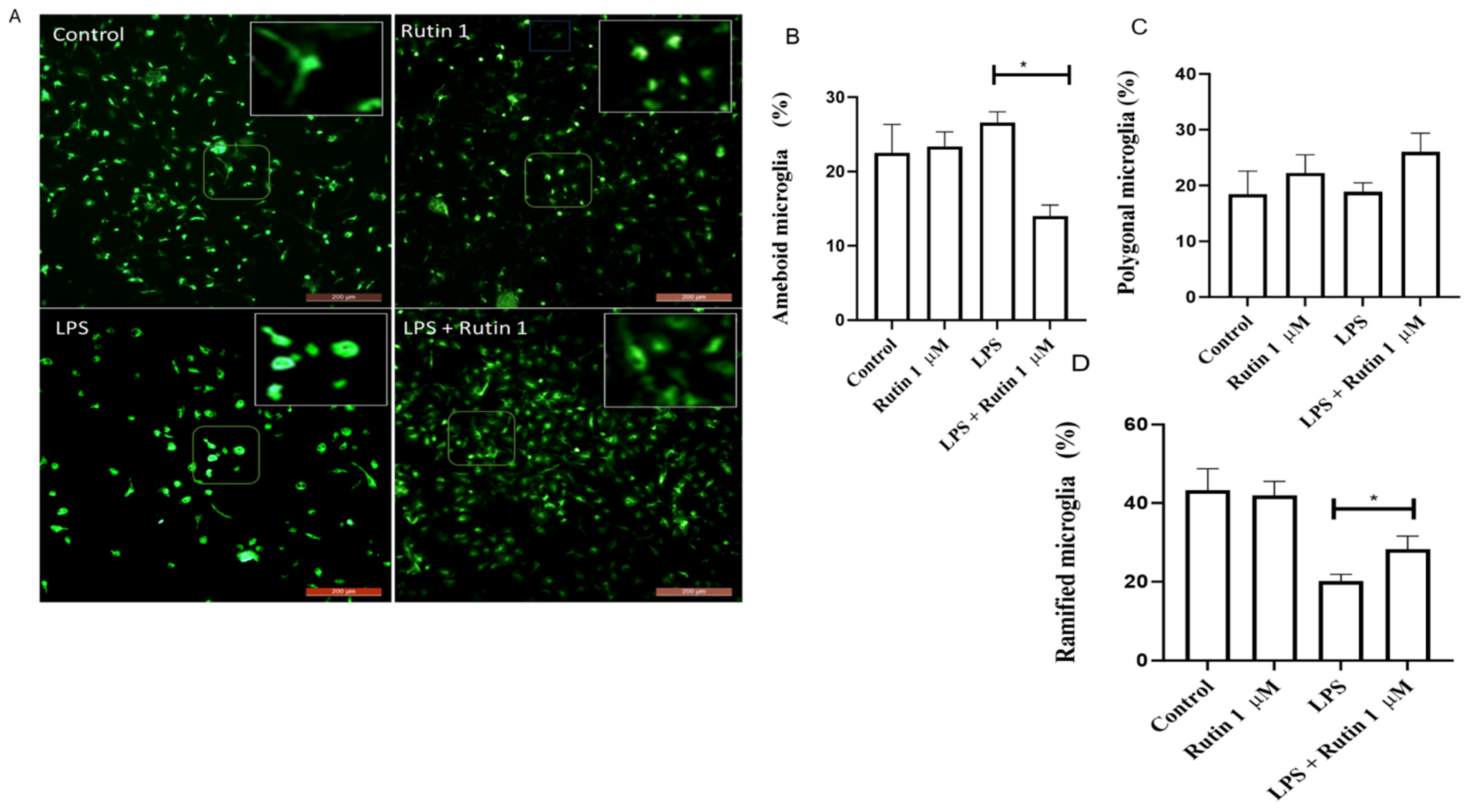
First, to characterize the effects of rutin treatment on the morphology of stimulated microglia, immunocytochemistry for the Iba-1 marker was performed after 24 h treatment with the flavonoid (1 μM), in association or not with the stimulus LPS (1 µg/mL), to induce an inflammatory response. The possible different microglia phenotypes (ramified, polygonal, or ameboid) were quantified (Figure 1A–D). In control cultures (vehicle DMSO) (Figure 1A–D), there was a predominance of microglia with ramified phenotype. LPS treatment induced a significant increase in the proportion of microglia presenting an amoeboid-activated phenotype. In contrast, co-treatment with LPS and rutin (1 μM) reduced the proportion of ameboid microglia compared to the control. Moreover, under this condition, the proportion of microglia with ramified phenotype was increased compared with cultures exposed to LPS alone. The proportion of cells with the intermediary polygonal phenotype was not affected by treatments.

3.2. Rutin Alters Inflammatory Marker Expression in Microglia

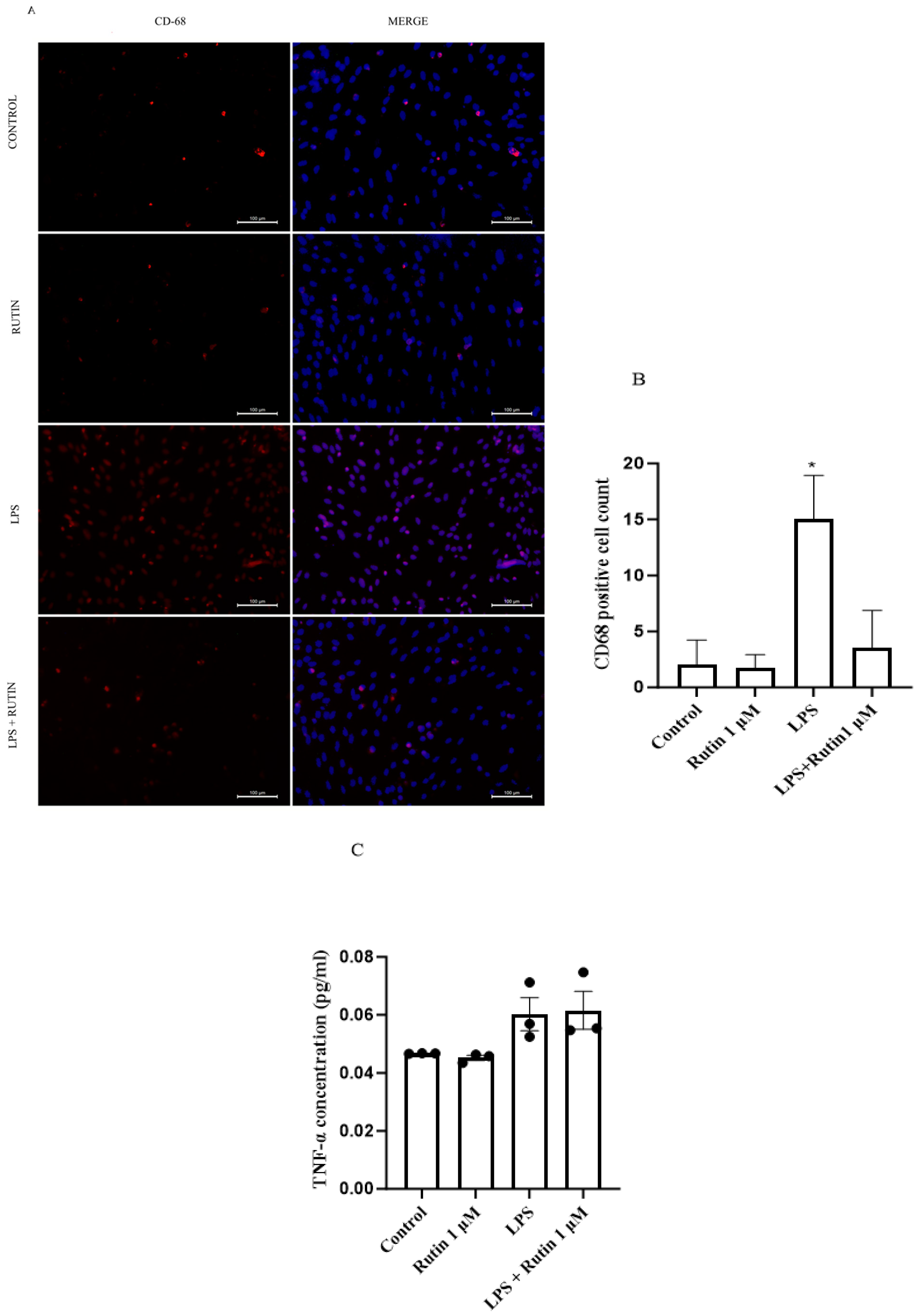
To characterize the M1 microglia phenotype, cells were subjected to control conditions (DMSO), rutin (1 µm), and LPS (1 µm) for 24 h. Subsequently, the cells were analyzed by immunocytochemistry assays using the CD-68 marker. No increase in CD68 marker was observed in the control (DMSO) or rutin-treated conditions. In contrast, LPS treatment significantly increased CD68 expression, indicating an inflammatory response (2 A and B). Nevertheless, in the concomitant condition (LPS 1 µm + rutin 1 µm), rutin was able to protect the cells by reducing the inflammatory process figure. The conditioned medium was then collected and analyzed by ELISA. Rutin treatment alone decreased TNF-α levels, and LPS alone increased; however, these changes were not statistically significant. In the co-treatment condition, rutin did not protect against LPS-induced damage (Figure 2C).

3.3. Rutin Regulates the Inflammatory Response in Rat Microglia

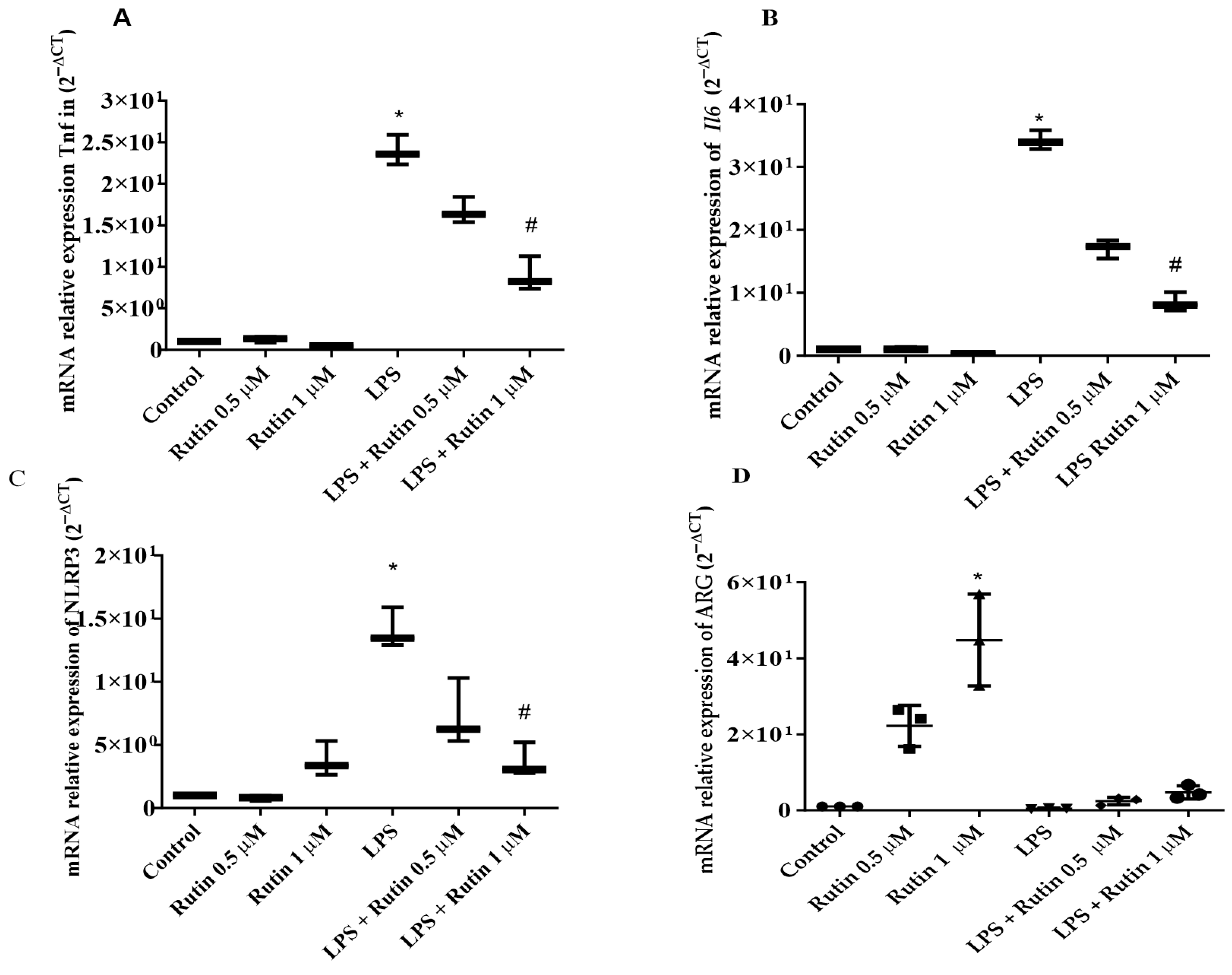
Analyzing the regulation of gene expression is fundamental to understanding how cells adapt to inflammatory stimuli. Thus, to characterize the effect of rutin in microglia exposed to LPS (1 µg/mL), RT-qPCR was used to measure mRNA expression of the inflammatory mediators interleukin 6 (IL-6), tumor necrosis factor (TNF), the NOD-like receptor protein 3 (NLRP3), and arginase (ARG), a key enzyme that acts as an anti-inflammatory, immunosuppressive factor. LPS-stimulated microglia showed a significative increase in the expression of mRNA for TNF-alpha (33.93), as well as in the expression of mRNA for IL-6 (23.57) and NLRP3 (13.46), compared to the control condition after 24 h (Figure 3). However, microglia co-treated with LPS (1 µg/mL) and rutin (0.5 or 1 μM) for 24 h showed no significant change in the expression of mRNA for TNF (Figure 3A), IL-6, (Figure 3B) and NLRP3 (Figure 3C) compared to LPS alone. In addition, the treatment with 1 μM rutin increased expression of mRNA for ARG (44.75) compared to control conditions (Figure 3D).

3.4. Rutin Upregulates GDNF and SHH/Gli1 in Microglial Cells

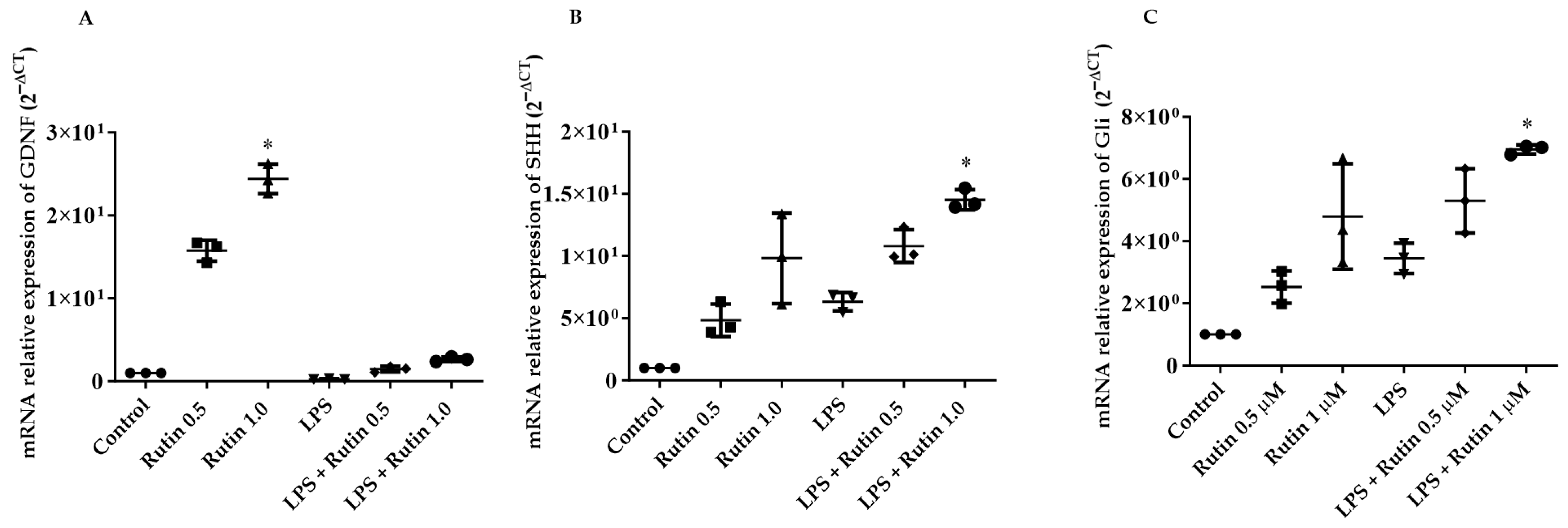
Numerous studies have evidenced the de novo expression of glial cell line-derived neurotrophic factor (GDNF) by glial cells in injured brains [17], and the growth factor is the one of the most potent neuroprotective molecule tested in cellular and animal models of PD [18]. In this sense, we investigated the effects of rutin on GDNF expression in LPS-stimulated microglia. Also, we analyzed the expression of components of the SHH/Gli-1 pathway that play a key role in the recovery of neuronal function through the modulation of inflammation. An increase in the levels of mRNA for GDNF was observed in microglia treated with 1 μM rutin when compared with the control cultures (Figure 4A), not evidenced in microglia treated with 0.5 μM rutin or LPS. Moreover, co-treatment with LPS and rutin (1 µM) increased mRNA levels of SHH (14.52 ± 0.81) and GLI-1 (5.29 ± 1.03), compared to microglia in control conditions. On the other hand, treatment with 0.5 μM rutin or LPS did not induce significant changes in mRNA levels for SHH or GLI-1 compared to control cultures (Figure 4B,C).

3.5. Rutin Modulates Microglial Secretome to Reduce Cytotoxicity in PC-12 Cells

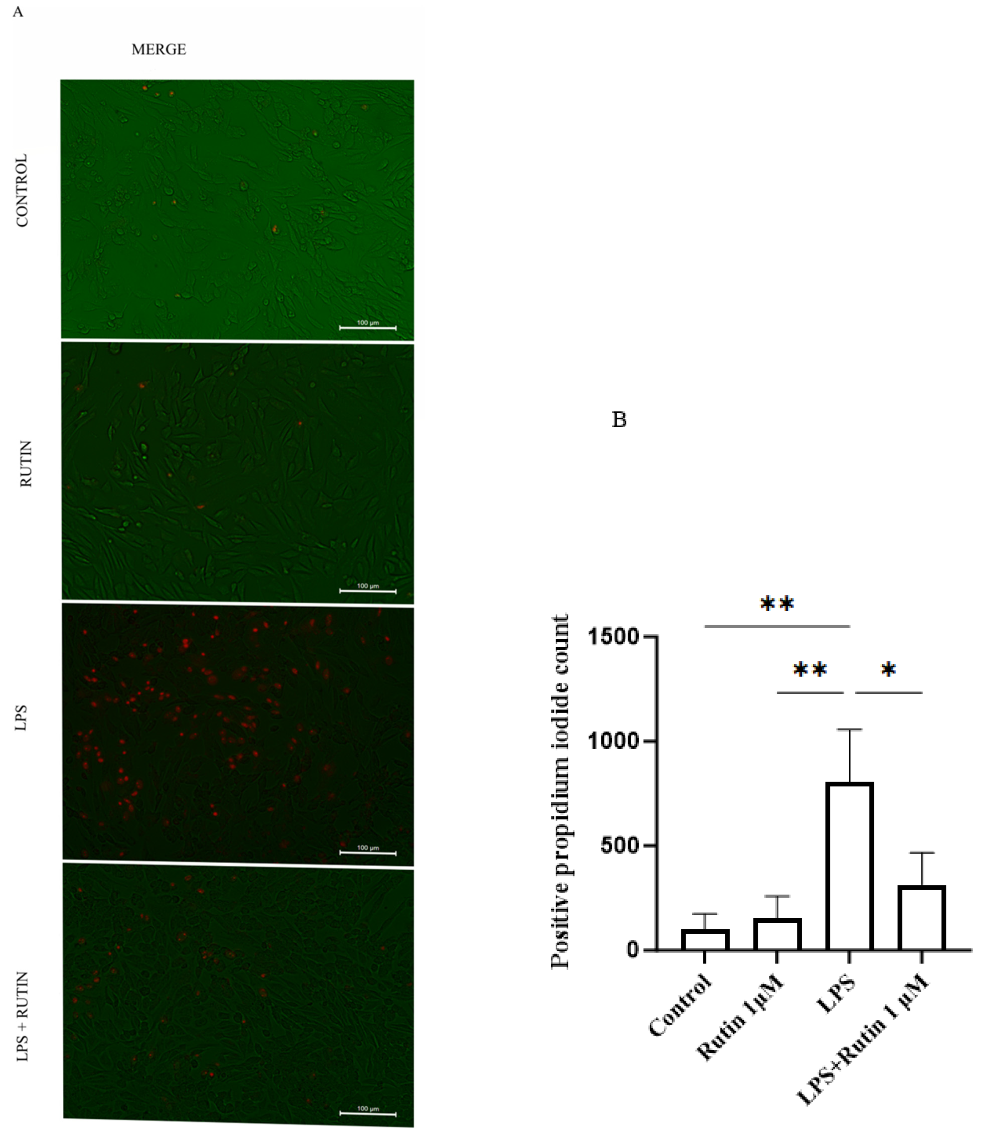
To analyze the effect of microglial conditioned medium on PC-12, cells were treated for 24 h. Conditioned medium from control (DMSO) and rutin (1 µM) groups showed no cytotoxicity effects. In contrast, PC12 cells treated with MC from LPS-stimulated microglia showed a significant 8-fold increase in propidium iodide-positive cells compared to the control. Notably, the treatment with MC from the LPS 1 µM and rutin 1 µM group attenuated this effect, reducing cytotoxicity by ~5-fold compared to LPS-treated MC (Figure 5A,B).

4. Discussion

Iba1 is a calcium-binding protein constitutively expressed in microglia in the central nervous system [19]. In the present study, rutin prevented the LPS-induced reduction in the microglial density and the increase in amoeboid microglia, suggesting attenuation of microglia reactivity and cytotoxicity. For instance, rutin 50 µM has been shown to induce an anti-inflammatory phenotype characterized by an increase in CD150 and CD206 markers, but not in Arginase 1 [14]. In contrast, an increase in Arginase 1 observed in our study, using lower concentrations of rutin, highlighting its potential protective effect as Arginase 1-positive microglia are involved in Aβ plaque reduction during IL-1β-dependent neuroinflammation [20].
Microglia reactivity and neuroinflammation are some of the most important events associated with neurodegeneration [21,22]. Consistently, we observed an increase in the mRNA levels of inflammatory TNF, IL6, and NLRP3 in LPS-treated microglia. Furthermore, the treatment with rutin mitigated the inflammatory response induced by LPS. The regulatory effect of rutin in microglia stimulated with LPS has been reported [23,24]. However, the molecular mechanism underlying the anti-inflammatory response is not fully understood. It is well-defined that LPS stimulates inflammatory response via Toll-like receptor 4 (TLR4), which initiates the via of TLR4/NF-κB/NLRP3 inflammasome activation [25]. The effect of rutin in the regulation of NLRP3 in the CNS has been reported in a model of spinal cord injury in rats [26]. However, evidence of the effect of rutin in the NLRP3 in brain microglia is missing. TLR4 may be a key molecular target since rutin presents a strong binding affinity to TLR4 and inhibits the Toll-like receptor 4/nuclear factor-kappa B signaling pathway in BV2 cells [22].
The beneficial effects of microglia polarization towards an anti-inflammatory phenotype can be attributed to their capacity to produce neurotropic mediators that support remyelination and regeneration. Distinct roles of neurotrophic factors in maintaining normal brain function, neuroprotection, or neuronal differentiation [27] have been well characterized, among them, the work in [28]. Previously, we demonstrated that the mRNA expression of GDNF was improved by rutin in an animal model of PD [14,16]. In line with those findings, we observed that rutin increased mRNA expression for GDNF in microglia associated with modulation of phenotype after inflammatory stimulus, supporting the hypothesis that this neurotrophic factor can be involved in its neuroprotective potential.
SHH is a protein linked to some MAPK pathways. It makes up a family of flags for the hedgehog pathway, which can be active in glial cells during brain development or in adulthood and/or in precursor cells. There is evidence that the Hedgehog signaling pathway is involved in the response after injury to the brain and this process occurs due to the activation of astrocytes [29]. Another study revealed that activated microglia showed positive expression for SHH in animals that received MPTP or in primary culture exposed to LPS [30]. The results obtained in the present study revealed a substantial increase in SHH mRNA in microglia exposed to LPS and treated with rutin. These findings can be related to the observation by [31], which showed an increase in SHH expression induced by epigallocatechin gallate (EGCG) and minocycline as an effect related to their anti-inflammatory actions. GLI-1 is a transcriptional regulator of the canonical and non-canonical Hedgehog pathway, which acts as the final effector of SHH and other important molecular pathways. In this sense, GLI-1 regulates pro-proliferative and pro-survival genes [6,9,32]. Here, we showed an increase in SHH and Gli1 in microglia treated with LPS plus rutin. The modulation of SHH/GLI-1 by rutin has not previously been shown. We suggest that the activation of Nrf2/Shh signaling cascade must be further investigated as a molecular mechanism of rutin since there is evidence of this role in the effect of resveratrol, another neuroprotective flavonoid [33]. Furthermore, the role of Nrf2 in the protective action of rutin in microglia and neural cells is well established [34,35].
The CD-68 marker is expressed in inflammatory processes by stimuli such as LPS, which was used in this study as a positive control, in microglia. It was observed that treatment with LPS resulted in a significant increase in CD68 levels, while concomitant treatment resulted in a reduction. The findings of this study, through rtqPCR, showed that LPS treatment increased inflammasome and TNF levels. These CD68 data showed that LPS increases and rutin reduces expression, an indication of protection. This effect was also observed in concomitant treatment, with rutin inhibiting the effect of LPS. Studies [36] showed that microglia under conditioned medium conditions of PC-12 treated with LPS in combination with AF-MeOH (MC-LPS + AF-MeOH), compared to control cultures exposed to MC of PC12 cells subjected to inflammatory damage with LPS, had reduced CD68 expression.
To evaluate the effect of microglia conditioned medium on PC12 cells, the IP intercalating dye was used to attach to the DNA of damaged or dead cells. It was observed that treatment of PC12 cells with CM from microglia treated with LPS significantly increased the number of nonviable PC12 cells, but cell treated with the CM from microglia treated with LPS in association with rutin, the cytotoxic effect of LPS was inhibited. A factor that may have contributed to the number of positive IP cells is the fact that LPS induces an inflammatory effect in microglia, leading to the participation of pro-inflammatory cytokines such as TNF. On the other hand, rutin induces the microglia regulatory response, which may be indicative of a reduction in positive IP cells in concomitant treatment. In addition, the SHH pathway may also be contributing to the process of reducing the expected number of damaged PC-12 cells.

5. Conclusions

We conclude that rutin modulates LPS-induced inflammatory response in microglia associated with the improvement of GDNF and the SHH/GLI-1. Further investigations need to be performed to clarify the role of the Sonic Hedgehog/GLI pathway in the pharmacological effect of rutin in models of study of CNS injury.
Exposure of PC12 neuronal cells to the CM from microglia exposed to LPS and treated with rutin attenuated LPS-induced toxicity, demonstrating a protective effect. Further studies are necessary to better characterize protective components of microglia secretoma modulated by the flavonoid rutin under inflammatory conditions.

Author Contributions

Conceptualization, S.L.C. and V.D.A.d.S.; Methodology and data curation, É.N.S., L.d.S.F. and J.M.P.B.; Writing—original draft, É.N.S. and J.M.P.B.; Writing—review and editing, M.R.d.S., J.M.P.B. and V.D.A.d.S.; Formal analysis, A.M.P. and M.d.F.D.C.; Supervision and resources, S.L.C., and V.D.A.d.S.; Project administration, S.L.C. and V.D.A.d.S. Funding acquisition S.L.C. and V.D.A.d.S. All authors have read and agreed to the published version of the manuscript.

Funding

This study was funded by grants from the National Council for Scientific and Technological Development (CNPq) (Process No. 407833/2023-4); V.D.A.d.S. (Process Nº 303882/2022-0), and S.L.C. (Process Nº 312388/2021-7) are CNPq Research Fellows. S.L.C., V.D.A.d.S. and M.F.D.C. are members of the Brazilian National Institute of Science and Technology of Glia (INCT/CNPq-iGLIA).

Institutional Review Board Statement

Ethical Approval: Rat microglial cells were obtained in accordance with the local Ethics Committee for Animal Experimentation of the Institute of Health Sciences, Federal University of Bahia (CEUA protocol nº 6731220818, approved on 20 March 2019).

Informed Consent Statement

Not applicable.

Data Availability Statement

Data is contained within the article.

Acknowledgments

We would like to thank the Postgraduate Program in Immunology and the Laboratory of Neurochemistry and Cell Biology of the Federal University of Bahia. We thank the Coordination for the Improvement of Higher Education Personnel–CAPES for the post-graduation support.

Conflicts of Interest

The authors declare no conflicts of interest.

References

  1. Çınar, E.; Tel, B.C.; Şahin, G. Neuroinflammation in Parkinson’s Disease and its Treatment Opportunities. Balk. Med. J. 2022, 39, 318–333. [Google Scholar] [CrossRef] [Scilit] [PubMed] [PubMed Central]
  2. Han, J.; Zhang, Z.; Zhang, P.; Yu, Q.; Cheng, Q.; Lu, Z.; Zong, S. The roles of microglia and astrocytes in neuroinflammation of Alzheimer’s disease. Front. Neurosci. 2025, 19, 1575453. [Google Scholar] [CrossRef] [Scilit] [PubMed] [PubMed Central]
  3. von Tobel, J.S.; Antinori, P.; Zurich, M.G.; Rosset, R.; Aschner, M.; Glück, F.; Scherl, A.; Monnet-Tschudi, F. Repeated exposure to Ochratoxin A generates a neuroinflammatory response, characterized by neurodegenerative M1 microglial phenotype. Neurotoxicology 2014, 44, 61–70. [Google Scholar] [CrossRef] [Scilit] [PubMed]
  4. Nakagawa, Y.; Chiba, K. Papel da polarização M1/M2 da micróglia na recaída e remissão de transtornos e doenças psiquiátricas. Pharmaceuticals 2014, 7, 1028–1048. [Google Scholar] [CrossRef] [Scilit]
  5. Ma, H.; Li, H.; Zhang, Y.; Zhou, Y.; Liu, H.; Xu, H.; Zhu, L.; Zhang, G.; Wang, J.; Li, Z.; et al. Microglia Exhibit Distinct Heterogeneity Rather than M1/M2 Polarization within the Early Stage of Acute Ischemic Stroke. Aging Dis. 2023, 14, 2284–2302. [Google Scholar] [CrossRef] [Scilit] [PubMed] [PubMed Central]
  6. Zhang, H.; Su, Y.; Qu, Z.; Zhang, C.; Ma, S.; Li, X.; Wang, Y. Sonic Hedgehog Mediates High Frequency-Dependent Deep Brain Stimulation for the Correction of Motor Deficits in a Parkinson’s Disease Model. Neurosci. Bull. 2024, 40, 1732–1738. [Google Scholar] [CrossRef] [Scilit] [PubMed] [PubMed Central]
  7. Liao, H.; Huang, J.; Liu, J.; Zhu, H.; Chen, Y.; Li, X.; Wen, J.; Yang, Q. Sirt1 regulates microglial activation and inflammation following oxygen-glucose deprivation/reoxygenation injury by targeting the Shh/Gli-1 signaling pathway. Mol. Biol. Rep. 2023, 50, 3317–3327. [Google Scholar] [CrossRef] [Scilit] [PubMed] [PubMed Central]
  8. Tsuboi, K.; Shults, C.W. Intrastriatal injection of sonic hedgehog reduces behavioral impairment in a rat model of Parkinson’s disease. Exp. Neurol. 2002, 173, 95–104. [Google Scholar] [CrossRef] [Scilit] [PubMed]
  9. Lai, S.W.; Chen, J.H.; Lin, H.Y.; Liu, Y.S.; Tsai, C.F.; Chang, P.C.; Lu, D.Y.; Lin, C. Regulatory Effects of Neuroinflammatory Responses Through Brain-Derived Neurotrophic Factor Signaling in Microglial Cells. Mol. Neurobiol. 2018, 55, 7487–7499. [Google Scholar] [CrossRef] [Scilit] [PubMed]
  10. Marrelli, M. Medicinal Plants. Plants 2021, 10, 1355. [Google Scholar] [CrossRef] [Scilit] [PubMed]
  11. Negahdari, R.; Bohlouli, S.; Sharifi, S.; Maleki Dizaj, S.; Rahbar Saadat, Y.; Khezri, K.; Jafari, S.; Ahmadian, E.; Gorbani Jahandizi, N.; Raeesi, S. Therapeutic benefits of rutin and its nanoformulations. Phytother. Res. 2021, 35, 1719–1738. [Google Scholar] [CrossRef] [Scilit] [PubMed]
  12. Ullah, A.; Munir, S.; Badshah, S.L.; Khan, N.; Ghani, L.; Poulson, B.G.; Emwas, A.H.; Jaremko, M. Important Flavonoids and Their Role as a Therapeutic Agent. Molecules 2020, 25, 5243. [Google Scholar] [CrossRef] [Scilit] [PubMed]
  13. Yoon, C.-S. Natural Products in the Treatment of Neuroinflammation at Microglia: Recent Trend and Features. Cells 2025, 14, 571. [Google Scholar] [CrossRef] [Scilit]
  14. da Silva, A.B.; Coelho, P.L.C.; Amparo, J.A.O.; de Almeida Carneiro, M.M.A.; Borges, J.M.P.; dos Santos Souza, C.; Costa, M.D.F.D.; Mecha, M.; Rodriguez, C.G.; da Silva, V.D.A.; et al. The flavonoid rutin modulates microglial/macrophage activation to a CD150/CD206 M2 phenotype. Chem. Biol. Interact. 2017, 274, 89–99. [Google Scholar] [CrossRef] [Scilit] [PubMed]
  15. Ferreira, R.S.; Teles-Souza, J.; dos Santos Souza, C.; Pereira, É.P.; de Araújo, F.M.; da Silva, A.B.; Castro e Silva, J.H.; Nonose, Y.; Núñez-Figueredo, Y.; de Assis, A.M.; et al. Rutin improves glutamate uptake and inhibits glutamate excitotoxicity in rat brain slices. Mol. Biol. Rep. 2021, 48, 1475–1483. [Google Scholar] [CrossRef] [Scilit]
  16. De Araújo, F.M.; Frota, A.F.; de Jesus, L.B.; Cuenca-Bermejo, L.; Ferreira, K.M.S.; Santos, C.C.; Soares, E.N.; Souza, J.T.; Sanches, F.S.; Costa, A.C.S.; et al. Protective Effects of Flavonoid Rutin Against Aminochrome Neurotoxicity. Neurotox. Res. 2023, 41, 224–241. [Google Scholar] [CrossRef] [Scilit]
  17. Lu, W.; Chen, Z.; Wen, J. Flavonoids and ischemic stroke-induced neuroinflammation: Focus on the glial cells. Biomed. Pharmacother. 2024, 170, 115847. [Google Scholar] [CrossRef] [Scilit]
  18. Dos Santos, B.L.; Dos Santos, C.C.; Soares, J.R.P.; da Silva, K.C.; de Oliveira, J.V.R.; Pereira, G.S.; de Araújo, F.M.; Costa, M.F.D.; David, J.M.; da Silva, V.D.A.; et al. The Flavonoid Agathisflavone Directs Brain Microglia/Macrophages to a Neuroprotective Anti-Inflammatory and Antioxidant State via Regulation of NLRP3 Inflammasome. Pharmaceutics 2023, 15, 1410. [Google Scholar] [CrossRef] [Scilit] [PubMed] [PubMed Central]
  19. Ito, D.; Tanaka, K.; Suzuki, S.; Dembo, T.; Fukuuchi, Y. Enhanced expression of Iba1, ionized calcium-binding adapter molecule 1, after transient focal cerebral ischemia in rat brain. Stroke 2001, 32, 1208–1215. [Google Scholar] [CrossRef] [Scilit]
  20. Cherry, J.D.; Olschowka, J.A.; O’Banion, M.K. Neuroinflammation and M2 microglia: The good, the bad, and the inflamed. J. Neuroinflammation 2014, 11–98. [Google Scholar] [CrossRef] [Scilit]
  21. Deczkowska, A.; Keren-Shaul, H.; Weiner, A.; Colonna, M.; Schwartz, M.; Amit, I. Disease-Associated Microglia: A Universal Immune Sensor of Neurodegeneration. Cell 2018, 173, 1073–1081. [Google Scholar] [CrossRef] [Scilit] [PubMed]
  22. Lang, G.P.; Li, C.; Han, Y.Y. Rutin pretreatment promotes microglial M1 to M2 phenotype polarization. Neural Regen. Res. 2021, 16, 2499–2504. [Google Scholar] [CrossRef] [Scilit] [PubMed]
  23. Lu, Y.C.; Yeh, W.C.; Ohashi, P.S. LPS/TLR4 signal transduction pathway. Cytokine 2008, 42, 145–151. [Google Scholar] [CrossRef] [Scilit] [PubMed]
  24. Wu, J.; Maoqiang, L.; Fan, H.; Zhenyu, B.; Qifang, H.; Xuepeng, W.; Liulong, Z. Rutin attenuates neuroinflammation in spinal cord injury rats. J. Surg. Res. 2016, 203, 331–337. [Google Scholar] [CrossRef] [Scilit]
  25. Meimei, C.; Fei, Z.; Wen, X.; Huangwei, L.; Zhenqiang, H.; Rongjun, Y.; Qiang, Z.; Qiuyang, L.; Xiaozhen, L.; Yuan, Y.; et al. Taxus chinensis (Pilg.) Rehder fruit attenuates aging behaviors and neuroinflammation by inhibiting microglia activation via TLR4/NF-κB/NLRP3 pathway. J. Ethnopharmacol. 2025, 337, 118943. [Google Scholar] [CrossRef] [Scilit]
  26. Lang, Y.; Chu, F.; Shen, D.; Zhang, W.; Zheng, C.; Zhu, J.; Cui, L. Role of Inflammasomes in Neuroimmune and Neurodegenerative Diseases: A Systematic Review. Mediat. Inflamm. 2018, 2018, 1549549. [Google Scholar] [CrossRef] [Scilit]
  27. Weissmiller, A.M.; Wu, C. Current advances in using neurotrophic factors to treat neurodegenerative disorders. Transl. Neurodegener. 2012, 1, 14. [Google Scholar] [CrossRef] [Scilit]
  28. Yoshida, S.; Yamamoto, N.; Wada, N.; Tomokiyo, A.; Hasegawa, D.; Hamano, S.; Mitarai, H.; Monnouchi, S.; Yuda, A.; Maeda, H. GDNF From Human Periodontal Ligament Cells Treated With Pro-Inflammatory Cytokines Promotes Neurocytic Differentiation of PC12 Cells. J. Cell Biochem. 2017, 118, 699–708. [Google Scholar] [CrossRef] [Scilit]
  29. Allahyari, R.V.; Clark, K.L.; Shepard, K.A.; Garcia, A.D.R. Sonic hedgehog signaling is negatively regulated in reactive astrocytes after forebrain stab injury. Sci. Rep. 2019, 9, 565. [Google Scholar] [CrossRef] [Scilit] [PubMed]
  30. Bermúdez-Muñoz, O.M. Sonic Hedgehog (SHH) pathway in the adult brain: Key signaling for astrocyte reactivation and brain repair. Actual. Biológicas 2016, 38, 197–209. [Google Scholar] [CrossRef] [Scilit]
  31. Lee, J.H.; Chung, Y.C.; Bok, E.; Lee, H.; Huh, S.H.; Lee, J.E.; Jin, B.K.; Ko, H.W. Injury-stimulated Sonic hedgehog expression in microglia contributes to neuroinflammatory response in the MPTP model of Parkinson’s disease. Biochem. Biophys. Res. Commun. 2017, 482, 980–986. [Google Scholar] [CrossRef] [Scilit]
  32. Bhardwaj, K.; Jha, A.; Roy, A.; Kumar, H. The crucial role of VPS35 and SHH in Parkinson’s disease: Understanding the mechanisms behind the neurodegenerative disorder. Brain Res. 2024, 1845, 149204. [Google Scholar] [CrossRef] [Scilit] [PubMed]
  33. Mastrangelo, E.; Milani, M. Role and inhibition of GLI1 protein in cancer. Lung Cancer 2018, 9, 35–43. [Google Scholar] [CrossRef] [Scilit] [PubMed]
  34. Liu, J.; Liao, H.; Chen, Y.; Zhu, H.; Li, X.; Liu, J.; Xiang, Q.; Zeng, F.; Yang, Q. Resveratrol Inhibits Oxidative Stress and Regulates M1/M2-Type Polarization of Microglia via Mediation of the Nrf2/Shh Signaling Cascade after OGD/R Injury In Vitro. J. Pers. Med. 2022, 12, 2087. [Google Scholar] [CrossRef] [Scilit] [PubMed]
  35. Lai, X.; Zhang, Y.; Wu, J.; Shen, M.; Yin, S.; Yan, J. Rutin Attenuates Oxidative Stress Via PHB2-Mediated Mitophagy in MPP(+)-Induced SH-SY5Y Cells. Neurotox. Res. 2023, 41, 242–255. [Google Scholar] [CrossRef] [Scilit]
  36. Nunes, C.d.J.; Santos, C.C.; Soares, E.N.; Lima, I.S.; Alves, U.V.; Lanna, E.; Batista, R.; do Nascimento, R.P.; Costa, S.L. Methanolic Extract and Brominated Compound from the Brazilian Marine Sponge Aplysina fulva Are Neuroprotective and Modulate Inflammatory Profile of Microglia. Mar. Drugs 2024, 22, 235. [Google Scholar] [CrossRef] [Scilit]
Figure 1. Effects of rutin on microglia morphology following inflammatory stimuli. (A) Photomicrography of cortical microglia assessed by immunofluorescence for Iba1in control cultures (0.01% DMSO) or treated for 24 h with 1 µM of rutin, LPS (1 µg/mL) or LPS plus rutin; bar scale: 200 μm. Quantification of total microglia and the phenotypes at the different conditions: amoeboid (B) polygonal (C) and ramified (D). The data are presented as medians ± percentiles (n = 3) (one-way ANOVA followed by the Kruskal–Wallis and Dunn post-test); * p < 0.05 statistical significance compared LPS with LPS and rutin.
Neuroglia 07 00015 g001
Figure 2. Effects of rutin on CD68 expression and TNF-α levels in LPS-stimulated microglia. (A) Representative photomicrographs of CD68 immunostaining in microglial cultures for 24 h under control conditions (DMSO), exposed to rutin (1 µM), LPS (1 µM) and exposed to LPS and rutin; labeled in blue (DAPI) and red (CD-68) scale bar = 100 µm. (B) Quantification of CD-68-positive microglia in each condition. (C) TNF-α levels in the microglial secretome. Results are presented as medians ± percentiles (n = 3) (one-way ANOVA followed by one sample t-test); * p < 0.05 statistical significance compared with the control and rutin and LPS.
Neuroglia 07 00015 g002
Figure 3. Effects of rutin on the immune profile of microglia. Microglial cells treated with LPS (1 μg/mL) and/or treated with rutin (0.5 and 1μM) or under control conditions (DMSO at 0.01%) for 24 h. (A) Evaluation of TNF mRNA expression. (B) Evaluation of IL6 mRNA expression. (C) Evaluation of mRNA expression for NLRP3. (D) Evaluation mRNA expression for ARG. The data are presented as medians ± percentiles (n = 3) (one-way ANOVA followed by the Kruskal–Wallis and Dunn post-test); * (p < 0.05) statistical significance compared with the control; # (p < 0.05) statistical significance compared to LPS.
Neuroglia 07 00015 g003
Figure 4. Effects of rutin on GDNF (A), and SHH (B) and Gli1 (C) mRNA expression in microglia cells. Microglia sensitized with LPS (1 μM) and/or treated with rutin (0.5 and 1 μM) or under control conditions (DMSO at 0.01% as vehicle) for 24 h. Expression of GDNF mRNA. (A) Evaluation of mRNA expression for GDNF. Data are presented as medians (n = 3), and analysis of variance was performed using the Kruskal–Wallis test and Dunn post-test. Statistical significance * p < 0.0273 compared with control. (B) Evaluation of mRNA expression for SHH. (C) Evaluation of mRNA expression for Gly1. The data are presented as medians ± percentiles (n = 3) (one-way ANOVA followed by the Kruskal–Wallis and Dunn post-test). * (p < 0.05) statistical significance compared to the control.
Neuroglia 07 00015 g004
Figure 5. Effects of microglia secretome on PC-12 viability. PC-12 cells were exposed to microglial conditioned medium for 48 h, under control (DMSO) rutin (1 µM), LPS (1 µM) and concomitant (LPS 1 µM + rutin 1 µM) conditions. (A) Representative photomicrographs obtained by phase-contrast microscopy coupled with fluorescence of microglia cultures under different conditions; scale bar = 100 μm; nonviable cells appear stained with the typical red of the DNA-intercalating exclusion dye, propidium iodide. (B) Quantification of cell death by propidium iodide. Results are expressed as a percentage of the control (MC-DMSO), considered 100% (n = 3) (one-way ANOVA, one sample t-test); ** p < 0.05, statistical significance compared to the control. ** p < 0.05, statistical significance compared between rutin and LPS. * p < 0.05, statistical significance compared between LPS and LPS + rutin.
Neuroglia 07 00015 g005
Disclaimer/Publisher’s Note: The statements, opinions and data contained in all publications are solely those of the individual author(s) and contributor(s) and not of MDPI and/or the editor(s). MDPI and/or the editor(s) disclaim responsibility for any injury to people or property resulting from any ideas, methods, instructions or products referred to in the content.