Article

Sustainable Biogenic Synthesis of High-Performance CaO/NiO Nanocomposite for Antimicrobial, Antioxidant, and Antidiabetic Applications

by
Saravanan Priyadharshini
1,
Muniappan Ayyanar
2,
Ravichandran Krishnasamy
3,
Manimaran Sundarraj
3,
Gabriela Sandoval-Hevia
4,
Arun Thirumurugan
5,* and
Natarajan Chidhambaram
1,*
1
Department of Physics, Rajah Serfoji Government College (Autonomous) (Affiliated to Bharathidasan University, Tiruchirappalli), Thanjavur 613005, Tamil Nadu, India
2
PG & Research Department of Botany, AVVM Sri Pushpam College (Autonomous), Bharathidasan University, Poondi, Thanjavur 613503, Tamil Nadu, India
3
PG and Research Department of Physics, Thanthai Hans Roever College (Affiliated to Bharathidasan University, Tiruchirappalli), Perambalur 621220, Tamil Nadu, India
4
Departamento de Quimica, Facultad de Ciencias Naturales, Matemáticas y del Medio Ambiente, Universidad Tecnologica Metropolitana, Las Palmeras 3360, Ñuñoa 7800003, Chile
5
Sede Vallenar, Universidad de Atacama, Costanera #105, Vallenar 1612178, Chile
*
Authors to whom correspondence should be addressed.
Ceramics 2025, 8(2), 46; https://doi.org/10.3390/ceramics8020046
Submission received: 12 March 2025 / Revised: 20 April 2025 / Accepted: 21 April 2025 / Published: 29 April 2025
(This article belongs to the Special Issue Ceramics Containing Active Molecules for Biomedical Applications)

Abstract

Herein, we present in-depth investigations of the biological activities of a CaO/NiO nanocomposite synthesized via a sustainable eco-friendly approach, utilizing Citrus limonium fruit extract as a natural stabilizing and facilitating agent. The efficacy of the nanocomposite is compared with those of individual CaO and NiO nanoparticles. X-ray diffraction analysis confirms the cubic phase of CaO as well as NiO within a unified matrix, demonstrating a refined crystallite size of 48 nm, which is smaller than that of the individual nanoparticles. FTIR study substantiates the occurrence of strong Ca-O-Ni-O bonds, along with CO32−, C–H, and CH2 bonds. The CaO, NiO, and CaO/NiO samples exhibit bandgap values of 1.70, 3.46, and 3.44 eV, respectively. Surface morphology analysis reveals that CaO/NiO holds a well-defined heterostructure with porous morphology. An XPS study confirms that Ca and Ni elements exist in the 2+ oxidation state in the CaO/NiO. The nanocomposite exhibits superior antibacterial activity, with inhibition zones of 24.3 mm against Bacillus subtilis and 20.6 mm against Salmonella typhi, and MIC values of 23.4 and 46.8 µg/mL, respectively. It also demonstrates strong antioxidant potential, with IC50 values of 96.8 ± 0.4 µg/mL (DPPH) and 91.8 ± 0.1 µg/mL (superoxide anion). Furthermore, it shows the lowest IC50 for α-amylase (98.6 ± 0.7 µg/mL) and strong α-glucosidase inhibition (81.96 ± 0.5 µg/mL). Consequently, this insightful study reveals how biogenic synthesis helps develop high-performance multifunctional CaO/NiO nanocomposites for biomedical applications.

Graphical Abstract

1. Introduction

Environmental pollution, including air, water, and soil contamination, is a critical global concern that poses significant threats to human health. Pollutants, such as particulate matter, sulfur dioxide, nitrogen oxides, and heavy metals, can create reactive oxygen species and some ROS are free radicals. This may disrupt cellular functions and lead to oxidative stress [1]. Further, prolonged exposure to these pollutants from industrial emissions, vehicle exhaust, and agricultural runoff intensifies free radical generation in the human body, aggravating health conditions. Particularly, air pollution is harmful due to its ability to penetrate deep into the respiratory system, triggering inflammatory responses and increasing reactive oxygen/nitrogen species production. Similarly, contaminants in water and soil, including toxic heavy metals and pathogens, contribute to systemic oxidative stress, which plays a key role in bacterial infections, oxidative damage, and metabolic disorders [2]. Therefore, the development and implementation of effective antioxidant and antibacterial strategies are critical to combat the detrimental effects of environmental pollution.
Bionanomaterials offer a promising new frontier in medicine, with the potential to revolutionize treatment for pollution-related diseases. These nanoscale materials, engineered from biocompatible components, can be designed for the targeted delivery of therapeutic agents [3]. For instance, they can function as scavengers of harmful free radicals and mitigate oxidative stress. They can also deliver potent antibacterial drugs directly to infection sites, combating drug-resistant bacteria [4,5,6]. Furthermore, they hold promise for targeted insulin delivery, potentially restoring metabolic balance in diabetic patients. This is not merely theoretical; researchers are actively developing innovative nanomedicines with enhanced stability and efficacy. While challenges remain, the potential of bionanomaterials to address the intricate interplay between pollution, infection, and metabolic disease is substantial. They offer not only the prospect of effective treatment but also renewed hope for improved health outcomes in a world grappling with environmental toxins [7].
The unique properties of metal oxide nanoparticles and nanocomposites have recently led numerous researchers to create low-cost, sustainable, eco-friendly, and time-efficient methods for creating technologically significant nanomaterials for therapeutic applications [8,9]. Biogenic synthesis, also known as green synthesis, has drawn more attention than other synthesis pathways used to create nanoparticles for biological applications. This is mostly because of its accessibility, ease of use, affordability, and environmentally benign methodology [10]. In addition to being economically beneficial, the biological synthesis of nanoparticles provides natural reducing, capping, and stabilizing chemicals that stop nanoparticles from clumping together. Promising instruments for biomedical applications, green-produced metal oxide nanoparticles, and nanocomposites have shown significant antibacterial, antioxidant, and anti-inflammatory properties [11].
Among the several research studies on metal oxide nanoparticles for biomedical purposes and as free radical scavengers, many have utilized green synthesis methods employing different plant-derived extracts [10]. Calcium oxide (CaO) is a widely used inorganic compound. It is a white, alkaline, and crystalline solid material [12]. The controlled synthesis of CaO parades the form calcium carbonate (CaCO3) and calcium hydroxide Ca(OH)2 metaphase, which improve its biological properties by improving biocompatibility and antibacterial properties [13]. Numerous natural sources contain these calcium carbonates, which can be found in several different forms, including aragonite and calcite [14]. In 2018, the worldwide production of Ca(OH)2 and CaO was approximately 420 million tons [15]. These compounds are special in material selection because of their many uses. A classic Ayurvedic bhasma that falls under Shudhvargya Dravya is Kukutanda twak Bhasma (KB). Shudhavarg is composed of calcium in the form of its compounds or salts. Calcium-rich drugs originate from minerals and animals, making them Sudhavargadravyas, and they are also characterized by their white color. Bhasma (ash) contains calcium oxide or hydroxide as its key constituents and is considered suitable for drug delivery applications due to its nanometric size, biocompatibility, and bioactive properties. KB enhances calcium absorption in the intestines and has been traditionally used in treating tuberculosis. S. Sinha et al. synthesized Kukkutandatwak Bhasma (KB) from hen’s eggshells using lemon juice through a low-cost, eco-friendly calcination process [16]. The prepared KB (CaO) exhibited antifungal properties as a nanomedicine.
What truly sets NiO nanoparticles apart from other metal oxide nanoparticles is their compelling combination of cost-effectiveness, non-toxicity, and excellent stability as conductive materials. NiO nanoparticles have a wide bandgap of 3.6–4.0 eV [17] They are ideal candidates for a multitude of applications. Beyond their established roles, NiO nanoparticles are also being explored for groundbreaking medical uses, including imaging, drug delivery, biomedical detection, and antibacterial treatments. Hsuan Heng et al. prepared NiO NPs via microwave irradiation using lemon peel extract as a natural reducing agent, demonstrating their promising antibacterial properties [18].
Considering their exceptional properties, calcium oxide and nickel oxide have driven the development of a novel CaO/NiO nanocomposite through a green method for biomedical applications. To achieve this, we utilized Citrus limonium juice, a member of the Rutaceae family, as a green synthesis agent. Lemon juice is rich in natural antioxidants, including vitamin C, flavonoids, citric acid, and limonene, all of which exhibit potent antimicrobial, anti-inflammatory, antioxidant, and immune-boosting properties [19]. Despite the promising properties of CaO and NiO, reports on binary nanocomposite systems synthesized through eco-friendly green methods remain scarce, particularly for biomedical applications. Moreover, the antidiabetic potential of a CaO/NiO binary composite system has not been extensively explored. To address these gaps, we synthesized a novel CaO/NiO nanocomposite using a sustainable green synthesis approach and characterized it using various analytical techniques. Furthermore, we systematically evaluated its antioxidant, antibiotic, and antidiabetic activities. For comparison, pristine CaO and NiO nanoparticles were also prepared.

2. Experimental Methods

2.1. Synthesis of CaO, NiO, and CaO/NiO Samples

The lemon extract preparation was previously reported in our earlier work [20]. For the synthesis of CaO nanoparticles, 7.8 g of calcium nitrate tetrahydrate was solubilized in 50 mL of pulp-free lemon extract (initial pH 2) and heated to 80 °C under continuous stirring for one hour. During this process, the solution gradually changed to light yellow, and the pH increased to 4. The mixture was then aged at room temperature for 48 h to allow nanoparticle formation. After removing the supernatant, the precipitate was dried at 100 °C for three hours, ground into fine powder, and calcined at 550 °C for two hours, resulting in ash-white CaO nanoparticles. NiO nanoparticles were synthesized following the same method adopted for CaO synthesis using nickel nitrate hexahydrate. The dried NiO product initially appeared light green but changed to greyish-black after calcination at 550 °C.
For the synthesis of the CaO/NiO nanocomposite, 7.8 g of calcium nitrate tetrahydrate and 8.7 g of nickel nitrate hexahydrate were separately dissolved in 50 mL of freshly prepared lemon extract under magnetic stirring for 15 min to ensure complete precursor dissolution. The two solutions were then slowly combined while stirring to prevent rapid reactions and heated at 80 °C for 1 h to ensure uniform mixing. During this process, the pH gradually increased to 5. The mixture was then aged at room temperature for 48 h, dried at 100 °C for 3 h, and ground into a fine powder. Before calcination, the dried product appeared brownish, but after being heated at 550 °C for 2 h, it turned dark grey.

2.2. Characterization Techniques

The structural features of the samples were analyzed using a D2 Phaser (Bruker, Singapore, Singapore) X-ray diffractometer with a Cu-Kα radiation source (λ = 1.5406 Å), operating at 30 kV and 10 mA. The diffraction patterns were recorded in the 2θ range of 10.002°–80.010° with a 0.02° step size. Functional groups in the samples were identified through spectra recorded on a Perkin Elmer Spectrum II instrument (PerkinElmer Inc., Waltham, MA, USA) in the 400–4000 cm−1 range with a resolution of 1.00 cm−1. The optical behavior was studied using a SHIMADZU/UV2600 spectrophotometer (Shimadzu Corporation, Kyoto, Japan) in diffuse reflectance mode (UV-DRS). Spectra were recorded in the wavelength range of 200–800 nm. The existence of the individual elements in the composite was examined with a Model K-Alpha-KAN9954133 spectrometer (Thermofisher Scientific, Waltham, MA, USA) with a monochromatic Al-Kα source (hυ = 1486.6 eV). Morphological characteristics were observed using a VEGA3, TESCAN (Brno, Czech Republic) scanning electron microscope, which was operated at an accelerating voltage of 30 kV with a working distance of 6.83 mm. Imaging was performed using a secondary electron (SE) detector in resolution mode with a dwell time of 100 µs. The spot size was 12.39 nm, and the scan speed was set to high resolution. The chamber pressure was maintained at 89.82 mPa while the elemental composition was determined with a BRUKER Nano, GmbH, D-12489 instrument (Berlin, Germany). The analysis was carried out at 30 kV primary energy with a take-off angle of 35° and an azimuthal 45°. A silicon drift detector (XFlash 5010) (Berlin, Germany), with 0.45 mm thickness and a Si dead layer of 0.029 mm, was used.

2.3. Biological Activity Assessment

The biological activities of the as-synthesized CaO, NiO, and CaO/NiO nanocomposites were evaluated to determine their antimicrobial, antioxidant, and antidiabetic properties. The detailed methodologies are described in the following sections.

2.3.1. Antimicrobial Activity by Disc Diffusion Method

The antimicrobial efficiency of the as-prepared samples was observed through the disc diffusion method [21]. The bacterial isolates used in this study were procured from the Microbial Type Culture Collection (MTCC) at the CSIR-Institute of Microbial Technology (IMTECH), Chandigarh, India. Initially, the samples of different concentrations (50, 100, 150, and 200 µg/mL) in tween-80 were prepared, and ciprofloxacin, amoxicillin, the standard antibiotic, was used as a control. Secondly, the bacterial inoculum was made by inoculating each tested bacterium in 5 mL of Mueller-Hinton broth medium. After that, the bacterial culture was swabbed into the medium, and the medium was permitted to solidify for 25 min. The Whatman No. 1 sterile filter paper disc (6 mm diameter) was drenched and dipped into the different concentrations of diluted nanoparticles and then placed onto the inoculated bacterial culture medium. Finally, all the plates were incubated at 37 °C for 24 h for the zone formation. The antibacterial activity was assessed by measuring the zone of inhibition (ZOI) around the discs.

2.3.2. Minimum Inhibitory Concentration (MIC)

The MIC analysis was accomplished using the 96-well microdilution method [22]. In brief, the synthesized samples at different concentrations (750 to 0.73 µg/mL) in tween-80 were prepared and 50 µL of each of the samples was added to the 96 well plates by using a two-fold microdilution method, followed by the serial dilution (50 µL) of freshly prepared muller broth, which was poured into the well plates and incubated for 24 h. Further, 50 µL of bacterial inoculum at 106 CFU/mL was added to each well and incubated at 37 °C for 24 h. After the incubation period, 20 µL of 0.5 mg/mL INT (Iodonitrotetrazolium) was added to each well and incubated at 37 °C for 30 min. Amoxycillin (30 to 0.029 µg/mL) was used as a positive control. The color change (red-pink) indicated the inhibition of bacterial growth.

2.3.3. Antioxidant Activity by DPPH Radical Scavenging Activity

The antioxidant activity of the as-synthesized samples was assessed via DPPH assay with some modifications [23]. In this process, various concentrations (950, 475, 237.5, 118.75, and 59.37 µg/mL) of the nanoparticle suspensions were prepared by using sonication. The prepared samples were added to the well plates, followed by serial dilution. A 0.1 mM DPPH solution was prepared with the methanolic solution. Then, each well was loaded with 100 µL of freshly prepared DPPH solution. The reaction mixture of the prepared samples was incubated in the dark place at 27 °C for 20 min. After that, a change in color from purple to pale yellow was detected, and the absorbance was monitored at 517 nm. All the evaluations were performed in triplicate. Ascorbic acid was used as a standard to compare the scavenging activity of the samples. The radical scavenging activity was determined by using the following equation.
P e r c e n t a g e   o f   I n h i b i t i o n = C o n t r o l   o p t i c a l   d e n s i t y S a m p l e   o p t i c a l   d e n s i t y C o n t r o l   o p t i c a l   d e n s i t y × 100
The amount or quantity of samples required to inhibit 50% of activity was calculated using the IC50 value. It was determined using Microsoft Excel. The obtained % inhibition values were plotted against different concentrations of the nanocomposite to generate a dose–response curve. A non-linear regression model (logistic/sigmoidal curve) was applied to fit the data points, and the equation of the trendline was used to determine the IC50 value by solving for the concentration corresponding to 50% inhibition [24,25].

2.3.4. Antioxidant Activity by Superoxide Anion Radical Scavenging

In this assay, the p-Nitroblue tetrazolium (NBT) chloride reduction method is used to assess the superoxide anion scavenging activity of the as-synthesized samples [26]. Initially, for the preparation, a 3 mL reaction mixture containing 20 µg riboflavin, 12 mM EDTA, and 0.1 mg NBT in 50 mM sodium phosphate buffer (pH 7.6) was mixed with 100 µL of various concentrations of nanoparticles. After the incubation at 25 °C for 5 min, the absorbance was monitored at 517 nm. The superoxide radical scavenging ability was determined by using the following equation,
S c a v e n g i n g   a c t i v i t y ( % ) = C o n t r o l   o p t i c a l   d e n s i t y S a m p l e   o p t i c a l   d e n s i t y C o n t r o l   o p t i c a l   d e n s i t y × 100

2.3.5. Antidiabetic Activity by α-Amylase Inhibitory Assay

Here, 100 μL of different concentrations of synthesized samples [27], 1% of starch in 20 mM phosphate buffer containing 6 mM sodium chloride, which has a pH of 6.9, were incubated for 10 min at 25 °C. After the incubation period, porcine pancreatic α-amylase enzyme (0.5 mg/mL) in 100 μL was mixed into the test tubes, 100 µL of dinitro salicylic acid reagent was added to stop the enzymatic reaction, and the mixture was kept under incubation at 100 °C for 5 min. After the enzymatic reaction, the samples were cooled at room temperature. Further, a 50 µL reaction mixture was removed from each test tube and transferred into the well microplates. For the dilution process, 200 µL of distilled water was added to each well. The absorbance was measured at 405 nm. The acarbose was used as a positive control. The α-Amylase inhibitory activity was calculated as follows,
P e r c e n t a g e   o f   a c t i v i t y = A b s o r b a n c e   o f   n a n o p a r t i c l e s A b s o r b a n c e   o f   c o n t r o l × 100

2.3.6. Antidiabetic Activity by α-Glucosidase Inhibitory Assay

Fifty µL of different concentrations of diluted synthesized samples in 100 mM sodium phosphate buffer (pH 6.9) were mixed with 50 µL of 5 mM p-nitrophenyl-α-D-glucopyranoside solution in phosphate buffer and incubated at 37 °C for 5 min [27]. Phosphate buffer (100 µL) containing 0.1 U/mL α-glucosidase was added to each well. After 30 min, the absorbance was measured at 405 nm. Acarbose was used as a positive control. α-Glucosidase inhibitory activity was calculated as follows,
P e r c e n t a g e   o f   a c t i v i t y = A b s o r b a n c e   o f   n a n o p a r t i c l e s A b s o r b a n c e   o f   c o n t r o l × 100

2.4. Statistical Analysis

The antioxidant and antidiabetic activity experiments were performed in triplicate, and the results were expressed as the mean ± standard deviation. The IC50 value, representing the concentration required to inhibit 50% of enzyme activity (for α-amylase and α-glucosidase) or free radical scavenging (for antioxidant activity), was calculated and expressed as the percentage of inhibitory potential and antioxidant capacity. Statistical analysis was conducted using Dunnett’s multiple comparison test, and graphical representations such as XRD, FTIR, UV-DRS, and XPS were generated and drawn by using Origin software (version 8.5).

3. Results and Discussion
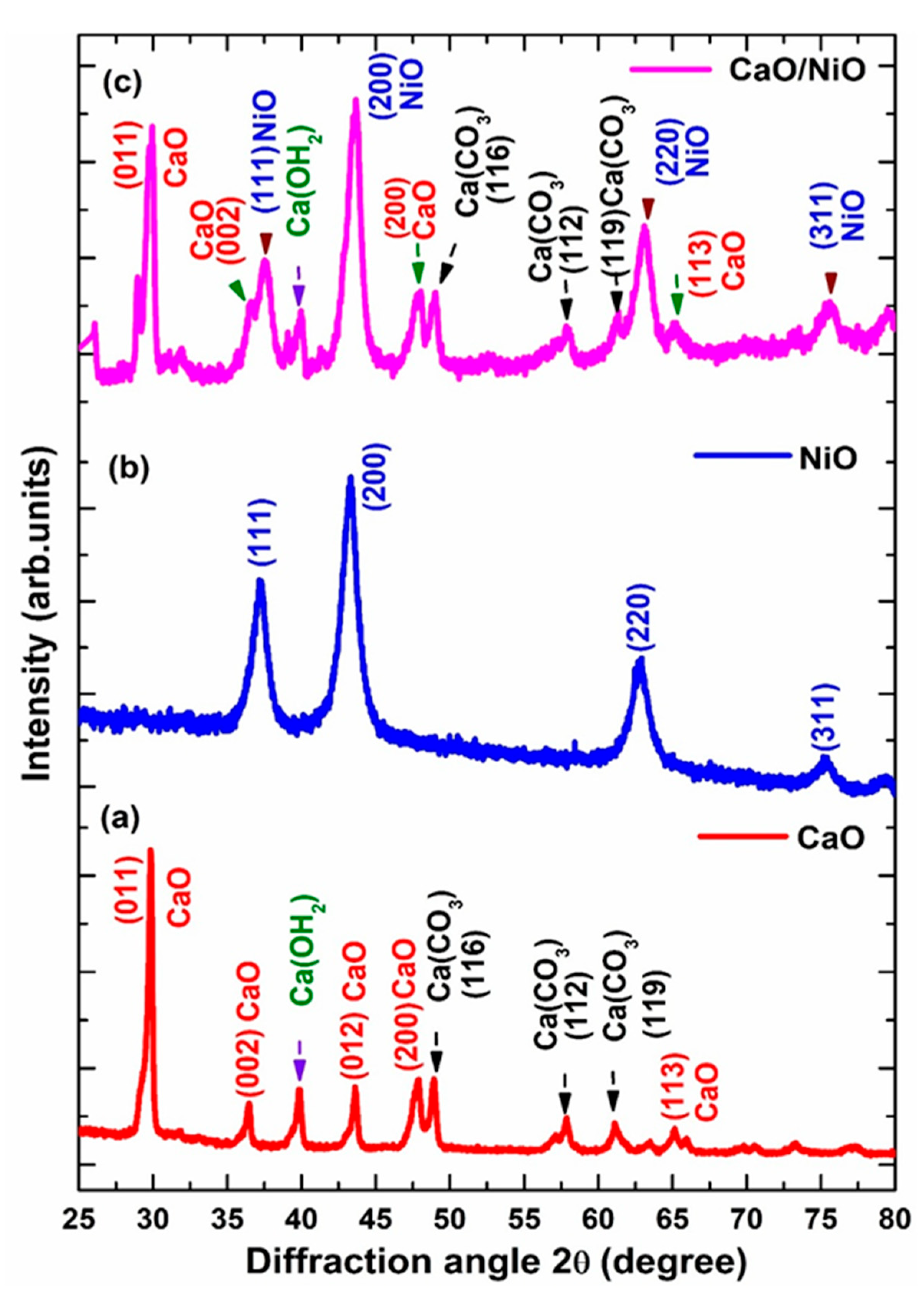
3.1. XRD Analysis

Figure 1a presents the XRD pattern of the CaO nanoparticles. Characteristic peaks are observed at 2θ values of 29.9°, 36.4°, 43.6°, and 65.3°, corresponding to the (011), (002), (012), and (113) planes, respectively. These diffraction peaks are consistent with the cubic structure of CaO and are in good agreement with the JCPDS card number 00-028-0775. In addition to the characteristic CaO peaks, some peaks were observed at 2θ values of 39.8°, 47.9°, 48.9°, 57.9°, 61.2°, 73.2°, and 77.4°. These peaks suggest the presence of trace amounts of calcium hydroxide (Ca(OH)2) and calcium carbonate (CaCO3) impurities [28]. These peaks likely arise from partial hydration and carbonation of the CaO during synthesis. This phenomenon, the formation of these secondary phases, is well-documented in the literature, as discussed by Amal et al. [29]. The XRD patterns of the NiO sample depicted in Figure 1b display prominent peaks at 2θ = 37.2°, 43.2°, 62.9°, and 75.5° indexed to the (111), (200), (220), and (311) planes, respectively, confirming the cubic structure of nickel oxide. This is in agreement with the JCPDS card no. 01-078-0429. In the diffraction pattern of the CaO/NiO nanocomposite depicted in Figure 1c, the presence of both CaO and NiO is evident along with CaCO3 and Ca(OH)2. The typical NiO peaks at 2θ = 37.2°, 43.2°, 61.2°, and 75.5° and the CaO is identified by the presence of peaks at 2θ = 29.9° and 36.4°, which are closely matched with those observed in the individual oxide patterns and correspond to the JCPDS cards mentioned above, confirming the retention of the cubic structures of both CaO and NiO in the composite. Furthermore, the presence of calcium carbonate (CaCO3) was discerned through reflections at 2θ = 48.8°, 57.7°, and 61.2°, corresponding to the (116), (119), and (119) planes, respectively. This is attributable to the facile carbonation of CaO upon exposure to atmospheric CO2, a ubiquitous phenomenon in calcium-based nanomaterials [30]. The presence of the aforementioned Ca(OH)2 and CaCO3 impurity peaks is also observed in the composite matrix, suggesting the retention of these secondary phases within the nanocomposite structure. S. Sinha et al. noted that Ca(OH)2 phases can form due to exposure to humidity in air [31]. However, a detailed analysis of the peak intensities reveals that the NiO peaks are significantly more prominent than those of CaO in the composite pattern. It is important to note that some degree of peak overlap is observed in the composite pattern. This observation, along with the previously discussed evidence, further confirms the formation of the CaO/NiO nanocomposite, as indicated by the mixed overlapping diffraction pattern of CaO/NiO in a single matrix.
To determine the average crystalline size of the biosynthesized samples, Scherrer’s formula was applied,
D = K λ β cos θ  
where λ—X-ray wavelength (1.5406 Å), β—Full-Width half maximum, K—shape factor (0.9), and θ—Bragg’s diffraction angle. The calculated average crystallite sizes were 51, 92, and 48 nm for CaO, NiO, and CaO/NiO, respectively. Notably, the crystallite size of the nanocomposite is smaller than that of the individual CaO and NiO nanoparticles. This size reduction may introduce lattice strain, defects, or changes in nucleation dynamics [32]. Smaller crystallite sizes generally lead to a higher surface area, which could enhance the composite’s reactivity. Based on the above discussion, we observed that the diminution of nanoparticle size is likely a consequence of the structural evolution during controlled calcination. During calcination at 550 °C, the decomposition and oxidation of organic components lead to the formation of CaO and NiO [20]. The Ca2+ and Ni2+ ions initially coordinated with oxygen-containing compounds in the lemon extract remain in the system and contribute to the nucleation and growth of these oxides. These oxides possess slightly different crystal lattice structures, and as CaO and NiO domains nucleate and grow, lattice mismatch occurs at their interfaces. This mismatch generates strain, manifesting as compressive stress due to the incompatibility of their lattice structures. The compressive stress at the interface restricts the free growth of crystallites, leading to smaller particle sizes in the nanocomposite [20]. It is important to mention that the uniform mixing procedure using the traditional green synthesis protocol ensures the effective incorporation of Ca2+ and Ni2+ ions with oxides in the complex matrix, corroborating the formation of the CaO/NiO nanocomposite.

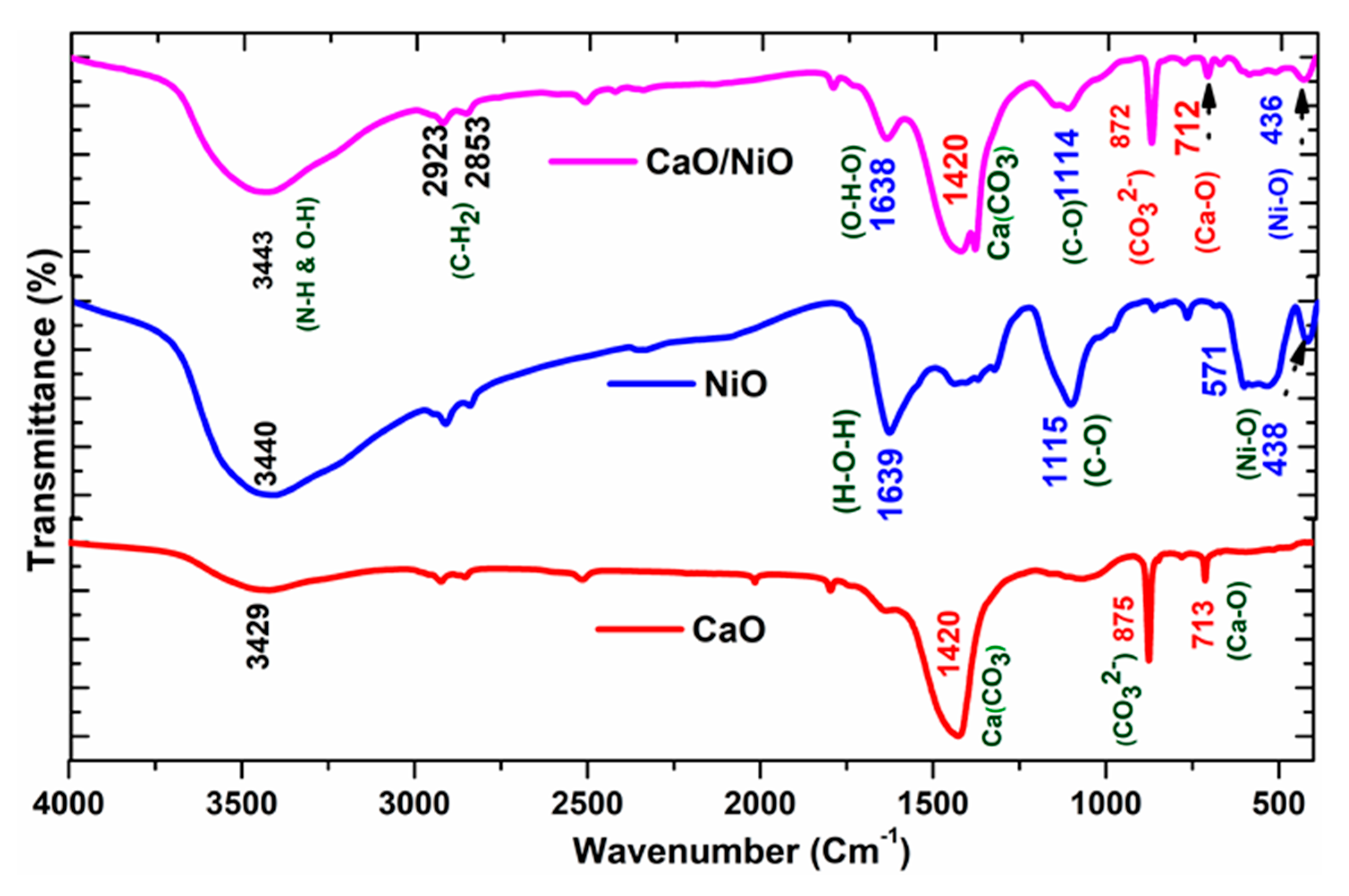
3.2. Functional Group Analysis

Figure 2 presents the FTIR spectra of the prepared samples. In the infrared spectrum of CaO nanoparticles, weak vibrational bands are observed at 713 and 875 cm−1 corresponding to O–Ca–O bending vibrations and the CO32− out-of-plane deformation mode of calcite (Ca–CO3), respectively. These observations are consistent with the earlier study by Gupta et al. [31]. A vibrational band centered at 1420 cm−1 was attributed to the doubly degenerate planar bending vibration of CO32−, confirming the persistence of carbonate groups even after calcination. This observation aligns with earlier findings of Abbas Ibrahim Hussein et al. [32]. These results reveal the partial retention of CaCO3 phases within the Ca–O structure. The FTIR spectrum of NiO nanoparticles displayed characteristic bands at 438 and 570 cm−1, indicative of metal–oxygen Ni–O bonding [33,34,35]. The appearance of a vibrational mode at 1119 cm−1 is a C-O stretching vibration [35], while a distinct absorption band at 1638 cm1 is allotted to the bending vibrations of adsorbed water molecules (H–O–H) from the atmosphere, which corroborates from the reported findings [36,37,38]. The FTIR spectrum of the CaO/NiO nanocomposite exposed bands at 436 cm1 and 712 cm1, corresponds to Ni–O and Ca–O stretching vibrations of the Ni–O–Ca–O structure, respectively [39,40]. These bands matched with the IR spectra of pure NiO and CaO, confirming the effective formation of the CaO/NiO nanocomposite. Further, the small frequency vibrational band noted at 1120 cm−1 was linked to the C-O stretching of carbonate species or carboxylic acid [41,42]. The 1420 cm−1 peak for Ca(CO3) and 1639 cm−1 peak for adsorbed water molecules are also present in the composite sample. Then, broad bands were observed at approximately 3429, 3440, and 3443 cm−1 in the FTIR spectra of CaO, NiO, and CaO/NiO, respectively, indicating the presence of N-H groups, which overlap with the O-H stretching vibrations [43,44]. These bands arise from adsorbed water molecules H–O bound to the surface of the samples [40,45]. This indicates the hydrophilic nature of our materials. The CaO/NiO nanocomposite also signifies the presence of atmospheric CO2 [46]. Weak absorption peaks at 2924 and 2859 cm−1 evidence the traces of C–H stretching vibrations of alkene groups [47]. During the oxidation process, these groups underwent structural rearrangement leading to the formation of CH2 bending or C–H of out-of-plane bending vibrations associated with aromatic rings; these are the characteristics of polyphenols and phenolic compounds, respectively [48,49,50]. The above results and discussion clearly reveal the formation of the CaO/NiO nanocomposite along with carbonate peaks, polyphenols, and alkene compounds, indicating a synergistic structural arrangement in the nanocomposite. This FTIR analysis validates the successful fusion of the CaO/NiO nanocomposite using lemon extract, ensuring the structural integrity, which will initiate the potential bioactivity of the material.

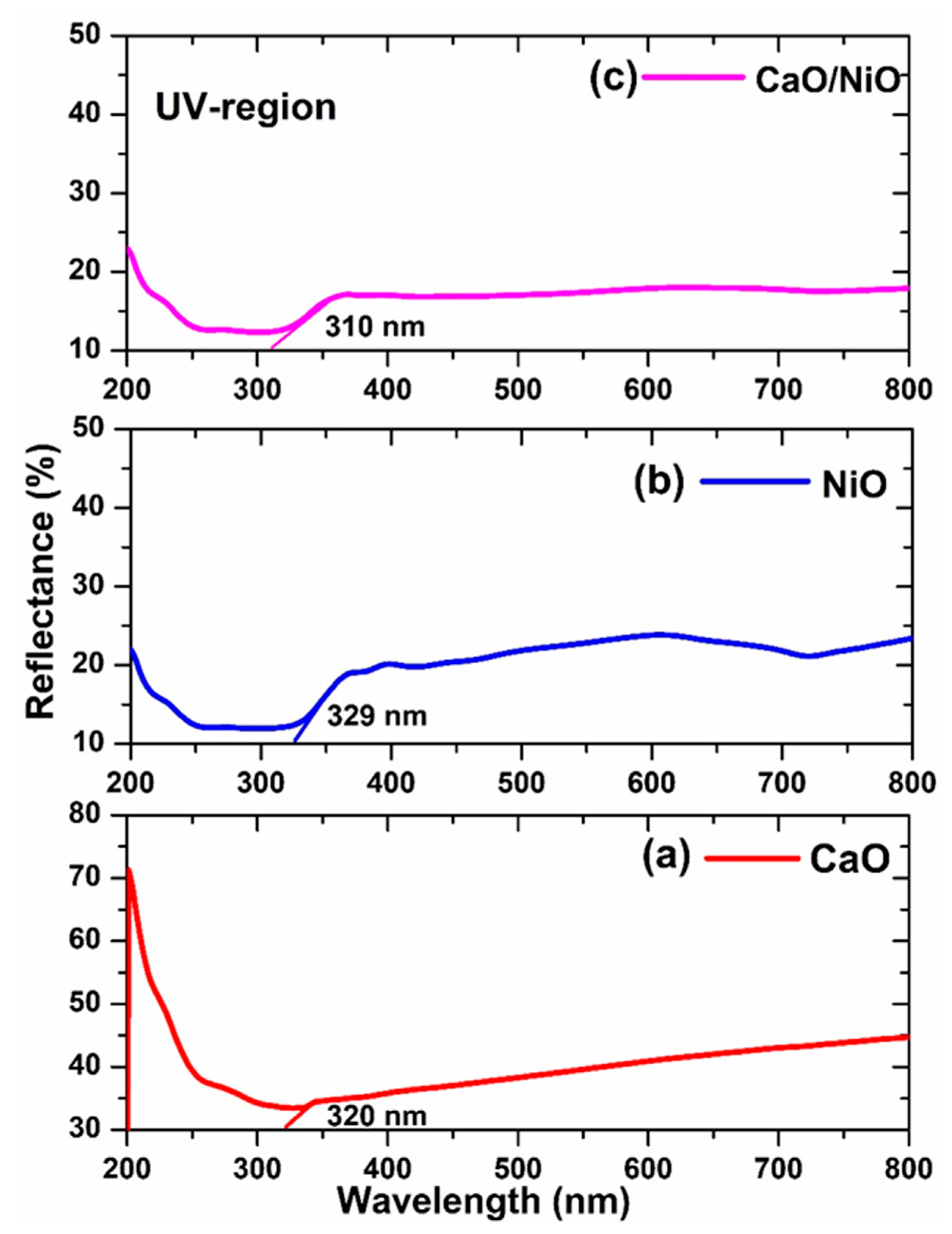
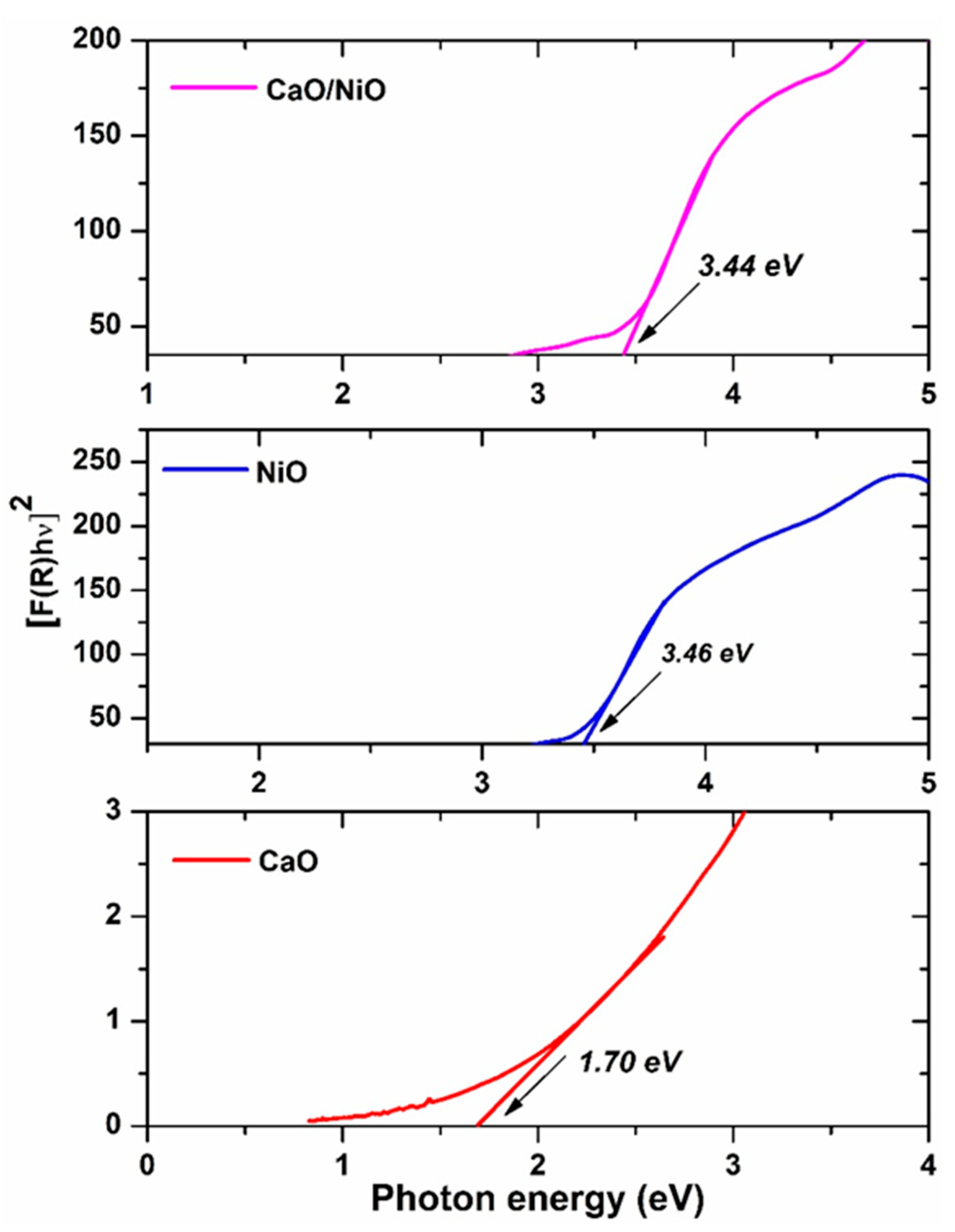
3.3. UV–Visible Diffuse Reflectance Studies

UV–Vis DRS investigations were performed to elucidate the photophysical behavior of the prepared samples. The reflectance spectra of CaO, NiO, and CaO/NiO are shown in Figure 3a–c. It exhibits high absorption (low reflectance) for the CaO, NiO, and CaO/NiO samples in the UV region, specifically within 200–320, 200–329, and 200–300 nm, respectively. Correspondingly, it reflects the percentage of light in the visible and near-infrared regions about 45% for CaO, 24% for NiO, and 20% for CaO/NiO. Here, we noted that there is a noticeable change in the reflectance behavior of the CaO/NiO nanocomposite compared to the individual oxides. Its lower reflectance in the visible and near-infrared regions indicates enhanced light absorption, which may be attributed to structural distortions, defect formation, or changes in crystallite size. These factors can influence the optical properties by modifying the band structure and electronic transitions. Additionally, the shift in the absorption range of the composite compared to CaO and NiO suggests an alteration in its band gap energy, possibly due to lattice strain or changes in particle distribution. To apprise the optical bandgap energy (Eg) of the CaO, NiO, and CaO/NiO samples, Tauc’s plots were used. The plot was drawn between [F(R) hν]2 on the y-axis against hν on the x-axis using reflectance data, which are depicted in Figure 4. To analyze the diffuse reflectance data, the Kubelka–Munk (K-M) function was applied.
The Kubelka–Munk function, F(R), is defined as follows:
F R = 1 R 2 2 R
Tauc’s relation is,
F R h ν = h ν E g n
where R—reflectance of the sample, υ—Frequency of the incident photon, and (1 − R)2/2R—the absorption coefficient. The value n depends on the type of electronic transition, were n = 2 for indirect transition and n = 1/2 for direct transition. The bandgap energy (Eg) was estimated by extrapolating the linear region [F(R) hν]2 vs. hυ plot to the x-axis [51,52]. The optical bandgap of the CaO, NiO, and CaO/NiO were assessed to be 1.70, 3.46, and 3.44 eV, respectively [53,54]. Notably, the bandgap of CaO (1.7 eV) is significantly lower compared to the NiO and CaO/NiO composites. A similar band gap value for CaO was reported by D. Mathivanan et al. who observed a bandgap of 1.8 eV for biosynthesized calcium hydroxide nanoparticles using Andrographis echioides leaf extract [55]. However, in this case, the presence of secondary phases with OH2 and CO3 entities may have influenced the optical properties of CaO, leading to a reduced bandgap value. In contrast, the CaO/NiO composite exhibits an increased bandgap, which may be attributed to alteration in electronic interactions between Ca2+ and Ni2+ ions in the composite sample, which arise due to defect formation and charge redistribution. This interaction modifies the electronic structure, creating new interface states and oxygen vacancies, which introduce localized energy levels within the band gap [56]. Furthermore, lattice distortions at the interface alter the band edge positions, contributing to the observed band gap reduction. Additionally, the observed optical changes are consistent with the XRD results, where peak broadening and shifts indicate lattice strain and crystallite size reduction. These structural modifications can generate additional defects, further influencing charge transfer mechanisms and light absorption properties. Collectively, these factors enhance the nanocomposite’s light absorption efficiency, improving its potential for redox activity-related applications.

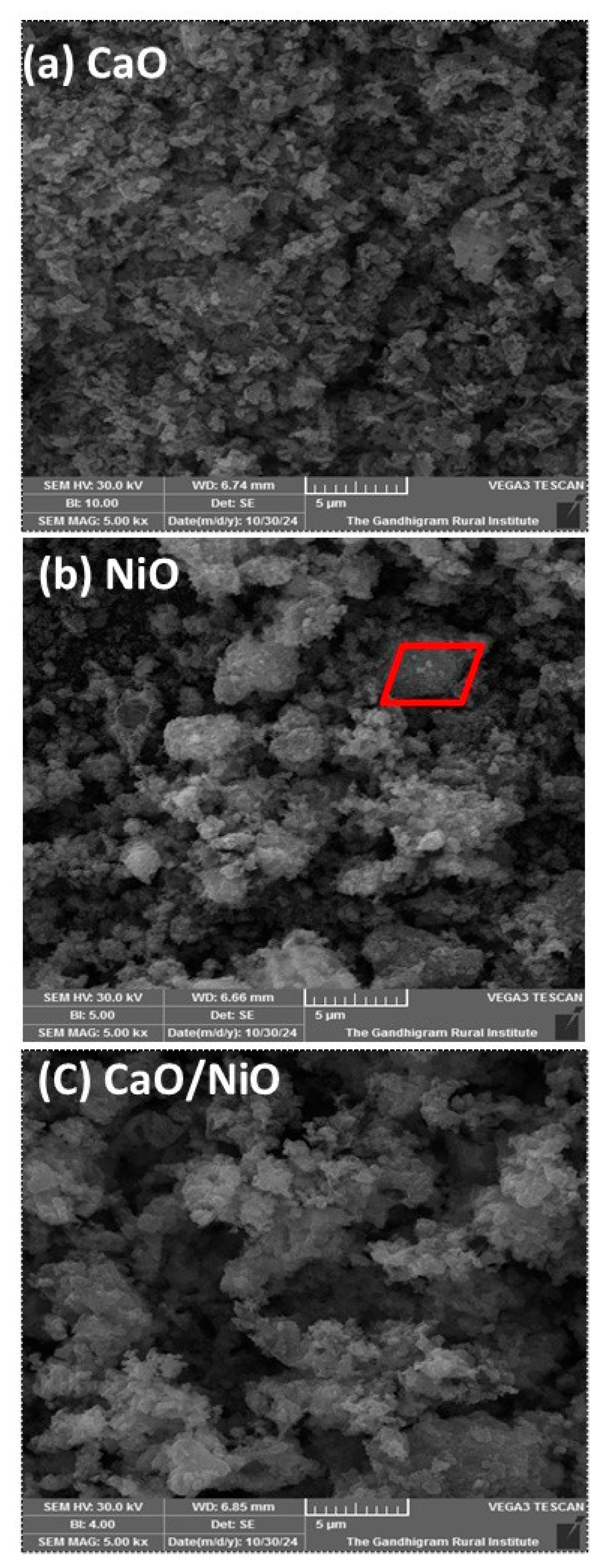
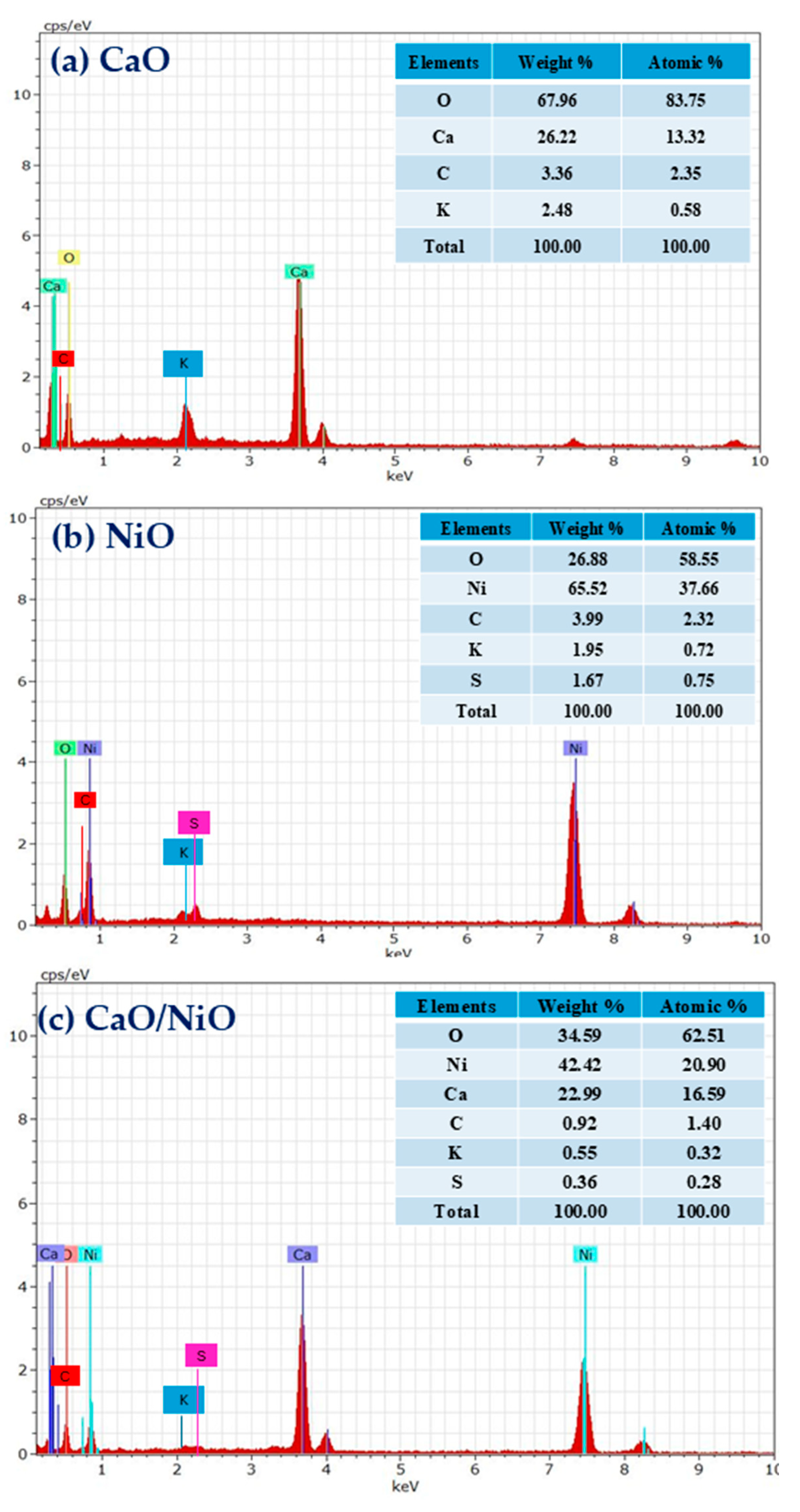
3.4. Morphological Analysis
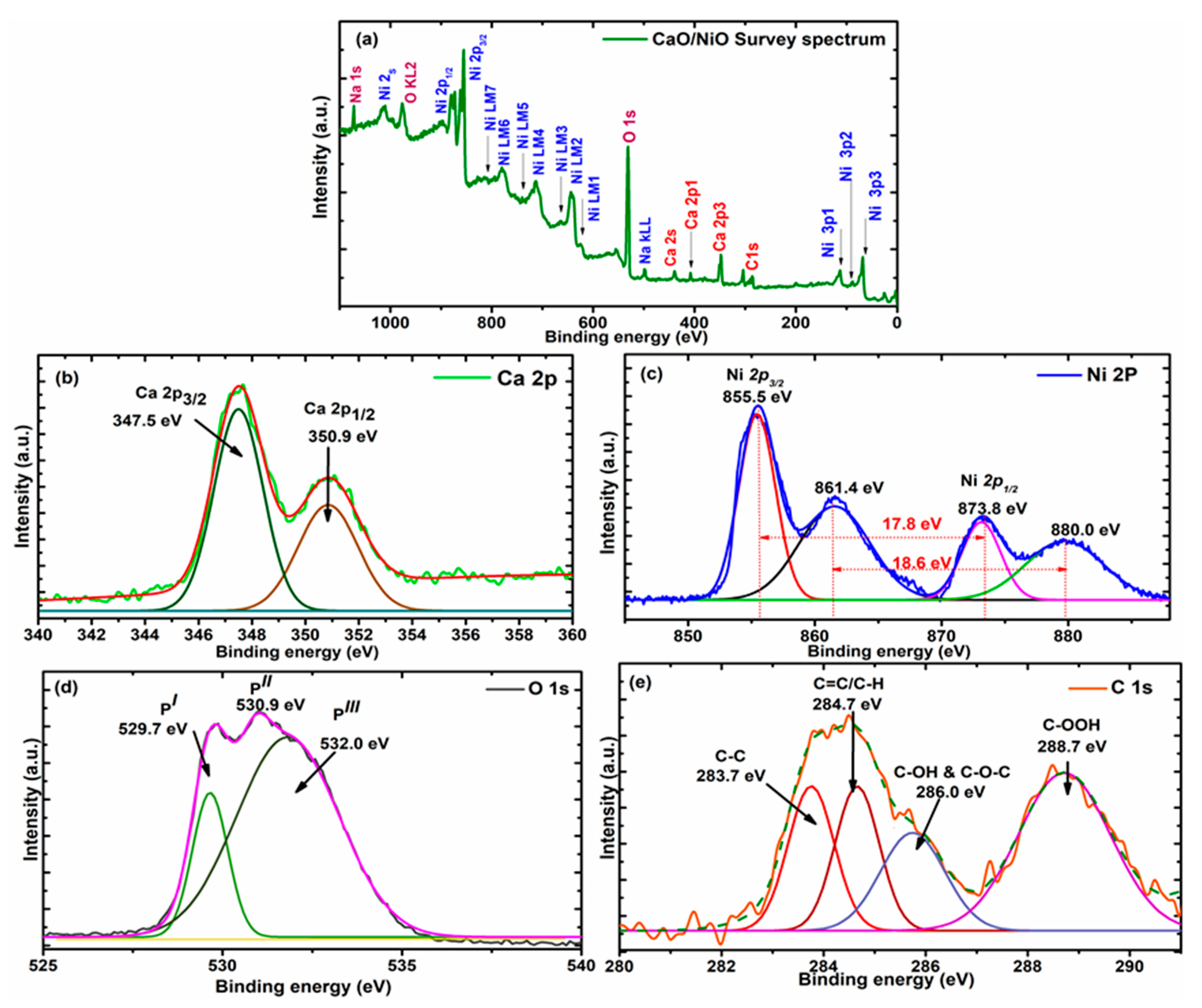
Microstructure analysis and surface morphology of the biosynthesized CaO, NiO, and CaO/NiO nanoparticles were systematically examined using scanning electron microscopy. The SEM images were captured at a magnification of 5 µm and presented in Figure 5a–c. Based on the SEM images, the micrograph of CaO nanoparticles displayed in Figure 5a reveals the accumulation of irregularly shaped tiny particles that are distributed uniformly. Interestingly, some of these particles on their surface exhibit a distinct spherical morphology [57]. This morphological property closely resembles the spherical and porous characteristics reported in earlier studies by M. Cabrere-Penna et al., who analyzed characteristics of calcium oxyhydroxide particles [45], and by Mohammad Amin Alavi et al., who studied ultrasonic-assisted Ca(OH)2 and CaO structure [58]. Figure 5b shows the SEM image of the NiO nanoparticles exhibiting an arrangement of larger particles with irregular morphologies with interspersed voids with notable agglomerates. Here, distinct rhombohedral and cubic shapes are identifiable in certain regions, reflecting the unique crystallographic structures of NiO [59]. Furthermore, residues of extract moieties are also visible and adhere to the circumstances of the sample. In contrast, from Figure 5c, the SEM image of the CaO/NiO nanocomposite sample reveals a heterostructure morphology with larger cavities and voids throughout the surface. The heterostructure morphology is defined, as seen at the bottom of the image, and some of them are as large as they look, similar to what we observed in the NiO image, and some of the spherical particles that adhere to the surface are likely due to residual Ca(OH)2 and Ca(CO3). This is consistent with the FTIR and XRD spectra results, which showed characteristic peaks of calcium-containing carbonate compounds. These voids provide enlarged exposure with active sites that enable the redox activity [60]. This is evidence that the CaO/NiO nanoparticles have a high surface area. The observed results from the SEM image of the CaO/NiO nanocomposite exhibit a unique morphology with porosity and high surface area, suggesting that this CaO/NiO nanocomposite exhibits captivating bioactivities. Figure 6a–c presents the EDX spectra of CaO, NiO, and CaO/NiO samples, confirming the elemental composition of the synthesized materials. The spectra indicate the occurrence of Ca and O in CaO, Ni and O in NiO, and a combination of Ca, Ni, and O in the CaO/NiO nanocomposite. In the case of CaO nanoparticles (Figure 6a), the EDX spectrum displays prominent peaks corresponding to calcium (Ca) and oxygen (O). The atomic composition was determined to be 86.65% oxygen and 13.35% calcium, while the weight percentages were calculated as 67.96% for oxygen and 26.22% for calcium. In addition to these primary elements, minor signals for carbon (3.36 wt%, 2.35 at%) and potassium (2.48 wt%, 0.58 at%) were also present. Similarly, the EDX spectrum of NiO nanoparticles (Figure 6b) exhibited clear peaks for nickel (Ni) and oxygen (O). The atomic percentages were found to be 58.55% for oxygen and 37.66% for nickel, while the weight percentages were 26.88% for oxygen and 65.52% for nickel. Moreover, small quantities of carbon (3.99 wt%, 2.32 at%), potassium (1.95 wt%, 0.72 at%), and sulfur (1.67 wt%, 0.75 at%) were identified in the sample. Meanwhile, for the CaO/NiO nanocomposite (Figure 6c), the EDX spectrum confirmed the combined presence of calcium, nickel, and oxygen, which indicates the successful formation of the composite structure. The atomic composition was calculated as 20.90% nickel, 16.59% calcium, and 62.51% oxygen, and the corresponding weight percentages were 42.42% for nickel, 22.99% for calcium, and 34.59% for oxygen. In addition, the spectrum of the CaO/NiO composite also exposed minor traces of carbon (0.92 wt%, 1.40 at%), potassium (0.35 wt%, 0.25 at%), sulfur (0.33 wt%, 0.29 at%), and sodium (0.39 wt%, 0.48 at%) in the CaO/NiO composite, signifying minor surface contamination in the sample. From the EDX analysis, it was observed that the presence of trace elements, including C, K, S, and Na, is likely attributable to residual compounds from the lemon extract, which persist in the sample after calcination. These trace elements, along with Ca, Ni, and O, may contribute to the biological activity of the CaO/NiO nanocomposite, potentially enhancing its antimicrobial, antioxidant, or other bioactive properties.

3.5. XPS Analysis

The XPS analysis was carried out to comprehend the oxidation states of the biosynthesized CaO/NiO nanocomposite sample. The XPS survey scan spectrum of CaO/NiO nanocomposite, displayed in Figure 7a, confirms the presence of distinct peaks corresponding to Ca 2p3, Ni 2p, Ni LMM, O KLL, C 1s, Na 1s, and O 1s, indicating the existence of calcium, nickel, and oxygen elements in their respective binding energy states. The deconvoluted Ca 2p core level spectrum depicted in Figure 7b reveals the spin–orbit doublet splitting of Ca 2p3/2 and Ca 2p1/2 corresponding to the binding energies of 347.5 and 350.9 eV, respectively. The peak at 347.5 eV represents the existence of the calcium with carbonate CaCO3 species [61]. This is known to be the partial surface carbonation. This may be caused due to the interaction of the prepared CaO/NiO nanocomposite sample with atmospheric CO2, which is adsorbed on the surface of the sample after the completion of the annealing. The binding energy values of carbonate CaCO3 states are already reported in the study by G. Vanthana Sree et al. [62]. Moreover, the coexisting peak at 350.9 eV is attributed to the Ca-O bonding. The peak values at the Ca 2p level in our study are consistent with the previously reported values for CaO nanoparticles [63]. The distance of separation between these two peaks was 3.4 eV, which correlates with the presence of calcium in the 2+ oxidation state in the composite with Ca-O-H species [64]. Figure 7c displays the deconvoluted core level binding energy spectrum of the Ni 2p. The resemblance of two binding energy peaks at 855.5 eV and 873.8 eV correspond to Ni 2p1/2 and Ni 2p3/2, respectively. Moreover, their respective satellite peaks have also arisen due to the shake-up process appearing between 861.4 and 880 eV. The spin–orbit splitting between these two states of Ni 2p1/2 and Ni 2p3/2 is 19 eV, which also correlates with the earlier reports [18,40]. The deconvoluted XPS spectrum of O 1s signals is illustrated in Figure 7d. The three deconvoluted parts are named P′, P″, and P‴. The peak P′ at 529.7 eV is from Ni2+ and Ca2+ related to the Ni-O and Ca-O bonding of the cubic form of Ni and Ca with the lattice oxygen O2+ [40]. The peak P″ at 530.9 eV is attributed to Ni-O-Ca, whereas the peak P‴ observed at 532.0 eV is attributed to surface-adsorbed oxygen species, likely originating from Ni–O–H–Ca configurations. This indicates the presence of hydroxyl or carbon–oxygen functional groups, such as C–OH, C=O, and –COOH, which may arise due to residual carbonaceous species from the extract used during synthesis. These carbon–oxygen groups can interact with calcium and nickel ions, leading to the formation of interfacial species, such as Ni–OH–Ca, Ni–O–C, or Ni(OH)2. Additionally, the signal may also correspond to surface carbonates, including CaCO3. Therefore, the peak at 532.0 eV likely represents a combination of chemical states, including Ni(OH)2 and CaCO3, or it may be associated with a transition from O 1s states to unoccupied O 2p states hybridized with Ni 3d orbitals [65]. The deconvoluted XPS spectrum of C 1s, shown in Figure 7e, divulges the presence of various carbon-containing groups. The peak at 283.7 eV corresponds to C–C bonds, while the peak at 284.7 eV is assigned to C=C or C–H groups. The binding energy at 286.0 eV is attributed to C–OH and C–O–C species, and the peak at 288.7 eV corresponds to carboxylic-type groups, such as –COOH [66,67]. The hydroxyl (OH) groups and carboxyl compounds act as active sites, playing a crucial role in enhancing the nanocomposite’s reactivity. The presence of these active sites facilitates interactions with biological systems by improving hydrophilicity, promoting biomolecular adsorption, and contributing to reactive oxygen species (ROS) generation, thereby enhancing antibacterial efficacy [68,69]. Moreover, Table 1 represents the peak binding energies (BE), full width at half maximum (FWHM), peak areas, and atomic percentages of the detected elements in the nanocomposite samples. From the table, it was observed that oxygen (56.91%) is the most dominant element in the CaO/NiO nanocomposite, followed by nickel (21.86%) and carbon (11.51%). The presence of carbon is likely associated with organic residues in the sample. A small amount of sodium (3.13%) was also detected, which may indicate minor surface contamination introduced during material preparation. Furthermore, calcium was identified at 4.54%, confirming the expected composition of the nanocomposite. The observed elemental distribution confirms the successful synthesis of the CaO/NiO nanocomposite. Additionally, the presence of trace elements in this surface composition plays a supportive role in enhancing the material’s biological as well as cytotoxic and redox activity.

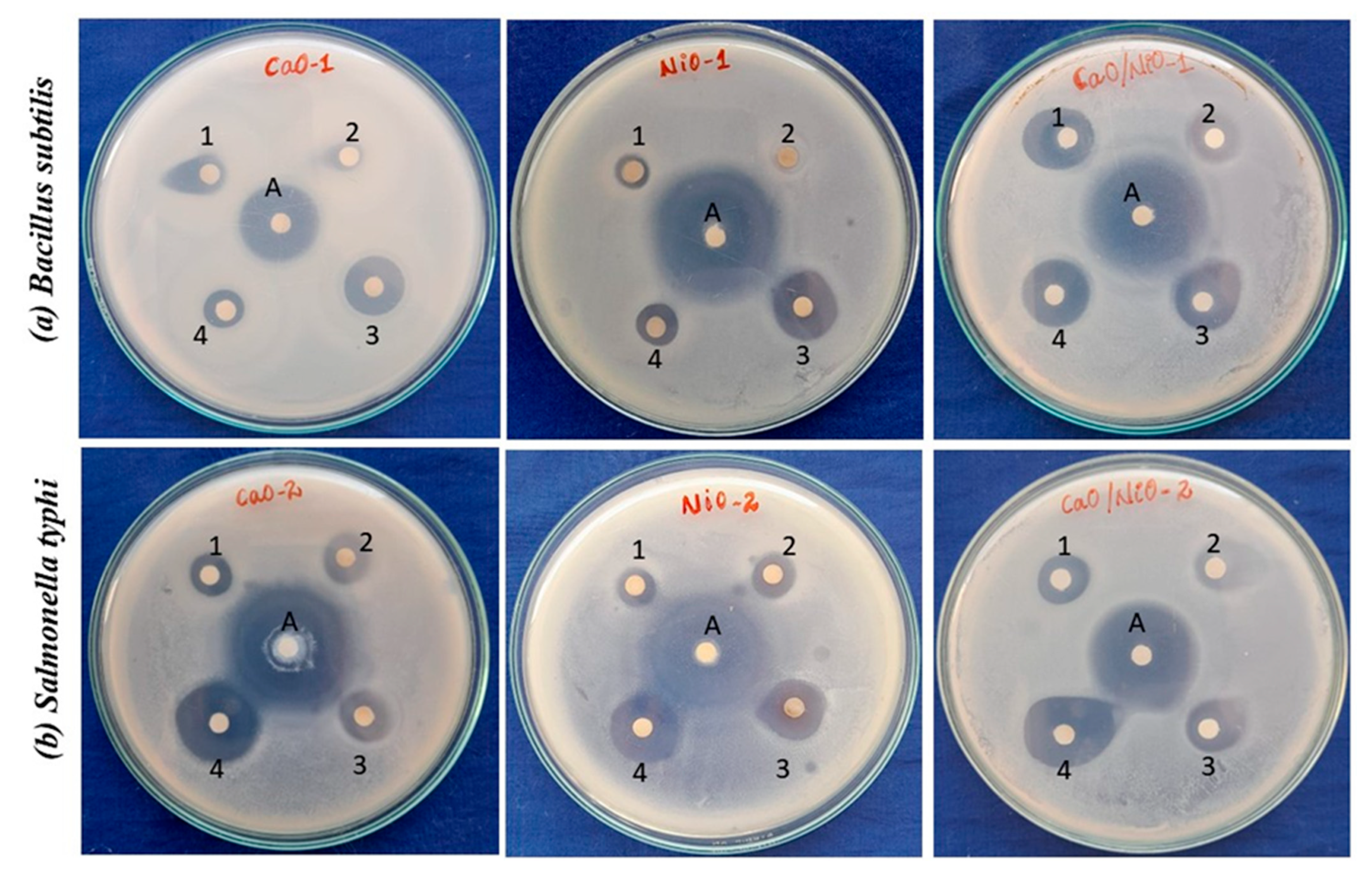
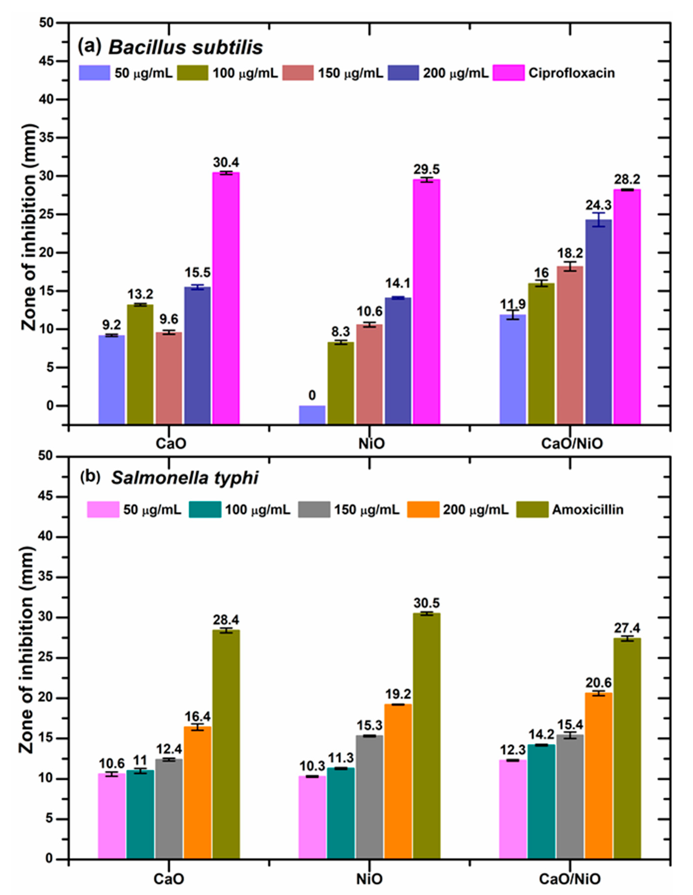
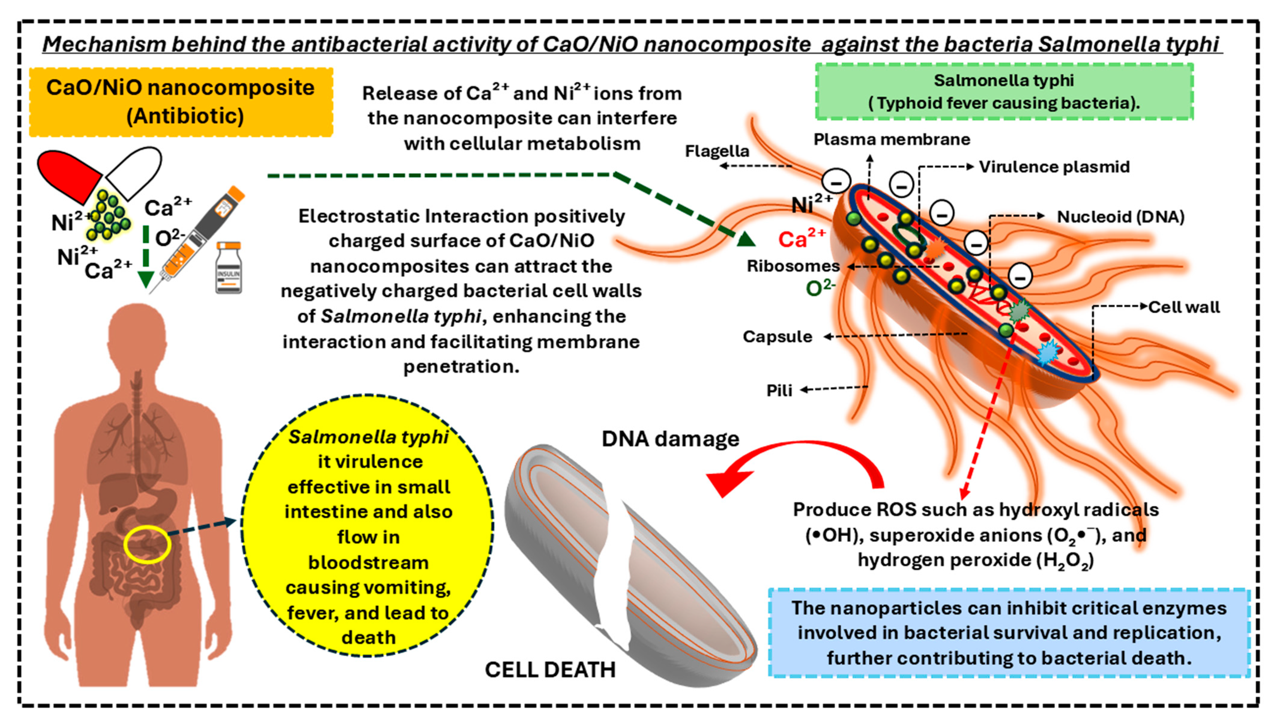
3.6. Antibacterial Assessment

The antibacterial action of CaO, NiO, and CaO/NiO samples was assessed against Bacillus subtilis (Gram-positive) and Salmonella typhi (Gram-negative) bacterial species using the standard disc diffusion protocol and minimum inhibitory concentration (MIC) assessments. The bacterial strains were prudently selected based on their clinical and environmental consequence. Bacillus subtilis is a well-known model organism for Gram-positive bacteria, often associated with food spoilage and biofilm formation whereas Salmonella typhi is a challenging Gram-negative pathogen responsible for typhoid fever, which is a pretentious and significant menace to public health due to its increasing resistance to conventional antibiotics. This selection allowed us to comprehensively evaluate the broad-spectrum antibacterial potential of the synthesized nanomaterials.
Figure 8 provides a visual representation of the zones of inhibition observed against the tested bacteria. The diameter of the zones, produced by the CaO, NiO, and CaO/NiO samples at various concentrations are presented in Figure 9. Across all the concentrations tested, the samples exhibited moderate to high zones of inhibition surrounding the disc. Specifically, at the highest concentration of 200 µg/mL, against Bacillus substilis, the CaO/NiO composite shows significantly greater bactericidal activity, exhibiting a zone of inhibition of 24.3 mm. This was substantially larger than the zones produced by CaO (15.5 mm) and NiO (14.1 mm). This enhanced activity was consistently observed against Salmonella typhi as well. The CaO/NiO composite again exhibited markedly superior bactericidal activity (20.6 mm) compared to CaO (16.4 mm) and NiO (19.2 mm). These results prove the CaO/NiO nanocomposite with surface carbonates and hydroxides demonstrates enhanced antibacterial activity compared to CaO and NiO individually, as evidenced by the larger zones of inhibition. This observation supports the broader trend of carbonate-containing materials exhibiting antibacterial properties, as exemplified by the work of Z. Jannah et al., who observed significant antibacterial activity in CaCO3/MgO composites against E. coli and S. aureus, corresponding to 31.96 mm and 32.26 mm inhibition zones, respectively [70].
The MIC refers to the lowest concentration of a substance required to prevent visible bacterial growth. Each bacterial strain was cultured in an appropriate nutrient broth and incubated with serial dilutions of the samples, ranging from high to low concentrations. The MIC values, presented in Table 2, demonstrate the enhanced antibacterial performance of the CaO/NiO nanocomposite compared to the individual components. Specifically, against Salmonella typhi, the nanocomposite exhibited an MIC of 46.8 µg/mL, significantly lower than the 750 µg/mL concentration for CaO and 187.5 µg/mL concentration for NiO. The notable MIC value of 23.4 µg/mL achieved by the CaO/NiO nanocomposite against Bacillus subtilis signifies a dramatic enhancement in antibacterial efficacy compared to the individual components, such as CaO, which exhibits an MIC of 750 µg/mL concentration, and NiO, with an MIC of 375 µg/mL concentration. The nanocomposite required significantly lower concentrations to inhibit bacterial growth. This substantial reduction in the MIC values accentuates the synergistic effect of combining CaO and NiO, leading to a potent antimicrobial agent against Bacillus subtilis. This outcome suggests that the nanocomposite could be a promising candidate for applications requiring robust bacterial inhibition, particularly against Gram-positive bacteria like Bacillus subtilis. The enhanced bacterial growth resistance activity of the CaO/NiO nanocomposite can be ascribed to the collaborative effects of the combined properties of the material with redox properties.
The antibacterial efficiency of CaO/NiO binary metal oxide nanocomposites is primarily influenced by factors, such as size, specific surface area, and morphology. However, the exact mechanisms underlying their inhibitory action against bacteria are not yet fully understood. Limited studies suggest that electrostatic interactions between the negatively charged bacterial cell membrane and the positively charged functional groups present in the sample play a crucial role in their bactericidal activity. In addition to preventing bacterial growth, this interaction causes ROS to be produced, which eventually results in cell death. It is noteworthy that Bacillus subtilis (MTCC 3055) and Salmonella typhi (MTCC 3224) are two clinically relevant bacterial strains that differ in their structural and physiological characteristics. B. subtilis is a Gram-positive bacterium known for its thick peptidoglycan layer and the absence of an outer membrane. This robust peptidoglycan provides structural support but is more accessible to external agents, including nanoparticles [71]. On the other hand, S. typhi is a Gram-negative bacterium characterized by a thinner peptidoglycan layer surrounded by an outer membrane composed of lipopolysaccharides (LPS). These structural differences influence how each type of bacterium responds to CaO/NiO nanocomposites and determine their level of susceptibility to the nanomaterials [72]. The possible mechanisms involved in the antibacterial activity of CaO/NiO nanocomposite against the salmonella typhi are portrayed in Figure 10. The antibacterial action of CaO/NiO nanocomposites may include the following: i) the release of Ca2+ and Ni2+ ions from the nanocomposite, which penetrate bacterial cells and interact with negatively charged components, causing microbial death [73] and ii) when the nanomaterials are exposed to light with photon energy exceeding their band gap, electrons from the valence band (e) are excited to the conduction band, leaving holes (h+) in the valence band. These charge carriers exhibit strong redox potential, contributing to bacterial inactivation. The electrons in the conduction band react with oxygen molecules, forming superoxide anions (O2•−) through a reductive process. These superoxide anions then interact with water molecules, generating reactive oxygen species, such as hydrogen peroxide (H2O2), which damages bacterial membranes. Meanwhile, holes in the valence band extract electrons from water or hydroxyl ions (OH), leading to the formation of hydroxyl radicals (OH), highly reactive neutral species produced through an oxidative process. H2O2, a potent oxidizing agent, effectively kills bacteria, while hydroxyl radicals and superoxide anions, though unable to penetrate bacterial membranes, remain in direct contact with the outer surface, causing extensive damage to proteins, lipids, and DNA [74].

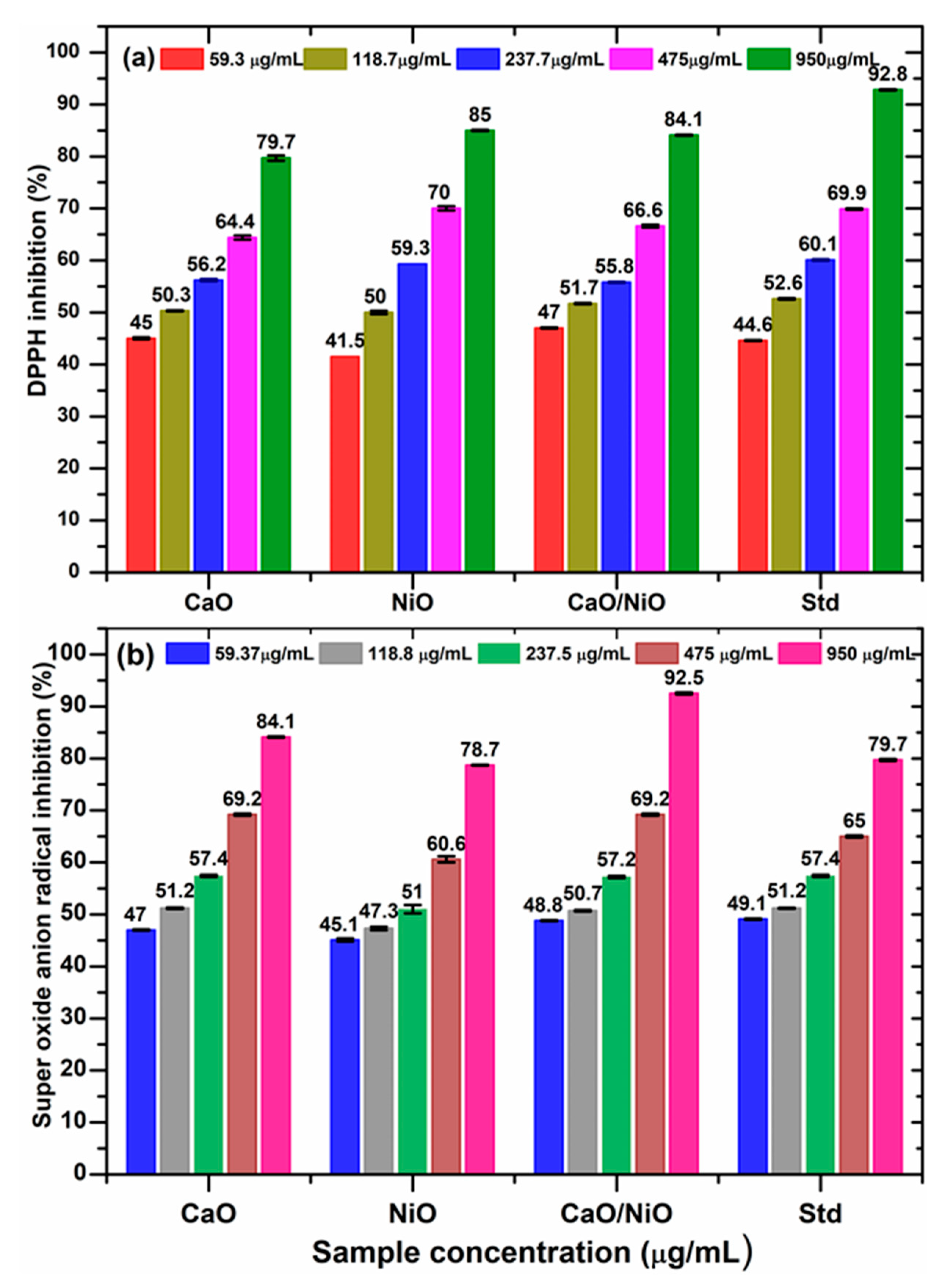
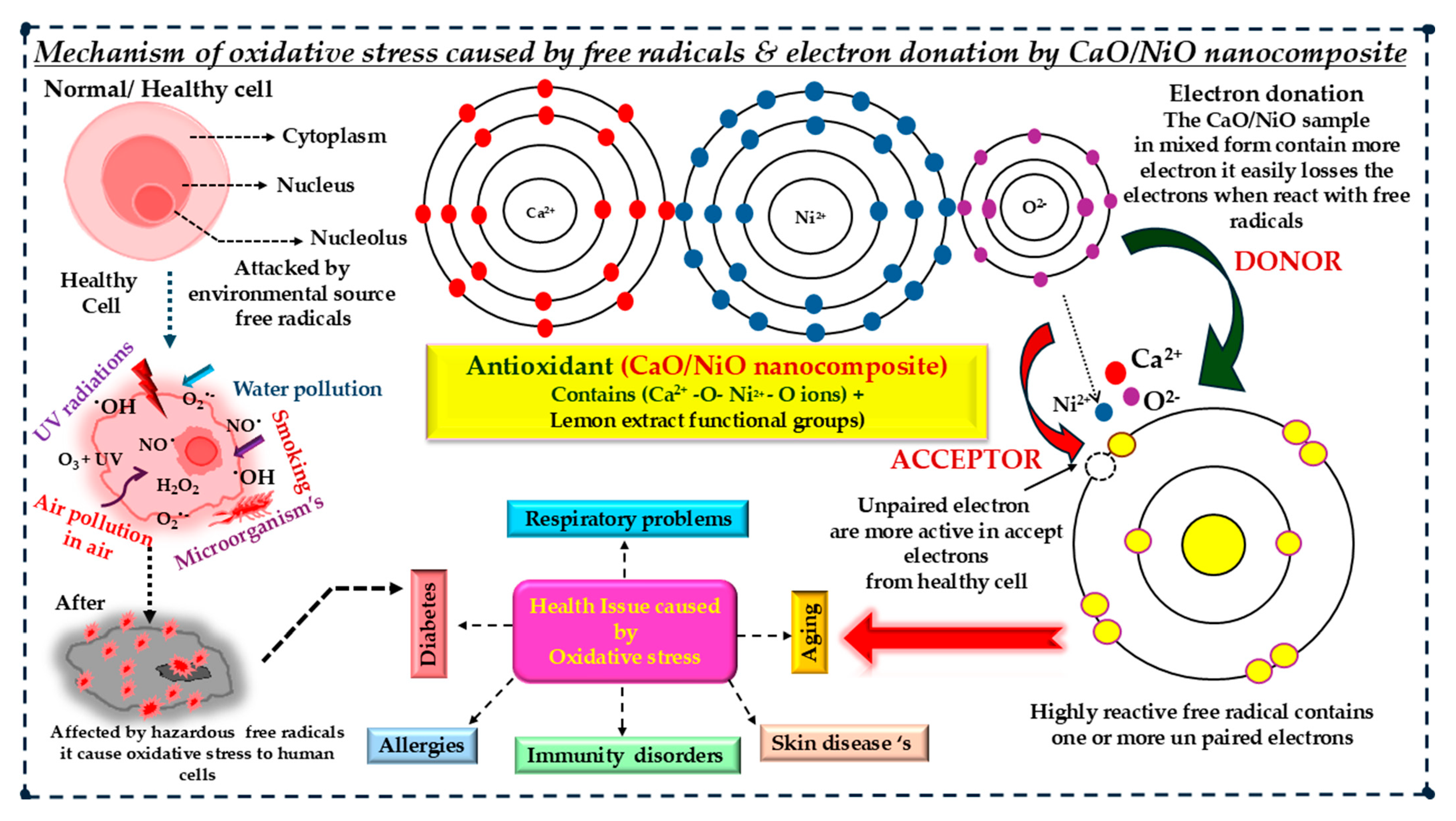
3.7. Antioxidant Activity

In vitro methods provide a valuable way to evaluate antioxidant activities, but their results do not always render directly to biological systems or reliably predict in vivo performance. To gain a comprehensive understanding, multiple methods are often necessary. In this study, the antioxidant potential of synthesized CaO, NiO, and CaO/NiO samples was assessed using 2,2-Diphenyl-1-picrylhydrazyl (DPPH) scavenging assays across a concentration range of 59.3 to 950 µg/mL are drawn and displayed in Figure 11a. The calculated values reveal that radical scavenging activity increases with the concentration of samples. Compared to CaO and NiO nanoparticles, the CaO/NiO nanocomposite shows better antioxidant activity. Here, we noted which concentration shows the 50% radical scavenging profile. As we observed from the results, all the samples show 50% or equal to 50 percentage activity at a concentration of 118 µg/mL. At the highest concentration of 950 µg/mL, all the samples exhibit a promising radical scavenging ability. In this study, the CaO/NiO reveals potential free radical scavenging activity of 84.1% noticeably higher than the CaO. Table 3 shows the sample concentration required for 50% activity of the DPPH radicals. The requirement of the CaO/NiO nanocomposite sample is 96.8 ± 0.4 µg/mL, which is very close to the performance of the standard antioxidant ascorbic acid 92.8 ± 1.4 µg/mL. In comparison, CaO and NiO show an IC50 value of 121.2 ± 0.6 and 119.6 ± 1.9 µg/mL, respectively, indicating slightly less efficiency in scavenging DPPH radicals.
The percentage inhibition of superoxide anion radical scavenging activity (O2−) of the CaO, NiO, and CaO/NiO nanocomposites is depicted in Figure 11b. The CaO sample expresses a 51% inhibition at 118 µg/mL, and NiO requires 237 µg/mL to achieve 50% inhibition. The CaO/NiO nanocomposite shows 48.8% at just 59 µg/mL and also achieves 50.7% inhibition at 118 µg/mL. It is noteworthy to mention that at the highest tested concentration of about 950 µg/mL, the composite exhibited a 92% effect; this permanence is greater when compared to the individual metal oxides CaO and NiO. The estimated IC50 values listed in Table 3 further confirmed the same observation for the better result for the composite with CaO, NiO, and the CaO/NiO nanocomposites, showing values of 79.8 ± 0.7 µg/mL, 194.2 ± 1.2 and 91.8 ± 0.1 µg/mL, respectively. Although the standard drug butylated hydroxytoluene (BHT) displayed the lowest IC50 value 59.2 ± 0.1 µg/mL, the CaO/NiO nanocomposite proved astonishing antioxidant efficiency, particularly at higher concentrations, with its eminence lying in its potential as a robust free radical scavenger.
In a healthy human body, cells serve as the fundamental units of life, playing a crucial role in various biological functions. Each cell contains a cytoplasm, a nucleus, and a nucleolus, all enclosed within a well-structured membrane. These cells maintain body homeostasis by regulating metabolic processes, supporting immune responses, and facilitating intercellular communication. However, in today’s world, increasing industrialization and urbanization have led to severe air pollution, introducing harmful toxins into the environment. Prolonged exposure to these pollutants can negatively impact cellular health, leading to various diseases. To counteract the harmful effects of ROS, antioxidants play a vital role in neutralizing oxidative stress and protecting cellular integrity. Among various antioxidant materials, our synthesized CaO/NiO nanocomposite has shown promising potential. Figure 12 illustrates the possible mechanism behind the oxidative stress caused by free radicals and the antioxidant activity of CaO/NiO nanocomposite. When administered as an oral medication or therapeutic agent, this nanocomposite actively scavenges free radicals and reduces oxidative damage. The enhanced surface properties of CaO/NiO, particularly the presence of surface carbonate species due to lemon extract-mediated green synthesis, contribute to its strong antioxidant activity. The Ni2+ and Ca2+ metal oxides present in the nanocomposite act as electron donors, effectively pairing with the reactive oxygen species to neutralize their harmful effects. This process helps restore cellular balance, preventing further damage and promoting overall health.

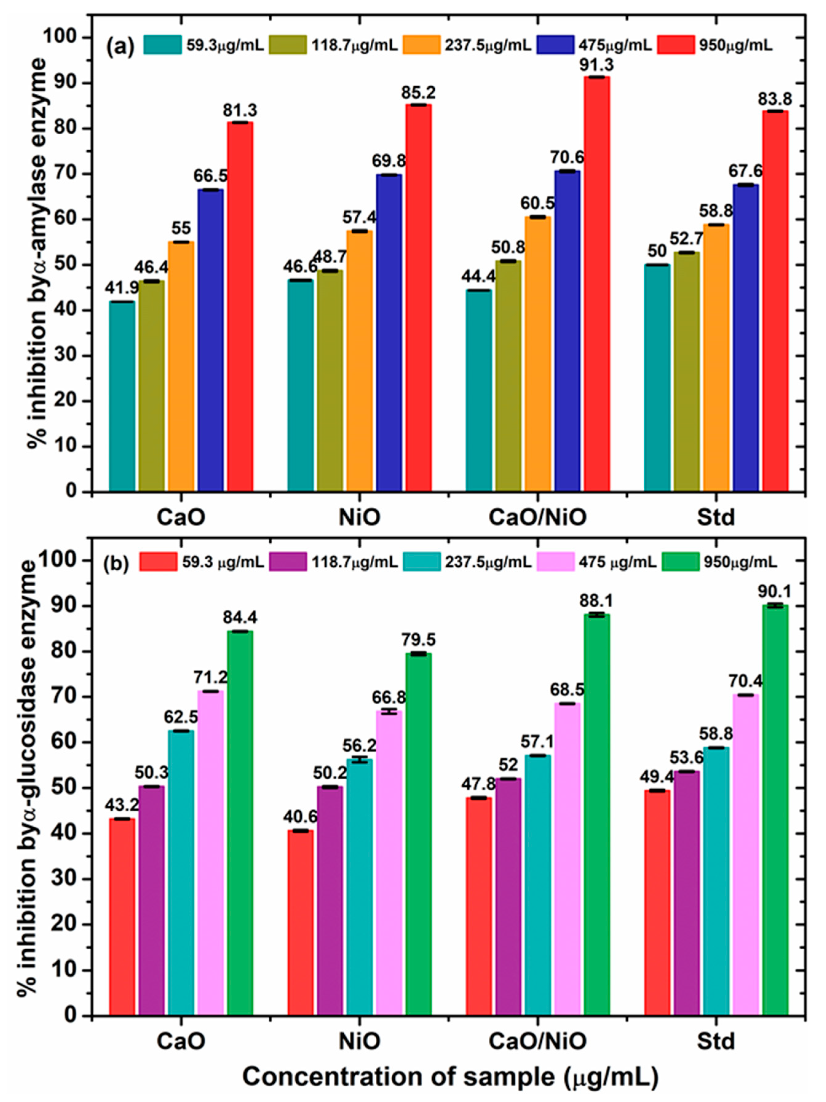
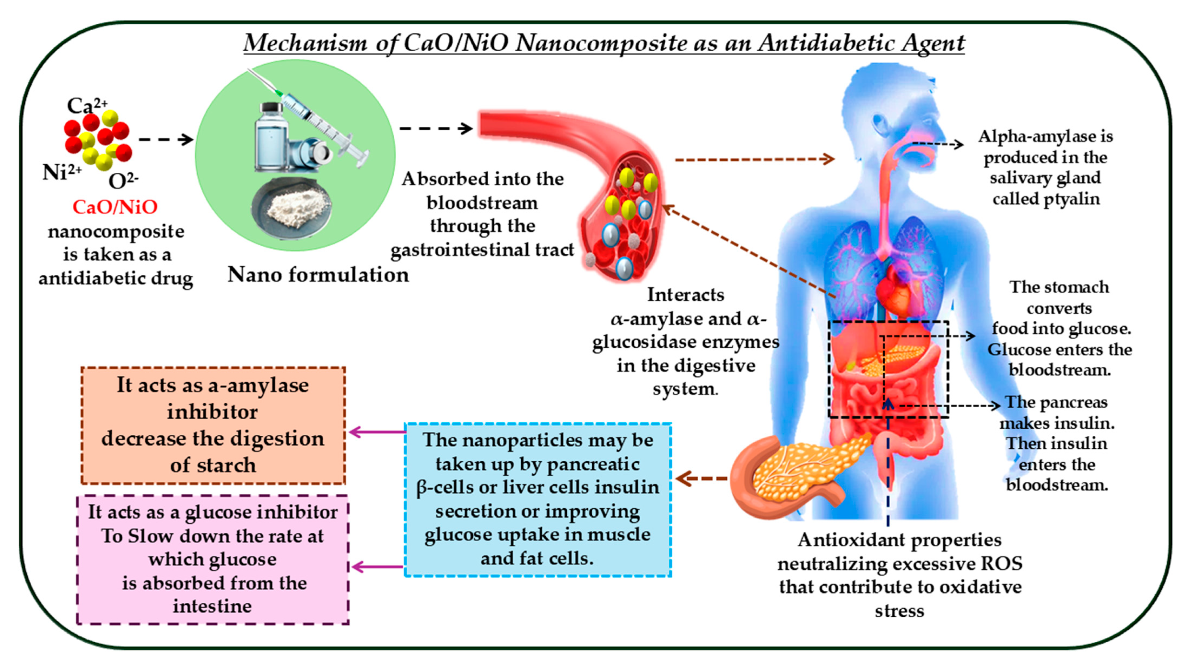
3.8. Antidiabetic Activity

Diabetes is one of the most widespread and enduring diseases worldwide. Currently, diabetes ranks as the fifth most considerable health concern globally. Its rapid growth has made it a pressing problem, often referred to as a modern epidemic disease. It affects millions of people across the world [75]. Although treatments have improved, managing diabetes is still a tough challenge. Current diabetes treatments typically require high doses of medication, which can lead to several challenges. These drugs have limited effectiveness, meaning they do not always work for everyone. They may have low solubility, making it difficult for the body to absorb the medication properly. Additionally, patients can experience varying responses to the same treatment, with some having better results than others. These factors contribute to the complexity of diabetes management and highlight the need for more personalized and effective treatment options [75]. Here, the in vitro α-amylase and α-glucosidase enzyme inhibition activity of the biosynthesized CaO, NiO, and CaO/NiO samples was scientifically evaluated. These activities were compared with the standard drug, acarbose. The concentration-based enzyme inhibition activity of the synthesized samples was observed and calculated using a standard formula, and the results are presented in Figure 13. The enzyme quenching reports of the samples show a clear difference in the antidiabetic potential of the synthesized samples. Among them, the CaO/NiO nanocomposite stands out with significantly better α-amylase and α-glucosidase enzyme inhibition compared to the individual CaO and NiO nanoparticles.
In this study, our focus was to determine the concentration at which 50% enzyme inhibition was achieved, as well as the concentration that exhibited the highest inhibitory activity. For alpha-amylase inhibition, as depicted in Figure 13a, the CaO demonstrates a 55% inhibition at 237.5 µg/mL, indicating a moderate ability to inhibit this enzyme. The NiO, on the other hand, shows 48.7% inhibition at a lower concentration of 118.7 µg/mL, which highlights its relatively strong activity at lower doses. Interestingly, the CaO/NiO nanocomposite exhibited 50.8% inhibition at the same concentration of 118.7 µg/mL, suggesting a slight enhancement of inhibitory activity due to the synergistic interaction between CaO and NiO. When the concentration was increased to 950 µg/mL, all samples displayed significantly improved inhibition rates. The CaO exhibits an inhibition of 81.3%, while NiO shows a slightly higher inhibition of 85.2%. The CaO/NiO nanocomposite reveals the highest inhibition of 91.3%, surpassing the inhibition of acarbose, a standard α-amylase inhibitor, which recorded an inhibition of 83.8%. These results highlight the superior potential of the CaO/NiO nanocomposite as an effective inhibitor of alpha-amylase, particularly at higher concentrations.
In the case of alpha-glucosidase inhibition, as shown in Figure 13b, similar results were observed. At the concentration of 118.7 µg/mL, CaO exhibits a 50.3% inhibition, closely followed by NiO at 50.2%, demonstrating comparable activity levels. The CaO/NiO nanocomposite, at the same concentration, exhibits a slightly lower inhibition rate of 47.8%, which is near the 50% inhibition threshold. This indicates that while the individual oxides show effective inhibition, the nanocomposite may not significantly outperform them at lower concentrations. However, as the concentration increased to 950 µg/mL, the inhibition rates improved markedly. The CaO demonstrates 84.4% inhibition, while NiO shows 79.5%. The CaO/NiO nanocomposite exhibits the highest inhibition rate of 88.1%, showcasing its effectiveness in inhibiting alpha-glucosidase at higher concentrations. These results accentuate the potential of the CaO/NiO nanocomposite as a promising material for controlling postprandial blood glucose levels in diabetic patients. The enhanced activity of the nanocomposite compared to individual metal oxides further demonstrates the advantages of combining materials to achieve superior enzyme inhibition. The IC50 concentration values are also evaluated to obtain the significant potential of the synthesized sample values, as they provide a quantitative measure of the sample’s efficiency in inhibiting specific enzymes linked to diabetes management as listed in Table 4. The CaO/NiO nanocomposite exhibits the lowest IC50 for α-amylase 98.6 ± 0.7 µg/mL and exposes a strong inhibition for α-glucosidase 81.96 ± 0.5 µg/mL compared to individual metal oxides while the standard inhibitor acarbose exhibited superior IC50 values for both α-amylase 32.8 ± 0.4 µg/mL and α-glucosidase 47.0 ± 0.8 µg/mL; the performance of the CaO/NiO nanocomposite was nearly matched with the potential of the standard drug. The results also reveal that NiO displays a better α-amylase inhibition of 105.2 ± 3.4 µg/mL compared to CaO 176.7 ± 0.6 µg/mL, while CaO demonstrated stronger α-glucosidase inhibition of 81.7 ± 1.9 µg/mL than NiO 151.2 ± 2.9 µg/mL. However, there are only a few reports available on the synthesis of calcium oxide (CaO) and nickel oxide (NiO) nanoparticles with antidiabetic activity [4,76,77]. Our study adds valuable insight into the potential of these materials as effective antidiabetic agents. The synthesis of CaO/NiO nanocomposite demonstrated enhanced inhibitory activity against key enzymes involved in glucose metabolism, namely α-amylase and α-glucosidase. Additionally, research into the scalability and cost-effectiveness of synthesizing these nanoparticles could facilitate their translation from bench to bedside, potentially revolutionizing diabetes treatment options.
Figure 14 illustrates the possible mechanism of antidiabetic activity of the CaO/NiO nanocomposite. When the CaO/NiO nanocomposite is taken as a drug or as a neoformation, the nanoparticles in the composite enter the body, and they slow down the breakdown of carbohydrates by blocking enzymes like α-amylase and α-glucosidase [78]. This helps control blood sugar spikes after meals. At the same time, they act as antioxidants, reducing harmful ROS molecules that can damage cells, including insulin-producing β-cells. They may also help the body use insulin better by improving glucose uptake in muscles and other tissues [79]. Overall, these nanocomposites could be useful in managing type 2 diabetes by keeping blood sugar levels stable and protecting cells from damage.

4. Conclusions

The CaO/NiO nanocomposite was successfully synthesized via an eco-friendly biogenic approach. Comprehensive characterization analyses were conducted to investigate its structural, morphological, and chemical properties, including oxidation states. The XRD analysis of the CaO/NiO nanocomposite confirms the coexistence of the cubic structures of CaO and NiO in a single matrix. A noticeable reduction in crystallite size was observed, with the CaO/NiO nanocomposite exhibiting a crystallite size of 48 nm, which is smaller than those of CaO and NiO. The FTIR studies corroborate the formation of Ca-O–Ni–O bonds besides the CaCO3 functional bonds. The bandgap energy value of the CaO/NiO is 3.44 eV and it has a good UV absorption property. The agglomerations with porous heterostructure observed in the CaO/NiO sample have a mix-up form of adherence of the spherical CaO onto the rhombohedral shape of NiO with more voids. The XPS analysis confirmed the presence of calcium and nickel in their respective oxidation states, with distinct peaks for Ca 2p and Ni 2p, signifying the successful formation of CaO and NiO phases in the composite. The deconvoluted O 1s spectrum revealed the presence of surface hydroxyl groups (Ca–OH and Ni–OH), which contribute to the enhanced reactivity of the nanocomposite. The CaO/NiO nanocomposite exhibits a notable zone of inhibition of 24.3 mm against Bacillus subtilis and 20.6 mm against Salmonella typhi at 200 µg/mL, significantly outperforming individual CaO and NiO nanoparticles. It also demonstrated remarkable antioxidant activity, achieving 92% superoxide anion radical scavenging at 950 µg/mL, indicating strong free radical neutralization potential. Furthermore, the CaO/NiO nanocomposite exhibited superior antidiabetic activity, with enzyme inhibition rates of 91.3% for α-amylase and 88.1% for α-glucosidase at 950 µg/mL, surpassing both individual metal oxides and the standard inhibitor acarbose and the lowest IC50 values of 98.6 ± 0.7 µg/mL for α-amylase and 81.96 ± 0.5 µg/mL for α-glucosidase. These results substantiate the potential of the biosynthesized CaO/NiO nanocomposite as a formidable candidate for combating life-threatening microbial infections, oxidative stress-related damage, and diabetes-related complications.

Author Contributions

Conceptualization, S.P., A.T. and N.C.; methodology, S.P., A.T. and N.C.; investigation, formal analysis, writing—original draft, S.P. and N.C.; writing—review and editing, S.P., M.A., R.K., M.S., G.S.-H., A.T. and N.C.; supervision, N.C. All authors have read and agreed to the published version of the manuscript.

Funding

This research received no external funding.

Institutional Review Board Statement

Not applicable.

Informed Consent Statement

Not applicable.

Data Availability Statement

The original contributions presented in this study are included in the article. Further inquiries can be directed to the corresponding author.

Acknowledgments

The author SP gratefully acknowledges the Principal and Head of the Department of Physics, Rajah Serfoji Government College (Autonomous), Thanjavur, for their encouragement and support.

Conflicts of Interest

The authors declare no conflicts of interest.

References

  1. Sierra-Vargas, M.P.; Montero-Vargas, J.M.; Debray-García, Y.; Vizuet-de-Rueda, J.C.; Loaeza-Román, A.; Terán, L.M. Oxidative Stress and Air Pollution: Its Impact on Chronic Respiratory Diseases. Int. J. Mol. Sci. 2023, 24, 853. [Google Scholar] [CrossRef] [PubMed]
  2. Leni, Z.; Künzi, L.; Geiser, M. Air Pollution Causing Oxidative Stress. Curr. Opin. Toxicol. 2020, 20–21, 1–8. [Google Scholar] [CrossRef]
  3. Hu, X.; Zhang, Y.; Ding, T.; Liu, J.; Zhao, H. Multifunctional Gold Nanoparticles: A Novel Nanomaterial for Various Medical Applications and Biological Activities. Front. Bioeng. Biotechnol. 2020, 8, 990. [Google Scholar] [CrossRef] [PubMed]
  4. Samad, N.; Ejaz, U.; Kousar, S.; Al-Mutairi, A.A.; Khalid, A.; Amin, Z.S.; Bashir, S.; Al-Hussain, S.A.; Irfan, A.; Zaki, M.E.A. A Novel Approach to Assessing the Antioxidant and Anti-Diabetic Potential of Synthesized Calcium Carbonate Nanoparticles Using Various Extracts of Ailanthus Altissima. Front. Chem. 2024, 12, 1345950. [Google Scholar] [CrossRef]
  5. Mazher, M.; Ishtiaq, M.; Hamid, B.; Haq, S.M.; Mazhar, A.; Bashir, F.; Mazhar, M.; Mahmoud, E.A.; Casini, R.; Alataway, A.; et al. Biosynthesis and Characterization of Calcium Oxide Nanoparticles from Citrullus Colocynthis Fruit Extracts; Their Biocompatibility and Bioactivities. Materials 2023, 16, 2768. [Google Scholar] [CrossRef]
  6. Dai, Y.; Guo, Y.; Tang, W.; Chen, D.; Xue, L.; Chen, Y.; Guo, Y.; Wei, S.; Wu, M.; Dai, J.; et al. Reactive Oxygen Species-Scavenging Nanomaterials for the Prevention and Treatment of Age-Related Diseases. J. Nanobiotechnol. 2024, 22, 252. [Google Scholar] [CrossRef]
  7. Foulkes, R.; Man, E.; Thind, J.; Yeung, S.; Joy, A.; Hoskins, C. The Regulation of Nanomaterials and Nanomedicines for Clinical Application: Current and Future Perspectives. Biomater. Sci. 2020, 8, 4653–4664. [Google Scholar] [CrossRef]
  8. Vallejo Castaño, S.; Callagon La Plante, E.; Shimoda, S.; Wang, B.; Neithalath, N.; Sant, G.; Pilon, L. Calcination-Free Production of Calcium Hydroxide at Sub-Boiling Temperatures. RSC Adv. 2021, 11, 1762–1772. [Google Scholar] [CrossRef]
  9. Anbu, P.; Gopinath, S.C.B.; Saravanakumar, K.; Vijayakumar, S.; Ramanathan, S.; Wang, M.-H. Bimetallic Mixed Metal Oxide (CuO/NiO) in Fusion with Nitrogen-Doped Graphene Oxide: An Alternate Approach for Developing Potential Biocarrier. J. Environ. Chem. Eng. 2021, 9, 105781. [Google Scholar] [CrossRef]
  10. Singh, H.; Desimone, M.F.; Pandya, S.; Jasani, S.; George, N.; Adnan, M.; Aldarhami, A.; Bazaid, A.S.; Alderhami, S.A. Revisiting the Green Synthesis of Nanoparticles: Uncovering Influences of Plant Extracts as Reducing Agents for Enhanced Synthesis Efficiency and Its Biomedical Applications. Int. J. Nanomed. 2023, 18, 4727–4750. [Google Scholar] [CrossRef]
  11. Szczyglewska, P.; Feliczak-Guzik, A.; Nowak, I. Nanotechnology–General Aspects: A Chemical Reduction Approach to the Synthesis of Nanoparticles. Molecules 2023, 28, 4932. [Google Scholar] [CrossRef] [PubMed]
  12. Rashad, A.M. A Concise on the Effect of Calcium Oxide on the Properties of Alkali-Activated Materials: A Manual for Civil Engineers. Int. J. Concr. Struct. Mater. 2023, 17, 72. [Google Scholar] [CrossRef]
  13. Niu, Y.-Q.; Liu, J.-H.; Aymonier, C.; Fermani, S.; Kralj, D.; Falini, G.; Zhou, C.-H. Calcium Carbonate: Controlled Synthesis, Surface Functionalization, and Nanostructured Materials. Chem. Soc. Rev. 2022, 51, 7883–7943. [Google Scholar] [CrossRef] [PubMed]
  14. Jimoh, O.A.; Ariffin, K.S.; Hussin, H.B.; Temitope, A.E. Synthesis of Precipitated Calcium Carbonate: A Review. Carbonates Evaporites 2018, 33, 331–346. [Google Scholar] [CrossRef]
  15. Vallejo Castaño, S.; Callagon La Plante, E.; Shimoda, S.; Wang, B.; Neithalath, N.; Sant, G.; Pilon, L. Calcination-Free Production of Calcium Hydroxide at Sub-Boiling Temperatures. RSC Adv. 2021, 11, 1762–1772. [Google Scholar] [CrossRef]
  16. Sinha, T.; Ahmaruzzaman, M.; Adhikari, P.P.; Bora, R. Green and Environmentally Sustainable Fabrication of Ag-SnO2 Nanocomposite and Its Multifunctional Efficacy As Photocatalyst and Antibacterial and Antioxidant Agent. ACS Sustain. Chem. Eng. 2017, 5, 4645–4655. [Google Scholar] [CrossRef]
  17. Wang, G.; Zheng, J.; Xu, B.; Zhang, C.; Zhu, Y.; Fang, Z.; Yang, Z.; Shang, M.-H.; Yang, W. Tailored Electronic Band Gap and Valance Band Edge of Nickel Oxide via P-Type Incorporation. J. Phys. Chem. C 2021, 125, 7495–7501. [Google Scholar] [CrossRef]
  18. Heng, M.H.; Win, Y.F.; Cheah, E.S.G.; Chan, Y.B.; Rahman, M.K.; Sultana, S.; Tey, L.-H.; Wong, L.S.; Djearamane, S.; Akhtaruzzaman, M.; et al. Microwave-Assisted Green Synthesis, Characterization, and in Vitro Antibacterial Activity of NiO Nanoparticles Obtained from Lemon Peel Extract. Green Process. Synth. 2024, 13, 20240071. [Google Scholar] [CrossRef]
  19. Ben Hsouna, A.; Sadaka, C.; Generalić Mekinić, I.; Garzoli, S.; Švarc-Gajić, J.; Rodrigues, F.; Morais, S.; Moreira, M.M.; Ferreira, E.; Spigno, G.; et al. The Chemical Variability, Nutraceutical Value, and Food-Industry and Cosmetic Applications of Citrus Plants: A Critical Review. Antioxidants 2023, 12, 481. [Google Scholar] [CrossRef]
  20. Priyadharshini, S.; Jasmine Jecintha Kay, S.; Ayyanar, M.; Jenipher, C.; Thirumurugan, A.; Isaac, R.S.R.; Chidhambaram, N. A Cleaner and Multifunctional Citrus Limonium-Assisted ZnO/MgO Nanocomposite Synthesis: Extensive Investigations on Antibacterial, Antioxidant, and Antidiabetic Applications. Mater. Today Commun. 2024, 41, 110620. [Google Scholar] [CrossRef]
  21. Khan, F.U.; Khan, Z.U.H.; Ma, J.; Khan, A.U.; Sohail, M.; Chen, Y.; Yang, Y.; Pan, X. An Astragalus Membranaceus Based Eco-Friendly Biomimetic Synthesis Approach of ZnO Nanoflowers with an Excellent Antibacterial, Antioxidant and Electrochemical Sensing Effect. Mater. Sci. Eng. C 2021, 118, 111432. [Google Scholar] [CrossRef] [PubMed]
  22. Darwish, R.M.; Salama, A.H. Study the Effect of Conjugate Novel Ultra-Short Antimicrobial Peptide with Silver Nanoparticles against Methicillin Resistant S. aureus and ESBL E. coli. Antibiotics 2022, 11, 1024. [Google Scholar] [CrossRef] [PubMed]
  23. Hosny, M.; Fawzy, M.; Abdelfatah, A.M.; Fawzy, E.E.; Eltaweil, A.S. Comparative Study on the Potentialities of Two Halophytic Species in the Green Synthesis of Gold Nanoparticles and Their Anticancer, Antioxidant and Catalytic Efficiencies. Adv. Powder Technol. 2021, 32, 3220–3233. [Google Scholar] [CrossRef]
  24. Hazekawa, M.; Nishinakagawa, T.; Kawakubo-Yasukochi, T.; Nakashima, M. Evaluation of IC50 Levels Immediately after Treatment with Anticancer Reagents Using a Real-time Cell Monitoring Device. Exp. Ther. Med. 2019. [Google Scholar] [CrossRef]
  25. Noreen, H.; Semmar, N.; Farman, M.; McCullagh, J.S.O. Measurement of Total Phenolic Content and Antioxidant Activity of Aerial Parts of Medicinal Plant Coronopus Didymus. Asian Pac. J. Trop. Med. 2017, 10, 792–801. [Google Scholar] [CrossRef]
  26. Amalraj, S.; Krupa, J.; Sriramavaratharajan, V.; Mariyammal, V.; Murugan, R.; Ayyanar, M. Chemical Characterization, Antioxidant, Antibacterial and Enzyme Inhibitory Properties of Canthium Coromandelicum, a Valuable Source for Bioactive Compounds. J. Pharm. Biomed. Anal. 2021, 192, 113620. [Google Scholar] [CrossRef]
  27. Karthick, V.; Zahir, A.A.; Ayyanar, M.; Amalraj, S.; Anbarasan, K.; Rahuman, A.A.; Kadaikunnan, S.; Kamaraj, C.; Thiruvengadam, M.; Gangapriya, P.; et al. Optimization and Characterization of Eco-Friendly Formulated ZnO NPs in Various Parameters: Assessment of Its Antidiabetic, Antioxidant and Antibacterial Properties. Biomass Convers. Biorefin 2024, 14, 24567–24581. [Google Scholar] [CrossRef]
  28. Shirsath, S.R.; Bhanvase, B.A.; Sonawane, S.H.; Gogate, P.R.; Pandit, A.B. A Novel Approach for Continuous Synthesis of Calcium Carbonate Using Sequential Operation of Two Sonochemical Reactors. Ultrason. Sonochem 2017, 35, 124–133. [Google Scholar] [CrossRef]
  29. Amal, R.; Nadeem, R.; Intisar, A.; Rouf, H.; Hussain, D.; Kousar, R. An Insight into the Catalytic Properties and Process Optimization of Fe, Ni Doped Eggshell Derived CaO for a Green Biodiesel Synthesis from Waste Chicken Fat. Catal. Commun. 2024, 187, 106848. [Google Scholar] [CrossRef]
  30. Amir, A.; Bibi, S.; Nawaz, A.; Ibrahim, A.; Ashiq, M.N.; Saeed, M.Q.; Hamza, M.; Rasheed, H.M.Z.; Islam, B. In Vitro Antibacterial Response of ZnO-MgO Nanocomposites at Various Compositions. Int. J. Appl. Ceram. Technol. 2021, 18, 1417–1429. [Google Scholar] [CrossRef]
  31. Gupta, U.; Singh, V.K.; Kumar, V.; Khajuria, Y. Experimental and Theoretical Spectroscopic Studies of Calcium Carbonate (CaCO3). Materials Focus 2015, 4, 164–169. [Google Scholar] [CrossRef]
  32. Hussein, A.I.; Ab-Ghani, Z.; Che Mat, A.N.; Ab Ghani, N.A.; Husein, A.; Ab. Rahman, I. Synthesis and Characterization of Spherical Calcium Carbonate Nanoparticles Derived from Cockle Shells. Appl. Sci. 2020, 10, 7170. [Google Scholar] [CrossRef]
  33. Alam, M.W.; BaQais, A.; Mir, T.A.; Nahvi, I.; Zaidi, N.; Yasin, A. Effect of Mo Doping in NiO Nanoparticles for Structural Modification and Its Efficiency for Antioxidant, Antibacterial Applications. Sci. Rep. 2023, 13, 1328. [Google Scholar] [CrossRef] [PubMed]
  34. Minisha, S.; Johnson, J.; Mohammad Wabaidur, S.; Gupta, J.K.; Aftab, S.; Siddiqui, M.R.; Lai, W.-C. Synthesis and Characterizations of Fe-Doped NiO Nanoparticles and Their Potential Photocatalytic Dye Degradation Activities. Sustainability 2023, 15, 14552. [Google Scholar] [CrossRef]
  35. Agnes, J.; Selvakumar, M.S.; Prem Anand, D. Synthesis and Characterization of Chitosan Encapsulated Nickel Oxide Nanoparticles Modified with Folic Acid. Mater. Today Proc. 2022, 66, 1658–1663. [Google Scholar] [CrossRef]
  36. Sabouri, Z.; Akbari, A.; Hosseini, H.A.; Darroudi, M. Facile Green Synthesis of NiO Nanoparticles and Investigation of Dye Degradation and Cytotoxicity Effects. J. Mol. Struct. 2018, 1173, 931–936. [Google Scholar] [CrossRef]
  37. Alshatwi, M.S.; Alburaih, H.A.; Alghamdi, S.S.; Alfadhil, D.A.; Alshehri, J.A.; Aljamaan, F.A. Iron-Doped Nickel Oxide Nanoparticles Synthesis and Analyzing Different Properties. Adv. Sci. Technol. Eng. Syst. J. 2021, 6, 1422–1426. [Google Scholar] [CrossRef]
  38. Khodair, Z.T.; Ibrahim, N.M.; Kadhim, T.J.; Mohammad, A.M. Synthesis and Characterization of Nickel Oxide (NiO) Nanoparticles Using an Environmentally Friendly Method, and Their Biomedical Applications. Chem. Phys. Lett. 2022, 797, 139564. [Google Scholar] [CrossRef]
  39. Witoon, T. Characterization of Calcium Oxide Derived from Waste Eggshell and Its Application as CO2 Sorbent. Ceram. Int. 2011, 37, 3291–3298. [Google Scholar] [CrossRef]
  40. Vijaya Kumar, P.; Jafar Ahamed, A.; Karthikeyan, M. Synthesis and Characterization of NiO Nanoparticles by Chemical as Well as Green Routes and Their Comparisons with Respect to Cytotoxic Effect and Toxicity Studies in Microbial and MCF-7 Cancer Cell Models. SN Appl. Sci. 2019, 1, 1083. [Google Scholar] [CrossRef]
  41. Jadhav, V.; Bhagare, A.; Wahab, S.; Lokhande, D.; Vaidya, C.; Dhayagude, A.; Khalid, M.; Aher, J.; Mezni, A.; Dutta, M. Green Synthesized Calcium Oxide Nanoparticles (CaO NPs) Using Leaves Aqueous Extract of Moringa oleifera and Evaluation of Their Antibacterial Activities. J. Nanomater. 2022, 2022, 9047507. [Google Scholar] [CrossRef]
  42. Zenkin, K.; Durmuş, S.; Emre, D.; Bilici, A.; Yılmaz, S. Salvia Officinalis Leaf Extract-Stabilized NiO NPs, ZnO NPs, and NiO@ZnO Nanocomposite: Green Hydrothermal Synthesis, Characterization and Supercapacitor Application. Biomass Convers. Biorefin 2024. [Google Scholar] [CrossRef]
  43. El-Belely, E.F.; Farag, M.M.S.; Said, H.A.; Amin, A.S.; Azab, E.; Gobouri, A.A.; Fouda, A. Green Synthesis of Zinc Oxide Nanoparticles (ZnO-NPs) Using Arthrospira Platensis (Class: Cyanophyceae) and Evaluation of Their Biomedical Activities. Nanomaterials 2021, 11, 95. [Google Scholar] [CrossRef] [PubMed]
  44. Araya-Castro, K.; Chao, T.-C.; Durán-Vinet, B.; Cisternas, C.; Ciudad, G.; Rubilar, O. Green Synthesis of Copper Oxide Nanoparticles Using Protein Fractions from an Aqueous Extract of Brown Algae Macrocystis Pyrifera. Processes 2020, 9, 78. [Google Scholar] [CrossRef]
  45. Cabrera-Penna, M.; Rodríguez-Páez, J.E. Calcium Oxyhydroxide (CaO/Ca(OH)2) Nanoparticles: Synthesis, Characterization and Evaluation of Their Capacity to Degrade Glyphosate-Based Herbicides (GBH). Adv. Powder Technol. 2021, 32, 237–253. [Google Scholar] [CrossRef]
  46. Imtiaz, A.; Farrukh, M.A.; Khaleeq-ur-rahman, M.; Adnan, R. Micelle-Assisted Synthesis of Al 2 O 3 ·CaO Nanocatalyst: Optical Properties and Their Applications in Photodegradation of 2,4,6-Trinitrophenol. Sci. World J. 2013, 2013, 641420. [Google Scholar] [CrossRef]
  47. Panicker, N.J.; Sahu, P.P. Green Reduction of Graphene Oxide Using Phytochemicals Extracted from Pomelo Grandis and Tamarindus Indica and Its Supercapacitor Applications. J. Mater. Sci. Mater. Electron. 2021, 32, 15265–15278. [Google Scholar] [CrossRef]
  48. Uddin, S.; Safdar, L.B.; Anwar, S.; Iqbal, J.; Laila, S.; Abbasi, B.A.; Saif, M.S.; Ali, M.; Rehman, A.; Basit, A.; et al. Green Synthesis of Nickel Oxide Nanoparticles from Berberis Balochistanica Stem for Investigating Bioactivities. Molecules 2021, 26, 1548. [Google Scholar] [CrossRef]
  49. Hossain, A.; Abdallah, Y.; Ali, M.A.; Masum, M.M.I.; Li, B.; Sun, G.; Meng, Y.; Wang, Y.; An, Q. Lemon-Fruit-Based Green Synthesis of Zinc Oxide Nanoparticles and Titanium Dioxide Nanoparticles against Soft Rot Bacterial Pathogen Dickeya Dadantii. Biomolecules 2019, 9, 863. [Google Scholar] [CrossRef]
  50. Muhaymin, A.; Mohamed, H.E.A.; Hkiri, K.; Safdar, A.; Azizi, S.; Maaza, M. Green Synthesis of Magnesium Oxide Nanoparticles Using Hyphaene Thebaica Extract and Their Photocatalytic Activities. Sci. Rep. 2024, 14, 20135. [Google Scholar] [CrossRef]
  51. Nilavazhagan, S.; Anbuselvan, D.; Santhanam, A.; Chidhambaram, N. Effect of an Alkali Hydroxide Concentration on the Structural, Optical, and Surface Morphological Properties of ZnO Nanoparticles. Appl. Phys. A 2020, 126, 279. [Google Scholar] [CrossRef]
  52. Kay, S.J.J.; Chidhambaram, N.; Thirumurugan, A.; Shanavas, S.; Sakthivel, P.; Rimal Isaac, R.S. Stoichiometric Balancing of Bismuth Ferrite-Perovskite Nanoparticles: Comparative Investigations on Biogenic versus Conventional Chemical Synthesis. J. Mater. Sci. Mater. Electron. 2023, 34, 2034. [Google Scholar] [CrossRef]
  53. Suvith, V.S.; Devu, V.S.; Philip, D. Facile Synthesis of SnO2/NiO Nano-Composites: Structural, Magnetic and Catalytic Properties. Ceram. Int. 2020, 46, 786–794. [Google Scholar] [CrossRef]
  54. Song, L.; Zhang, S.; Chen, B.; Sun, D. Highly Active NiO–CaO Photocatalyst for Degrading Organic Contaminants under Visible-Light Irradiation. Catal. Commun. 2009, 10, 421–423. [Google Scholar] [CrossRef]
  55. Mathivanan, D.; Kirankumar, V.S.; Sumathi, S.; Suseem, S.R. Facile Biosynthesis of Calcium Hydroxide Nanoparticles Using Andrographis Echioides Leaf Extract and Its Photocatalytic Activity Under Different Light Source. J. Clust. Sci. 2018, 29, 167–175. [Google Scholar] [CrossRef]
  56. Weldekirstos, H.D.; Habtewold, B.; Kabtamu, D.M. Surfactant-Assisted Synthesis of NiO-ZnO and NiO-CuO Nanocomposites for Enhanced Photocatalytic Degradation of Methylene Blue Under UV Light Irradiation. Front. Mater. 2022, 9, 832439. [Google Scholar] [CrossRef]
  57. Degfie, T.A.; Mamo, T.T.; Mekonnen, Y.S. Optimized Biodiesel Production from Waste Cooking Oil (WCO) Using Calcium Oxide (CaO) Nano-Catalyst. Sci. Rep. 2019, 9, 18982. [Google Scholar] [CrossRef]
  58. Amin Alavi, M.; Morsali, A. Ultrasonic-Assisted Synthesis of Ca(OH) 2 and CaO Nanostructures. J. Exp. Nanosci. 2010, 5, 93–105. [Google Scholar] [CrossRef]
  59. Rajith Kumar, C.R.; Betageri, V.S.; Nagaraju, G.; Pujar, G.H.; Suma, B.P.; Latha, M.S. Photocatalytic, Nitrite Sensing and Antibacterial Studies of Facile Bio-Synthesized Nickel Oxide Nanoparticles. J. Sci. Adv. Mater. Devices 2020, 5, 48–55. [Google Scholar] [CrossRef]
  60. Basavaraju, B.; Nagaraja, S.; Banagar, A.R.; Srinivasa, C.V.; Ramesh, B.T.; Ramdan, D.; Ammarullah, M.I. Influence of Suspended Cenospheres on the Mechanical Characteristics and Wear Loss of Natural Fiber-Reinforced Hybrid Composites: Implications for Biomedical Applications and Sustainable Material Management. RSC Adv. 2024, 14, 33332–33344. [Google Scholar] [CrossRef]
  61. Granados, M.L.; Poves, M.D.Z.; Alonso, D.M.; Mariscal, R.; Galisteo, F.C.; Moreno-Tost, R.; Santamaría, J.; Fierro, J.L.G. Biodiesel from Sunflower Oil by Using Activated Calcium Oxide. Appl. Catal. B 2007, 73, 317–326. [Google Scholar] [CrossRef]
  62. Vanthana Sree, G.; Nagaraaj, P.; Kalanidhi, K.; Aswathy, C.A.; Rajasekaran, P. Calcium Oxide a Sustainable Photocatalyst Derived from Eggshell for Efficient Photo-Degradation of Organic Pollutants. J. Clean. Prod. 2020, 270, 122294. [Google Scholar] [CrossRef]
  63. Chatla, A.; Almanassra, I.W.; Kochkodan, V.; Laoui, T.; Alawadhi, H.; Atieh, M.A. Efficient Removal of Eriochrome Black T (EBT) Dye and Chromium (Cr) by Hydrotalcite-Derived Mg-Ca-Al Mixed Metal Oxide Composite. Catalysts 2022, 12, 1247. [Google Scholar] [CrossRef]
  64. Wei, X.; Chen, S.; Rong, J.; Sui, Z.; Wang, S.; Lin, Y.; Xiao, J.; Huang, D. Improving the Ca(II) Adsorption of Chitosan via Physical and Chemical Modifications and Charactering the Structures of the Calcified Complexes. Polym. Test. 2021, 98, 107192. [Google Scholar] [CrossRef]
  65. Kitchamsetti, N.; Ramteke, M.S.; Rondiya, S.R.; Mulani, S.R.; Patil, M.S.; Cross, R.W.; Dzade, N.Y.; Devan, R.S. DFT and Experimental Investigations on the Photocatalytic Activities of NiO Nanobelts for Removal of Organic Pollutants. J. Alloys Compd. 2021, 855, 157337. [Google Scholar] [CrossRef]
  66. Chen, X.; Wang, X.; Fang, D. A Review on C1s XPS-Spectra for Some Kinds of Carbon Materials. Fuller. Nanotub. Carbon Nanostruct. 2020, 28, 1048–1058. [Google Scholar] [CrossRef]
  67. Ramasamy, V.; Sathishpriya, T.; Thenpandiyan, E.; Suresh, G.; Sagadevan, S. Eco-Friendly Synthesis of Cadmium-Loaded CaCO3/PMMA Nanocomposite for an Enhanced Photocatalytic Activity for Removal of Organic Dyes in the Aqueous Solution. Biomass Convers. Biorefin 2025, 15, 3585–3597. [Google Scholar] [CrossRef]
  68. Li, N.; Luo, Z.; Zeng, C.; Chen, L.; Yang, H.; Gong, S. Antibacterial Activity and Possible Mechanisms of One-Step Synthetic Laminated Flower-like Nickelous(II) Hydroxide. SN Appl. Sci. 2019, 1, 931. [Google Scholar] [CrossRef]
  69. Darwish, N.; Ashani, M.; Lewis, I.A.; Husein, M.M. Controlled Carbonation of Ca (OH) 2 Surface and Its Application as an Antibacterial Particle. Colloids Surf. A Physicochem. Eng. Asp. 2024, 682, 132852. [Google Scholar] [CrossRef]
  70. Jannah, Z.; Mubarok, H.; Syamsiyah, F.; H Putri, A.A.; Rohmawati, L. Preparation of Calcium Carbonate (from Shellfish)/Magnesium Oxide Composites as an Antibacterial Agent. IOP Conf. Ser. Mater. Sci. Eng. 2018, 367, 012005. [Google Scholar] [CrossRef]
  71. Rajagopal, M.; Walker, S. Envelope Structures of Gram-Positive Bacteria. In Protein and Sugar Export and Assembly in Gram-Positive Bacteria; Springer: Berlin/Heidelberg, Germany, 2015; pp. 1–44. [Google Scholar]
  72. Jahan, F.; Chinni, S.V.; Samuggam, S.; Reddy, L.V.; Solayappan, M.; Su Yin, L. The Complex Mechanism of the Salmonella Typhi Biofilm Formation That Facilitates Pathogenicity: A Review. Int. J. Mol. Sci. 2022, 23, 6462. [Google Scholar] [CrossRef] [PubMed]
  73. Ravichandran, K.; Chidhambaram, N.; Gobalakrishnan, S. Copper and Graphene Activated ZnO Nanopowders for Enhanced Photocatalytic and Antibacterial Activities. J. Phys. Chem. Solids 2016, 93, 82–90. [Google Scholar] [CrossRef]
  74. Ravichandran, K.; Chidhambaram, N.; Arun, T.; Velmathi, S.; Gobalakrishnan, S. Realizing Cost-Effective ZnO:Sr Nanoparticles@graphene Nanospreads for Improved Photocatalytic and Antibacterial Activities. RSC Adv. 2016, 6, 67575–67585. [Google Scholar] [CrossRef]
  75. Hazarika, M.; Boruah, P.K.; Pal, M.; Das, M.R.; Tamuly, C. Synthesis of Pd-rGO Nanocomposite for the Evaluation of In Vitro Anticancer and Antidiabetic Activities. ChemistrySelect 2019, 4, 1244–1250. [Google Scholar] [CrossRef]
  76. Haritha, V.; Gowri, S.; Janarthanan, B.; Faiyazuddin, M.; Karthikeyan, C.; Sharmila, S. Biogenic Synthesis of Nickel Oxide Nanoparticles Using Averrhoa Bilimbi and Investigation of Its Antibacterial, Antidiabetic and Cytotoxic Properties. Inorg. Chem. Commun. 2022, 144, 109930. [Google Scholar] [CrossRef]
  77. UR, S.; CR, R.K.; MS, K.M.; Betageri, V.S.; MS, L.; Veerapur, R.; Lamraoui, G.; Al-Kheraif, A.A.; Elgorban, A.M.; Syed, A.; et al. Biogenic Synthesis of NiO Nanoparticles Using Areca Catechu Leaf Extract and Their Antidiabetic and Cytotoxic Effects. Molecules 2021, 26, 2448. [Google Scholar] [CrossRef]
  78. Dewanjee, S.; Chakraborty, P.; Mukherjee, B.; De Feo, V. Plant-Based Antidiabetic Nanoformulations: The Emerging Paradigm for Effective Therapy. Int. J. Mol. Sci. 2020, 21, 2217. [Google Scholar] [CrossRef]
  79. Puri, A.; Mohite, P.; Maitra, S.; Subramaniyan, V.; Kumarasamy, V.; Uti, D.E.; Sayed, A.A.; El-Demerdash, F.M.; Algahtani, M.; El-kott, A.F.; et al. From Nature to Nanotechnology: The Interplay of Traditional Medicine, Green Chemistry, and Biogenic Metallic Phytonanoparticles in Modern Healthcare Innovation and Sustainability. Biomed. Pharmacother. 2024, 170, 116083. [Google Scholar] [CrossRef]
Figure 1. XRD profiles of (a) CaO, (b) NiO, and (c) CaO/NiO samples.
Ceramics 08 00046 g001
Figure 2. FTIR spectra of CaO, NiO, and CaO/NiO samples.
Ceramics 08 00046 g002
Figure 3. UV–Vis DRS spectra of (a) CaO, (b) NiO, and (c) CaO/NiO samples.
Ceramics 08 00046 g003
Figure 4. Optical bandgap energies of CaO, NiO, and CaO/NiO samples.
Ceramics 08 00046 g004
Figure 5. SEM micrograph of (a) CaO, (b) NiO, and (c) CaO/NiO samples.
Ceramics 08 00046 g005
Figure 6. EDX spectrum of (a) CaO, (b) NiO, and (c) CaO/NiO samples.
Ceramics 08 00046 g006
Figure 7. (a) XPS survey spectrum of CaO/NiO nanocomposite and high-resolution spectrum of (b) Ca 2p, (c) Ni 2p, (d) O 1s, (e) C 1s.
Ceramics 08 00046 g007
Figure 8. Antibacterial activity of CaO, NiO, and CaO/NiO samples against (a) Bacillus subtilis and (b) Salmonella typhi. The notations 1, 2, 3, and 4 correspond to sample concentrations of 50, 100, 150, and 200 µg/mL, respectively. The notation “A” represents the standard antibiotics, ciprofloxacin and amoxicillin, used for comparison.
Ceramics 08 00046 g008
Figure 9. Inhibition zones of CaO, NiO, and CaO/NiO nanocomposite against Bacillus subtilis (a) and Salmonella typhi (b).
Ceramics 08 00046 g009
Figure 10. Mechanism of CaO/NiO nanocomposite-mediated ROS generation and its impact on Salmonella typhi viability.
Ceramics 08 00046 g010
Figure 11. The antioxidant activity of CaO, NiO, and CaO/NiO nanocomposite compared with the standard BHT and ascorbic acid against (a) DPPH and (b) superoxide anion free radical scavenging activity.
Ceramics 08 00046 g011
Figure 12. Mechanism of oxidative stress caused by free radicals and electron donation by CaO/NiO nanocomposite.
Ceramics 08 00046 g012
Figure 13. The antidiabetic enzyme inhibitory effect of CaO, NiO, and CaO/NiO samples against (a) alpha-amylase enzyme and (b) alpha-glucosidase enzyme.
Ceramics 08 00046 g013
Figure 14. Diagrammatic representation of the possible mechanism of antidiabetic activity of the CaO/NiO nanocomposite.
Ceramics 08 00046 g014
Table 1. XPS peak parameters and surface elemental compositions of CaO/NiO nanocomposite.
Elements Peak BEFWHM (eV)Area (P) CP (eV)Atomic %
C 1s285.91.48169,65711.51
Ca 2p3347.292.52253,832.84.54
O 1s531.324.372,030,659.3456.91
Ni 2p855.534.284,138,711.0621.86
Na 1s1072.322.55224,719.313.13
Table 2. MIC values of the CaO, NiO, and CaO/NiO samples against Salmonella typhi (MTCC 3224) and Bacillus subtilis (MTCC 3055).
MIC Values in µg/mL
SamplesSalmonella typhi (µg/mL)Bacillus subtilis (µg/mL)
CaO750750
NiO187.5375
CaO/NiO46.823.4
Standard0.468 #0.234 *
#—Ciprofloxacin *—Amoxycillin.
Table 3. In vitro antioxidant of the CaO, NiO, and CaO/NiO samples.
Sample/AssayDPPH (IC50, µg/mL)SO (IC50, µg/mL)
CaO121.2 ± 0.679.8 ± 0.7
NiO119.6 ± 1.9194.2 ± 1.2
CaO/NiO96.8 ± 0.491.8 ± 0.1
Standard92.8 ± 1.4 *59.2 ± 0.1 #
*—Ascorbic acid, #—Butylated hydroxytoluene (BHT).
Table 4. Antidiabetic activity (IC50, µg/mL) of CaO, NiO, and CaO/NiO samples.
Sample/Assayα-Amylase (IC50, µg/mL)α-Glucosidase (IC50, µg/mL)
CaO176.7 ± 0.681.7 ± 1.9
NiO105.2 ± 3.4151.2 ± 2.9
CaO/NiO98.6 ± 0.781.9 ± 0.5
Standard32.8 ± 0.4 #47.0 ± 0.8 #
#—Acarbose.
Disclaimer/Publisher’s Note: The statements, opinions and data contained in all publications are solely those of the individual author(s) and contributor(s) and not of MDPI and/or the editor(s). MDPI and/or the editor(s) disclaim responsibility for any injury to people or property resulting from any ideas, methods, instructions or products referred to in the content.

