Article

Tagetes erecta—Mediated Green Synthesis of ZnO–Ag Nanocomposites: Characterization and Dual Applications in Solar Photocatalytic Degradation and Antibacterial Activity

by
Juan R. López-López
1,
Miguel A. Hernández-Chávez
1,
María de J. López-López
2,
Armando Tejeda-Ochoa
3,
Maritza E. Cervantes-Gaxiola
1,
Jesús R. Parra-Unda
4,
Gladymar G. Valenzuela-Ramírez
4,
Héctor Flores-Villaseñor
5,
Nidia León-Sicairos
5,
Adrián Canizalez-Roman
5,
José M. Herrera-Ramírez
3,* and
Perla F. Méndez-Herrera
1,*
1
Faculty of Chemical and Biological Sciences, Autonomous University of Sinaloa, Culiacán 80013, Sinaloa, Mexico
2
Faculty of Veterinary Medicine and Animal Science, Culiacán 80260, Sinaloa, Mexico
3
Research Center for Advanced Materials (CIMAV), Chihuahua 31136, Chihuahua, Mexico
4
Public Health Research Unit “Dr. Kaethe Willms”, Faculty of Chemical and Biological Sciences, Autonomous University of Sinaloa, Culiacán 80013, Sinaloa, Mexico
5
Faculty of Medicine, Autonomous University of Sinaloa, Culiacán 80019, Sinaloa, Mexico
*
Authors to whom correspondence should be addressed.
Ceramics 2025, 8(2), 45; https://doi.org/10.3390/ceramics8020045
Submission received: 23 March 2025 / Revised: 16 April 2025 / Accepted: 23 April 2025 / Published: 29 April 2025
(This article belongs to the Special Issue Ceramics Containing Active Molecules for Biomedical Applications)

Abstract

This study presents the green synthesis and comprehensive characterization of ZnO–Ag nanocomposites using an eco-friendly approach that incorporates aqueous Tagetes erecta extract via the co-precipitation method. The research systematically evaluates the effect of silver concentration (0.1–0.5%) on material properties and dual applications: solar photocatalytic degradation of methylene blue and antibacterial activity against both Gram-positive (Staphylococcus aureus) and Gram-negative (Escherichia coli) bacteria. Advanced characterization techniques, including UV-Vis, XRD, TEM, FTIR, and TGA, confirmed the successful formation of crystalline nanocomposites with spherical and hemispherical morphologies, consisting of hexagonal wurtzite ZnO and face-centered cubic Ag phases. Results demonstrate that strategic silver incorporation significantly enhances ZnO photocatalytic activity by improving charge separation and reducing recombination rates, with the ZnO–Ag (0.3%) nanocomposite exhibiting optimal performance, achieving complete methylene blue degradation within 25 min under solar irradiation. Antibacterial assays showed efficacy against the bacteria used, with a significantly stronger bactericidal effect against S. aureus than E. coli, especially for ZnO–Ag (0.2%) at a 250 μg/mL concentration. This study highlights the synergistic effect between ZnO, Ag, and bioactive compounds from Tagetes erecta, offering a sustainable approach for developing multifunctional nanomaterials with significant potential in environmental remediation and antibacterial applications.

Graphical Abstract

1. Introduction

Water is essential for life and is fundamental in developing agriculture and industry. Nevertheless, as noted by the World Health Organization (WHO) [1], at least 1.7 billion individuals worldwide depend on drinking water sources that are tainted by feces, leading to diseases such as diarrhea, cholera, dysentery, poliomyelitis, and typhoid fever. Concurrently, the extensive use of organic dyes in various industrial sectors, including plastics, printing, pharmaceuticals, food, cosmetics, and leather manufacturing, has significantly increased environmental pollution, creating urgent challenges for water resource management.
In response to these challenges, semiconductor photocatalysis technology has attracted widespread research interest due to its effectiveness in degrading organic pollutants and persistent dyes in wastewater. Various semiconductor materials, including ZnO, Fe₂O₃, and TiO₂, have been extensively studied for their photocatalytic capabilities under different irradiation conditions [2,3,4,5]. Zinc oxide (ZnO) has received significant attention among various photocatalysts due to its exceptional properties: high photosensitivity, elevated excitonic binding energy, cost-effectiveness, straightforward synthesis methods, and environmental compatibility [4]. Moreover, ZnO demonstrates strong antibacterial activity against a wide variety of microorganisms, including S. aureus, E. coli, Salmonella, Listeria monocytogenes, and the fungus Fusarium, making it particularly relevant today when facing multi-drug resistant strains [6,7,8].
Despite these advantages, the practical applications of ZnO face limitations due to the rapid recombination of photoexcited electron-hole pairs and its narrow light response range. A promising approach to address these challenges involves modifying the ZnO structure with silver (Ag) [9]. Research has demonstrated that photoexcited electrons from the semiconductor can become trapped in Ag, facilitating the formation of holes that generate hydroxyl radicals. These radicals subsequently react with organic species, leading to their degradation [10] and enabling the photochemical elimination of bacteria such as Escherichia coli [11].
However, developing novel, safe, environmentally friendly, and cost-effective synthesis methods for preparing metal–metal oxide nanocomposites remains a critical need. Bioactive plant compounds are increasingly utilized in ZnO nanoparticle synthesis within the green synthesis paradigm. Plants contain valuable phytochemicals—including terpenoids, flavones, aldehydes, amides, and carboxylic acids—that function as reducing and capping agents when treated with metal ion solutions, facilitating controlled nanoparticle formation.
Researchers have effectively reported the green synthesis of ZnO–Ag nanocomposites mediated by various plant extracts, including Valeriana officinalis L. [12], T. vulgaris leaf [13], potato peel [10], Gongura [14], trigonella foenum-graecum [15], Cannabis sativa [16], Macrotyloma uniflorum [17], Tetradenia riperia [18], and Cassava starch [19]. These studies demonstrate the versatility and effectiveness of plant-mediated synthesis approaches.
Mexican marigold (Tagetes erecta), an ornamental plant belonging to the Asteraceae family native to Mexico, produces bright yellow, brownish-yellow, or orange flowers rich in chemical compounds, including polyphenolic acids, flavonoids, carotenoids, terpenes, and thiophenes. T. erecta flower extract has demonstrated remarkable biological properties, such as antibacterial, antimutagenic, antiviral, anti-inflammatory, antitumor, anti-immunostimulating, insecticidal, nematocidal, and analgesic activities [20].
Building on this foundation, the present study focuses on the synthesis of ZnO–Ag nanocomposites using Tagetes erecta flower extract. The effects of the aqueous extract and varying silver doping percentages on ZnO nanostructure formation and properties were systematically analyzed. Additionally, the dual functionality of these synthesized ZnO–Ag nanocomposites was investigated through their solar photocatalytic performance in methylene blue dye degradation and their antibacterial efficacy against both Escherichia coli (E. coli) and Staphylococcus aureus (S. aureus), providing insights into their potential environmental and biomedical applications.

2. Materials and Methods

All reagents and materials used in the experimental procedures were utilized as received, without further purification. Zinc acetate (98%), ethanol (95.2%), and silver nitrate (99.73%) were obtained from FagaLab (Mocorito, Mexico). Gentamicin sulfate (G4918) and linezolid (PHR1885) were sourced from Sigma-Aldrich (St. Louis, MO, USA). Tagetes erecta flowers were purchased from Tierra de Colores (Mexico). Culture media, including McConkey agar (Difco, Detroit, MI, USA), Luria–Bertani (LB) agar (10 g/L tryptone, 5 g/L yeast extract, 10 g/L sodium chloride, 15 g/L bacto agar; Difco Laboratories, Detroit, MI, USA), LB broth (10 g/L tryptone, 5 g/L yeast extract, 10 g/L sodium chloride; Difco Laboratories, Detroit, MI, USA), and Mueller–Hinton broth (Oxoid, Basingstoke, UK), were prepared according to the manufacturers’ instructions.

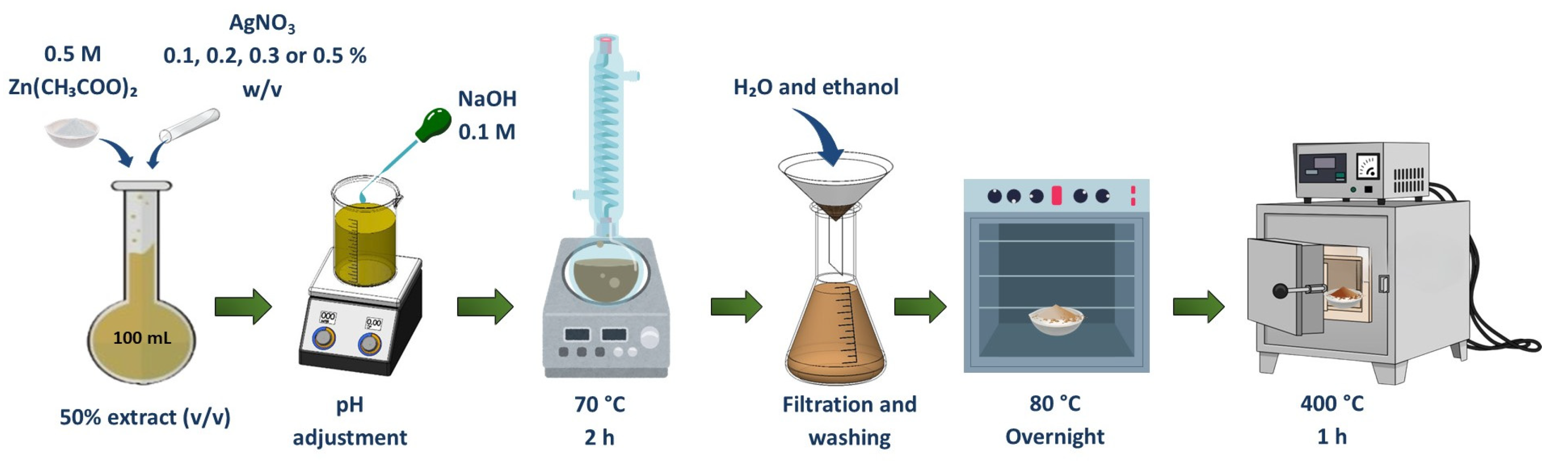
2.1. Synthesis of ZnO–Ag Nanocomposites

The aqueous extract of Tagetes erecta was prepared carefully. First, 39.5 g of dried and ground flowers were combined with 750 mL of distilled water in a temperature-controlled vessel maintained at 60 °C for 30 min under constant agitation. This temperature was selected to extract bioactive compounds without thermal degradation. Following extraction, the mixture was allowed to cool naturally to room temperature to prevent potential degradation of heat-sensitive compounds. The solution was subsequently filtered to eliminate plant remains, yielding an extract presumed to be rich in phytochemicals, such as polyphenols, flavonoids, and terpenoids, based on previous studies of Tagetes erecta [21,22]. The filtered extract was kept at 4 °C to preserve its stability and bioactivity for further experiments [23,24].
For the synthesis of ZnO–Ag nanocomposites (Figure 1), a modified co-precipitation approach was employed using 0.5 M zinc acetate as the primary zinc precursor and silver nitrate at varying concentrations (0.1, 0.2, 0.3, and 0.4% w/v) to achieve controlled silver incorporation. The pH of the reaction mixture was carefully adjusted to 10 using an appropriate alkaline solution, creating adequate conditions for controlled nucleation and growth of the nanoparticles. The solution was then subjected to a controlled heating and stirring regimen at 70 °C for 2 h, allowing for the formation of uniformly dispersed nanocomposites through the reduction of metal ions by the bioactive compounds present in the Tagetes erecta extract. The resulting precipitate was collected through filtration and thoroughly washed multiple times with distilled water and ethanol to remove unreacted precursors and excess plant metabolites. The purified material was dried overnight in an oven at 80 °C to remove residual moisture. A final heat treatment at 400 °C for 1 h was performed to achieve crystallization and eliminate remaining organic components, resulting in well-defined crystalline ZnO–Ag nanocomposites with varying silver concentrations [23]. The materials were characterized at the end of the entire synthesis process, after the powder underwent thermal treatment.

2.2. Characterization of ZnO–Ag Nanocomposites

2.2.1. UV-Vis Spectroscopy

The materials were characterized by several techniques. The formation of the nanomaterials was confirmed by UV-Vis spectroscopy. For this purpose, the materials were previously dispersed in distilled water (0.01 g in 100 mL) with ultrasonic treatment for 30 min. Subsequently, the absorbance spectra were recorded over a wavelength range of 600 to 300 nm using a VE-5100UV spectrophotometer (Velab, Mexico City, Mexico).

2.2.2. X-Ray Diffraction (XRD)

The X-ray diffraction (XRD) patterns for ZnO nanoparticles were collected using an X’Pert3 MRD X-ray diffractometer made by Panalytical (Malvern Panalytical, Almelo, NL), which utilized Cu-Kα (λ = 0.1542 nm) monochromatic radiation. The analysis spanned the 2θ range of 20 to 80 degrees, with a step size of 0.05. The nanoparticles’ average crystallite size (D) was determined using the Scherrer method [25].
D = 0.9 λ β c o s ( θ )
where λ represents the X-ray wavelength, β denotes the full width at half-maximum intensity, and θ is the diffraction angle. The Scherrer method is instrumental in assessing crystallite size by leveraging the broadening of XRD peaks.

2.2.3. Transmission Electron Microscopy (TEM)

Transmission electron microscopy (TEM) was performed using a Hitachi 7700 microscope (Hitachi, Tokyo, Japan) to study the morphology of the synthesized ZnO nanoparticles and to obtain the selected area electron diffraction (SAED) patterns. The microscope was operated at 100 kV. The identification of Miller indices on SAED was facilitated by the Crystallographic Tool Box (CrystBox) software (Version 1.10), utilizing the ring analysis mode (ring GUI) [26]. This analytical approach allowed for a detailed exploration of the nanoparticles’ morphology and provided a precise means of identifying crystallographic information through advanced software analysis.

2.2.4. Fourier Transform Infrared (FTIR)

Fourier transform infrared (FTIR) spectra were obtained from 4000 to 440 cm−1 in a System GX developed by Perkin Elmer (Spectrum 10, Waltham, MA, USA).

2.2.5. Thermogravimetric Analysis (TGA)

Thermogravimetric analysis (TGA) was performed in a Q600 equipment brand TA Instruments (New Castle, DE, USA) from ambient temperature up to 800 °C with a heating rate of 10 °C/min in air atmosphere.

2.3. Photocatalytic Performance of ZnO–Ag Nanocomposites

To comprehensively evaluate the photocatalytic activity under solar irradiation conditions, methylene blue (MB) was selected as a model organic contaminant due to its structural stability and widespread use as a standard photodegradation reference. The experiments were conducted using a precisely controlled MB concentration of 10 mg/L, with a catalyst/volume ratio of 1 g/L in a total solution volume of 300 mL to ensure uniform dispersion and maximum light absorption.
Before solar exposure, the catalyst-dye suspension was subjected to a dark equilibration period of 45 min under continuous magnetic stirring to establish adsorption–desorption equilibrium between the nanocomposite surfaces and MB molecules. This critical pre-equilibration step ensures that adsorption effects do not confound subsequent photocatalytic degradation measurements. Following equilibration, the reaction vessels were positioned in direct sunlight with carefully monitored irradiation intensity averaging 1259.2 ± 50.62 W/m2, maintaining a consistent light exposure across all experimental trials.
Aliquots were systematically collected at predetermined time intervals to track degradation kinetics. Each sample was immediately centrifuged at 12,000 rpm for 3 min to effectively separate the nanoparticulate catalysts from the solution, preventing light scattering interference during subsequent spectrophotometric analysis. The supernatants were then analyzed using UV-Vis spectrophotometry over the wavelength range of 450–750 nm, with particular focus on the characteristic MB absorption peak at 664 nm for precise quantification of the remaining dye concentration.
The photocatalytic degradation efficiency was calculated using Equation (2),
P h o t o d e g r a t a d i o n   e f f i c i e n c y   % = C 0 C x C 0 × 100
where C0 correspond to the initial concentration of methylene blue and Cx is the concentration at time t during solar irradiation.

2.4. Antibacterial Activity of ZnO–Ag Nanocomposites

2.4.1. Strains and Bacterial Culture Media

Escherichia coli ATCC 25922 was obtained from ATCC, Manassas, VA, USA (https://www.atcc.org/products/25922, accessed on 26 March 2025), a standardized clinical isolate widely used as a reference strain in antibacterial susceptibility testing, and was cultivated on selective McConkey agar (Difco™) at 37 °C for 24 h under aerobic conditions. This medium allows for the selective isolation and differentiation of Gram-negative enteric bacteria. Simultaneously, Staphylococcus aureus ATCC 25923 was obtained from ATCC, Manassas, VA, USA (https://www.atcc.org/products/25923, accessed on 26 March 2025), a quality control strain recommended by the Clinical and Laboratory Standards Institute (CLSI), and was cultured on Luria–Bertani (LB) agar under identical incubation parameters to ensure optimal growth of this Gram-positive pathogen.
For subsequent antibacterial assays, both bacterial strains were subcultured in nutrient-rich LB broth and incubated at 37 °C for 18 h with continuous orbital shaking to ensure uniform bacterial suspension. Before antibacterial testing, the overnight cultures were transferred to fresh LB broth and incubated at 37 °C for another 2 h to obtain bacteria in their mid-log growth phase. This careful timing ensures that the bacterial populations tested were in their most metabolically active and susceptible state, providing more consistent and reproducible antibacterial assessment results.

2.4.2. ZnO–Ag Nanocomposites Evaluated in the Antibacterial Assays

The study systematically evaluated a comprehensive range of nanocomposites with varying silver concentrations. The following materials were prepared and characterized for antibacterial testing: pure ZnO synthesized with 50% Tagetes erecta extract (labeled as ZnO) and four ZnO–Ag nanocomposites with incrementally increasing silver concentrations: ZnO plus 0.1% Ag in 50% extract (ZnO–Ag 0.1%), ZnO plus 0.2% Ag in 50% extract (ZnO–Ag 0.2%), ZnO plus 0.3% Ag in 50% extract (ZnO–Ag 0.3%), and ZnO plus 0.5% Ag in 50% extract (ZnO–Ag 0.5%). This systematic variation in silver content allowed for the precise determination of concentration-dependent antibacterial effects and the identification of optimal silver loading for maximum bactericidal activity.

2.4.3. Inhibition of Bacterial Growth in the Presence of Nanoparticles

To test the bactericidal activity of the nanoparticles, a microdilution test was performed according to CLSI standard methods; approximately 2 × 10⁸ CFU/mL of each strain was added to 96-well microplates (Corning Inc., New York, NY, USA) containing Muller–Hinton broth, after which volumes of nanoparticle solutions at final concentrations of 150 µg/mL, 200 µg/mL, and 250 µg/mL were evaluated. Growth controls of Mueller–Hinton, untreated bacteria, and the antibiotics gentamicin (10 µg/mL) and linezolid (30 µg/mL) were used as growth inhibition controls. Cultures were incubated at 37 °C with constant shaking for 4 h and 8 h, and optical density was measured at 660 nm every 30 min for 4 h and 8 h of incubation. Finally, the percentage of viable cells relative to viable bacteria obtained in the Mueller–Hinton medium was calculated. All experiments were performed in triplicate.

3. Results and Discussion

3.1. Characterization of ZnO NPs

3.1.1. UV-Vis Spectroscopy

The UV-Vis absorption spectra (Figure 2) show a characteristic absorption band for ZnO between 363 and 370 nm, which corresponds to its intrinsic band gap transition. In the case of ZnO–Ag nanocomposites, a slight shoulder appears from 400 nm in addition to the ZnO absorption band. This feature is attributed to the surface plasmon resonance (SPR) of silver nanoparticles.
Moreover, the intensity of the SPR band increases progressively with higher silver content, consistent with the increasing concentration of Ag in the nanocomposites. This behavior suggests that AgNPs were effectively anchored on the ZnO surface and that their plasmonic response is concentration-dependent.
The slight red-shift and broadening of the ZnO absorption band observed in some samples may also indicate possible interactions between ZnO and Ag nanoparticles, affecting the local electronic environment [27].

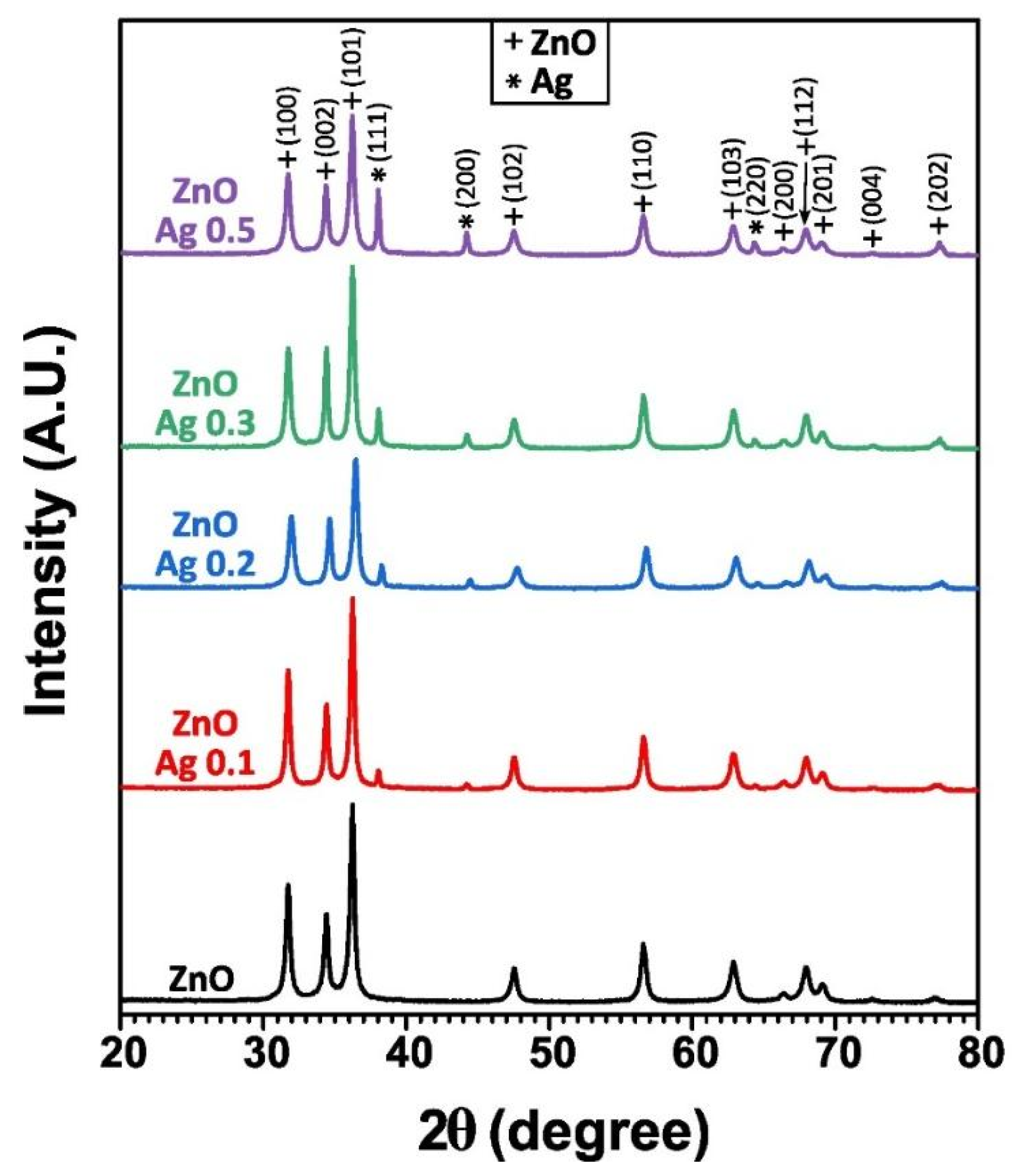
3.1.2. X-Ray Diffraction (XRD)

Figure 3 displays the XRD patterns of pure ZnO and ZnO–Ag nanoparticles synthesized with different silver concentrations. The diffraction peaks represented with “+” correspond to the hexagonal wurtzite phase of ZnO (JCPDS card No. 70-0206), showing well-defined and sharp diffraction peaks. In the ZnO–Ag samples, additional diffraction peaks appear at 2θ ≈ 38.1°, 44.3°, and 64.5°, corresponding to the (111), (200), and (220) planes of face-centered cubic (FCC) metallic silver (JCPDS card No. 87-0718). The intensities of these peaks increase with silver content, suggesting enhanced crystallization of metallic silver.
No shift in the ZnO diffraction peaks is observed with increasing silver content, even at 0.5%, indicating that silver is not incorporated into the ZnO lattice but remains as a separate phase. This observation is consistent with findings by Liu et al. [28], who concluded that the introduction of silver does not distort the ZnO crystal lattice or significantly alter the crystallite size. The coexistence of distinct ZnO and Ag peaks further supports the structural stability of the ZnO. It was concluded that the introduction of silver does not result in the distortion of ZnO structure.
Notably, these silver-related diffraction signals, even at the lowest concentration (0.1%), suggest that the phytochemical constituents in the Tagetes erecta extract effectively promote the reduction of silver ions to their metallic state during synthesis.
The average crystallite size estimated using the Scherrer equation ranges from 16.76 nm (ZnO–Ag 0.2%) to 19.45 nm (ZnO–Ag 0.1%), as shown in Table 1. Although the smallest crystallite size is observed for the 0.2% Ag sample, no direct correlation is observed between the crystallite size and the photocatalytic or antibacterial performance. This suggests that factors beyond crystallinity, such as surface area, Ag distribution, or reactive site availability, may play a more significant role in functional performance.

3.1.3. Transmission Electron Microscopy (TEM)

Figure 4 presents TEM micrographs of ZnO and ZnO–Ag nanocomposites at varying silver concentrations (Figure 4a–e). The images reveal nanoparticles with predominantly spherical and hemispherical morphologies with moderate polydispersity and a tendency to agglomerate. Micrographs did not show silver nanoparticles, likely because of their ultrafine size and low concentration (≤0.5%). Further confirmation of the crystalline structure is provided by the SAED pattern analysis (Figure 4f), which displays concentric diffraction rings attributed to the hexagonal wurtzite phase of ZnO. These patterns confirm the polycrystalline nature of the synthesized materials and are in excellent agreement with the XRD results. Notably, no distinct diffraction rings corresponding to metallic silver are observed in the SAED, which supports the hypothesis that silver is either highly dispersed, present in small quantities, or not detected due to the limited volume analyzed in the TEM.
Table 1 presents the average particle size and crystallite size (from XRD) for each sample. The ZnO–Ag 0.2% sample exhibited the smallest average particle size (17.39 ± 6.45 nm) and crystallite size (16.76 ± 1.84 nm), while the ZnO–Ag 0.1% sample showed the largest. A general trend of size reduction is observed with increasing Ag content up to 0.2%, although this trend is not strictly linear.
The TEM-derived particle sizes are slightly larger than the crystallite sizes obtained from XRD, which is expected due to polycrystalline aggregation or surface capping layer formed during the green synthesis using Tagetes erecta extract. Despite the variations in particle and crystallite size, no consistent correlation was found between particle/crystallite size and photocatalytic or antibacterial performance. For instance, ZnO–Ag 0.3% exhibits enhanced photocatalytic activity despite having a larger crystallite size (18.81 ± 1.26 nm) compared to ZnO–Ag 0.5% (17.17 ± 0.69 nm). This observation reinforces that functional properties are influenced by surface phenomena, Ag–ZnO interfacial interaction, or electronic modifications, rather than only particle size.

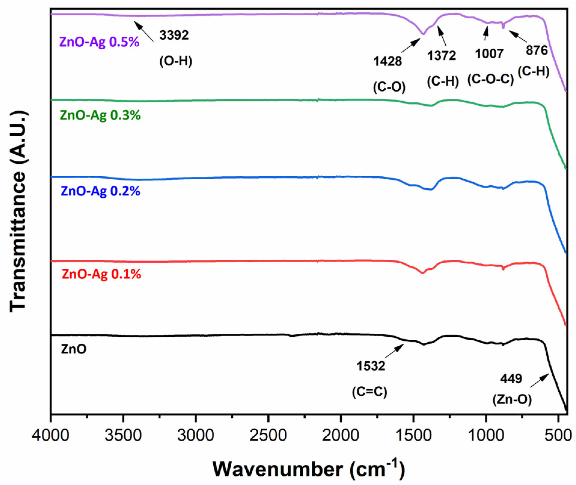
3.1.4. Fourier Transform Infrared (FTIR) Spectroscopy

Figure 5 displays the FTIR spectra of pure ZnO and ZnO–Ag nanocomposites synthesized using 50% Tagetes erecta extract. The spectra reveal characteristic absorption bands at 3392, 1532, 1428, 1372, 1007, 876, and 449 cm−1, corresponding to functional groups from both the botanical extract and the ZnO structure. The weak band at 3392 cm−1 is attributed to the stretching vibration of hydroxyl groups (O–H) present in phenolic compounds, flavonoids, and adsorbed water molecules on the ZnO surface [29]. The prominent signal at 1532 cm−1 corresponds to C=C vibrations in aromatic rings, indicative of flavonoids and phenolic compounds that function as effective stabilizing agents during nanoparticle formation [29]. The absorption bands at 1428 cm−1 and 1372 cm−1 are associated with C–O and C–H vibrations in carboxylates and polyphenols, respectively, which can form coordination complexes with both Ag and Zn ions, thereby promoting nanoparticle stabilization and controlled growth [30]. The signal at 1007 cm−1 is characteristic of C–O–C bonds in carbohydrates and ethers, suggesting the presence of organic residues from the plant extract that contribute to the capping process [31]. The band at 876 cm−1 corresponds to C–H out-of-plane vibrations in aromatic compounds, further confirming the role of polyphenols and flavonoids in the stabilization of the nanoparticles [32]. Finally, the sharp band at 449 cm−1 is characteristic of the Zn–O bond vibration, definitively confirms the formation of ZnO in its wurtzite crystalline phase [33]. Notably, the incorporation of silver induces subtle but significant changes in band intensities, particularly due to its interaction with functional groups from both the botanical extract and the ZnO crystal structure.

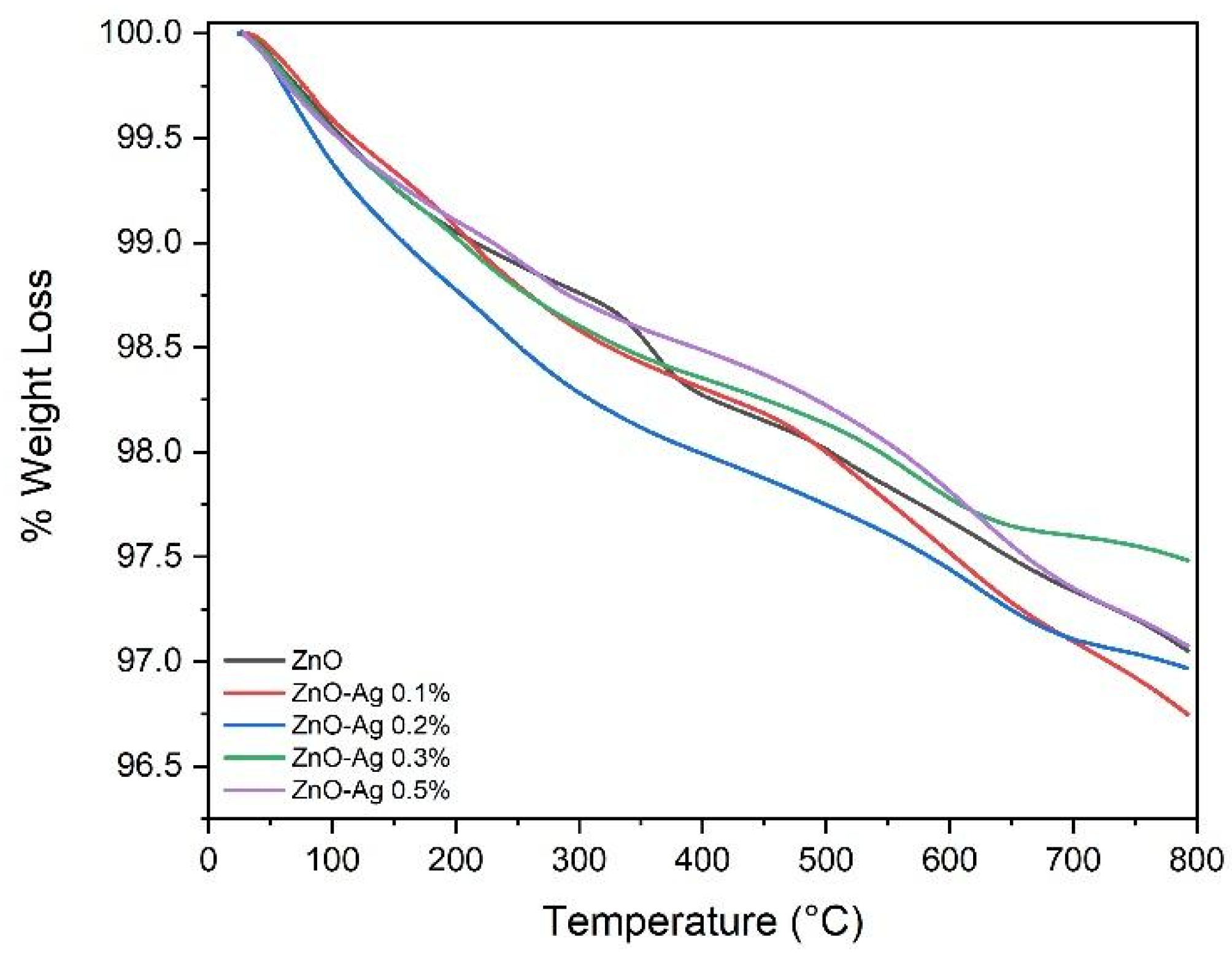
3.1.5. Thermogravimetric Analysis

Figure 6 presents the TGA thermograms of the synthesized ZnO–Ag nanocomposites over a temperature range of 35 °C to 800 °C. The results indicate an average mass loss of only 2.764 ± 0.11%, demonstrating the exceptional thermal stability of the synthesized nanomaterials. This minimal weight reduction is attributed primarily to the elimination of physiosorbed water molecules and the thermal decomposition of residual volatile organic compounds from the Tagetes erecta extract that remained adsorbed on the nanoparticle surfaces. The high thermal stability observed across all samples suggests effective calcination during synthesis and confirms the predominance of inorganic ZnO and Ag phases in the final nanocomposites. This thermal behavior is particularly advantageous for applications requiring exposure to elevated temperatures, such as photocatalytic processes under intense solar irradiation.

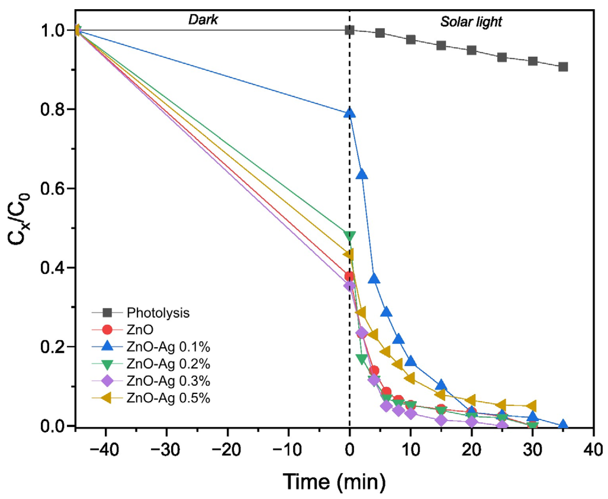
3.2. Photocatalytic Performance

Various industries, including cosmetics, paper, tanneries, food, and pharmaceuticals, generate dye-containing effluents, with textile factories being among the largest consumers of synthetic dyes. These dyes can significantly impact aquatic ecosystems, affecting flora and fauna. Key consequences include reduced dissolved oxygen levels, light penetration obstruction, alteration of the photosynthesis process, eutrophication, formation of recalcitrant compounds, and potential carcinogenic and mutagenic effects.
Several technologies, including biological or physicochemical treatments, have been explored to address this issue. However, since 2 to 50% of synthetic dyes are highly persistent and cannot be effectively removed using conventional methods, advanced oxidation processes (AOPs) have been proposed as a viable alternative. In this sense, photocatalysis has emerged as an efficient strategy for degrading these compounds, with TiO2 and ZnO being among the most widely used photocatalysts.
Figure 7 shows the Cx/C0 behavior of the synthesized materials. The initial 45 min represents the equilibrium phase of the adsorption-desorption processes between the dye and the catalyst before solar exposure. The results indicate that silver incorporation enhances the photocatalytic activity of ZnO, which can be attributed to improved charge carrier separation and a reduced electron-hole recombination rate facilitated by silver.
Based on the experimental data and assuming a pseudo-first-order kinetic reaction (Equation (3)), the data fitting was performed using OriginPro 2018 software to calculate the kinetic constant for each system (Table 2),
l n   C 0 C X = k a p p t
where C0 is the initial methylene blue concentration (10 mg/L), Cx is the residual dye concentration in solution after a specific time (mg/L), kapp is the rate constant (min−1), and t is the exposure time to sunlight (min).
Table 2 compares the photocatalytic performance of ZnO and ZnO–Ag nanocomposites. It is observed that silver enhances the response of the material by increasing its reaction rate (from 0.1 to 0. 3%), which can be attributed to the fact that the metallic nanoparticles reduce recombination processes. It has been reported that silver nanoparticles act as trap centers in the forbidden band, effectively trapping electrons. Additionally, the inclusion of metallic ions and the green synthesis (including residual phytochemicals) promotes the formation of defects in the semiconductor nanoparticles. These defects are closely related to recombination processes, and by reducing recombination, they help increase photocatalytic efficiency [10]. However, an excessive amount of silver can block the active sites of the ZnO, reducing the available spaces for adsorption and pollutant degradation, as seen when the silver concentration is increased to 0.5% [34]. This effect may also be due to the formation of a photogenerated electron-hole pair complex, which can decrease photocatalytic activity. Therefore, to optimize photocatalytic performance, finding the ideal silver concentration in ZnO is crucial.

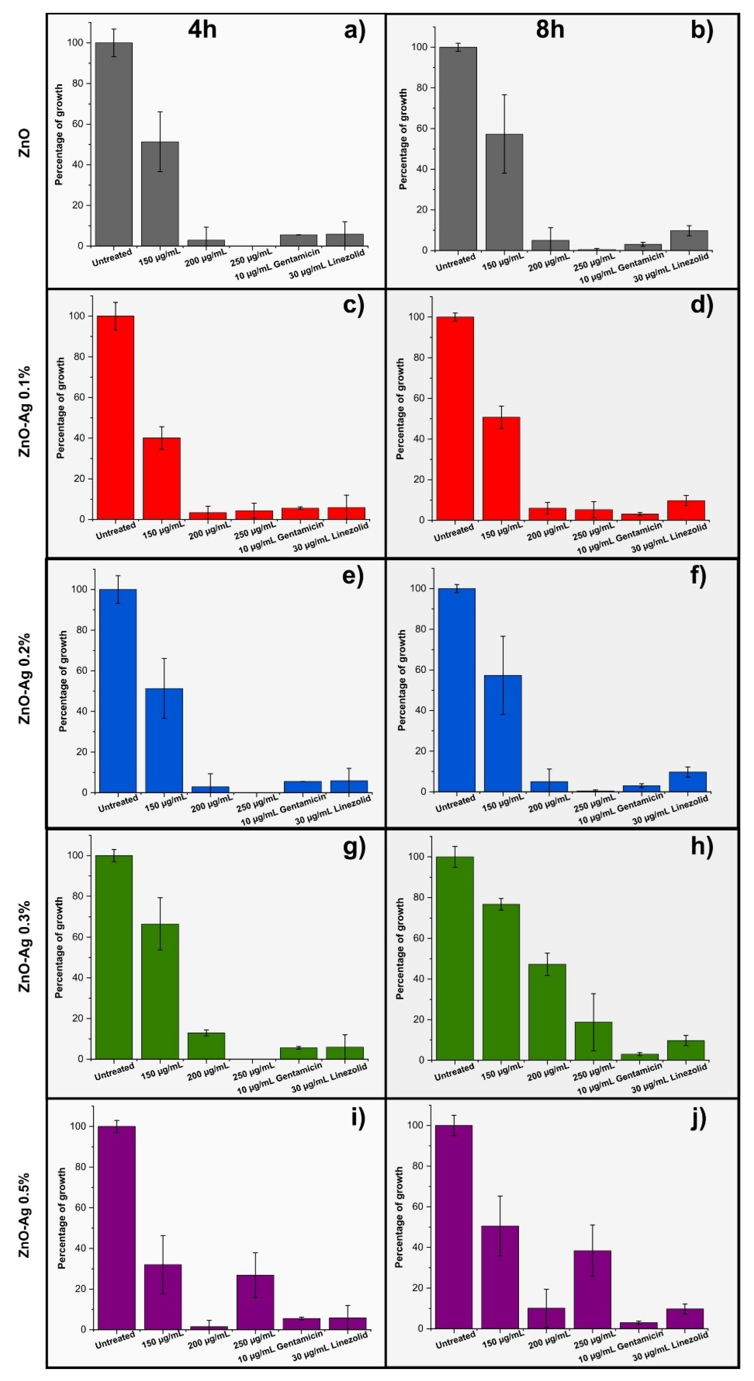
3.3. Antibacterial Activity

Infectious bacterial diseases have presented a considerable and ongoing challenge in healthcare environments for years [35]. Escherichia coli (E. coli) is responsible for various infections, including neonatal meningitis, urinary tract infections, hemorrhagic colitis, Crohn’s disease, and gastroenteritis. Similarly, Staphylococcus aureus (S. aureus) can lead to severe conditions such as septic arthritis, osteomyelitis, and endocarditis [36]. Tagetes erecta (marigold) is a plant used since ancient times for various purposes in medicine and agriculture. The leaves are employed as an antiseptic agent for kidney disorders and muscular pain. The flowers are used in febrile conditions, epileptic disorders, gastrointestinal issues, and liver ailments to eliminate scabies and treat ocular diseases [37]. The synthesis of metal nanoparticles (NPs) has recently become a significant focus due to their chemical, physical, and biological applications across various fields. The increased reactivity of NPs, which range from 1 to 100 nm, is attributed to their large surface area, making them appealing for therapeutic uses at different dosages and drug delivery [36]. Zinc oxide (ZnO) nanoparticles are considered safe, low in toxicity, biocompatible, cost-effective, and environmentally friendly materials. However, ZnO NPs and Ag exhibit potential biocidal activity as they possess inherent antibacterial properties [38]. Therefore, the synthesis of ZnO nanoparticles with Tagetes erecta extract and Ag could be considered a potential antibiotic therapy.
The antibacterial activity of ZnO with Ag and marigold flower extract was tested on Gram-positive (S. aureus) and Gram-negative (E. coli) bacteria (ATCC strains) (Figure 8 and Figure 9).

3.3.1. Antibacterial Activity of ZnO and Ag with Marigold Flower Extract Against S. aureus ATCC 14923

ZnO and Ag were diluted in marigold flower extract and tested for antibacterial activity against S. aureus and E. coli (Figure 8 and Figure 9). As for S. aureus ATCC 25923 (Figure 8), ZnO (200 and 250 µg/mL) with 50% marigold flower extract (without Ag) inhibited more than 95% of S. aureus cultures, while about 50% at 150 µg/mL at 4 h (51.32% and 2.93% of growth with 150 and 200 µg/mL, respectively) and 8 h (57.31%, 5.01%, and 0.43% of growth with 150, 200, and 250 µg/mL, respectively) (Figure 8a,b). Similar results were obtained in ZnO with 0.1 and 0.2 Ag in marigold flower extract at 50%, 200, and 250 µg/mL, inhibiting more than 95% of S. aureus cultures at both incubation times. In comparison, 150 µg/mL with 0.1 Ag showed about 60% of inhibition at 4 h incubation (40.09% of growth) and approximately 50% (50.73% of growth) at 8 h and 150 µg/mL with 0.2 Ag inhibited around 50% (51.32% of growth) at 4 h and around 45% (57.31% of growth) at 8 h of S. aureus cultures compared to untreated (Figure 8c–f). In ZnO with 0.3 Ag in 50% extract, 250 µg/mL inhibited 100% of S. aureus, 200 µg/mL around 90% (12.86% of growth), and 150 µg/mL around 35% (66.49% of growth) at 4 h, while 250 µg/mL approximately 80% (18.75% of growth), 200 µg/mL around of 50% (47.24% of growth), and 150 µg/mL around 20% (76.69% of growth) at 8 h compared to untreated S. aureus (Figure 8g,h). The last concentration was ZnO with 0.5 Ag in 50% extract, in which 200 µg/mL had the best antibacterial activity (more than 95% inhibition; 1.46% of growth), followed by 250 µg/mL (around 75% of inhibition; 26.87% of growth) and 150 µg/mL approximately 70% of inhibition (32% of growth) at 4 h, while at 8 h 200 µg/mL inhibited around 90% (9.34% of growth) of S. aureus cultures, 250 µg/mL around 60% (38.40% of growth), and approximately 50% (50.58% of growth) at 150 µg/mL compared to untreated (Figure 8i,j).

3.3.2. Antibacterial Activity of ZnO and Ag with Marigold Flower Extract Against E. coli ATCC 25922

The antibacterial activity of ZnO and Ag with marigold flower extract was also tested against E. coli ATCC 25922 (Figure 9). In E. coli ATCC 25922, only 250 µg/mL of ZnO in 50% marigold flower extract inhibited around 65% of bacteria (34.12% of growth was observed) at 4 h and 100% at 8 h compared to the untreated. The remaining concentrations (200 and 150 µg/mL) inhibited about 20% of E. coli growth at both incubation times (Figure 9a,b). No antibacterial activity was observed for the three concentrations of ZnO with 0.1 Ag in 50% of the extract against E. coli strains ATCC 25922 (more than 70% growth is monitored) (Figure 9c,d). Only 250 µg/mL ZnO with 0.2 Ag in 50% extract showed antibacterial activity at 4 h (75% inhibition) and 8 h (100% inhibition) compared to the untreated, while the rest of the treatments inhibited only 20% of the E. coli cultures at both incubation times (Figure 9e,f). In the case of ZnO with 0.3 Ag in 50% of the extract, the best antibacterial activity was 250 µg/mL with inhibition more than 95% at 4 h and around 90% at 8 h (0.66% and 11.91% of growth, respectively), followed by 200 µg/mL with around 55% at 4 h and more 50% at 8 h (44.05% and 48% of growth, respectively) and 150 µg/mL, around 15% at 4 h, and more than 30% at 8 h (84.25% and 68.64% of growth, respectively) (Figure 9g,h). The last tested concentration of ZnO was 0.5 Ag in 50% of the extract. The best inhibitory activity was observed at 4 h with 200 µg/mL (more than 95% inhibition; 1.94% of growth), followed by 250 µg/mL (around 85% of inhibition; 14.38% of growth) and 150 µg/mL with more than 40% of inhibition (58.33% of growth), while at 8 h, the treatments decreased their antibacterial activity, 250 µg/mL around 75% of inhibition (24.75% of growth), 200 µg/mL (more than 55% of inhibition; 52.72% of growth), and 150 µg/mL (around 15%of inhibition) (Figure 9i,j).
Antibiotics play an essential role in the treatment of infectious diseases caused by bacteria and fungi. Unfortunately, there has been an increase in the emergence of bacteria resistant to these types of treatments. Metallic nanoparticles have generated interest in their therapeutic applications [36] and plant extracts such as Tagetes erecta due to their antibacterial, antifungal, and anti-inflammatory properties [37].
The healing qualities of medicinal herbs are linked to their secondary metabolites, which serve as a defense strategy against pathogens. These substances exhibit various health-enhancing effects, including antimicrobial, antioxidant, anticancer, and anti-inflammatory activities [39]. Extract of Tagetes erecta leaves has been used for the synthesis of silver nanoparticles, which exhibited potent antibacterial activity against Staphylococcus aureus, with better activity observed against Escherichia coli [36]. In our data, the compounds showed higher activity against Staphylococcus aureus than Escherichia coli. In this context, the importance of the Tagetes erecta extract and silver activity is highlighted. This activity could be enhanced in the future by designing a multitarget treatment.
Zavaleta et al. [40] demonstrated the antibacterial activity of ZnO nanoparticles against Staphylococcus aureus ATCC 25923 at various concentrations, specifically at a minimum concentration of 1.2 mg/mL, which is higher than the active concentration found in our study, where activity was established at a greater concentration of 250 µg/mL. This activity was even superior to the commercial drugs Gentamicin and Linezolid. The antibacterial activity of these nanoparticles is attributed to their small size, large surface area, and minimal toxic effects on human cells at low concentrations [40]. Zinc oxide (ZnO) is a valuable inorganic compound known for its potential antimicrobial properties due to its photocatalytic abilities [35,38]. Nano-sized ZnO and its derivative biomaterials have surfaces that produce free radicals when exposed to light. These active radicals have been shown to suppress microorganisms effectively. In addition, they are non-toxic, biocompatible, cost-effective, environmentally friendly, and transparent, which makes them suitable for innovative medical applications [41,42].
The antibacterial activity of silver nanoparticles stabilized with copolymers has been studied in Escherichia coli ATCC 25922, Kanamycin-resistant Escherichia coli, and Staphylococcus aureus ATCC 25923. A concentration of 10.8 µg/mL completely inhibited the growth of Escherichia coli in a 4 h incubation period. Escherichia coli was more sensitive than Staphylococcus aureus to the evaluated nanoparticles, which were even more active than the antibiotics kanamycin and ampicillin [43]. Although higher concentrations were evaluated in our study, our compounds were more active for Staphylococcus aureus ATCC 25923 to Escherichia coli ATCC 25922 at 4 and 8 h of incubation. In this context, the nanoparticles complement, calendula (Tagetes erecta) extract, and ZnO may present a predisposition to Gram-positive bacteria for the nanoparticles stabilized with copolymers mentioned, which were more active for Gram-negative bacteria. Numerous studies have demonstrated the effective application of silver nanoparticles against both Gram-positive and Gram-negative bacteria [39]. Silver carries a positive charge, which interacts with negatively charged biomolecules such as phosphorus and sulfur, the primary components of cell membranes, proteins, and DNA bases [44].
Silver ions are consistently released from silver nanoparticles, which can be regarded as a mechanism for killing microbes. Due to their electrostatic attraction, silver ions readily adhere to the cell wall and cytoplasmic membrane, as they are more closely linked with sulfur-containing proteins [45,46].
ZnO nanoparticles with plant extracts have been synthesized and characterized, showing an effect on the analyzed microbes, particularly against Gram-positive bacteria [47]. According to our data, the most effective concentrations of the treatment under study were 250 µg/mL, followed by 200 µg/mL. Although a decrease in the growth percentage was observed compared to the untreated bacteria, better antibacterial activity was noted against the Gram-positive bacterium S. aureus, consistent with the previously mentioned study.
The antibacterial efficacy of ZnO NPs facilitated by plant extracts is more effective against bacterial and fungal infections and human diseases. It has been found that plants such as Aloe barbadensis, Plectranthus amboinicus, Sedum alfredii Hance, Cassia auriculata, and Pretence blossoms are involved in the synthesis of NPs [48]. The synthesized green ZnO NPs are effective antibacterial agents for both Gram-positive and Gram-negative bacteria, such as Mycobacterium tuberculosis, Bacillus subtilis, Salmonella typhimurium, Staphylococcus aureus, Streptococcus pyogenes, Escherichia coli, and Klebsiella pneumoniae [49]. Through a variety of mechanisms, ZnO NPs exhibit noteworthy antibacterial characteristics.
The generation of reactive oxygen species (ROS), including superoxide anions (O2•−) and hydroxyl radicals (OH), is an essential phenomenon in nature. These highly reactive ROS can damage DNA, proteins, and the membranes of bacterial cells, ultimately leading to cell death. Additionally, upon exposure to UV light, ZnO nanoparticles (ZnO NPs) act as photocatalysts, producing reactive species that further disrupt bacterial cell walls and membranes [50].
Adding ZnO to form nanoparticles like those proposed in this study could help develop new drugs with potential mechanisms of action depending on their components and bacterial targets.
Table 3 compares the photocatalytic and antibacterial properties reported in this article and those reported in other literature.
A comparative analysis of ZnO–Ag nanocomposites (Table 3) synthesized via various methods indicates that the synthesis route and experimental parameters strongly influence photocatalytic and antibacterial activities. Photocatalytic degradation of methylene blue (MB) dye demonstrates that green synthesis approaches, particularly those employing plant extracts such as Tagetes erecta and Punica granatum [54], can achieve high degradation efficiencies, reaching 98–100% under sunlight irradiation. Remarkably, the nanocomposites developed in the present study exhibit superior performance compared to previously reported in terms of degradation efficiency and reaction time. This enhanced photocatalytic activity is likely attributable to silver content, phytochemicals in the extract of Tagetes erecta, and the pH used in the degradation tests.
Regarding antibacterial activity, ZnO–Ag nanocomposites substantially inhibit E. coli and S. aureus growth. The nanocomposites developed in this work achieved up to 100% inhibition for E. coli and 95% for S. aureus at 250 µg/mL after 8 h (ZnO–Ag 0.2%). These results are comparable or superior to those reported in previous studies, even those using higher bacterial concentrations or different synthesis techniques. Such performance underscores the potential of the synthesized nanocomposite as an efficient dual-functional material for environmental and biomedical applications.

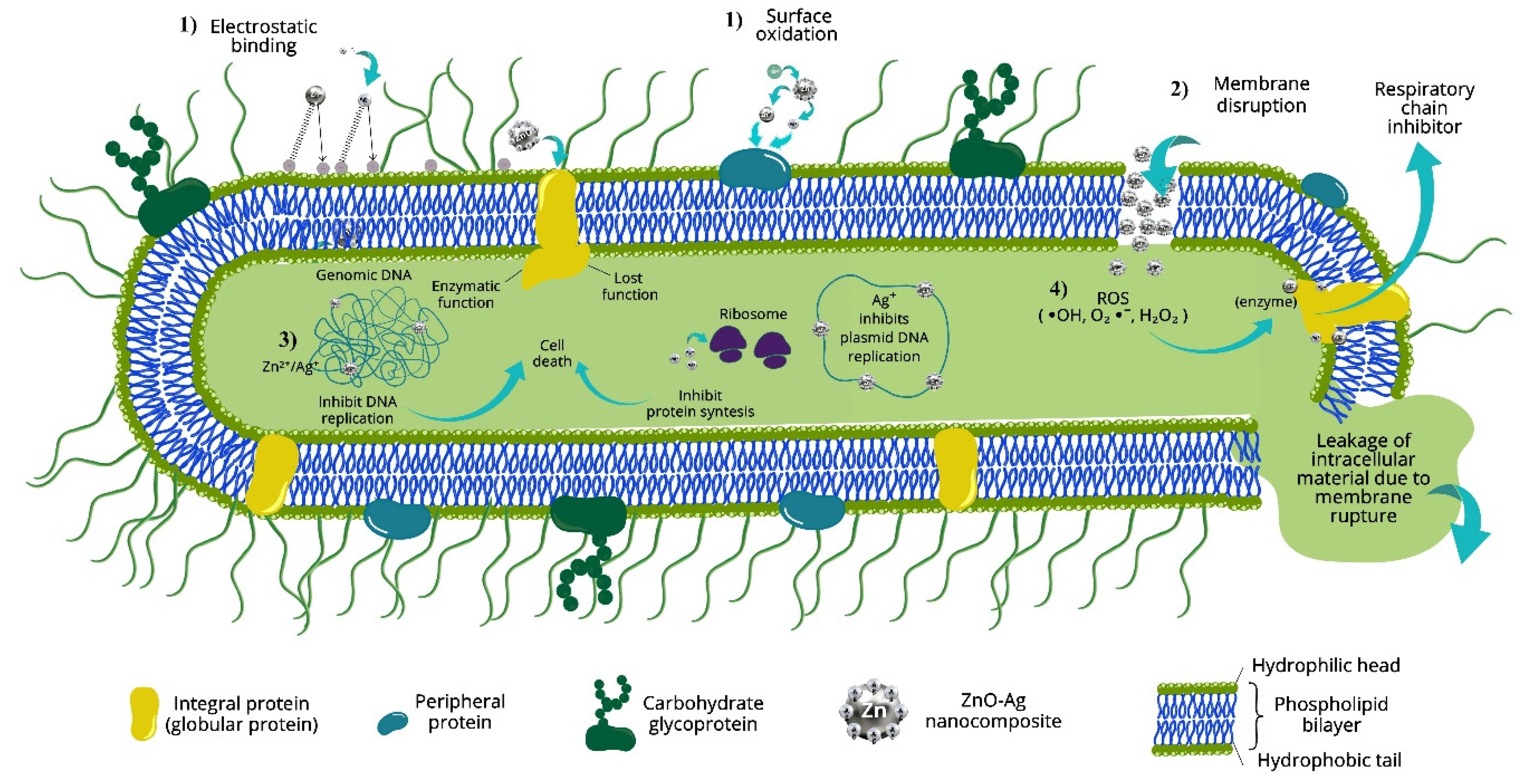
3.4. Proposed Mechanism of Photocatalytic and Antibacterial Activity of Prepared Nanocomposites

Figure 10 and Figure 11 show the proposed mechanism of the ZnO–Ag nanocomposites regarding their photocatalytic and antibacterial activity.
The bactericidal mechanism of zinc oxide–silver (ZnO–Ag) nanoparticles is complex and multifactorial, involving synergistic processes that alter bacterial cell structure and function. The main mechanisms supported by scientific studies are described below (Figure 10):
  • Physical damage to the cell membrane. ZnO–Ag nanoparticles adhere to the surface of bacteria through electrostatic interactions between the released Zn2+ and Ag+ ions and the negatively charged bacterial cell wall [58]. This interaction can cause physical damage to the cell membrane, such as deformation, loss of integrity, and even cell lysis. Electron microscopic studies have demonstrated morphological alterations in bacteria such as E. coli, including cell elongation and membrane rupture [59,60].
  • Intracellular inclusion of nanoparticles. Once the nanoparticles penetrate the cell, leakage from the cytoplasm occurs, causing membrane shrinkage and loss of cellular functionality. This leads to bacterial cell death by structural collapse [18].
  • Damage to bacterial DNA by interaction with Zn2+ and Ag+ ions. Nanoparticles, mainly because of their silver content, can interact directly with bacterial DNA, inhibiting its replication and affecting gene expression and cell division [61,62].
  • Damage due to interaction with reactive oxygen species (ROS). Under exposure to light, ZnO–Ag nanocomposites generate ROS (free radicals) such as •OH, O₂•⁻, and H₂O₂. These species attack membrane lipids, structural proteins, and DNA, inducing severe oxidative stress and causing cell death [15,63].
The mechanism of photodegradation of ZnO–Ag nanoparticles can be illustrated by the following main phases (Figure 11):
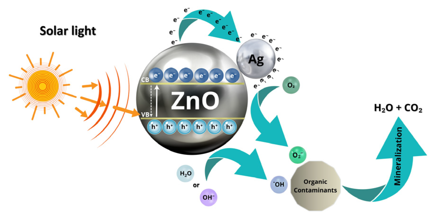
  • Excitation of ZnO–Ag nanoparticles under light irradiation. When ZnO–Ag nanoparticles are irradiated with light (UV or visible), electronic excitation occurs in the ZnO semiconductor (with a bandgap of about 3.37 eV). Photons with sufficient energy excite electrons from the valence band (VB) to the conduction band (CB), generating free electrons (e) in the CB and positive holes (h) in the VB, as indicated in the following expression [64]:
    ZnO + hν → e (BC) + h+ (BV)
  • Role of silver (Ag) as an electron sink. The incorporation of silver nanoparticles into ZnO forms a heterojunction that significantly improves the photocatalytic performance since silver acts as an electron sink, capturing electrons from the conduction band of ZnO and reducing the e/h+ recombination [16]:
    e (BC, ZnO) → Ag
  • Generation of reactive oxygen species (ROS). Electrons transferred to silver can reduce molecular oxygen adsorbed on the surface, forming superoxide radicals, while holes in the valence band of ZnO can oxidize water molecules or hydroxyl ions to generate hydroxyl radicals, as follows:
    Oxygen reduction:
    O2 + eO2
    Oxidation of water or hydroxyl ions:
    H2O + h+OH + H+
    OH + h+OH
  • Degradation of organic pollutants. The generated radicals attack organic pollutant molecules, such as dyes or pharmaceutical compounds, oxidizing them to carbon dioxide, water, and other harmless products:
    Contaminant + OH → Intermediates → CO2 + H2O
This synergistic mechanism between ZnO and Ag results in more efficient photocatalysis, broadening the spectral absorption range and improving charge separation, which is crucial for pollutant degradation and wastewater treatment applications.

4. Conclusions

This study presents results on the potential environmental and biomedical applications of nanocomposites synthesized by a simple and environmentally friendly technique. Eco-friendly synthesis of ZnO–Ag nanocomposites resulted in spherical and hemispherical nanoparticles, with hexagonal ZnO–Ag wurtzite-like and cubic Ag face-centered phases. XRD and TEM analyses confirmed increased crystallinity and homogeneity with increasing Ag content, with crystallite sizes ranging from 16.76 to 19.45 nm. In addition, the presence of bioactive phytochemicals in the extract played a key role in reducing and stabilizing Zn and Ag ions, favoring the controlled growth of the nanocomposites.
Photocatalytic tests evidenced a direct relationship between the incorporation of Ag and the improvement in the degradation performance of methylene blue under solar irradiation. In particular, the ZnO–Ag nanocomposite (0.3%) showed the highest photocatalytic activity, achieving complete degradation of methylene blue in only 25 min, with a rate constant of 0.20912 min−1. This improvement is attributed to the role of Ag as an electron trap, which reduces electron-hole recombination and enhances charge carrier separation. However, by increasing the Ag content to 0.5%, a decrease in performance was observed, possibly due to Ag nanoparticles blocking active sites or generating recombination centers. This highlights the importance of optimizing the Ag concentration to maximize photocatalytic efficiency.
In antibacterial experiments, all nanocomposites demonstrated bactericidal activity against Staphylococcus aureus (Gram-positive) and Escherichia coli (Gram-negative), with a more pronounced effect observed against S. aureus. The ZnO–Ag nanocomposite (0.5%) achieved almost complete inhibition at 200 µg/mL concentration over 4 h. While the ZnO–Ag nanocomposite (0.2%) inhibits more than 95% of S. aureus and 100% of E. Coli at 250 µg/mL concentration over 8 h, the enhanced antibacterial effect is attributed to the synergistic interaction between the ability of ZnO to generate reactive oxygen species (ROS), the membrane-altering properties of Ag, and the bioactive compounds present in T. erecta. Together, these compounds alter bacterial cell membranes, induce oxidative stress, and inhibit growth, especially in Gram-positive bacteria, whose outer membrane is less complex.
Nevertheless, although these results are promising, it is important to recognize certain limitations. First, the study used standard bacterial strains under controlled in vitro conditions, which may not fully reflect the real environment or clinical settings. Also, the photocatalytic activity was evaluated with only one dye (methylene blue), so further studies are required to analyze its performance against a broader range of pollutants.
Generally, this green synthesis route offers a sustainable and scalable approach to producing multifunctional nanomaterials. ZnO–Ag nanocomposites stand out for their great potential in two key applications: solar-powered wastewater treatment and their use as antibacterial agents. Future research could focus on their integration into filtration membranes or disinfectant surfaces and extend testing to multi-resistant pathogens and complex mixtures of contaminants present in real wastewater.

Author Contributions

Conceptualization, J.R.L.-L. and P.F.M.-H.; methodology, J.R.L.-L., A.T.-O., J.M.H.-R., M.A.H.-C., H.F.-V., N.L.-S., A.C.-R. and P.F.M.-H.; validation J.R.L.-L., A.T.-O., J.M.H.-R., H.F.-V., A.C.-R. and P.F.M.-H.; formal analysis, A.T.-O., J.M.H.-R., M.E.C.-G., H.F.-V., N.L.-S., A.C.-R., M.d.J.L.-L., J.R.P.-U., G.G.V.-R. and P.F.M.-H.; investigation, J.R.L.-L., A.T.-O., J.M.H.-R., M.d.J.L.-L., M.A.H.-C., H.F.-V., A.C.-R. and P.F.M.-H.; resources, J.M.H.-R., H.F.-V., N.L.-S., A.C.-R. and P.F.M.-H.; data curation, A.T.-O., J.M.H.-R., M.E.C.-G., H.F.-V., A.C.-R., J.R.P.-U., G.G.V.-R. and P.F.M.-H.; writing—original draft preparation, A.T.-O., J.M.H.-R., M.E.C.-G., H.F.-V., A.C.-R., J.R.P.-U., G.G.V.-R. and P.F.M.-H.; writing—review and editing, J.R.L.-L., A.T.-O., J.M.H.-R., M.E.C.-G., M.d.J.L.-L., H.F.-V., A.C.-R., J.R.P.-U., G.G.V.-R. and P.F.M.-H.; visualization, J.R.L.-L. and P.F.M.-H.; supervision, J.M.H.-R. and P.F.M.-H.; project administration, P.F.M.-H.; funding acquisition, J.R.L.-L. and P.F.M.-H. All authors have read and agreed to the published version of the manuscript.

Funding

This research was funded by the Program for the Promotion and Support of Research Projects (PRO-A8-002) of Universidad Autónoma de Sinaloa.

Institutional Review Board Statement

Not applicable.

Informed Consent Statement

Not applicable.

Data Availability Statement

The original contributions presented in this study are included in the article. Further inquiries can be directed to the corresponding author.

Acknowledgments

The authors thank R.A. Ochoa-Gamboa, D. Lardizabal-Gutiérrez, M. Román-Aguirre, Y. P. Ahumada-Santos, and J.E. Ledezma-Silla for their technical support.

Conflicts of Interest

The authors declare no conflicts of interest.

References

  1. WHO. Drinking-Water. Available online: https://www.who.int/news-room/fact-sheets/detail/drinking-water (accessed on 3 March 2025).
  2. Bharathi, D.; Thiruvengadam Nandagopal, J.G.; Rajamani, R.; Pandit, S.; Kumar, D.; Pant, B.; Pandey, S.; Kumar Gupta, P. Enhanced photocatalytic activity of St-ZnO nanorods for methylene blue dye degradation. Mater. Lett. 2022, 311, 131637. [Google Scholar] [CrossRef]
  3. Anucha, C.B.; Altin, I.; Bacaksiz, E.; Stathopoulos, V.N. Titanium dioxide (TiO2)-based photocatalyst materials activity enhancement for contaminants of emerging concern (CECs) degradation: In the light of modification strategies. Chem. Eng. J. Adv. 2022, 10, 100262. [Google Scholar] [CrossRef]
  4. Le, V.T.; Doan, V.D.; Le, T.T.N.; Dao, M.U.; Vo, T.-T.T.; Do, H.H.; Viet, D.Q.; Tran, V.A. Efficient photocatalytic degradation of crystal violet under natural sunlight using Fe3O4/ZnO nanoparticles embedded carboxylate-rich carbon. Mater. Lett. 2021, 283, 128749. [Google Scholar] [CrossRef]
  5. Zhang, X.; Wang, J.; Dong, X.-X.; Lv, Y.-K. Functionalized metal-organic frameworks for photocatalytic degradation of organic pollutants in the environment. Chemosphere 2020, 242, 125144. [Google Scholar] [CrossRef] [PubMed]
  6. Kim, J.H.; Yu, D.; Eom, S.H.; Kim, S.H.; Oh, J.; Jung, W.K.; Kim, Y.M. Synergistic Antibacterial Effects of Chitosan-Caffeic Acid Conjugate against Antibiotic-Resistant Acne-Related Bacteria. Mar. Drugs 2017, 15, 167. [Google Scholar] [CrossRef] [PubMed]
  7. Mishra, A.; Pradhan, D.; Halder, J.; Biswasroy, P.; Rai, V.K.; Dubey, D.; Kar, B.; Ghosh, G.; Rath, G. Metal nanoparticles against multi-drug-resistance bacteria. J. Inorg. Biochem. 2022, 237, 111938. [Google Scholar] [CrossRef]
  8. Brayner, R.; Ferrari-Iliou, R.; Brivois, N.; Djediat, S.; Benedetti, M.F.; Fievet, F. Toxicological impact studies based on Escherichia coli bacteria in ultrafine ZnO nanoparticles colloidal medium. Nano Lett. 2006, 6, 866–870. [Google Scholar] [CrossRef]
  9. Ghosh, T.; Das, A.B.; Jena, B.; Pradhan, C. Antimicrobial effect of silver zinc oxide (Ag-ZnO) nanocomposite particles. Front. Life Sci. 2015, 8, 47–54. [Google Scholar] [CrossRef]
  10. Alharthi, F.A.; Alghamdi, A.A.; Al-Zaqri, N.; Alanazi, H.S.; Alsyahi, A.A.; Marghany, A.E.; Ahmad, N. Facile one-pot green synthesis of Ag–ZnO Nanocomposites using potato peeland their Ag concentration dependent photocatalytic properties. Sci. Rep. 2020, 10, 20229. [Google Scholar] [CrossRef]
  11. Adhikari, S.; Banerjee, A.; Eswar, N.K.; Sarkar, D.; Madras, G. Photocatalytic inactivation of E. coli by ZnO–Ag nanoparticles under solar radiation. RSC Adv. 2015, 5, 51067–51077. [Google Scholar] [CrossRef]
  12. Yeganeh-Faal, A.; Bordbar, M.; Negahdar, N.; Nasrollahzadeh, M. Green synthesis of the Ag/ZnO nanocomposite using Valeriana officinalis L. root extract: Application as a reusable catalyst for the reduction of organic dyes in a very short time. IET Nanobiotechnol. 2017, 11, 669–676. [Google Scholar] [CrossRef]
  13. Zare, M.; Namratha, K.; Alghamdi, S.; Mohammad, Y.H.E.; Hezam, A.; Zare, M.; Drmosh, Q.A.; Byrappa, K.; Chandrashekar, B.N.; Ramakrishna, S.; et al. Novel Green Biomimetic Approach for Synthesis of ZnO-Ag Nanocomposite; Antimicrobial Activity against Food-borne Pathogen, Biocompatibility and Solar Photocatalysis. Sci. Rep. 2019, 9, 8303. [Google Scholar] [CrossRef] [PubMed]
  14. Jadhav, P.; Shinde, S.; Suryawanshi, S.S.; Teli, S.B.; Patil, P.S.; Ramteke, A.A.; Hiremath, N.G.; Prasad, N.R. Green AgNPs Decorated ZnO Nanocomposites for Dye Degradation and Antimicrobial Applications. Eng. Sci. 2020, 12, 79–94. [Google Scholar] [CrossRef]
  15. Noohpisheh, Z.; Amiri, H.; Farhadi, S.; Mohammadi-Gholami, A. Green synthesis of Ag-ZnO nanocomposites using Trigonella foenum-graecum leaf extract and their antibacterial, antifungal, antioxidant and photocatalytic properties. Spectrochim. Acta Part A Mol. Biomol. Spectrosc. 2020, 240, 118595. [Google Scholar] [CrossRef]
  16. Chauhan, A.; Verma, R.; Kumari, S.; Sharma, A.; Shandilya, P.; Li, X.; Batoo, K.M.; Imran, A.; Kulshrestha, S.; Kumar, R. Photocatalytic dye degradation and antimicrobial activities of Pure and Ag-doped ZnO using Cannabis sativa leaf extract. Sci. Rep. 2020, 10, 7881. [Google Scholar] [CrossRef] [PubMed]
  17. Sali, R.K.; Pujar, M.S.; Patil, S.; Sidarai, A.H. Green Synthesis of ZnO and Ag-ZnO Nanoparticles using Macrotyloma Uniflorum: Evaluation of Antibacterial Activity. Adv. Mater. Letters. 2021, 12, 1–7. [Google Scholar] [CrossRef]
  18. Mtavangu, S.G.; Machunda, R.L.; van der Bruggen, B.; Njau, K.N. In situ facile green synthesis of Ag–ZnO nanocomposites using Tetradenia riperia leaf extract and its antimicrobial efficacy on water disinfection. Sci. Rep. 2022, 12, 15359. [Google Scholar] [CrossRef]
  19. Primo, J.d.O.; Horsth, D.F.; Correa, J.d.S.; Das, A.; Bittencourt, C.; Umek, P.; Buzanich, A.G.; Radtke, M.; Yusenko, K.V.; Zanette, C.; et al. Synthesis and Characterization of Ag/ZnO Nanoparticles for Bacteria Disinfection in Water. Nanomaterials 2022, 12, 1764. [Google Scholar] [CrossRef]
  20. Chitrakar, B.; Zhang, M.; Bhandari, B. Edible flowers with the common name “marigold”: Their therapeutic values and processing. Trends Food Sci. Technol. 2019, 89, 76–87. [Google Scholar] [CrossRef]
  21. Rivas-Garcia, L.; Crespo-Antolin, L.; Forbes-Hernandez, T.Y.; Romero-Marquez, J.M.; Navarro-Hortal, M.D.; Arredondo, M.; Llopis, J.; Quiles, J.L.; Sanchez-Gonzalez, C. Bioactive Properties of Tagetes erecta Edible Flowers: Polyphenol and Antioxidant Characterization and Therapeutic Activity against Ovarian Tumoral Cells and Caenorhabditis elegans Tauopathy. Int. J. Mol. Sci. 2024, 25, 280. [Google Scholar] [CrossRef]
  22. Burlec, A.F.; Pecio, L.; Kozachok, S.; Mircea, C.; Corciova, A.; Verestiuc, L.; Cioanca, O.; Oleszek, W.; Hancianu, M. Phytochemical Profile, Antioxidant Activity, and Cytotoxicity Assessment of Tagetes erecta L. Flowers. Molecules 2021, 26, 1201. [Google Scholar] [CrossRef] [PubMed]
  23. López-López, J.; Tejeda-Ochoa, A.; López-Beltrán, A.; Herrera-Ramírez, J.; Méndez-Herrera, P. Sunlight Photocatalytic Performance of ZnO Nanoparticles Synthesized by Green Chemistry Using Different Botanical Extracts and Zinc Acetate as a Precursor. Molecules 2021, 27, 6. [Google Scholar] [CrossRef] [PubMed]
  24. López-López, J.R.; Tejeda-Ochoa, A.; Cervantes-Gaxiola, M.E.; Herrera-Ramirez, J.M.; Méndez-Herrera, P.F. Photocatalytic activity of ZnO nanoparticles synthesized from zinc nitrate and botanical extracts of neem, chrysanthemum, Mexican marigold and shiitake mushroom. J. Chem. Technol. Biotechnol. 2023, 98, 1810–1818. [Google Scholar] [CrossRef]
  25. Klug, H.P.; Alexander, L.E. X-Ray Diffraction Procedures: For Polycrystalline and Amorphous Materials; Wiley: Hoboken, NJ, USA, 1974. [Google Scholar]
  26. Klinger, M.; Jäger, A.J.A.C. Crystallographic Tool Box (CrysTBox): Automated tools for transmission electron microscopists and crystallographers. J. Appl. Crystallogr. 2015, 48, 2012–2018. [Google Scholar] [CrossRef]
  27. Mohammadzadeh Kakhki, R.; Tayebee, R.; Ahsani, F. New and highly efficient Ag doped ZnO visible nano photocatalyst for removing of methylene blue. J. Mater. Sci.: Mater. Electron. 2017, 28, 5941–5952. [Google Scholar] [CrossRef]
  28. Liu, Y.; Zhang, Q.; Xu, M.; Yuan, H.; Chen, Y.; Zhang, J.; Luo, K.; Zhang, J.; You, B. Novel and efficient synthesis of Ag-ZnO nanoparticles for the sunlight-induced photocatalytic degradation. Appl. Surf. Sci. 2019, 476, 632–640. [Google Scholar] [CrossRef]
  29. Balogun, S.W.; James, O.O.; Sanusi, Y.K.; Olayinka, O.H. Green synthesis and characterization of zinc oxide nanoparticles using bashful (Mimosa pudica), leaf extract: A precursor for organic electronics applications. SN Appl. Sci. 2020, 2, 504. [Google Scholar] [CrossRef]
  30. Moezzi, A.; McDonagh, A.M.; Cortie, M.B. Zinc oxide particles: Synthesis, properties and applications. Chem. Eng. J. 2012, 185-186, 1–22. [Google Scholar] [CrossRef]
  31. Jayachandran, A.; Aswathy, T.R.; Nair, A.S. Green synthesis and characterization of zinc oxide nanoparticles using Cayratia pedata leaf extract. Biochem. Biophys. Rep. 2021, 26, 100995. [Google Scholar] [CrossRef]
  32. Sharma, V.K.; Filip, J.; Zboril, R.; Varma, R.S. Natural inorganic nanoparticles–formation, fate, and toxicity in the environment. Chem. Soc. Rev. 2015, 44, 8410–8423. [Google Scholar] [CrossRef]
  33. Baruah, S.; Dutta, J. Hydrothermal growth of ZnO nanostructures. Sci. Technol. Adv. Mater. 2009, 10, 013001. [Google Scholar] [CrossRef] [PubMed]
  34. Mao, T.; Liu, M.; Lin, L.; Cheng, Y.; Fang, C. A Study on Doping and Compound of Zinc Oxide Photocatalysts. Polymers 2022, 14, 4484. [Google Scholar] [CrossRef]
  35. Karthikeyan, C.; Tharmalingam, N.; Varaprasad, K.; Mylonakis, E.; Yallapu, M.M. Biocidal and biocompatible hybrid nanomaterials from biomolecule chitosan, alginate and ZnO. Carbohydr. Polym. 2021, 274, 118646. [Google Scholar] [CrossRef] [PubMed]
  36. Maji, A.; Beg, M.; Das, S.; Aktara, M.N.; Nayim, S.; Patra, A.; Islam, M.M.; Hossain, M. Study on the antibacterial activity and interaction with human serum albumin of Tagetes erecta inspired biogenic silver nanoparticles. Process Biochem. 2020, 97, 191–200. [Google Scholar] [CrossRef]
  37. Camacho-Campos, C.; Pérez-Hernández, Y.; Valdivia-Ávila, A.; Ramírez-Pérez, H.L.; Gómez-Brisuela, L. Propiedades fitoquímicas y antibacterianas de extractos de Tagetes erecta L. (Asteraceae). Rev. Cuba. De Química 2019, 31, 53–64. [Google Scholar]
  38. Karthikeyan, C.; Varaprasad, K.; Akbari-Fakhrabadi, A.; Hameed, A.S.H.; Sadiku, R. Biomolecule chitosan, curcumin and ZnO-based antibacterial nanomaterial, via a one-pot process. Carbohydr. Polym. 2020, 249, 116825. [Google Scholar] [CrossRef]
  39. Bittner Fialová, S.; Rendeková, K.; Mučaji, P.; Nagy, M.; Slobodníková, L. Antibacterial Activity of Medicinal Plants and Their Constituents in the Context of Skin and Wound Infections, Considering European Legislation and Folk Medicine—A Review. Int. J. Mol. Sci. 2021, 22, 10746. [Google Scholar] [CrossRef]
  40. Zavaleta Espejo, G.; Saldaña Jiménez, J.; Jáuregui Rosas, S.R.; Pacherrez Gallardo, D.; Rivera Burgos, M.; Samanamud Moreno, F.V.; Perales Pérez, O.J. Efecto antibacteriano de nanopartículas de ZnO sobre Staphylococcus aureus y Salmonella typhi. Arnaldoa 2019, 26, 421–430. [Google Scholar] [CrossRef]
  41. Kaushik, M.; Niranjan, R.; Thangam, R.; Madhan, B.; Pandiyarasan, V.; Ramachandran, C.; Oh, D.-H.; Venkatasubbu, G.D. Investigations on the antimicrobial activity and wound healing potential of ZnO nanoparticles. Appl. Surf. Sci. 2019, 479, 1169–1177. [Google Scholar] [CrossRef]
  42. Wojnarowicz, J.; Chudoba, T.; Lojkowski, W. A Review of Microwave Synthesis of Zinc Oxide Nanomaterials: Reactants, Process Parameters and Morphoslogies. Nanomaterials 2020, 10, 1086. [Google Scholar] [CrossRef]
  43. Ji, H.; Zhou, S.; Fu, Y.; Wang, Y.; Mi, J.; Lu, T.; Wang, X.; Lü, C. Size-controllable preparation and antibacterial mechanism of thermo-responsive copolymer-stabilized silver nanoparticles with high antimicrobial activity. Mater. Sci. Eng. C 2020, 110, 110735. [Google Scholar] [CrossRef] [PubMed]
  44. More, P.R.; Pandit, S.; Filippis, A.D.; Franci, G.; Mijakovic, I.; Galdiero, M. Silver Nanoparticles: Bactericidal and Mechanistic Approach against Drug Resistant Pathogens. Microorganisms 2023, 11, 369. [Google Scholar] [CrossRef]
  45. Mendes, C.R.; Dilarri, G.; Forsan, C.F.; Sapata, V.d.M.R.; Lopes, P.R.M.; de Moraes, P.B.; Montagnolli, R.N.; Ferreira, H.; Bidoia, E.D. Antibacterial action and target mechanisms of zinc oxide nanoparticles against bacterial pathogens. Sci. Rep. 2022, 12, 2658. [Google Scholar] [CrossRef] [PubMed]
  46. Jin, J.; Wu, Y.; Liang, L.; Wei, Y.; Zheng, X.; Chen, Y. Altering sliver nanoparticles-induced inhibition to bacterial denitrification via visible light by regulating silver transformation and adaptive mechanism under anaerobic conditions. Chem. Eng. J. 2023, 452, 139268. [Google Scholar] [CrossRef]
  47. Nandi, A.; Giram, H.S.; Patel, V.P.; Mehera, R.; Das, S.; Choudhary, D.K.; Rahman, A.; Saha, D.; Chandra, P.; Singh, M.; et al. Single-step synthesis of ZnO nanoparticles using a phytosynthesis route and its characterization. Z. Fur Naturforschung A 2024, 79, 141–155. [Google Scholar] [CrossRef]
  48. Imparato, C.; Bifulco, A.; Silvestri, B.; Vitiello, G. Recent Advances in Endocrine Disrupting Compounds Degradation through Metal Oxide-Based Nanomaterials. Catalysts 2022, 12, 289. [Google Scholar] [CrossRef]
  49. Mohamed, A.A.; Abu-Elghait, M.; Ahmed, N.E.; Salem, S.S. Eco-friendly Mycogenic Synthesis of ZnO and CuO Nanoparticles for In Vitro Antibacterial, Antibiofilm, and Antifungal Applications. Biol. Trace Elem. Res. 2021, 199, 2788–2799. [Google Scholar] [CrossRef]
  50. Dey, S.; Mohanty, D.l.; Divya, N.; Bakshi, V.; Mohanty, A.; Rath, D.; Das, S.; Mondal, A.; Roy, S.; Sabui, R. A critical review on zinc oxide nanoparticles: Synthesis, properties and biomedical applications. Intell. Pharm. 2025, 3, 53–70. [Google Scholar] [CrossRef]
  51. Alzahrani, E.A.; Nabi, A.; Kamli, M.R.; Albukhari, S.M.; Althabaiti, S.A.; Al-Harbi, S.A.; Khan, I.; Malik, M.A. Facile Green Synthesis of ZnO NPs and Plasmonic Ag-Supported ZnO Nanocomposite for Photocatalytic Degradation of Methylene Blue. Water 2023, 15, 384. [Google Scholar] [CrossRef]
  52. Shelar, S.G.; Mahajan, V.K.; Patil, S.P.; Sonawane, G.H. Effect of doping parameters on photocatalytic degradation of methylene blue using Ag doped ZnO nanocatalyst. SN Appl. Sci. 2020, 2, 820. [Google Scholar] [CrossRef]
  53. Rabbani, M.; Shokraiyan, J.; Rahimi, R.; Amrollahi, R. Comparison of photocatalytic activity of ZnO, Ag-ZnO, Cu-ZnO, Ag, Cu-ZnO and TPPS/ZnO for the degradation of methylene blue under UV and visible light irradiation. Water Sci. Technol. 2021, 84, 1813–1825. [Google Scholar] [CrossRef] [PubMed]
  54. Momeni, A.; Meshkatalsadat, M.H.; Bakhtiari Shahin, B.; Mousavi, Y. Photodegradation of methylene blue by phytosynthesized Ag–ZnO nanocomposites. Hybrid Adv. 2023, 3, 100050. [Google Scholar] [CrossRef]
  55. Cuadra, J.G.; Scalschi, L.; Vicedo, B.; Guc, M.; Izquierdo-Roca, V.; Porcar, S.; Fraga, D.; Carda, J.B. ZnO/Ag Nanocomposites with Enhanced Antimicrobial Activity. Appl. Sci. 2022, 12, 5023. [Google Scholar] [CrossRef]
  56. Dutta, G.; Chinnaiyan, S.K.; Sugumaran, A.; Narayanasamy, D. Sustainable bioactivity enhancement of ZnO-Ag nanoparticles in antimicrobial, antibiofilm, lung cancer, and photocatalytic applications. RSC Adv. 2023, 13, 26663–26682. [Google Scholar] [CrossRef] [PubMed]
  57. Asamoah, R.B.; Annan, E.; Mensah, B.; Nbelayim, P.; Apalangya, V.; Onwona-Agyeman, B.; Yaya, A. A Comparative Study of Antibacterial Activity of CuO/Ag and ZnO/Ag Nanocomposites. Adv. Mater. Sci. Eng. 2020, 2020, 1–18. [Google Scholar] [CrossRef]
  58. Sinha, R.; Karan, R.; Sinha, A.; Khare, S.K. Interaction and nanotoxic effect of ZnO and Ag nanoparticles on mesophilic and halophilic bacterial cells. Bioresour. Technol. 2011, 102, 1516–1520. [Google Scholar] [CrossRef]
  59. Liu, Y.; He, L.; Mustapha, A.; Li, H.; Hu, Z.Q.; Lin, M. Antibacterial activities of zinc oxide nanoparticles against Escherichia coli O157:H7. J. Appl. Microbiol. 2009, 107, 1193–1201. [Google Scholar] [CrossRef]
  60. Padmavathy, N.; Vijayaraghavan, R. Interaction of ZnO nanoparticles with microbes-a physio and biochemical assay. J. Biomed. Nanotechnol. 2011, 7, 813–822. [Google Scholar] [CrossRef]
  61. Rai, M.K.; Deshmukh, S.D.; Ingle, A.P.; Gade, A.K. Silver nanoparticles: The powerful nanoweapon against multidrug-resistant bacteria. J. Appl. Microbiol. 2012, 112, 841–852. [Google Scholar] [CrossRef]
  62. Matai, I.; Sachdev, A.; Dubey, P.; Kumar, S.U.; Bhushan, B.; Gopinath, P. Antibacterial activity and mechanism of Ag-ZnO nanocomposite on S. aureus and GFP-expressing antibiotic resistant E. coli. Colloids Surf. B Biointerfaces 2014, 115, 359–367. [Google Scholar] [CrossRef]
  63. Saha, R.; Subramani, K.; Petchi Muthu Raju, S.A.K.; Rangaraj, S.; Venkatachalam, R. Psidium guajava leaf extract-mediated synthesis of ZnO nanoparticles under different processing parameters for hydrophobic and antibacterial finishing over cotton fabrics. Prog. Org. Coat. 2018, 124, 80–91. [Google Scholar] [CrossRef]
  64. Georgekutty, R.; Seery, M.K.; Pillai, S.C. A Highly Efficient Ag-ZnO Photocatalyst: Synthesis, Properties, and Mechanism. J. Phys. Chem. C 2008, 112, 13563–13570. [Google Scholar] [CrossRef]
Figure 1. ZnO and ZnO–Ag nanocomposite synthesis process.
Ceramics 08 00045 g001
Figure 2. Absorption spectra of ZnO and ZnO–Ag nanocomposites.
Ceramics 08 00045 g002
Figure 3. X-ray diffraction patterns of ZnO–Ag nanocomposites at varying silver concentrations (0–0.5%).
Ceramics 08 00045 g003
Figure 4. TEM micrographs of ZnO–Ag nanocomposites: (a) pure ZnO, (b) ZnO–Ag 0.1%, (c) ZnO–Ag 0.2%, (d) ZnO–Ag 0.3%, (e) ZnO–Ag 0.5%, and (f) representative indexed SAED pattern.
Ceramics 08 00045 g004
Figure 5. FTIR spectra pure ZnO and ZnO–Ag nanocomposites (0.1–0.5% Ag) showing characteristic absorption bands associated with functional groups from both the botanical extract components and ZnO crystal structure.
Ceramics 08 00045 g005
Figure 6. TGA thermograms of pure and ZnO–Ag nanocomposites (0.1–0.5% Ag), demonstrating thermal stability with minimal mass loss.
Ceramics 08 00045 g006
Figure 7. The Cx/C0 behavior of MB vs. time.
Ceramics 08 00045 g007
Figure 8. S. aureus ATCC 25923 was incubated in Mueller–Hinton medium and with ZnO panel (a,b). ZnO plus 0.1% Ag panel (c,d). ZnO plus 0.2% Ag panel (e,f). ZnO plus 0.3% Ag panel (g,h). ZnO plus 0.5% Ag panel (i,j). The optical density was measured at 660 nm every 30 min, with results presented for 4 and 8 h of incubation. All experiments were conducted in triplicate. The percentage of viable cells was determined in relation to the viable bacteria obtained in the Mueller–Hinton medium. The mean and standard deviation are indicated.
Ceramics 08 00045 g008
Figure 9. E. coli ATCC 25922 was incubated in Mueller–Hinton medium and with ZnO in panel (a,b). ZnO plus 0.1% Ag panel (c,d). ZnO plus 0.2% Ag panel (e,f). ZnO plus 0.3% Ag panel (g,h). ZnO plus 0.5% Ag panel (i,j). The optical density was measured at 660 nm every 30 min, with results presented for 4 and 8 h of incubation. All experiments were conducted in triplicate. The percentage of viable cells was determined in relation to the viable bacteria obtained in the Mueller–Hinton medium. The mean and standard deviation are indicated.
Ceramics 08 00045 g009
Figure 10. Proposed antibacterial mechanism of ZnO–Ag nanocomposites.
Ceramics 08 00045 g010
Figure 11. Proposed photocatalytic mechanism of ZnO–Ag nanocomposites.
Ceramics 08 00045 g011
Table 1. Average particle size and crystallite size of ZnO and ZnO–Ag nanoparticles.
SampleParticle Size (nm) ± SD 1Crystallite Size (nm) ± SD 1
ZnO23.02 ± 11.6117.40 ± 1.26
ZnO–Ag 0.1%24.13 ± 9.5219.45 ± 1.85
ZnO–Ag0.2%17.39 ± 6.4516.76 ± 1.84
ZnO–Ag 0.3%20.64 ± 6.0618.81 ± 1.26
ZnO–Ag 0.5%18.48 ± 5.6217.17 ± 0.69
1 Standard deviation.
Table 2. Kinetic constants and degradation parameters for solar photocatalysis of MB.
Materialkapp
(min−1)
R2Degradation
(%)
Degradation Time
(min)
Without catalyst (photolysis)0.00273 ± 5.3342 × 10−50.997339.2635
ZnO0.13423 ± 0.013330.926910030
ZnO–Ag 0.1%0.13502 ± 0.005320.9862210035
ZnO–Ag 0.2%0.15977 ± 0.018180.9061810030
ZnO–Ag 0.3%0.20912 ± 0.01530.963910025
ZnO–Ag 0.5%0.0752 ± 0.006480.930897.0145
Table 3. MB dye photocatalyst performance and kinetic growth reports on E. coli and S. aureus for ZnO–Ag nanocomposites.
Summary of Methylene Blue Dye Photodegradation from Different Research Papers
Synthesis ConditionsPhotocatalysis ConditionsDegradation ParametersReference
Green synthesis (Carthamus tinctorius L.) with 0.5 M zinc nitrate solution in 50 mL.
g of as-prepared ZnO NPs in water + 50 mL (0.1 M) of silver nitrate solution
MB (10 mg/L); UV lamp (400 W); 0.6 g/L; Vol. = 50 mL; pH = 898% at 60 min[51]
Modified Pechini co-precipitation method
0.5 M of zinc nitrate and 1 M of NaOH + silver nitrate
MB (10 mg/L); halogen lamp (500 W); 1 g/L; Vol. = 50 mL; pH = 895% was found for 10 mg/L for 4% Ag-doped ZnO at 100 min[52]
Precipitation (molar ratio Ag to ZnO of 4:96): 0.038 M of zinc nitrate and 0.0016 M of silver nitrateMB (10 mg/L); 400 W tungsten as a visible light source and 400 W high-pressure mercury lamps as a UV light;1 g/L; Vol. = 20 mL94.9% at 180 min at visible light[53]
Green synthesis (Punica granatum), 1.347 g zinc nitrate, and 5 wt% siver nitrate, pH = 10MB (5 mg/L); sunlight; 2 g/L; Vol. = 30 mL, pH = 1098% at 50 min[54]
Green synthesis (Tagetes erecta), 0.5 M zinc acetate, and (0.1, 0.2, 0.3, and 0.4% w/v) silver nitrate. pH = 10MB (10 mg/L); sunlight; 1 g/L; Vol. = 300 mL, pH = 10100% at 25 minThis work
Percentage growth of E. coli and S. aureus for different ZnO–Ag nanocomposites
Sol-gel method, 0.4 M of zinc acetate + ethanol + triethanolamine and 0-10% of silver acetateS. aureus ATCC 29213 and the E. coli NCIMB 9484 strains; 0–10% Ag; 106 CFU/mL of S. aureus and 105 CFU/mL of E. coli; m/v of ZnO–Ag not indicated1% of Ag reduces the percentage of living cells by more than 90%[55]
Green synthesis (Trichosanthes dioica), basic pH not specified.
0.2 M of zinc nitrate, and 0 or 0.1 M silver nitrate
1 × 10⁷ CFU/mL E. coli (ATCC 8739) S. aureus (ATCC 6538); 128 μg/mL for ZnO and 16 μg/mL for ZnO–AgE. coli and S. aureus biofilms were inhibited at 74.22% and 75.93%[56]
Wet chemical precipitation method, 0.1 M zinc acetate, pH 11, 0.001 M silver nitrate, and 0.004 M sodium borohydride6 × 107 CFU/mL; 0, 0.05, 0.1, 0.25, 0.5, and 1 mg/mL.91.7% and 89.3% E. coli and S. aureus for 1 mg/mL[57]
Green synthesis (Tagetes erecta), 0.5 M zinc acetate, and (0.1, 0.2, 0.3, and 0.4% w/v) silver nitrate. pH = 102 × 108 CFU/mL of each strain E. coli ATCC 25922 and S. aureus ATCC 25923; 150, 200, and 250 µg/mL>95% for S. aureus and 100% for E. Coli with 250 μg/mL at 8 hThis work
Disclaimer/Publisher’s Note: The statements, opinions and data contained in all publications are solely those of the individual author(s) and contributor(s) and not of MDPI and/or the editor(s). MDPI and/or the editor(s) disclaim responsibility for any injury to people or property resulting from any ideas, methods, instructions or products referred to in the content.

