
Mural Paintings Characterisation Using X-ray Fluorescence and Raman Spectroscopy—A Case Study: Nossa Senhora das Neves Chapel, Vilar de Perdizes, Galicia—North Portugal Euroregion
 , , , , ,
Abstract
:1. Introduction
2. Materials and Methods
3. Results
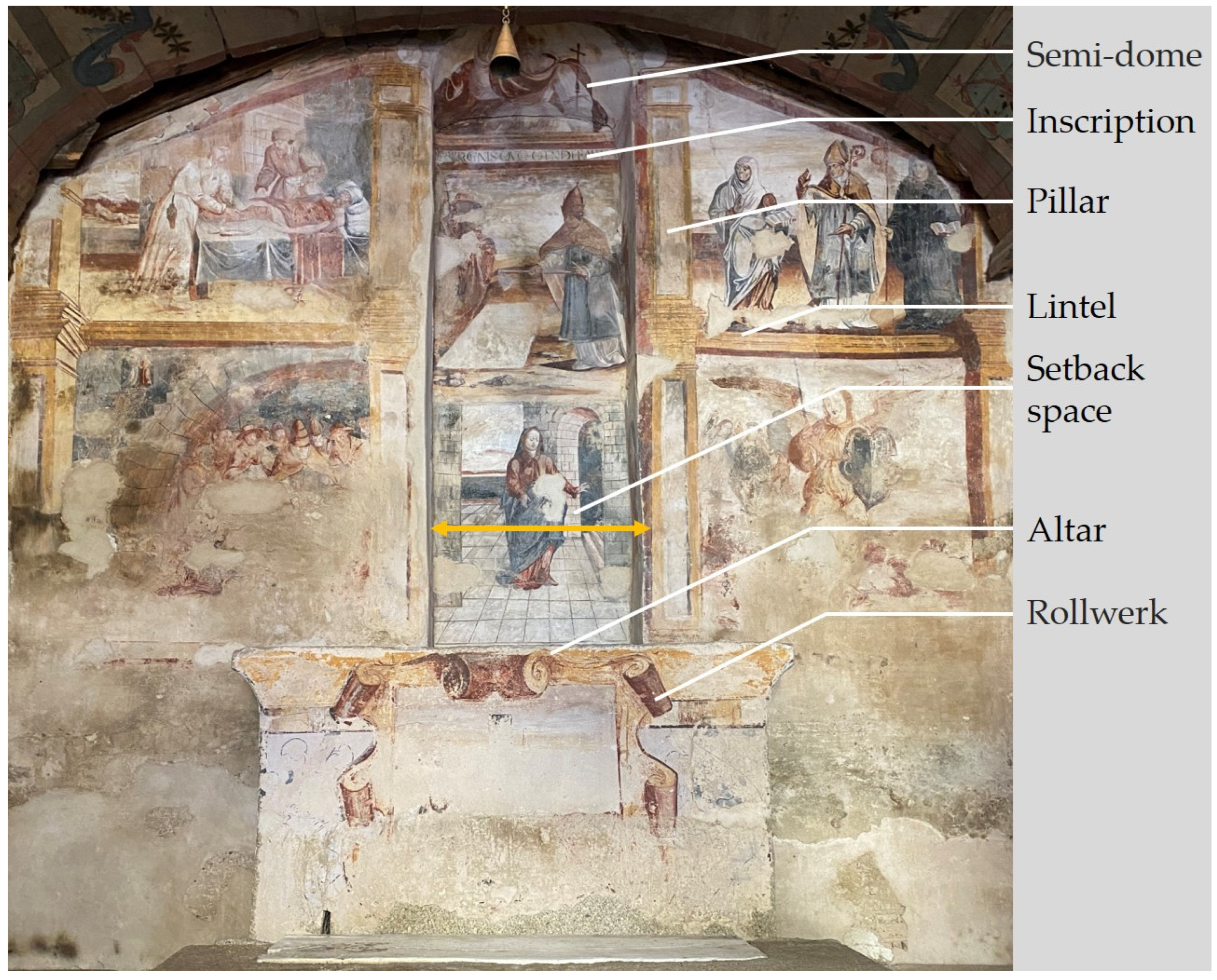
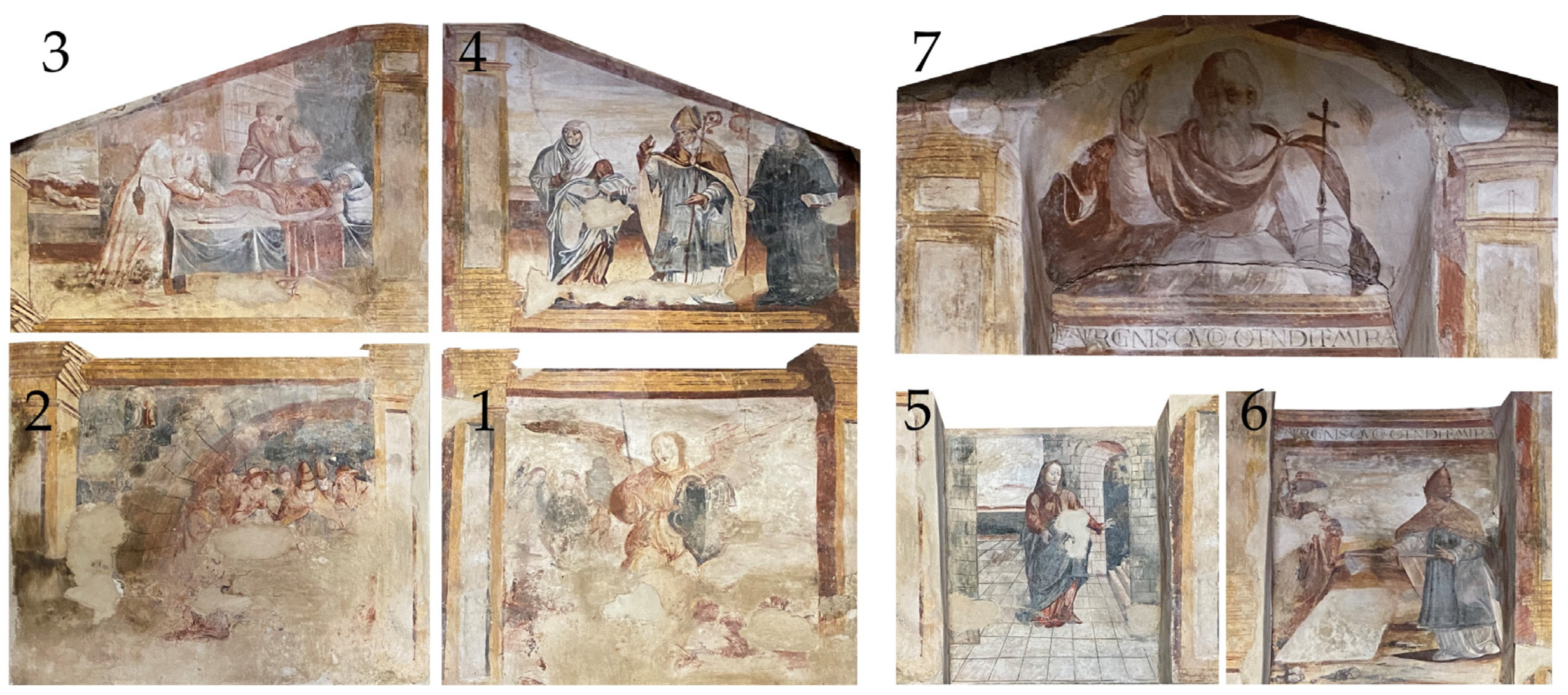
3.1. Mural Painting Iconographic Interpretation
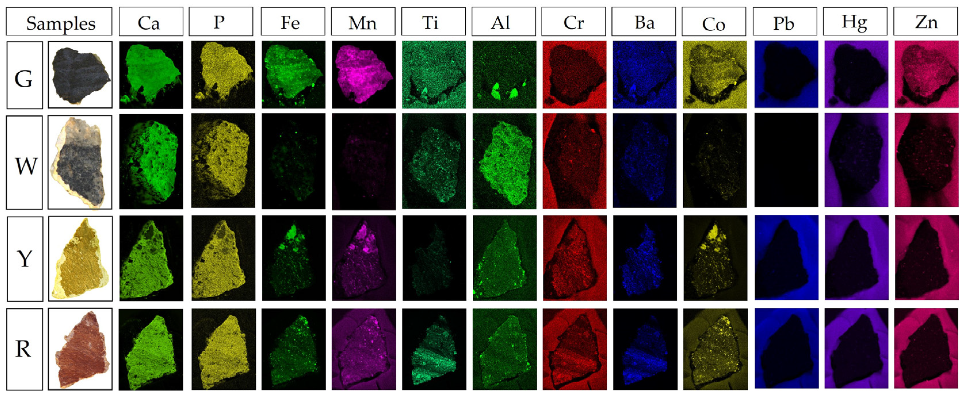
3.2. Micro X-ray Fluorescence Mapping
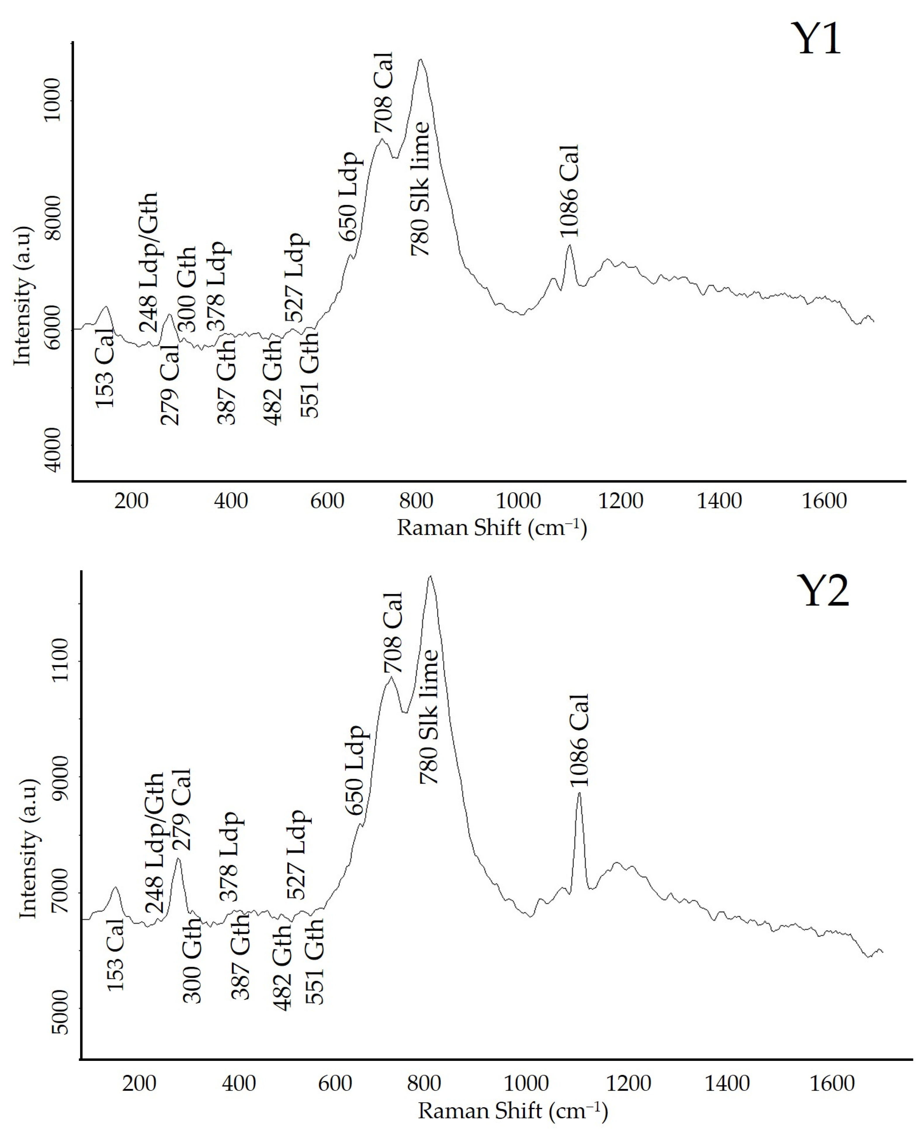
3.3. Portable i-Raman
3.3.1. Grey
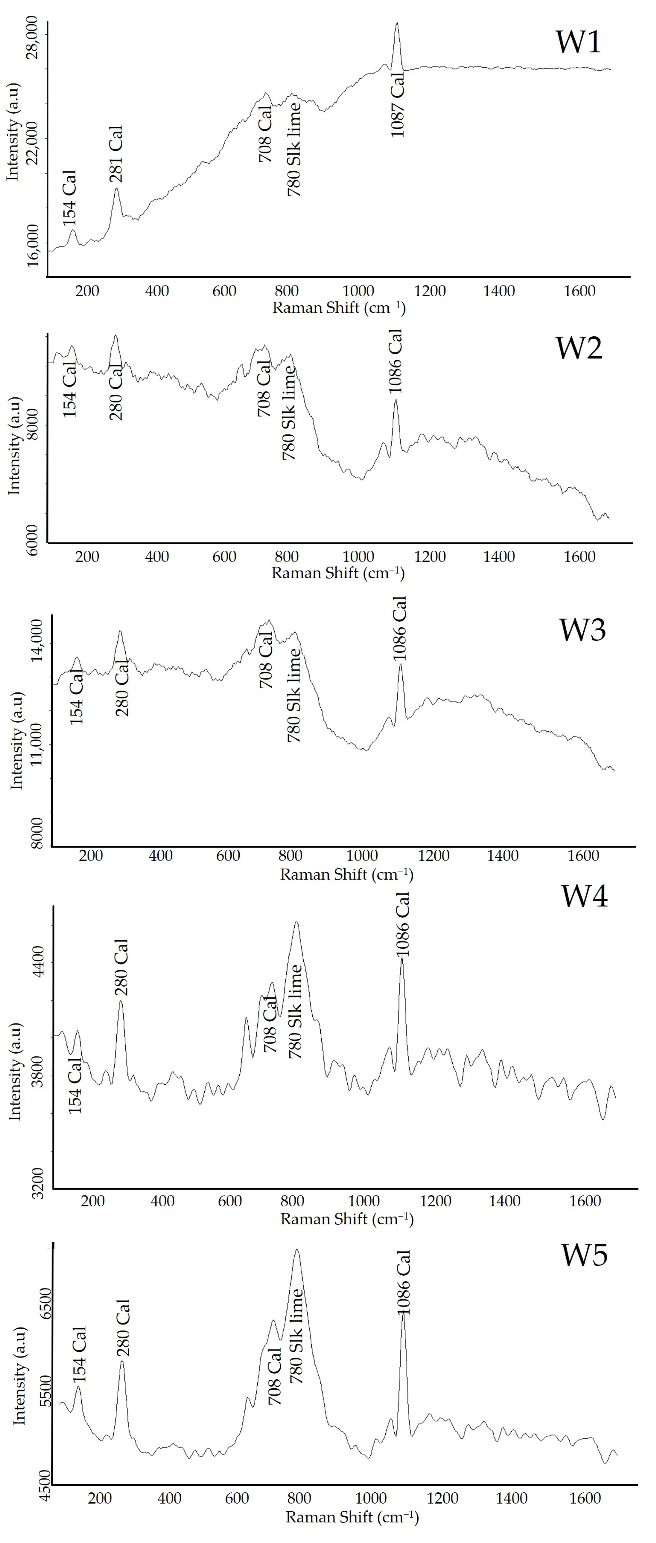
3.3.2. White
3.3.3. Yellow
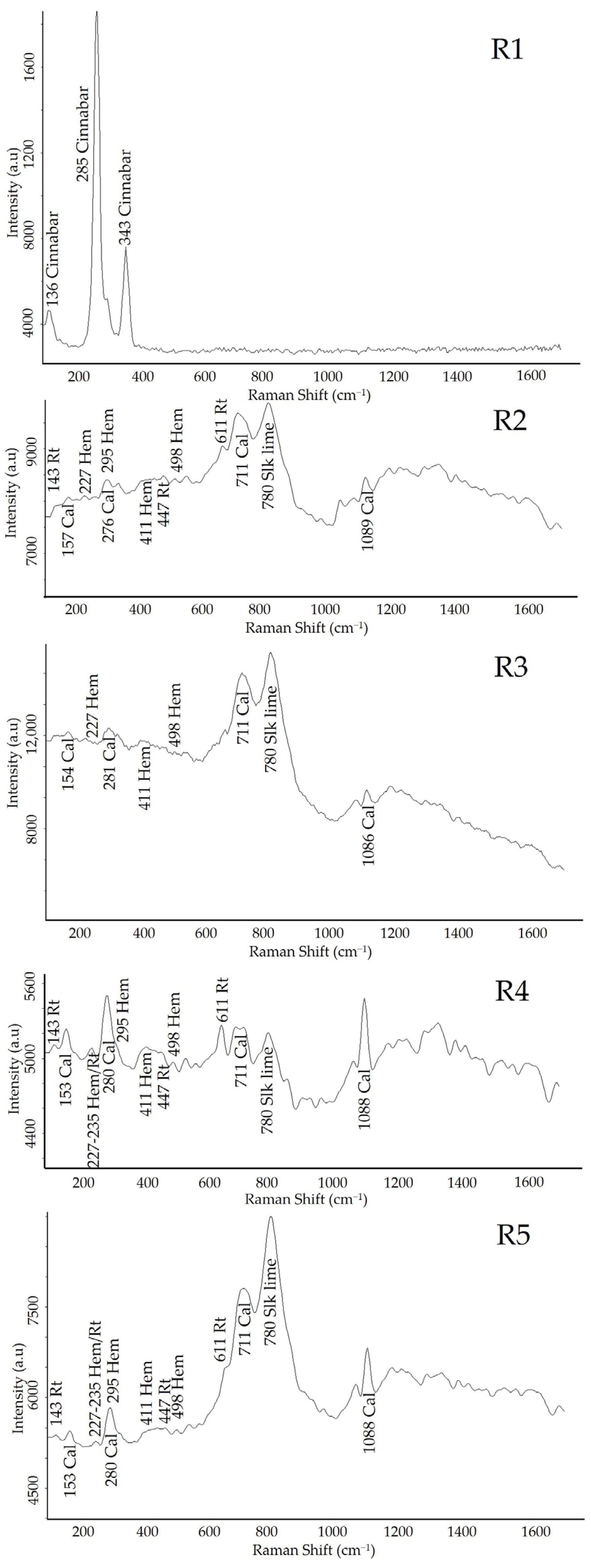
3.3.4. Red
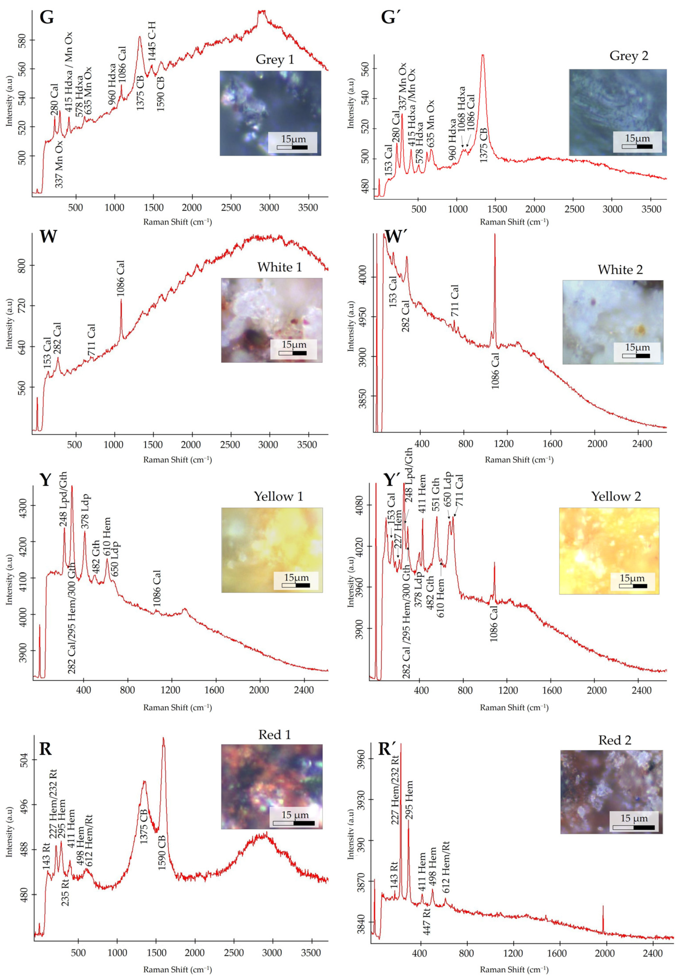
3.4. Confocal Raman Microscope
4. Discussion
5. Conclusions
Author Contributions
Funding
Data Availability Statement
Acknowledgments
Conflicts of Interest
References
- Hernanz, A.; Bratu, I.; Marutoiu, O.F.; Gavira-Vallejo, J.M.; Edwards, H.G.M. Micro-Raman spectroscopic investigation of external wall paintings from St. Dumitru’s Church, Suceava. Romania. Anal. Bioanal. Chem. 2008, 392, 263–268. [Google Scholar] [CrossRef]
- Li, Y.; Wang, F.; Ma, J.; He, K.; Zhang, M. Study on the pigments of Chinese architectural colored drawings in the Altar of Agriculture (Beijing, China) by portable Raman spectroscopy and ED-XRF spectrometers. Vib. Spectrosc. 2021, 116, 103291. [Google Scholar] [CrossRef]
- Marrocchino, E.; Telloli, C.; Paletta, M.G.; Leis, M.; Vaccaro, C. The mural paintings of the cloister in the Certosa di Calci, Pisa. J. Archaeol. Sci. 2022, 43, 103461. [Google Scholar] [CrossRef]
- Tsantini, E.; Minami, T.; Takahashi, K.; Cau Ontiveros, M.Á. Analysis of sulphur isotopes to identify the origin of cinnabar in the Roman wall paintings from Badalona (Spain). J. Archaeol. Sci. 2018, 18, 300–307. [Google Scholar] [CrossRef]
- Lofrumento, C.; Ricci, M.; Bachechi, L.; De Feo, D.; Castelluccie, M. The first spectroscopic analysis of Ethiopian prehistoric rock painting. J. Raman Spectrosc. 2012, 43, 809–816. [Google Scholar] [CrossRef]
- Veneranda, M.; Prieto-Taboada, N.; Fdez-Ortiz de Vallejuelo, S.; Maguregui, M.; Morillas, H.; Marcaida, I.; Castro, K.; Garcia-Diego, F.J.; Osanna, M.; Madariaga, J.M. In-situ multi-analytical approach to analyze and compare the degradation pathways jeopardizing two murals exposed to different environments (Ariadne House, Pompeii, Italy). Spectrochim. Acta Part A 2018, 203, 201–209. [Google Scholar] [CrossRef]
- Liu, Z.; Yang, R.; Wang, W.; Xu, W.; Zhang, M. Multi-analytical approach to the mural painting from an ancient tomb of Ming Dynasty in Jiyuan. China: Characterization of materials and techniques. Spectrochim. Acta Part A 2022, 279, 121419. [Google Scholar] [CrossRef]
- Priego, E.; Herráez, J.; Denia, J.L.; Navarro, P. Technical study for restoration of mural paintings through the transfer of a photographic image to the vault of a church. J. Cult. Heritage. 2022, 58, 112–121. [Google Scholar] [CrossRef]
- Buzgar, N.; Buzatu, A.; Apopei, A.I.; Cotiugă, V. In situ Raman spectroscopy at the Voroneţ Monastery (16th century, Romania): New results for green and blue pigments. Vib. Spectrosc. 2014, 72, 142–148. [Google Scholar] [CrossRef]
- He, J.; Zhou, W.; Hu, D.; Liu, S.; Otero, J.; Rodriguez-Navarro, C. A multi-analytical approach for the characterization of materials, manufacturing process and damage mechanisms of wall paintings in Samye Temple, Tibet. Dyes Pigm. 2022, 207, 110704. [Google Scholar] [CrossRef]
- Gil, M.; Carvalho, M.L.; Seruya, A.; Ribeiro, I.; Queralt, I.; Candeias, A.E.; Mirao, J. Limewashing paintings in Alentejo urban heritage: Pigment characterization and differentiation by WDXRF and XRD. Appl. Phys. A Mater. Sci. Process. 2007, 90, 49–54. [Google Scholar] [CrossRef]
- Gil, M.; Manso, M.; Pessanha, S.; Manhita, A.; Cardoso, A.; Nunes, M.; Relvas, C.; Monteiro, P.; Pereira, C.; Mirão, J.; et al. Old masters under the microscope. Technical and material comparison of a 17th c. mural and panel painting assigned to José de Escovar in Southern Portugal. Microchem. J. 2020, 153, 104396. [Google Scholar] [CrossRef]
- Salavessa, E.; Candeias, A.; Mirão, J.; Sousa, L.M.O.; Duarte, N.; Jalali, S.; Salgueiro, J. 19th century coloured stuccos and plasters from Grilos’ Church (Oporto, Portugal): Materials and techniques employed. Color Res. Appl. 2016, 41, 246–251. [Google Scholar] [CrossRef]
- Rosado, T.; Silva, M.; Dias, L.; Candeias, A.; Gil, M.; Mirão, J.; Pestana, J.; Caldeira, A.T. Microorganisms and the integrated conservation-intervention process of the renaissance mural paintings from Casas Pintadas in Évora—Know to act, act to preserve. J. King Saud Univ. Sci. 2017, 29, 478–486. [Google Scholar] [CrossRef]
- Freire-Lista, D.M.; Vidal Gonçalves, G.; Vazquez, P. Weathering Detection of Granite from Three Asynchronous Historical Quarries of Sabrosa Municipality (North Portugal). J. Cult. Heritage. 2022, 58, 199–208. [Google Scholar] [CrossRef]
- Freire-Lista, D.M.; Becerra Becerra, J.E.; Simões de Abreu, M. The historical quarry of Pena (Vila Real, north of Portugal): Associated cultural heritage and reuse as a geotourism resource. Resour. Policy. 2022, 75, 102528. [Google Scholar] [CrossRef]
- Campos, B.; Marco, A.; Freire-Lista, D.M.; Durãe, N.; Silvestre-Albero, J.; Silva, E.F.; Vieira, E.; Pintado, M.; Moreira, P.R. Rare Biogeochemical Phenomenon Associated to Manganese Patinas on Mural Painting and Granite Ashlars. Coatings. 2021, 11, 917. [Google Scholar] [CrossRef]
- Lourenço, P.B.; Roque, J.A. Simplified indexes for the seismic vulnerability of ancient masonry buildings. Constr. Build. Mater. 2006, 20, 200–208. [Google Scholar] [CrossRef]
- Freire-Lista, D.M.; Campos, B.B.; Moreira, P.; Ramil, A.; Lopez Díaz, A. Building Granite Characterisation, Construction Phases, Mason’s Marks and Glyptography of Nossa Senhora de Guadalupe Church, Mouçós e Lamares, Galicia-North Portugal Euroregion. Geoheritage. 2023, 15, 24. [Google Scholar] [CrossRef]
- Freire-Lista, D.M.; Campos, B.B.; Costa, R.; Sanjurjo-Sánchez, J. Main building granite of São Tiago de Folhadela Parish Church (North of Portugal). Petrography, glyptography, construction phases and decay by pyrite oxidation. Constr. Build. Mater. 2022, 350, 128904. [Google Scholar] [CrossRef]
- Dal Fovo, A.; Mazzinghi, A.; Omarini, S.; Pampaloni, E.; Ruberto, C.; Striova, J.; Fontana, R. Non-invasive mapping methods for pigments analysis of Roman mural paintings. J. Cult. Heritage 2020, 43, 311–318. [Google Scholar] [CrossRef]
- Oriols, N.; Salvadó, N.; Pradell, T.; Jiménez, N.; Juanhuix, J.; Buti, S. The three-tone system in Sant Climent de Taüll wall paintings: An imprint of medieval treatises. J. Cult. Heritage. 2023, 62, 114–123. [Google Scholar] [CrossRef]
- Pérez-Diez, S.; Caruso, F.; Frine Nardini, E.; Stollenwerk, M.; Maguregui, M. Secco painting technique revealed in non-restored Pompeian murals by analytical and imaging techniques. Microchem. J. 2023, 194, 109365. [Google Scholar] [CrossRef]
- Oriols, N.; Salvadó, N.; Pradell, T.; Butí, S. Amorphous calcium carbonate (ACC) in fresco mural paintings. Microchem. J. 2020, 154, 104567. [Google Scholar] [CrossRef]
- Costantini, I.; Aramendia, J.; Tomasini, E.; Castro, K.; Madariaga, J.M.; Arana, G. Detection of unexpected copper sulfate decay compounds on late Gothic mural paintings: Assessing the threat of environmental impact. Microchem. J. 2021, 169, 106542. [Google Scholar] [CrossRef]
- Ortega-Ramírez, J.; Bano, M.; Villa Alvarado, L.A.; Medellín Martínez, D.; Rivero-Chong, R.; Motolinía-Temol, C.L. High-resolution 3-D GPR applied in the diagnostic of the detachment and cracks in pre-Hispanic mural paintings at “Templo Rojo”, Cacaxtla, Tlaxcala, Mexico. J. Cult. Heritage. 2021, 50, 61–72. [Google Scholar] [CrossRef]
- Isola, D.; Bartoli, F.; Casanova Municchia, A.; Ju Lee, H.; Hye Jeong, S.; Chung, Y.J.; Caneva, G. Green biocides for the conservation of hypogeal mural paintings raised from Western and Eastern traditions: Evaluation of interference on pigments and substrata and multifactor parameters affecting their activity. J. Cult. Heritage. 2023, 61, 116–126. [Google Scholar] [CrossRef]
- Caneva, G.; Bartoli, F.; Fontani, M.; Mazzeschi, D.; Visca, P. Changes in biodeterioration patterns of mural paintings: Multi-temporal mapping for a preventive conservation strategy in the Crypt of the Original Sin (Matera, Italy). J. Cult. Heritage. 2019, 40, 59–68. [Google Scholar] [CrossRef]
- Tomasini, E.; Costantini, I.; Careaga, V.; Rua Landa, C.; Castro, K.; Madariaga, J.M.; Maier, M.; Siracusano, G. Identification of pigments and binders of a 17th century mural painting (Bolivia). New report on pigments associated with Andean minerals. J. Cult. Heritage. 2023, 62, 206–216. [Google Scholar] [CrossRef]
- Akyuz, S.; Akyuz, T.; Basaran, S.; Kocabas, I.; Gulec, A.; Cesmeli, H.; Ucar, B. FT-IR and EDXRF analysis of wall paintings of ancient Ainos Hagia Sophia Church. J. Mol. Struct. 2009, 924–926, 400–403. [Google Scholar] [CrossRef]
- Crupi, V.; Allodi, V.; Bottari, C.; D’Amico, F.; Galli, G.; Gessini, A.; La Russa, M.F.; Longo, F.; Majolino, D.; Mariotto, G.; et al. Spectroscopic investigation of Roman decorated plasters by combining FT-IR, micro-Raman and UV-Raman analyses. Vib. Spectrosc. 2016, 83, 78–84. [Google Scholar] [CrossRef]
- Lazic, V.; Fantoni, R.; Falzone, S.; Gioia, C.; Loreti, E.M. Stratigraphic characterization of ancient Roman frescos by laser induced breakdown spectroscopy and importance of a proper choice of the normalizing lines. Spectrochim. Acta Part B. 2020, 168, 105853. [Google Scholar] [CrossRef]
- Daniilia, S.; Minopoulou, E.; Demosthenous, F.D.; Karagiannis, G. A comparative study of wall paintings at the Cypriot monastery of Christ Antiphonitis: One artist or two. J. Archaeol. Sci. 2008, 35, 1695–1707. [Google Scholar] [CrossRef]
- Cerrato, E.J.; Cosano, D.; Esquivel, D.; Otero, R.; Jimémez-Sanchidrián, C.; Ruiz, J.R. A multi analytical study of a wall painting in the Satyr domus in Córdoba, Spain. Spectrochim. Acta Part A. 2020, 232, 118148. [Google Scholar] [CrossRef]
- Almaviva, S.; Fantoni, R.; Colao, F.; Piua, A.; Bisconti, F.; Fiocchi Nicolai, V.; Romani, M.; Cascioli, S.; Bellagamba, S. LIF/Raman/XRF non-invasive microanalysis of frescoes from St. Alexander catacombs in Rome. Spectrochim. Acta Part A Mol. Biomol. Spectrosc. 2018, 201, 207–215. [Google Scholar] [CrossRef]
- Fico, D.; Pennetta, A.; Rella, G.; Savino, A.; Terlizzi, V.; De Benedetto, G.E. A combined analytical approach applied to medieval wall paintings from Puglia (Italy): The study of painting techniques and its conservation state. J. Raman Spectrosc. 2016, 47, 321–328. [Google Scholar] [CrossRef]
- Ziemann, M.A.; Madariaga, J.M. Applications of Raman spectroscopy in art and archaeology. J. Raman Spectrosc. 2020, 52, 137–142. [Google Scholar] [CrossRef]
- Nosenko, V.V.; Yaremko, A.M.; Dzhagan, V.M.; Vorona, I.P.; Romanyuk, Y.A.; Zatovsky, I.V. Nature of some features in Raman spectra of hydroxyapatite-containing materials. J. Raman Spectrosc. 2016, 47, 726–730. [Google Scholar] [CrossRef]
- Bonnat, G.F.; Desimone, P.M.; Martínez, G.A.; Mazzanti, D.L. Raman spectroscopy study of ca 12.000 yrs cal BP lithic artifacts adhesions from archaeological sites in the Pampas region, Argentina. J. Archaeol. Sci. Rep. 2021, 38, 103101. [Google Scholar] [CrossRef]
- Bernardini, S.; Bellatreccia, F.; Casanova Municchia, A.; Della Ventura, G.; Sodo, A. Raman spectra of natural manganese oxides. J. Raman Spectrosc. 2019, 50, 873–888. [Google Scholar] [CrossRef]
- Gunasekaran, S.; Anbalagan, G.; Pandi, S. Raman and infrared spectra of carbonates of calcite structure. J. Raman Spectrosc. 2006, 37, 892–899. [Google Scholar] [CrossRef]
- Edwards, H.G.M.; Middleton, P.S.; Jorge Villar, S.E.; de Faria, D.L.A. Romano-British wall-paintings II: Raman spectroscopic analysis of two villa sites at Nether Heyford, Northants. Anal Chim Acta. 2003, 448, 211–221. [Google Scholar] [CrossRef]
- Guglielmi, V.; Andreoli, M.; Comite, V.; Baroni, A.; Fermo, P. The combined use of SEM-EDX, Raman, ATR-FTIR and visible reflectance techniques for the characterisation of Roman wall painting pigments from Monte d’Oro area (Rome): An insight into red, yellow and pink shades. Environ Sci Pollut Res. 2022, 29, 29419–29437. [Google Scholar] [CrossRef]
- Edwards, H.G.M. Analytical Raman spectroscopic discrimination between yellow pigments of the Renaissance. Spectrochim. Acta Part A 2011, 80, 14–20. [Google Scholar] [CrossRef]
- Botticelli, M.; Maras, A.; Candeias, A. μ-Raman as a fundamental tool in the origin of natural or synthetic cinnabar: Preliminary data. J. Raman Spectrosc. 2019, 51, 1470–1479. [Google Scholar] [CrossRef]
- Argote, D.L.; Torres, G.; Hernández-Padrón, G.; Ortega, V.; López-García, P.A.; Castaño, V.M. Cinnabar, hematite and gypsum presence in mural paintings in Teotihuacan, Mexico. J. Archaeol. Sci. Rep. 2020, 32, 102375. [Google Scholar] [CrossRef]
- Gao, Q.; Wu, X.; Fan, Y.; Zhou, X. Low temperature synthesis and characterization of rutile TiO2-coated mica–titania pigments. Dyes Pigm. 2012, 95, 534–539. [Google Scholar] [CrossRef]
- Tang, W.; Xu, W.; Zhong, M.; Zhang, Z. Slightly doped hydroxyapatite pigments of subtractive color with high near-infrared reflectance. J. Solid State Chem. 2023, 322, 123947. [Google Scholar] [CrossRef]
- Daveri, A.; Malagodi, M.; Vagnini, M. The bone black pigment identification by noninvasive, in situ infrared reflection spectroscopy. J. Anal. Methods Chem. 2018, 1–8, 6595643. [Google Scholar] [CrossRef]
- Sauer, G.R.; Zunic, W.B.; Durig, J.R.; Wuthier, R.E. Fourier transform Raman spectroscopy of synthetic and biological calcium phosphates. Calcif. Tissue Int. 1994, 54, 414–420. [Google Scholar] [CrossRef]
- Tomasini, E.; Siracusano, G.; Maier, M.S. Spectroscopic, morphological and chemical characterization of historic pigments based on carbon. Paths for the identification of an artistic pigment. Microchem. J. 2012, 102, 28–37. [Google Scholar] [CrossRef]
- Bordes, L.; Prinsloo, L.; Fullagar, R.; Sutikna, T.; Hayes, E.; Jatmiko, R.; Wahyu Saptomo, E.; Tocheri, M.; Roberts, R. Viability of Raman microscopy to identify micro-residues related to tool-use and modern contaminants on prehistoric stone artefacts. J. Raman Spectrosc. 2017, 48, 1212–1221. [Google Scholar] [CrossRef]
- Edwards, H.G.M.; Farwell, D.W.; de Faria, D.L.A.; Rozenberg, S. Raman spectroscopic study of red pigment and fresco fragments from King Herod’s Palace at Jericho. J. Raman Spectrosc. 1999, 30, 361–366. [Google Scholar] [CrossRef]
- Edwards, H.G.M.; Farwell, D.W.; de Faria, D.L.A.; Monteiro, A.M.F.; Afonso, M.C.; De Blasis, P.; Eggers, S. Raman spectroscopic study of 3000-year-old human skeletal remains from a sambaqui, Santa Catarina, Brazil. J. Raman Spectrosc. 2001, 32, 17–22. [Google Scholar] [CrossRef]
- Schmid, T.; Dariz, P. Shedding light onto the spectra of lime: Raman and luminescence bands of CaO, Ca (OH)2 and CaCO3. J. Raman Spectrosc. 2015, 46, 141–146. [Google Scholar] [CrossRef]
- Edwards, H.G.M.; Farwell, D.W. The conservational heritage of wall paintings and buildings: An FT- Raman spectroscopic study of prehistoric, Roman, mediaeval and Renaissance lime substrates and mortars. J. Raman Spectrosc. 2008, 39, 985–992. [Google Scholar] [CrossRef]
- Iriarte, M.; Hernanz, A.; Gavira-Vallejo, J.M.; Alcolea-González, J.; de Balbín-Behrmann, R. μ-Raman spectroscopy of prehistoric paintings from the El Reno cave (Valdesotos, Guadalajara, Spain). J. Archaeol. Sci. Rep. 2017, 14, 454–460. [Google Scholar] [CrossRef]
- Escribano, R.; Sloan, J.J.; Siddique, N.; Sze, N.; Dudev, T. Raman spectroscopy of carbon-containing particles. Vib. Spectrosc. 2001, 26, 179–186. [Google Scholar] [CrossRef]
- Tomasini, E.P.; Halac, E.B.; Reinoso, M.; Di Liscia, E.J.; Maier, M.S. Micro-Raman spectroscopy of carbon-based black pigments. J. Raman Spectrosc. 2012, 43, 1671–1675. [Google Scholar] [CrossRef]
- Neiman, M.K.; Balonis, M.; Kakoulli, I. Cinnabar alteration in archaeological wall paintings: An experimental and theoretical approach. Appl. Phys. 2015, 121, 915–938. [Google Scholar] [CrossRef]


| Colour | Sample | Location | Panel Number |
|---|---|---|---|
| Grey | G1 | Our Lady of the Snows veil | Panel 5 |
| G2 | Grey of the open space in the door | Panel 5 | |
| G3 | Tiles cement joint | Panel 5 | |
| G4 | Lintel inscription, letter D | Between panel 6 and 7 | |
| G5 | Lintel inscription, letter F | Between panel 6 and 7 | |
| White | W1 | Robe of woman holding a book | Panel 4 |
| W2 | Book page held by a woman | Panel 4 | |
| W3 | Bishop’s chasuble | Panel 4 | |
| W4 | God the Father’s clothing | Panel 7 | |
| W5 | God the Father’s clothing | Panel 7 | |
| Yellow | Y1 | Centre-right lintel capital | Between panel 4 and 6 |
| Y2 | Ground | Panel 6 | |
| Red | R1 | Skirt of Our Lady of the Snows | Panel 5 |
| R2 | Our Lady of the Snows top of dress | Panel 5 | |
| R3 | Centre-right pillar | Between panel 1 and 5 | |
| R4 | Centre-right pillar | Between panel 1 and 5 | |
| R5 | Red robe of God the Father | Panel 7 |
Disclaimer/Publisher’s Note: The statements, opinions and data contained in all publications are solely those of the individual author(s) and contributor(s) and not of MDPI and/or the editor(s). MDPI and/or the editor(s) disclaim responsibility for any injury to people or property resulting from any ideas, methods, instructions or products referred to in the content. |
© 2023 by the authors. Licensee MDPI, Basel, Switzerland. This article is an open access article distributed under the terms and conditions of the Creative Commons Attribution (CC BY) license (https://creativecommons.org/licenses/by/4.0/).
Share and Cite
Freire-Lista, D.M.; Vázquez, E.; Barreiro Castro, P.; Salavessa, E.; Costa, M.d.R.; Moreira, R.; López, A.J. Mural Paintings Characterisation Using X-ray Fluorescence and Raman Spectroscopy—A Case Study: Nossa Senhora das Neves Chapel, Vilar de Perdizes, Galicia—North Portugal Euroregion. Heritage 2023, 6, 7277-7292. https://doi.org/10.3390/heritage6120382
Freire-Lista DM, Vázquez E, Barreiro Castro P, Salavessa E, Costa MdR, Moreira R, López AJ. Mural Paintings Characterisation Using X-ray Fluorescence and Raman Spectroscopy—A Case Study: Nossa Senhora das Neves Chapel, Vilar de Perdizes, Galicia—North Portugal Euroregion. Heritage. 2023; 6(12):7277-7292. https://doi.org/10.3390/heritage6120382
Chicago/Turabian StyleFreire-Lista, David M., Ezequiel Vázquez, Pablo Barreiro Castro, Eunice Salavessa, Maria do Rosário Costa, Rafael Moreira, and Ana J. López. 2023. "Mural Paintings Characterisation Using X-ray Fluorescence and Raman Spectroscopy—A Case Study: Nossa Senhora das Neves Chapel, Vilar de Perdizes, Galicia—North Portugal Euroregion" Heritage 6, no. 12: 7277-7292. https://doi.org/10.3390/heritage6120382