Amyloid must be understood as any substance that presents a green birefringence under polarized light, after Congo red staining, a fibrillar structure in electron microscopy. The deposit and infiltration of this substance constitutes Amyloidosis. Although at first it was believed that amyloid was a single substance, it is currently known that the proteins that constitute it are different, all similar to each other from a morphological point of view, but different from a biochemical point of view (
Piacenza 1992). Various proteins of this type have been identified in various combinations (immunoglobulins). The most important are amyloid L (AL) and amyloid A (AA). L amyloidosis derives from light chain immunoglobulins and is therefore usually associated with an abnormal functioning of plasma cells (
Prakash 1995), both localized in the lungs and as part of a systemic pathology such as multiple myeloma or macroglobulinemia. Amyloidosis A derives from an acute serum phase reagent (AAS) synthesized in the liver. The latter can form in various conditions, connective tissue diseases (in particular rheumatoid arthritis), chronic infections (in particular tuberculosis), bronchiectasis and some neoplasms (such as Hodgkin's lymphoma). Due to the great variety of clinical and pathological manifestations, amyloidosis has been classified above all in relation to its biochemical origin. The traditional classification provides for four main forms of disease: Primary amyloidosis, in which no associated pathology is recognized or there is a concomitant disorder of plasma cells (most often multiple myeloma); secondary amyloidosis, in which there is an underlying or identifiable chronic inflammatory disease, such as tuberculosis, bronchiectasis, rheumatic disease; hereditary amyloidosis, a relatively rare form that can be localized in a specific tissue such as a nerve; Senile amyloidosis which affects many organs and tissues and is usually found in people over seventy (
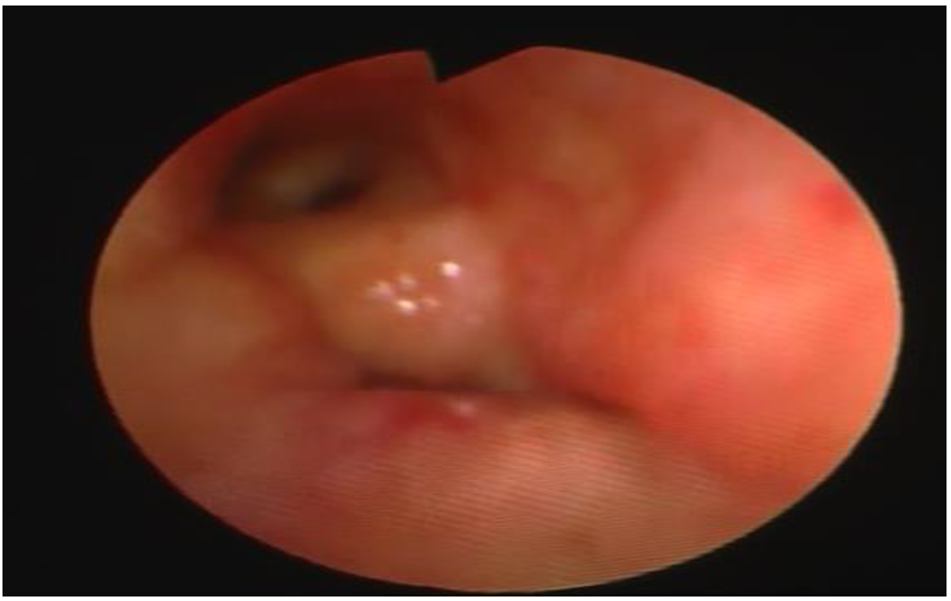
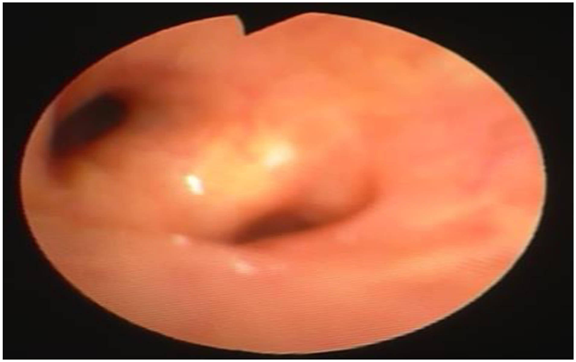
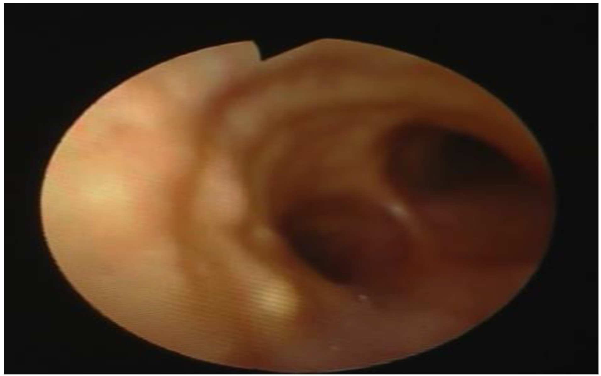
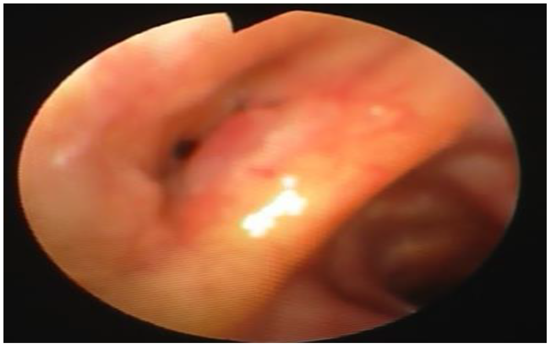
Fraser et al. 2006). The involvement of the respiratory tract can be isolated or integrated in the framework of a systemic amyloidosis that involves all organs. The localized form is the most frequent. Respiratory amyloidosis can take three main and characteristic forms: tracheobronchial, nodular parenchymal (the nodules can be solitary located at the periphery of the lung or more often multiple), diffuse parenchymal (almost exclusively affects the interstitium and is characterized by an infiltration of the alveolar wall by the amyloid substance, especially around the capillaries). The tracheobronchial form is probably the most frequent. It manifests itself with the presence of multiple submucosal plaques in the lumen of the trachea and bronchi or more rarely with a pseudotumoral endobronchial mass of a friable consistency that is easy to bleed, with an irregular surface, generally of a pinkish-yellowish color (
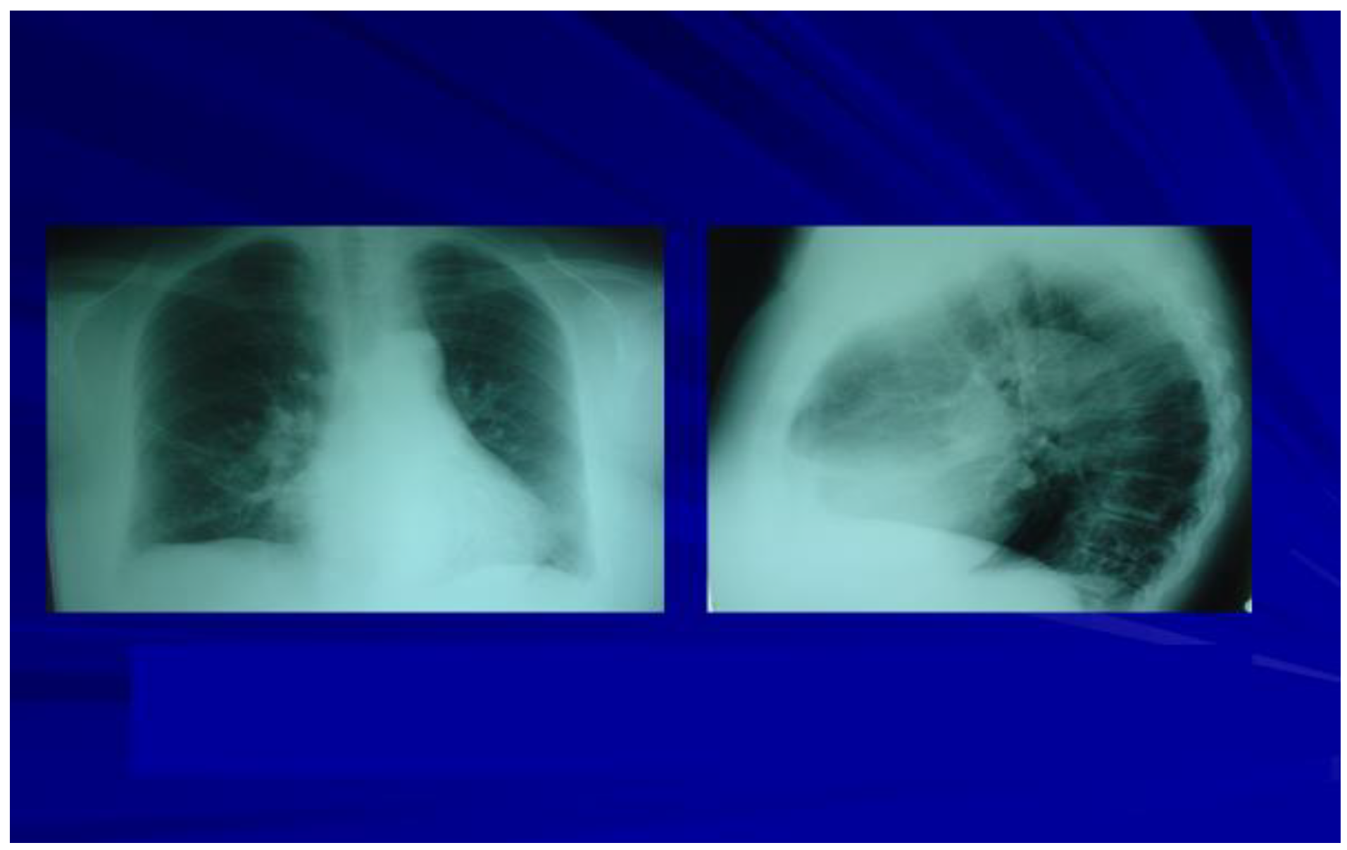
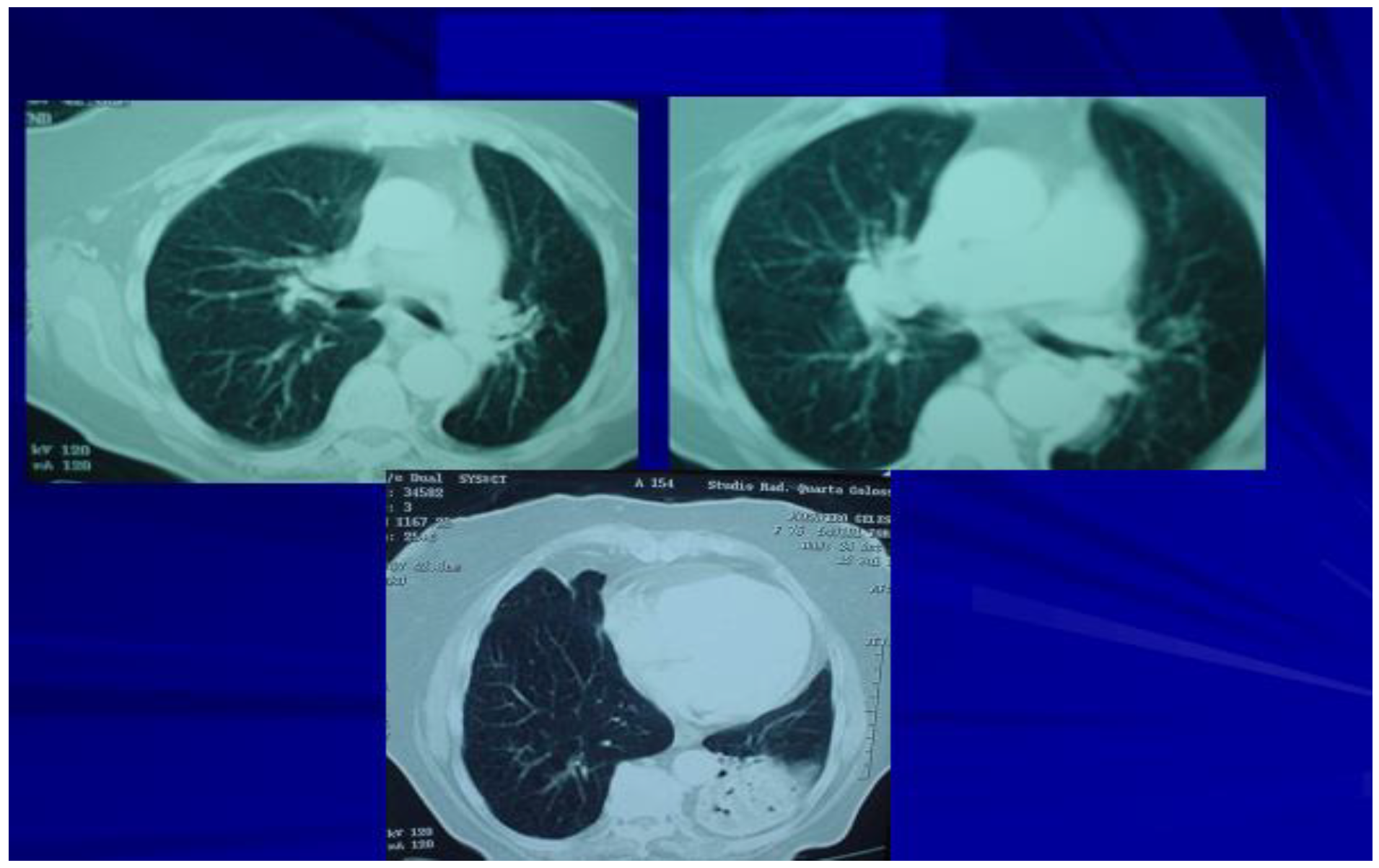
Piacenza 1992). Radiologically, the lesions of the respiratory tract are visible at the level of the trachea, the main bronchi and the lobar and segmental bronchi. They can be observed on standard radiological examination and better still on CT. They are characterized by a focal or diffuse thickening of the airways or they may present the appearance of multiple nodules that develop at the level of the tracheal wall, of the main, lobar, or segmental bronchi projecting into the lumen and can cause mild, moderate, or considerable entity. A rare and particular anomaly, called osteoplastic tracheopathy (characterized by diffuse or nodular ossification of the tracheobronchial mucosa) can be similar to tracheobronchial amyloidosis. As evidenced by some observations of amyloid deposits with histological studies of these tracheae, it can be said that osteoplastic tracheopathy is a late stage in the evolution of tracheobronchial amyloidosis (
Grenier 1993).