Next Article in Journal
Habeas data and patient self-determination
Previous Article in Journal
Tracheobronchial Amyloidosis
 
 
Journal of Interdisciplinary Research Applied to Medicine is published by MDPI from Volume 6 Issue 1 (2026). Previous articles were published by another publisher in Open Access under a CC-BY (or CC-BY-NC-ND) licence, and they are hosted by MDPI on mdpi.com as a courtesy and upon agreement with University of Salento.
Article

A Case of Retroperitoneal Fibroid and Literature Review

by
Alessandro D’Amuri
1,
Federica Floccari
2,
Ottavia D’Oria
3,4,
Sarah Gustapane
3 and
Andrea Tinelli
3,5,6,*
1
Anatomic Pathology Unit, "A. Perrino" Hospital, Brindisi, Italy
2
Clinic Pathology Unit, "L. Bonomo" Hospital, Andria (BT), Italy
3
Department of Obstetrics and Gynecology, “Veris delli Ponti” Hospital, Scorrano, Lecce, Italy
4
Department of Medical and Surgical Sciences and Translational Medicine, PhD Course in “Translational Medicine and Oncology”, Sapienza University, Rome, Italy
5
Division of Experimental, Endoscopic Surgery, Imaging, Technology and Minimally Invasive Therapy, Vito Fazzi Hospital, Lecce, Italy
6
Laboratory of Human Physiology, Phystech BioMed School, Faculty of Biological and Medical Physics, Moscow Institute of Physics and Technology (State University), Dolgoprudny, Moscow Region, Russia
*
Author to whom correspondence should be addressed.
J. Interdiscip. Res. Appl. Med. 2020, 4(2), 45-48; https://doi.org/10.1285/i25327518v4i2p45
Published: 31 December 2020

Abstract

Uterine fibroids or leiomyomas are the most common benign female genital tumors, although this pathology can also manifest itself outside the uterus, as in the case we present, with a retroperitoneal development. In this article, authors showed a rare case of 81-year-old women, with a right pelvic mass and lower abdominal discomfort. Basing on a preoperative radiological imaging, clinicians oriented to a provisional diagnosis of mesenchymal neoplasm of an uncertain origin. During surgery in the retroperitoneal space, it was detected a huge mass close to the uterus, beneath the right broad ligament. After the opening of the retroperitoneal spaces, surgeons enucleated a well-demarcated tumor, measuring 12 x 7 cm in diameter and histopathological findings demonstrated a rare retroperitoneal uterine leiomyoma.

1. Introduction

Uterine fibroids or leiomyomas are the most common benign gynecological tumors, usually originating and included in the uterus. This is one of the most common female diseases and one of the most common cause of hysterectomies (Sparic et al. 2016). Generally, the retroperitoneal origin of a fibroid is very unusual.
Retroperitoneal fibroid has a marked morphologic similarity to uterine leiomyoma, by virtue of hyaline change end trabecular pattern of growth (Yüksel et al. 2020)
Nevertheless, a tumor in this retroperitoneal region with a leiomyomatosic appearance, has a differential diagnosis with a uterine leiomyoma extending posteriorly, a well-differentiated leiomyosarcoma, benign and a malignant gastrointestinal stromal tumor (GIST) (Barnaś et al. 2019).
This differential diagnosis can lead to make the misdiagnosis, or challenge the strategy of treatment of tumor, shifting towards very invasive and radical treatments.

2. Case presentation

We report the case of an 81-year-old woman, hospitalized for diagnostic investigation after incidental finding of a pelvic mass on ultrasonography and a successive computerized tomography (CT) imaging scan, assessing the presence of a wide solid mass of size 12 x 7 cm in the pelvic cavity.
At radiological appearance, this mass showed regular boundary, with isolated intralesional calcifications, inhomogeneous vascularity and hypodense areas peripherally. A CT guided mass biopsy was performed, with 3 samples of size 5 mm each. Those samples showed stromal frustules immunoreactive for smooth muscle actin (SMA) and negative for S-100 protein, CKAE1/AE3 and CD34 so they were made of smooth muscle.
Thus, the entire mass was surgically enucleated in laparotomy and transported to the pathological anatomy unit for complete histological evaluation.
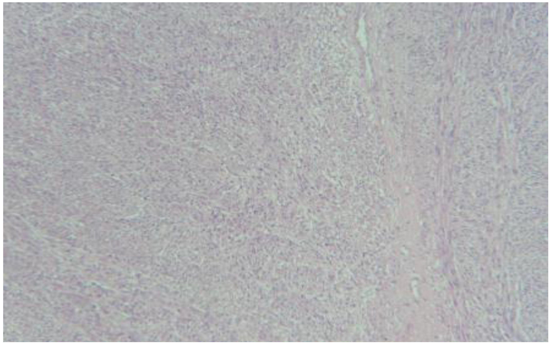
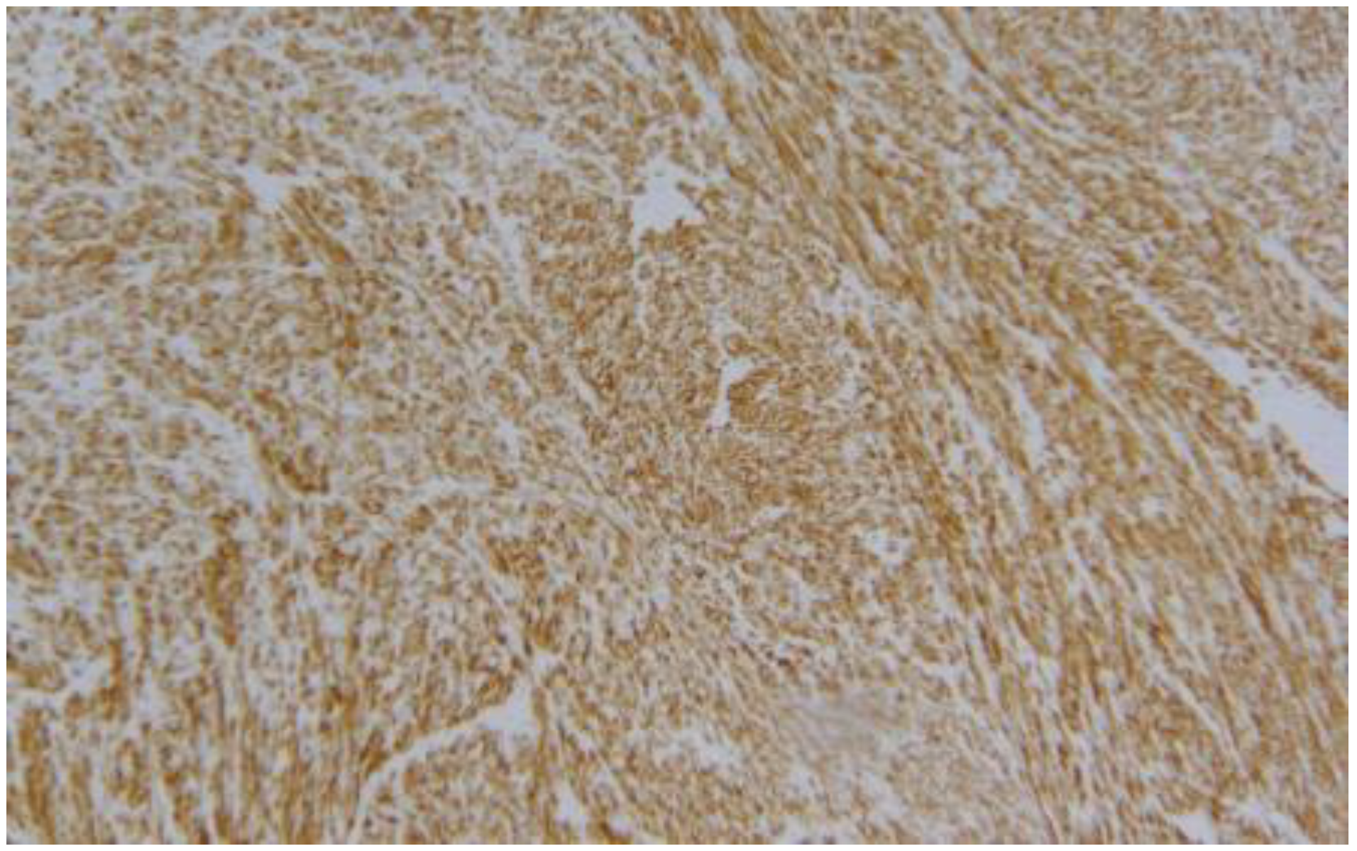
The morphological findings of the enucleated mass showed an irregular shape, with a compact, swirling and whitish cut surface (Figure 1). Hematoxylin and eosin staining revealed the intersecting fascicles of spindle cells (Figure 2), with no necrosis and mitotic activity. The immunohistochemical staining was positive for SMA and desmin (Figure 3) and negative for S-100 protein and CD117 (C-Kit). Therefore, the definitive diagnosis was a rare case of retroperitoneal fibroids or leiomyoma.
The patient’ postoperative course was uneventful, with a discharging after seven days after surgery. No several adverse events occurred. Patients follow up showed no evidence of recurrent retroperitoneal leiomyoma, until now.

3. Discussion

This clinical report represents a rare case of retroperitoneal leiomyoma, which has a hard preoperative diagnosis, by radiological imaging. A complete surgical removal is necessary for pathological diagnosis and treatment (Mahendru et al. 2012).
Uterine fibroids are the most common gynecological benign tumors, accounting up to 25% of the reproductive aged women, usually involving uterine body, but they may occur in any site of the uterus (Sparic et al. 2016).
They have an unusual growth pattern and the incidence among primary retroperitoneal tumors has been reported as very low rate up to 1.2% (Naykı et al. 2014), located in the pelvis in the 73% of cases (Poliquin, Victory, and Vilos 2008). Retroperitoneal tumor is a rare condition, and the incidence of malignant retroperitoneal tumors is higher than benign tumors (Tantitamit et al. 2015). Dalen et al. (Dalen et al. 2001) reported that the incidence of malignant retroperitoneal tumors was 80% of all primary non-visceral tumors in the retroperitoneum and sarcomas such as liposarcoma or leiomyosarcoma comprised one third of retroperitoneal malignant tumors.
Several theories of the pathogenesis of extrauterine leiomyoma including retroperitoneal leiomyoma were suggested as followed: benign metastasizing leiomyoma, disseminated peritoneal leiomyomatosis, intravenous dissemination, parasitic leiomyoma, and so on (Barnaś et al. 2019). Of these theories, "parasitic" leiomyoma was first described by Kelly and Cullen (Howard Atwood and Cullen 1909) and it was defined as completely separation of the pedunculated leiomyoma from uterus, with receiving their blood supply from another source.
Recently, as the increasing morcellation during myomectomy or hysterectomy, the theory of "iatrogenic" parasitic leiomyoma was suggested, as tumor growing by the fibroids’ seeding during morcellation (Mynbaev et al. 2017).
Anyway, retroperitoneal parasitic leiomyoma is very rare condition. There are few reports of "parasitic myoma" and almost of these literatures are just case reports or small series.
Kho and Nezhat reported 12 cases of parasitic myoma in a single institution for 8 years. Of 12 cases of parasitic myoma, only 2 cases were found retroperitoneal space in this report (Kho and Nezhat 2009).
After a huge multidisciplinary clinical discussion on its origin, we thought that our reported case of retroperitoneal fibroid was consistent with a parasitic leiomyoma, since it had a separate blood supply, neither from uterine artery nor from ovarian artery. But patient had no evidence of concurrently uterine leiomyoma or no history of remote hysterectomy for fibroids or myomectomy, so the possibility of "iatrogenic" parasitic leiomyoma was clinically rejected.
Preoperative diagnosis of retroperitoneal leiomyoma can be very changeling, because of the rarity of this tumor. Common symptoms of retroperitoneal fibroids include abdominal discomfort, fatigue, backache, dyspareunia, and urinary and bowel complaints (Poliquin, Victory, and Vilos 2008).
About radiological preoperative diagnosis, pelvic magnetic resonance imaging (MRI) with contrast medium, is the most reliable technique for evaluating a retroperitoneal leiomyoma and for assess the anatomical location. But it could be difficult to differentiate a leiomyoma from a leiomyosarcoma basing on radiological imaging, such as pelvic CT scan or pelvic MRI alone (Karray et al. 2018). Some features of these retroperitoneal masses show an extensive central necrosis, invasive growth pattern, and heterogeneous appearance. They are helpful to differentiate a leiomyosarcoma from a leiomyoma (Mynbaev et al. 2020). In the literature there are cases diagnosed with fine needle aspiration biopsy [(DURSUN et al. 2005). The use of preoperative antigens, particularly Ca 125 and carcinoembryionic antigens, could be useful in patients with large tumors; in literature a case of CA-125 was reported to be high in leiomyomas 15 cm and above (Bischof et al. 1992).
Anyway, histomorphologic diagnosis is always mandatory for such cases, so as it is mandatory to preoperatively diagnose the retroperitoneal tumor as accurately as possible, since it can lead to an appropriate surgical method to remove the mass, as the problem is how to proceed, whether to radically remove the mass in laparotomy or to remove only the fibroid in laparoscopy (Tinelli and Farghaly 2018; Kondo et al. 2011). Moreover, preoperative radiological assessment of tumor vascularization is also necessary.
Surgery seems to be the only valid therapeutic option. Particularly, open surgery was the most used strategy in many reported cases. Large tumor size at the moment of diagnosis could be a contraindication to laparoscopic or robotic approach. Kondo (Tinelli and Farghaly 2018) reported a case of retroperitoneal fibroid where resection was done by laparoscopic approach. Age of the woman influences the therapeutic approach (hysterectomy vs leiomyoma resection). Ultrasonography-guided or CT-guided fine-needle aspiration or biopsy may be histologically beneficial for the expectant management of asymptomatic patients refusing operation (Yüksel et al. 2020; L. Max et al. 2017).
Regular follow-up is mandatory because malignant transformation cannot be excluded (Jeong 2014; Paal and Miettinen 2001), but the prognosis of these patients is good.

4. Conclusions

The retroperitoneal uterine fibroid or leiomyoma is a rare condition, very difficult to diagnose preoperatively. This difficulty obviously also reflects on the surgical methodology to be applied in the removal: laparotomic radicality or minimally invasive surgery in case of fibroid (Billings, Folpe, and Weiss 2001).
A careful preoperative analysis of radiological imaging scan, by CT and MRI, or by intraoperative surgical and histological findings at frozen sections, may be helpful to diagnose accurately and to decide the strategy of treatment of retroperitoneal tumor.

References

  1. Barnaś, Edyta, Renata Raś, Joanna Skręt-Magierło, Mariusz Wesecki, Justyna Filipowska, Mariusz Książek, Andrzej Skręt, and Kazimierz Widenka. 2019. Natural History of Leiomyomas beyond the Uterus. Medicine 98, (25): e15877. [Google Scholar] [CrossRef] [Scilit]
  2. Billings, SD, AL Folpe, and SW Weiss. 2001. Do Leiomyomas of Deep Soft Tissue Exist? An Analysis of Highly Differentiated Smooth Muscle Tumors of Deep Soft Tissue Supporting Two Distinct Subtypes. Am J Surg Pathol 25, (9): 1134–42. [Google Scholar] [CrossRef] [Scilit]
  3. Bischof, P., M.A. Galfetti, J. Seydoux, J.U. von Hospenthal, and A. Campana. 1992. Peripheral CA 125 Levels in Patients with Uterine Fibroids. Human Reproduction 7, (1): 35–38. [Google Scholar] [CrossRef] [Scilit]
  4. Dalen, T, JW Coebergh, MK Casparie, CH Gimbrère, HJ Hoekstra, and BN Van Geel. 2001. Soft Tissue Sarcoma: The Predominant Primary Malignancy in the Retroperitoneum. Sarcoma 5: 5–8. [Google Scholar] [CrossRef] [Scilit]
  5. DURSUN, P., M. C. SALMAN, C. TASKIRAN, K. YÜCE, and A. AYHAN. 2005. Retroperitoneal Leiomyomatosis: A Case Report. International Journal of Gynecologic Cancer 15, (6). [Google Scholar] [CrossRef] [Scilit]
  6. Howard Atwood, Kelly, and Thomas Stephen Cullen. 1909. Myomata of the Uterus. In WB Saunders. Philadelphia. [Google Scholar]
  7. Jeong, Gui-Ae. 2014. Retroperitoneal Leiomyoma of the Uterus Mimicking Sarcoma in Perimenopausal Woman: Case Report. Journal of Menopausal Medicine 20, (3): 133. [Google Scholar] [CrossRef] [Scilit]
  8. Karray, O., R. Boulma, A. Abdi, A. Ben Miled, A. Dhaoui, N. Menif, K. Bellil, H. Khouni, and A. Chouchen. 2018. Management of a Giant Retroperitoneal Leiomyoma: A Case Report. Journal of Medical Case Reports 12, (1): 1–5. [Google Scholar] [CrossRef] [Scilit] [PubMed]
  9. Kho, Kimberly A., and Ceana Nezhat. 2009. Parasitic Myomas. Obstetrics & Gynecology 114, (3): 611–15. [Google Scholar] [CrossRef] [Scilit] [PubMed]
  10. Kondo, William, Revaz Botchorishvili, Fanny Desvignes, and Gérard Mage. 2011. Laparoscopic Management of a Pelvic Retroperitoneal Leiomyoma-Case Report. Gynecological Surgery 8, (2): 247–51. [Google Scholar] [CrossRef] [Scilit]
  11. L. Max, Almond, Warfield Adrian T, Desai Anant, Gourevitch David, and Ford Samuel J. 2017. Biphasic Malignant Tumours of the Abdominal Cavity. International Journal of Clinical Oncology. Springer Tokyo. [Google Scholar] [CrossRef] [Scilit]
  12. Mahendru, Rajiv, Geetinder Gaba, Shweta Yadav, Gurmeet Gaba, and Chinky Gupta. 2012. A Rare Case of Retroperitoneal Leiomyoma. Case Reports in Surgery 2012: 1–3. [Google Scholar] [CrossRef] [Scilit] [PubMed]
  13. Mynbaev, Ospan A., Antonio Malvasi, Sergei S. Simakov, and Andrea Tinelli. 2017. Comment on ‘Oestrogen-Induced Angiogenesis and Implantation Contribute to the Development of Parasitic Myomas after Laparoscopic Morcellation’. Reproductive Biology and Endocrinology. BioMed Central Ltd. [Google Scholar] [CrossRef] [Scilit]
  14. Mynbaev, Ospan A., Radmila Sparic, Michael Stark, Antonio Malvasi, Enrico Marinelli, Simona Zaami, and Andrea Tinelli. 2020. The Medical Device Applied to Uterine Fibroids Morcellation: Analysis of Critical Biological Issues and Drawbacks from A Medical-Legal Prospective. Current Pharmaceutical Design 26, (3): 318–25. [Google Scholar] [CrossRef] [Scilit] [PubMed]
  15. Naykı, Ümit, Cenk Naykı, Paşa Uluğ, Ismayil Yılmaz, Zeliha Cetin, Yusuf Yıldırım, Ümit Nayki, Yavuz Selim, and Mah Er. 2014. Case Report A Rare Case of a Giant Cystic Leiomyoma Presenting as a Retroperitoneal Mass. Iran J Reprod Med. Vol. 12. Shahid Sadoughi University of Medical Sciences and Health Services. [Google Scholar]
  16. Paal, E, and M Miettinen. 2001. Retroperitoneal Leiomyomas: A Clinicopathologic and Immunohistochemical Study of 56 Cases with a Comparison to Retroperitoneal Leiomyosarcomas. Am J Surg Pathol 25, (11): 1355–63. [Google Scholar] [CrossRef] [Scilit] [PubMed]
  17. Poliquin, Vanessa, Rahi Victory, and George A. Vilos. 2008. Epidemiology, Presentation, and Management of Retroperitoneal Leiomyomata: Systematic Literature Review and Case Report. Journal of Minimally Invasive Gynecology. Elsevier. [Google Scholar] [CrossRef] [Scilit]
  18. Sparic, Radmila, Ljiljana Mirkovic, Antonio Malvasi, and Andrea Tinelli. 2016. Epidemiology of Uterine Myomas: A Review. International Journal of Fertility and Sterility. Royan Institute (ACECR). [Google Scholar] [CrossRef] [Scilit]
  19. Tantitamit, Tanitra, Suttha Hamontri, Likit Rangsiratanakul, and Maysita Suksamarnwong. 2015. Pelvic Retroperitoneal Cellular Leiomyoma: A Case Report. Journal of the Medical Association of Thailand 98 (October): S160–64. [Google Scholar]
  20. Tinelli, Andrea, and Samir A. Farghaly. 2018. Morcellation of Occulted Sarcomas during Laparoscopic Myomectomy and Hysterectomy for Patients with Large Fibroid Uterus. Minerva Ginecologica. Edizioni Minerva Medica. [Google Scholar] [CrossRef] [Scilit]
  21. Yüksel, Dilek, Cigdem Kilic, Bahadır Kodal, Caner Cakir, Cigdem Güler Mesci, Nurettin Boran, and Fulya Kayıkçıoğlu. 2020. A RARE CASE OF A RETROPERITONEAL LEIOMYOMATOSIS. Journal of Gynecology Obstetrics and Human Reproduction April. , 101760. [Google Scholar] [CrossRef] [Scilit] [PubMed]
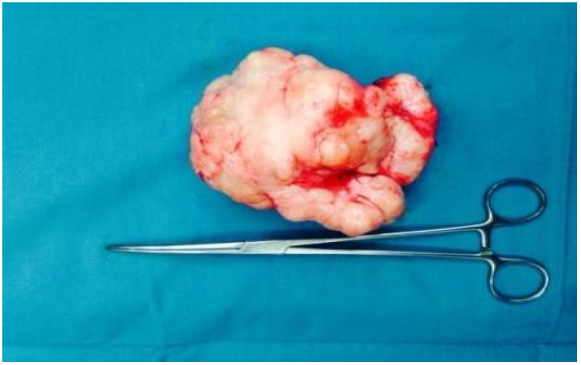
Figure 1. Grossly, it is well circumscribed mass, and the surface shows white whirling appearance.
Jdream 04 00045 g001
Figure 2. The tumor consists of intersecting fascicles of spindle cells (H & E, x100).
Jdream 04 00045 g002
Figure 3. Immunohistochemical stain for desmin revealed positivity in the tumor cells, consistent with smooth muscle tumor (x100).
Jdream 04 00045 g003