Next Article in Journal
Tracheobronchial Amyloidosis
Previous Article in Journal
Automatic detection of Voice Disorders: recent literature advancements
 
 
Journal of Interdisciplinary Research Applied to Medicine is published by MDPI from Volume 6 Issue 1 (2026). Previous articles were published by another publisher in Open Access under a CC-BY (or CC-BY-NC-ND) licence, and they are hosted by MDPI on mdpi.com as a courtesy and upon agreement with University of Salento.
Article

Prevention Strategy of Intrauterine Adhesions: update and future perspective

by
Sarah Gustapane
1,
Sara Cavalera
2,
Ottavia D’Oria
1,3 and
Andrea Tinelli
1,4,5,*
1
Department of Obstetrics and Gynecology, “Veris delli Ponti” Hospital, Scorrano, Lecce, Italy
2
Department of Orthopedics and traumatology, Area Nord AUSL Modena, Italy
3
Department of Medical and Surgical Sciences and Translation Medicine, Sant’Andrea University Hospital, PhD Course in “Translation Medicine and Oncology, Sapienza University of Rome, Italy
4
Division of Experimental Endoscopic Surgery, Imaging, Technology and Minimally Invasive Therapy, Vito Fazzi Hospital, Lecce, Italy
5
Adjunct Professor at the Laboratory of Human Physiology, Phystech BioMed School, Faculty of Biological & Medical Physics, Moscow Institute of Physics and Technology (State University), Dolgoprudny, Moscow Region, Russia
*
Author to whom correspondence should be addressed.
J. Interdiscip. Res. Appl. Med. 2020, 4(2), 31-40; https://doi.org/10.1285/i25327518v4i2p31
Published: 31 December 2020

Abstract

Intrauterine adhesions (IUAs) caused by endometrial injury have a serious impact on women’s fertility and morbility and involves a wide range of patients. Although the first case of IUAs was published in 1984 by Heinrich Fritsch, a full description of Asherman syndrome was done by Joseph Asherman. IUAs lead to a lot of complications in women, as the partial or complete closure of the uterine cavity, which may result in symptoms including abnormal menstruation, pelvic pain, recurrent pregnancy loss, secondary infertility, and pregnancy complications. Hysteroscopy, which has relegated blind curettage, is currently considered the gold standard diagnostic and therapeutic approach also as for outpatients. However, an integrated approach, including preoperative, intraoperative and postoperative procedures is needed to improve the reproductive outcome of the complex syndrome. In the post-operative care, the patient can benefit from some therapeutic and prophylactic methods used alone or in combination with each other. In this review, authors discuss on the efficacy of traditional methods for the prevention of complications of IUAs after surgery, such as hormonal therapy, physical barriers, vasodilators and antibiotics, as well as novel strategies such as stem cell therapy and novel therapeutic agents.

1. Introduction

Intrauterine adhesions (IUAs) are a result of mechanical or infectious injury to the basalis layer of the endometrium, caused by curettage, hysteroscopic surgery, uterine artery embolization, B Lynch sutures, abdominal myomectomy, hysteroscopic myomectomy, genital tuberculosis and surgical treatment of Mullerian anomalies (Doroftei et al. 2020).
In some women, the normal repair mechanisms of the endometrium are aberrant, including hypoxia, reduced neovascularization and altered expression of adhesion-associated cytokines, resulting in IUAs formation (Buttram et al. 1988). IUAs can lead to partial (Figure 1) or complete obliteration of the cervix and the uterine cavity, which may result in clinical sequelae including abnormal menstruation, amenorrhea, pelvic pain, infertility (caused by the obstruction of sperm transport into the cervix, impaired embryo migration within the uterine cavity and failure of embryo implantation (Dreisler and Kjer 2019)), recurrent pregnancy loss and pregnancy complications.
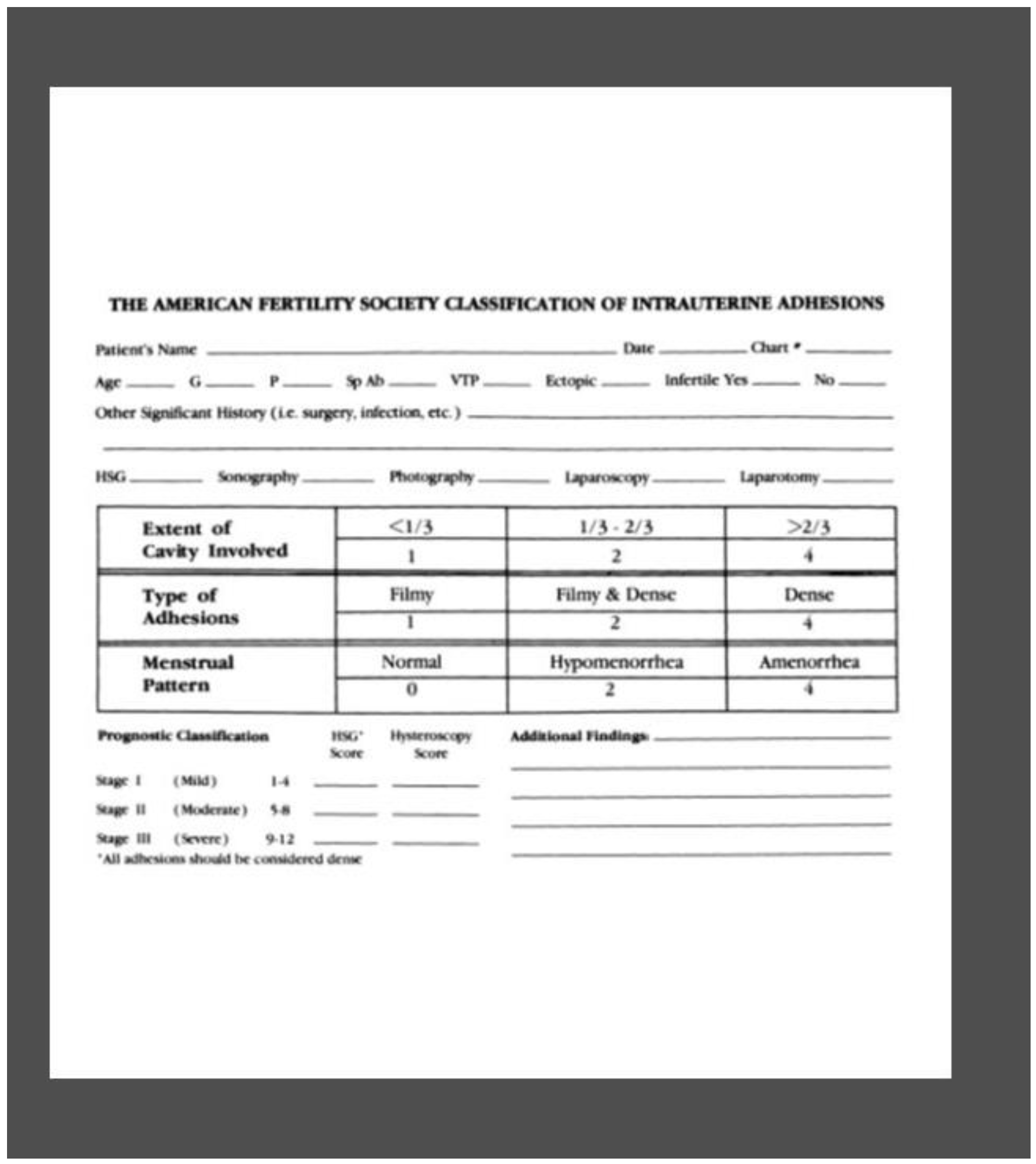
The traditionally widely used classification system of the IUAs is the American Fertility Society (Buttram et al. 1988) score (Figure 2), classifying IUAs in three stages: mild (grade I), moderate (grade II), and severe (grade III). Currently, Hysteroscopy can be considered the gold standard for diagnostic and therapeutic approach also for outpatients. The occurrence of new adhesions after primary hysteroscopic adhesiolysis is so much frequent and the recurrence rate is associated with the grade of adherences (Figure) as found by Hanstede et al, that reported 21%–25% recurrence with grade 1–2 adherences, 29.1% with grade 3, 38.5% with grade 4, and 41.9% with grade five (Bosteels et al. 2015).
Although numerous observational studies suggest potential benefit with the use of anti-adhesion therapies (intrauterine device or balloon, hormonal treatment, antibiotics, barrier gels or human amniotic membrane grafting) for decreasing IUAs, currently, there are no strong recommendation in favor of the use of anti-adhesion therapies after operative hysteroscopy. At present the effectiveness of the anti-adhesion treatment following operative hysteroscopy for decreasing IUAs remains uncertain as suggested the Cochrane Review of 2017, because of the low quality of the evidence (Hanstede et al. 2015).

1.2. Prevention of adhesion recurrence

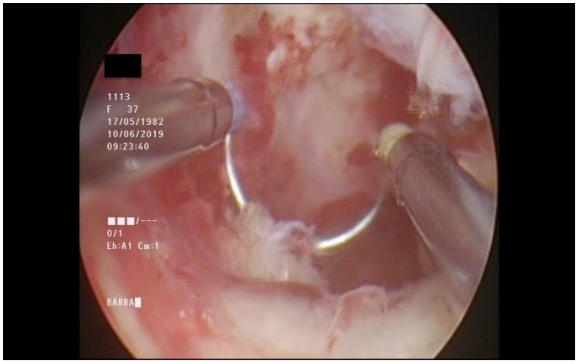
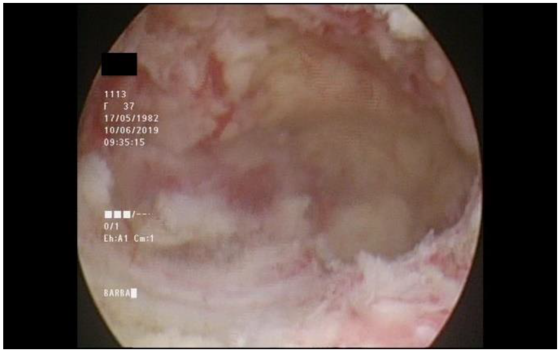
Adhesions recurrence after surgery is one of the most important factors which can delay reproductive outcome after IUA treatment. Adhesions recurrence rate is significantly higher in those cases where a severe AS is diagnosed (Figure 3 and Figure 4).
Several methods to prevent IUA reformations after surgery have been proposed. Nonetheless few comparative studies have been developed (Xu et al. 2018). This could be probably due to the multitude treatment approach adopted and particularly to the lack of a unified standardized classification system for IUA diagnostic characterization.

1.3. Intrauterine device

The intrauterine device (IUD) may provide a physical barrier between the uterine walls, separating the endometrial layers to prevent their fusion during the initial healing phase (Buttram et al. 1988).
The characteristics of IUD to prevent intrauterine adhesion formation should be the tolerability of the device, the suppression of IUA formation and the restoring of healing of the endometrium. There are several observational studies that recommended the insertion of a device after lysis of IUAs such as IUD and Foley catheter balloon after lysis of IUAs or septoplasty. There are different kinds of IUD (copper-containing IUD, T-shaped IUD, loop IUD) with particular characteristics and mechanism of actions, also, there are no sizes of IUD available for too large or too small uterine cavities (Kodaman and Arici 2007).

1.4. Intrauterine balloons

An intrauterine balloon stent is another mechanical method frequently used to prevent the reformation of adhesions. The Cook Medical balloon (Indianapolis, IN, USA) has designed a heartshaped intrauterine balloon for prevention of secondary intrauterine adhesions thanks to its triangular shape, which conforms to the configuration of a normal uterus and maintain separation at the margins of uterine cavity (March 2011).

1.5. Foley catheters

A standard pediatric Foley catheter is another commonly used method to prevent recurrence of IUAs.
In a randomized controlled trial, Lin et al compared the efficacy of intrauterine balloon (removed after 7 days) and IUD demonstrating similar efficacy (Lin et al. 2015).
Orhue et al compared an IUD with a pediatric Foley catheter and found that the catheter was a safer and more effective adjunctive method of treatment of IUA compared with the IUD. The persistent post-treatment amenorrhea and hypomenorrhea occurred less frequently in the Foley catheter group (18.6%) than in the IUD group (37.3%) (P<0.03), and the conception rate in the catheter group was 33.9% compared with 22.5% in the IUD group. The need for repeated treatment was also significantly less in the Foley catheter group (Orhue et al. 2003). Recently, Shi et al compared the efficacy of intermittent intrauterine balloon dilatation versus standard care in the prevention of adhesion reformation in two hundred patients with moderate to severe IUAs who underwent hysteroscopic adhesiolysis. In this randomized controlled trial, the balloon group received intrauterine balloon dilatation therapy at 2 weeks and 6 weeks after surgery, whereas the control group did not. A total of 191 patients successfully completed the study protocol (94 cases for the balloon group and 97 cases for the control group). According to hysteroscopic evaluation at the 8th week, the overall adhesion reformation rate was significantly lower in patients in the balloon group than patients in the control group (20.2% versus 40.2%, respectively; P < 0.05). This study shows that postoperative intermittent intrauterine balloon dilatation therapy can significantly reduce postoperative adhesion reformation and significantly increase menstruation flow (Shi et al. 2019).
Recently Huang at al have patented intrauterine stent of various sizes, flexible and thin, but at the moment their studies have been performed on a small number of patients with moderate or severe IUAs and so we have no encouraging data (Huang et al. 2020).

1.6. Anti-adhesion barrier

Hyaluronic acid-derived products showing a possible role in gynecologic surgery to prevent intra-abdominal IUAs, reducing the risk of adhesion recurrence after surgical treatment of IUAs (Guida et al. 2004; Tsapanos et al. 2002; Acunzo et al. 2003), but may not be suitable alone for endometrial surfaces due to a short half-life and weak attachment to the endometrium (Acunzo et al. 2003). The material usually needs to be used in combination with other devices.
Use of biodegradable gel surgical barriers is based on the principle of keeping adjacent wound surfaces mechanically separate (Renier et al. 2005). The exact mechanisms by which ACP (auto-cross-linked polysaccharide) and HACMC (sodium hyaluronate and carboxymethylcellulose gel) can reduce adhesion reformation are not well known but may be related to ‘hydro flotation’ or ‘siliconizing’ effects. Hyaluronic acid gel or polyethylene oxide-sodium carboxymethylcellulose gel for the prevention of intrauterine adherences have been investigated demonstrating conflicting results. Acunzo et al found a significant effect of hyaluronic acid compared to no treatment (14% versus 32%) (Acunzo et al. 2003). Instead, Lin et al demonstrated that the balloon and IUCD were more effective than hyaluronic acid (Lin et al. 2015). Ducarne et al compared application of ACP gel (30 women) versus no gel (24 women) at the end of an operative hysteroscopic procedure performed to treat myomas, polyps, uterine septa or IUAs, finding no statistically significant differences between comparison groups in the rate of adhesion formation, or in mean adhesion scores and severity of adhesions (Ducarme et al. 2006). Different results were obtained from the recent meta-analysis conducted by Fei et al. finding a significant reduction of the incidence of moderate and severe IUAs (RR 0.18, 95% CI: 0.07~0.47; p=0.0004) and an improvement in the pregnancy rate after miscarriage (RR 1.94, 95% CI 1.46~2.60; p<0.00001) with the use of hyaluronic acid gel (Zheng et al. 2020).

1.7. Human amniotic membrane grafting

Human amniotic membrane HAM is the innermost layer of the fetal membranes and possess many properties that make them suitable for use in regenerative medicine, such as low immunogenicity, anti-fibrotic, anti-inflammatory, angiogenic and anti-angiogenetic and anti-microbial properties (Gary and Jones, 2017). HAM acts as a biologically active mechanical barrier to suppress adhesion formation while promoting endometrial healing (Amer and Abd-El-Maeboud 2006), through regeneration of epithelium facilitating migration of epithelial cells, reinforcing adhesion of the basal epithelium, promoting epithelial cell differentiation (Meller and Tseng 1999), preventing cellular apoptosis (Hori et al. 2006), producing factors or creating a microenvironment for effective tissue repair and endometrial regeneration, possibly by stimulating endogenous stem cells (Padykula 1989). According to a randomized controlled trials of Zheng et al including 300 patients, which evaluated the ability of HAM to prevent the recurrence of IUAs after hysteroscopic adhesiolysis, the use of HAM increased menstrual blood volume (mean difference 6.15, 95% CI 4.20–8.11; P<0.001) but failed to improve the rate of intrauterine adhesion recurrence or spontaneous abortion (Zheng et al. 2018).
Yan et al in a network Meta-Analysis of randomized controlled trials has found a significant advantage with the use of freeze-dried amniotic agents plus a balloon to reduce IUAs recurrence and IUAs scores after adhesiolysis (Yan and Xu 2018).
A prospective randomized controlled trial conducted among 88 women with severe IUA who underwent hysteroscopic adhesiolysis analyzed the efficacy of freeze-dried amnion graft covered the balloon portion of the Foley catheter for prevention of IUAs. Also, this study concluded that the use of HAM was effective in improving menstruation, but the rates of IUAs reformation and pregnancy were not significantly different (Gan et al. 2017).

2. Medical therapy to restoration the endometrium

2.1. Vasodilators

In recent years, many studies described use of medications to increase vascular flow to endometrium such as aspirin, nitroglycerine and sildenafil citrate. But evidence was insufficient to show whether vasodilators increase the live birth rate (Gutarra-Vilchez et al. 2018).
Studies have demonstrated that aspirin combined with estrogen may significantly prevent the postoperative disease recurrent rate, improve endometrial receptivity and improve the conception rate by increasing endometrial blood supply and angiogenesis more effectively. The aspirin inhibits endometrial fibrosis by suppressing the TGF-1-Smad2/Smad3 pathways (Z. Zhang et al. 2020).
Zinger reported two cases of woman with history of a postpartum uterine curettage, inadequate endometrium thickness after surgical resection of IUAs that are treated with sildenafil citrate and with the results of having achieved pregnancy (Zinger et al. 2006). However, the number of women treated using these therapies remains small, and because all such treatment is off label, these medications cannot be endorsed outside of rigorous research protocols.

2.2. Antibiotics

There is no clear recommendation in the literature on whether it is necessary to use prophylactic antibiotics for minor operative procedures such as dilatation and curettage for evacuation of conceptive products, fractional curettage for abnormal uterine bleeding, hysterosalpingography for infertility evaluation and hysteroscopy for intrauterine cavity diagnosis and treatment. The Cochrane of 2013 regarding the prophylactic antibiotics for transcervical intrauterine procedures versus placebo concluded that there are no randomized controlled trials that asses the effects of prophylactic antibiotics on infection complications and therefore is not possible to draw any conclusions (Thinkhamrop et al. 2007). However, when obvious infection is seen, antibiotics are mandatory.
In India genital tuberculosis appears to be an important and common cause of IUA causing primary and secondary infertility with various grades of adhesions (Sharma et al. 2008) and so it is important to investigate the patients who come from those areas.

2.3. Hormonal therapy

Already in 1964 Wood and Pena hypothesized the beneficial effects of estrogen therapy on endometrial regeneration after surgical treatment for IUAs (Wood and Pena 1964). Postoperative treatment with estrogen in order to promote the regeneration of the endometrium has been recommended in several studies, either as estrogen only (Capella-Allouc et al. 1999; Dawood et al. 2010), either with IUD (March et al. 1978; Chen et al. 2017; Yu et al. 2016; Roy et al. 2014; Zikopoulos et al. 2004; Myers and Hurst 2012; Salma et al. 2014; Liu et al. 2019) or Foley catheter (Dawood et al. 2010; March et al. 1978; Salma et al. 2014). In several studies different regimens consisting of estrogen with or without a progestogen have been used (Kodaman and Arici 2007). There are no comparative studies that examine dosage, administration or combinations of hormones (Buttram et al. 1988). In a recent randomized study, 4 mg and 10 mg estradiol orally was compared. No superior effect of the high dosage was demonstrated (Liu et al. 2019). When comparing 2 mg and 6 mg in a prospective randomized trial, no benefit was demonstrated in the 6 mg arm. In the randomized controlled trials of Farhi et al, 60 women undergoing dilatation and curettage during the first trimester of pregnancy were allocated to receive estrogen combined with progestogen or no treatment (Farhi et al. 1993). The authors have found that women in the intervention group had a significantly thicker endometrium compared with women in the control group (8.4 with intervention vs 6.7 mm with no treatment; P = 0.02) and so they concluded that postoperative hormonal treatment may be useful for IUAs prevention following curettage. Nevertheless, but this study does not report the data about pregnancy rates and IUAs recurrence (Farhi et al. 1993). The systematic review of Johary et al, concluded that estrogen therapy, may be beneficial for women with IUAs, but as adjunctive therapy combined with other anti-adhesion strategies (Johary et al. 2014). Also, in three prospective randomized studies, the administration of oral estrogen did not reduce the risk of IUAs (Tonguc et al. 2010; Dabirashraft et al. 1996; Roy et al. 2014).

3. Future perspective

Recently, some experimental study has demonstrated that stem cells on rat models is a promising therapeutic approach for the regeneration of the inadequate endometrium. In particular, Zhao’s study aimed to identify exosomes derived from adipose-derived mesenchymal stem cells (ADSC-exo) and explore the therapeutic potential in IUA rat models. In IUA model, treatment with ADSC-exo maintained normal uterine structure, promoted endometrial regeneration and collagen remodeling, and enhanced the expression of integrin-β3, LIF, and VEGF. An improved receptivity of the regenerated endometrium was confirmed. Their findings demonstrated that ADSC-exo promoted endometrial regeneration and fertility restoration. It suggested that topical administration of ADSC-exo in uterus could be a promising strategy for patients suffering severe intrauterine adhesions and infertility (Zhao et al. 2020).
Another studies on rat model of Zhang at al evaluated urinary bladder matrix in order to improve endometrial regeneration, receptivity and fertility (H. Zhang et al. 2020).
Another promising research of Zhang SS, based on the synergistic effect of the well-known E2 and the Heparin-Poloxamer Hydrogel, revealed that administrating E2-HP hydrogel to injured uterus had a positive effect on endometrium regeneration in rat model (S. Zhang et al. 2020). In the only prospective study performed on humans by Santamaria et al. 16 women with IUA confirmed by hysteroscopy were treated with uterine intravascular infusion of bone marrow-derived stem cell (BMDSC). During the follow-up period, menstrual function returned to normal within 6 months after BMDSC infusion, with three spontaneous pregnancy and seven pregnancies after IVF and embryo transfer reported (Santamaria et al. 2016). These novel studies begin to open the door for further prospective research on human population.

4. Conclusions

Currently, there is no ideal method to prevent IUAs and it is difficult to standardize a therapy valid for all patients as IUAs is a heterogeneous syndrome with specific peculiarities.
Many devices, used alone or in combination, have been proposed to prevent IUAs formation after intrauterine procedures, but at present it is difficult to establish which approach is the best, due to the heterogeneity of the studies, the contrasting results reported, and the different outcomes investigated. To avoid the adhesions relapse, it would seem to be recommendable the use of balloon catheters and IUD with adjunctive estrogen therapy. The combination strategy that using physical barriers as the delivery carriers for therapeutics might provide new alternatives for the prevention of IUAs.
Recently several new methods have been patented to prevent IUAs but long-term results are not yet available. More research is needed to assess the best approach to prevent adhesions in order to increase reproductive chances and if pregnancy occurs to reduce obstetrics risk such as miscarriage, preterm birth, abnormal placentation, intrauterine growth restriction. Perhaps there is the key to improving the outcome of IUAs: the personalization of therapy and the evolution of biocompatible materials that are increasingly adaptable to specific needs.

References

  1. Acunzo, Giuseppe, Maurizio Guida, Massimiliano Pellicano, Giovanni Antonio Tommaselli, Attilio Di Spiezio Sardo, Giuseppe Bifulco, Domenico Cirillo, Alex Taylor, and Carmine Nappi. 2003. Effectiveness of Auto-Cross-Linked Hyaluronic Acid Gel in the Prevention of Intrauterine Adhesions after Hysteroscopic Adhesiolysis: A Prospective, Randomized, Controlled Study. Human Reproduction 18 (9): 1918–21. [Google Scholar] [CrossRef] [Scilit]
  2. Amer, Mohamed I., and Karim H. Abd-El-Maeboud. 2006. Amnion Graft Following Hysteroscopic Lysis of Intrauterine Adhesions. Journal of Obstetrics and Gynaecology Research 32 (6): 559–66. [Google Scholar] [CrossRef] [Scilit]
  3. Bosteels, Jan, Steven Weyers, Jenneke Kasius, Frank J. Broekmans, Ben Willem J. Mol, and Thomas M. D’Hooghe. 2015. Anti-Adhesion Therapy Following Operative Hysteroscopy for Treatment of Female Subfertility. Cochrane Database of Systematic Reviews. John Wiley and Sons Ltd. [CrossRef] [Scilit]
  4. Buttram, V. C., V. Gomel, A. Siegler, A. DeCherney, W. Gibbons, and C. March. 1988. The American Fertility Society Classifications of Adnexal Adhesions, Distal Tubal Occlusion, Tubal Occlusion Secondary to Tubal Ligation, Tubal Pregnancies, Mullerian Anomalies and Intrauterine Adhesions. Fertility and Sterility 49 (6): 944–55. [Google Scholar] [CrossRef] [Scilit] [PubMed]
  5. Capella-Allouc, Sylvie, Fadila Morsad, Catherine Rongières-Bertrand, Sabine Taylor, and Hervé Fernandez. 1999. Hysteroscopic Treatment of Severe Asherman’s Syndrome and Subsequent Fertility. Human Reproduction 14 (5): 1230–33. [Google Scholar] [CrossRef] [Scilit]
  6. Chen, Limei, Hongwei Zhang, Qing Wang, Feng Xie, Shujun Gao, Yu Song, Jing Dong, Hua Feng, Kangyun Xie, and Long Sui. 2017. Reproductive Outcomes in Patients With Intrauterine Adhesions Following Hysteroscopic Adhesiolysis: Experience From the Largest Women’s Hospital in China. Journal of Minimally Invasive Gynecology 24 (2): 299–304. [Google Scholar] [CrossRef] [Scilit]
  7. Dabirashraft, Hormoz, Kazem Mohammad, Nasrin Moghadami-Tabrizi, Kambiz Zandinejad, and Massoud Moghadami-Tabrizi. 1996. Is Estrogen Necessary after Hysteroscopic Incision of the Uterine Septum? Journal of the American Association of Gynecologic Laparoscopists 3 (4): 623–25. [Google Scholar] [CrossRef] [Scilit] [PubMed]
  8. Dawood, Ashraf, Ayman Al-Talib, and Togas Tulandi. 2010. Predisposing Factors and Treatment Outcome of Different Stages of Intrauterine Adhesions. Journal of Obstetrics and Gynaecology Canada 32 (8): 767–70. [Google Scholar] [CrossRef] [Scilit]
  9. Doroftei, Bogdan, Ana-Maria Dabuleanu, OvidiuDumitru Ilie, Radu Maftei, Emil Anton, Gabriela Simionescu, Theodor Matei, and Theodora Armeanu. 2020. Mini-Review of the New Therapeutic Possibilities in Asherman Syndrome—Where Are We after One Hundred and Twenty-Six Years? Diagnostics 10 (9): 706. [Google Scholar] [CrossRef] [Scilit] [PubMed]
  10. Dreisler, Eva, and Jens Joergen Kjer. 2019. Asherman’s Syndrome: Current Perspectives on Diagnosis and Management. International Journal of Women’s Health. Dove Medical Press Ltd. [CrossRef] [Scilit]
  11. Ducarme, G., C. Davitian, S. Zarrouk, M. Uzan, and C. Poncelet. 2006. Interest of Auto-crosslinked Hyaluronic Acid Gel in the Prevention of Intrauterine Adhesions after Hysteroscopic Surgery: A Case-control Study. J Gynecol Obstet Biol Reprod 35 (7): 691–95. [Google Scholar] [CrossRef] [Scilit]
  12. Farhi, Jacob, Itai Bar-Hava, Roy Homburg, Dov Dicker, and Zion Ben-Rafael. 1993. Induced Regeneration of Endometrium Following Curettage for Abortion: A Comparative Study. Human Reproduction 8 (7): 1143–44. [Google Scholar] [CrossRef] [Scilit]
  13. Gan, Lu, Hua Duan, Fu-Qing Sun, Qian Xu, Yi-Qun Tang, and Sha Wang. 2017. Efficacy of Freeze-Dried Amnion Graft Following Hysteroscopic Adhesiolysis of Severe Intrauterine Adhesions. International Journal of Gynecology & Obstetrics 137 (2): 116–22. [Google Scholar] [CrossRef] [Scilit] [PubMed]
  14. Gary, Katerina Jirsova, and L A Jones. n.d.FULL LENGTH REVIEW Amniotic Membrane in Ophthalmology: Properties, Preparation, Storage and Indications for Grafting-a Review. Cell and Tissue Banking 18. [Google Scholar] [CrossRef] [Scilit]
  15. Guida, Maurizio, Giuseppe Acunzo, Attilio Di Spiezio Sardo, Giuseppe Bifulco, Roberto Piccoli, Massimiliano Pellicano, Giuseppe Cerrota, Domenico Cirillo, and Carmine Nappi. 2004. Effectiveness of AutoCrosslinked Hyaluronic Acid Gel in the Prevention of Intrauterine Adhesions after Hysteroscopic Surgery: A Prospective, Randomized, Controlled Study. Human Reproduction 19 (6): 1461–64. [Google Scholar] [CrossRef] [Scilit]
  16. Gutarra-Vilchez, Rosa B., Xavier Bonfill Cosp, Demián Glujovsky, Andres Viteri-García, Fernando M. RunzerColmenares, and Maria José Martinez-Zapata. 2018. Vasodilators for Women Undergoing Fertility Treatment. Cochrane Database of Systematic Reviews. John Wiley and Sons Ltd. [CrossRef] [Scilit]
  17. Hanstede, Miriam M.F., Eva Van Der Meij, Laurien Goedemans, and Mark Hans Emanuel. 2015. Results of Centralized Asherman Surgery, 2003-2013. Fertility and Sterility 104 (6): 1561–1568.e1. [Google Scholar] [CrossRef] [Scilit]
  18. Hori, Junko, Mingcong Wang, Kazutaka Kamiya, Hiroshi Takahashi, and Norio Sakuragawa. 2006. Immunological Characteristics of Amniotic Epithelium. Cornea 25 (December): S53–58. [Google Scholar] [CrossRef] [Scilit] [PubMed]
  19. Huang, Huan, Lingxiao Zou, Aiqian Zhang, Xingping Zhao, Dabao Xu, and Min Xue. 2020. A Preliminary Study on a Patented Intrauterine Stent in the Treatment of Recurrent Intrauterine Adhesions with Poor Prognosis. Annals of Translational Medicine 8 (4): 57–57. [Google Scholar] [CrossRef] [Scilit]
  20. Johary, Jolinda, Min Xue, Xiaogang Zhu, Dabao Xu, and Prasad Palani Velu. 2014. Efficacy of Estrogen Therapy in Patients With Intrauterine Adhesions: Systematic Review. Journal of Minimally Invasive Gynecology. Elsevier. [CrossRef] [Scilit]
  21. Kodaman, Pinar H, and Aydin Arici. 2007. IntraUterine Adhesions and Fertility Outcome: How to Optimize Success? Current Opinion in Obstetrics and Gynecology 19 (3): 207–14. [Google Scholar] [CrossRef] [Scilit]
  22. Lin, Xiao Na, Feng Zhou, Min Ling Wei, Yang Yang, Ying Li, T. C. Li, and Song Ying Zhang. 2015. Randomized, Controlled Trial Comparing the Efficacy of Intrauterine Balloon and Intrauterine Contraceptive Device in the Prevention of Adhesion Reformation after Hysteroscopic Adhesiolysis. Fertility and Sterility 104 (1): 235–40. [Google Scholar] [CrossRef] [Scilit] [PubMed]
  23. Liu, Linlin, Xiaowu Huang, Enlan Xia, Xiaoyu Zhang, Tin Chiu Li, and Yuhuan Liu. 2019. A Cohort Study Comparing 4 Mg and 10 Mg Daily Doses of Postoperative Oestradiol Therapy to Prevent Adhesion Reformation after Hysteroscopic Adhesiolysis. Human Fertility 22 (3): 191–97. [Google Scholar] [CrossRef] [Scilit] [PubMed]
  24. March, Charles M. 2011. Management of Asherman’s Syndrome. In Reproductive BioMedicine Online, 23:63–76. Elsevier. [CrossRef] [Scilit]
  25. March, Charles M., Robert Israel, and Antonia D. March. 1978. Hysteroscopic Management of Intrauterine Adhesions. American Journal of Obstetrics and Gynecology 130 (6): 653–57. [Google Scholar] [CrossRef] [Scilit]
  26. Meller, D, and SC Tseng. 1999. Conjunctival Epithelial Cell Differentiation on Amniotic Membrane. | IOVS | ARVO Journals. Investigative Ophthalmology & Visual Science 40: 878–86. [Google Scholar]
  27. Myers, Erinn M., and Bradley S. Hurst. 2012. Comprehensive Management of Severe Asherman Syndrome and Amenorrhea. Fertility and Sterility 97 (1): 160–64. [Google Scholar] [CrossRef] [Scilit]
  28. Orhue, A. A.E., M. E. Aziken, and J. O. Igbefoh. 2003. A Comparison of Two Adjunctive Treatments for Intrauterine Adhesions Following Lysis. International Journal of Gynecology and Obstetrics 82 (1): 49–56. [Google Scholar] [CrossRef] [Scilit]
  29. Padykula, Helen A. 1989. Regeneration in the Primate Uterus. In Biology of the Uterus, 279–88. Springer US. pp. 279–88. [CrossRef] [Scilit]
  30. Renier, Davide, Pierangelo Bellato, Davide Bellini, Alessandra Pavesio, Daniele Pressato, and Anna Borrione. 2005. Pharmacokinetic Behaviour of ACP Gel, an Autocrosslinked Hyaluronan Derivative, after Intraperitoneal Administration. Biomaterials 26 (26): 5368–74. [Google Scholar] [CrossRef] [Scilit]
  31. Roy, Kallol Kumar, Neha Negi, Murali Subbaiah, Sunesh Kumar, Jai Bhagwan Sharma, and Neeta Singh. 2014. Effectiveness of Estrogen in the Prevention of Intrauterine Adhesions after Hysteroscopic Septal Resection: A Prospective, Randomized Study. Journal of Obstetrics and Gynaecology Research 40 (4): 1085–88. [Google Scholar] [CrossRef] [Scilit]
  32. Salma, Umme, Min Xue, Ali Sheikh Md Sayed, and Dabao Xu. 2014. Efficacy of Intrauterine Device in the Treatment of Intrauterine Adhesions. BioMed Research International. Hindawi Publishing Corporation. [CrossRef] [Scilit]
  33. Santamaria, Xavier, Sergio Cabanillas, Irene Cervelló, Cristina Arbona, Francisco Raga, Jaime Ferro, Julio Palmero, Jose Remohí, Antonio Pellicer, and Carlos Simón. 2016. Autologous Cell Therapy with CD133+ Bone Marrow-Derived Stem Cells for Refractory Asherman’s Syndrome and Endometrial Atrophy: A Pilot Cohort Study. Human Reproduction 31 (5): 1087–96. [Google Scholar] [CrossRef] [Scilit] [PubMed]
  34. Sharma, Jai Bhagwan, Kallol K. Roy, M. Pushparaj, Nupur Gupta, Sunesh Kumar Jain, Neena Malhotra, and Suneeta Mittal. 2008. Genital Tuberculosis: An Important Cause of Asherman’s Syndrome in India. Archives of Gynecology and Obstetrics 277 (1): 37–41. [Google Scholar] [CrossRef] [Scilit] [PubMed]
  35. Shi, X, SH Saravelos, Q Zhou, X Huang, E Xia, and TC Li. 2019. Prevention of Postoperative Adhesion Reformation by Intermittent Intrauterine Balloon Therapy: A Randomised Controlled Trial. BJOG: An International Journal of Obstetrics & Gynaecology 126 (10): 1259–66. [Google Scholar] [CrossRef] [Scilit]
  36. Thinkhamrop, Jadsada, M. Laopaiboon, and P. Lumbiganon. 2007. Prophylactic Antibiotics for Transcervical Intrauterine Procedures. Cochrane Database of Systematic Reviews. John Wiley and Sons Ltd. [CrossRef] [Scilit]
  37. Tonguc, Esra Ayşin, Turgut Var, Nafiye Yilmaz, and Sertac Batioglu. 2010. Intrauterine Device or Estrogen Treatment after Hysteroscopic Uterine Septum Resection. International Journal of Gynecology and Obstetrics 109 (3): 226–29. [Google Scholar] [CrossRef] [Scilit] [PubMed]
  38. Tsapanos, Vassilios S., Lavinia P. Stathopoulou, Vassiliki S. Papathanassopoulou, and Vassilios A. Tzingounis. 2002. The Role of Seprafilm? Bioresorbable Membrane in the Prevention and Therapy of Endometrial Synechiae. Journal of Biomedical Materials Research 63 (1): 10–14. [Google Scholar] [CrossRef] [Scilit]
  39. Wood, J, and G Pena. 1964. Treatment of Traumatic Uterine Synechiae. Int J Gynecol Obstet 9: 405–10. [Google Scholar]
  40. Xu, Wenzhi, Yuxue Zhang, Yang Yang, Songying Zhang, and Xiaona Lin. 2018. Effect of Early SecondLook Hysteroscopy on Reproductive Outcomes after Hysteroscopic Adhesiolysis in Patients with Intrauterine Adhesion, a Retrospective Study in China. International Journal of Surgery 50 (February): 49–54. [Google Scholar] [CrossRef] [Scilit] [PubMed]
  41. Yan, Yu, and Dongmei Xu. 2018. The Effect of Adjuvant Treatment to Prevent and Treat Intrauterine Adhesions: A Network Meta-Analysis of Randomized Controlled Trials.” Journal of Minimally Invasive Gynecology. Elsevier B.V. [CrossRef] [Scilit]
  42. Yu, Xiao, Liu Yuhan, Song Dongmei, Xia Enlan, and Li Tinchiu. 2016. The Incidence of Post-Operative Adhesion Following Transection of Uterine Septum: A Cohort Study Comparing Three Different Adjuvant Therapies. European Journal of Obstetrics and Gynecology and Reproductive Biology 201 (June): 61–64. [Google Scholar] [CrossRef] [Scilit] [PubMed]
  43. Zhang, Honghong, Qing Zhang, Jian Zhang, Fei Sheng, Shuang Wu, Fu Yang, and Wen Li. 2020. Urinary Bladder Matrix Scaffolds Improve Endometrial Regeneration in a Rat Model of Intrauterine Adhesions. Biomaterials Science 8 (3): 988–96. [Google Scholar] [CrossRef] [Scilit] [PubMed]
  44. Zhang, Si-Si, Xin-Xin Xu, Wei-Wei Xiang, Hui-Heng Zhang, Hui-Long Lin, Lai-En Shen, Qi Lin, Feng Lin, and Zhi-Yang Zhou. 2020. Using 17β-estradiol Heparin-poloxamer Thermosensitive Hydrogel to Enhance the Endometrial Regeneration and Functional Recovery of Intrauterine Adhesions in a Rat Model. The FASEB Journal 34 (1): 446–57. [Google Scholar] [CrossRef] [Scilit]
  45. Zhang, Zihui, Shuang Li, Jie Deng, Shaorong Yang, Ziwu Xiang, Hongyan Guo, Hongli Xi, Ming Sang, and Wei Zhang. 2020. Aspirin Inhibits Endometrial Fibrosis by Suppressing the TGF–Β1–Smad2/Smad3 Pathway in Intrauterine Adhesions. International Journal of Molecular Medicine 45 (5): 1351–60. [Google Scholar] [CrossRef] [Scilit]
  46. Zhao, S, W Qi, J Zheng, Y Tian, X Qi, D Kong, J Zhang, and X Huang. 2020. Exosomes Derived from Adipose Mesenchymal Stem Cells Restore Functional Endometrium in a Rat Model of Intrauterine Adhesions. Reprod Sci 27 (6): 1266–75. [Google Scholar] [CrossRef] [Scilit]
  47. Zheng, Fei, Xin Xin, Fei He, Jianyong Liu, and Yuechong Cui. 2020. Meta-analysis on the Use of Hyaluronic Acid Gel to Prevent Intrauterine Adhesion after Intrauterine Operations. Experimental and Therapeutic Medicine 19 (4): 2672–78. [Google Scholar] [CrossRef] [Scilit]
  48. Zheng, Fei, Bin Zhu, Yumo Liu, Ruifeng Wang, and Yuechong Cui. 2018. Meta-Analysis of the Use of Amniotic Membrane to Prevent Recurrence of Intrauterine Adhesion after Hysteroscopic Adhesiolysis. International Journal of Gynecology and Obstetrics. John Wiley and Sons Ltd. [CrossRef] [Scilit]
  49. Zikopoulos, Konstantinos A., Efstratios M. Kolibianakis, Peter Platteau, Luc de Munck, Herman Tournaye, Paul Devroey, and Michel Camus. 2004. Live Delivery Rates in Subfertile Women with Asherman’s Syndrome after Hysteroscopic Adhesiolysis Using the Resectoscope or the Versapoint System. Reproductive BioMedicine Online 8 (6): 720–25. [Google Scholar] [CrossRef] [Scilit]
  50. Zinger, Michael, James H. Liu, and Michael A. Thomas. 2006. Successful Use of Vaginal Sildenafil Citrate in Two Infertility Patients with Asherman’s Syndrome. Journal of Women’s Health 15 (4): 442–44. [Google Scholar] [CrossRef] [Scilit]
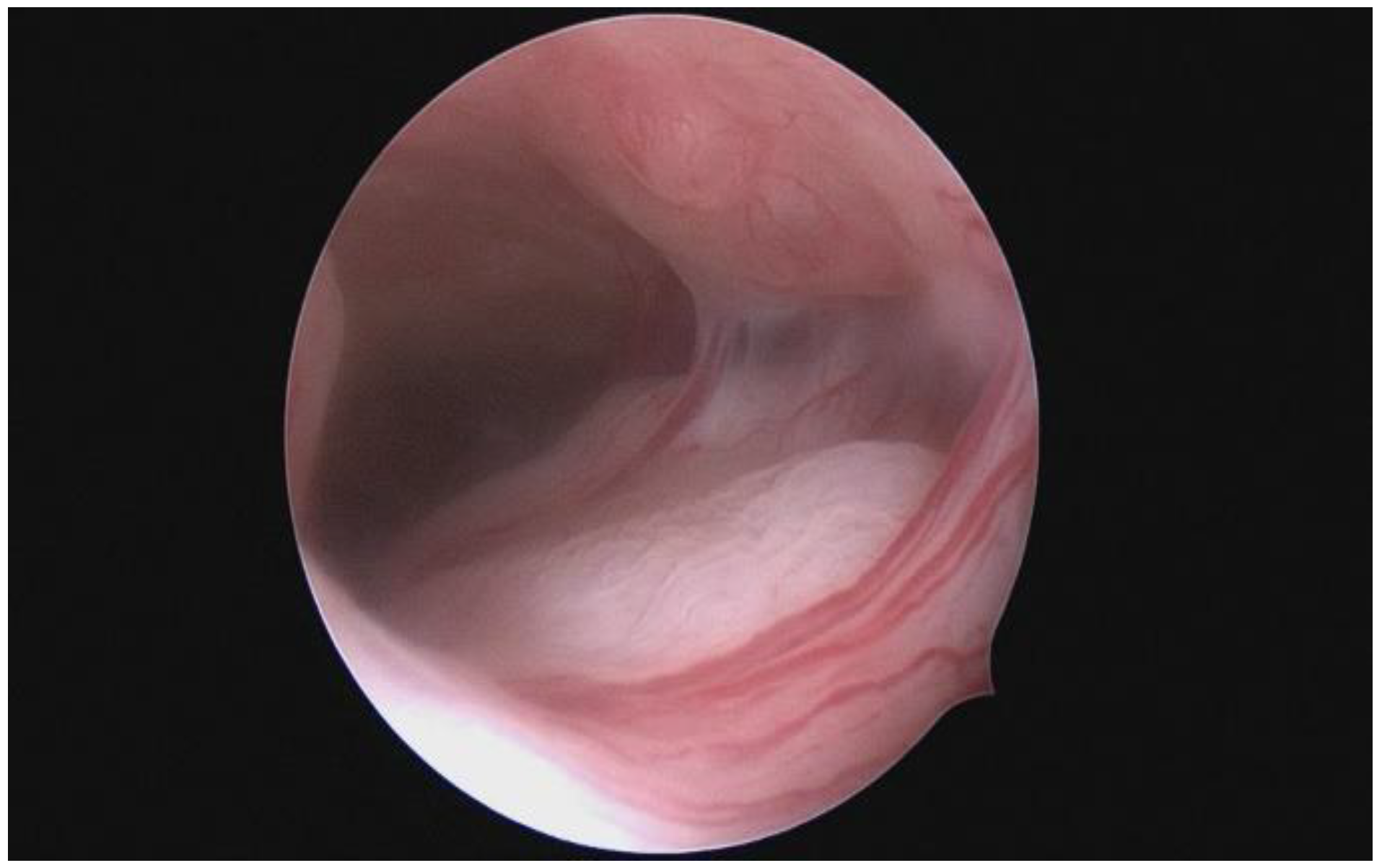
Figure 1. Uterine cavity partially occluded by adhesions.
Jdream 04 00031 g001
Figure 2. Classification system of the Intra Uterine Adhesions of the American Fertility Society (AFS).
Jdream 04 00031 g002aJdream 04 00031 g002b
Figure 3. The image shows a hysteroscopy with resectoscope for ablation of intrauterine adhesions in a patient with Asherman syndrome.
Jdream 04 00031 g003
Figure 4. Uterine cavity cleaned after ablation of intrauterine adhesions.
Jdream 04 00031 g004
Disclaimer/Publisher’s Note: The statements, opinions and data contained in all publications are solely those of the individual author(s) and contributor(s) and not of MDPI and/or the editor(s). MDPI and/or the editor(s) disclaim responsibility for any injury to people or property resulting from any ideas, methods, instructions or products referred to in the content.