Article

Development of Cu-Modified PVC and PU for Catalytic Generation of Nitric Oxide

1
School of Pharmacy and Biomolecular Sciences, University of Brighton, Brighton BN2 4GJ, UK
2
Surface Analysis Laboratory, School of Environment and Technology, University of Brighton, Brighton BN2 4GJ, UK
3
ANAMAD Ltd., Science Innovation Park, Science Park Square, Falmer, Brighton BN1 9SB, UK
*
Author to whom correspondence should be addressed.
Colloids Interfaces 2019, 3(1), 33; https://doi.org/10.3390/colloids3010033
Submission received: 22 December 2018 / Revised: 16 February 2019 / Accepted: 4 March 2019 / Published: 9 March 2019

Abstract

:
Nitric oxide (NO) generating surfaces are potentially promising for improving haemocompatibility of blood-contacting biomaterials. In the present report, Cu-modified poly(vinyl chloride) (PVC) and polyurethane (PU) were prepared via polydopamine (pDA)-assisted chelation. The copper content on the PVC and PU modified surfaces, assessed by inductively coupled plasma - optical emission spectrometry (ICP-OES), were about 3.86 and 6.04 nmol·cm−2, respectively. The Fourier-transform infrared spectroscopy (FTIR) and X-ray photoelectron spectroscopy (XPS) data suggest that copper is attached to the polymer surface through complex formation with pDA. The cumulative leaching of copper from modified PVC and PU during the five day incubation in phosphate buffered saline (PBS), measured by inductively coupled plasma mass spectrometry (ICP-MS), was about 50.7 ppb and 48 ppb, respectively which is within its physiological level. Modified polymers were tested for their ability to catalytically generate NO by decomposing of endogenous S-nitrosothiol (GSNO). The obtained data show that Cu-modified PVC and PU exhibited the capacity to generate physiological levels of NO which could be a foundation for developing new biocompatible materials with NO-based therapeutics.

1. Introduction

Poly(vinyl chloride) (PVC) and polyurethanes (PU) are the most commonly used polymers for blood-contacting applications [1]. The foremost important requirement for these polymers is the prevention of coagulation and platelet activation [1]. However, despite the progress that has been achieved in improving haemocompatibility of implantable biomaterials their long-term clinical applications still poses a problem [2]. The surface modification of the commercialized polymers is an efficient and cost effective approach to improving their haemocompatibility in comparison to creating new biomaterials [3]. During the recent decades, a number of biomaterials which release nitric oxide (NO) from the surface have been developed. NO has various physiological functions in the human body such as antiplatelet, anti-inflammatory, vasodilation [4,5] and inhibition of biofilm formation properties [6,7,8]. Healthy endothelial cells produce and release endogenously a NO flux of 0.5 4.0 × 10 10 mol·cm−2·min−1 [4]. Therefore, surfaces which release/generate NO locally are potentially promising for improving thromboresistance of biomaterials. One such approach to designing NO releasing surfaces is incorporation of exogenous NO donor molecules [5] and covalent binding of NO donors to the surface of various materials. However, biomaterials with covalently linked or embedded NO donors have several limitations such as the limited surface loading capacity of the NO donor, low NO donor stability, and formation of potentially toxic decomposition products such as N-nitrosoamines. The utility of NO-releasing biomaterials is limited to short-term contact biomedical devices [9,10,11,12]. S-nitrosothiols (RSNO) are a class of NO donors which are thought to serve as a reservoir and transporter of NO within biological systems [13]. The catalytic generation of NO from endogenous S-nitrosothiols at the blood/material interface was suggested as an alternative approach for the local NO delivery to the surface [6,14,15,16,17,18]. RSNOs are natural metabolites existing in the tissues and blood. Bearing in mind that RSNOs are continuously produced in the blood at concentrations ranging from 10 to 100 μM, they could be considered as an inexhaustible source of NO in vivo [19,20]. Therefore, designing NO-generating surfaces could be a potentially useful approach for long-term contact medical devices.
It has been reported that polymers with immobilized transition metal ions, primarily Cu 2 + and Fe 2 + [17,21], copper complexes or copper-containing metal–organic frameworks [6,8,14,15,16,17] catalytically decompose endogenous S-nitrosothiols with in situ generation of NO (Scheme 1). Copper ions catalysing RSNO decomposition are present in the blood and essential for the normal physiological function of the body.
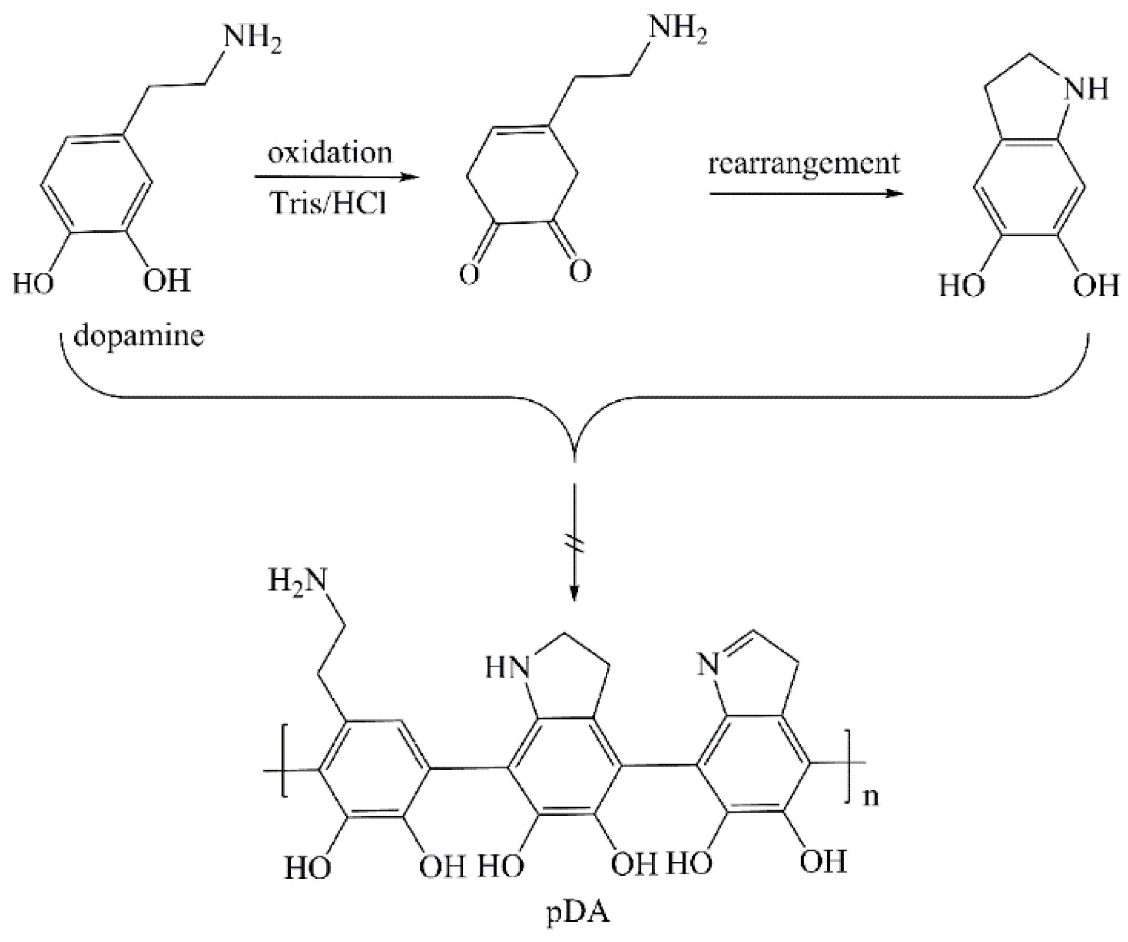
Depending on the substrate surface chemistry, different methods of copper immobilization on the surface have been proposed. Among them is a one-step incorporation of copper on the surface via catecholamines. Catecholamines are naturally occurring amines that function as neurotransmitters and hormones within the body. Dopamine (DA) (Scheme 1), one of catecholamines, has a half-life of a few minutes when circulating in the blood; it is secreted into urine after being broken down [22]. By self-polymerization DA can form a very thin layer of polydopamine (pDA) (Scheme 1) capable of attaching to various surfaces [23]. pDA exhibits stronger adhesion to the hydrophobic surfaces and adheres less strongly to the hydrophilic surfaces [24]. The metal ions attach to the surface via formation of complexes with pDA quinone and catechol units [25,26]. pDA provides a platform for further immobilization of biomolecules containing NH 2 and SH groups via Schiff base or Michael addition reactions [27,28]. According to literature, the structure of pDA is consists of a mixture of open-chain dopamine units and different oligomers with indole units with different degrees of saturation (Scheme 2).
Using the polydopamine-assisted chelation with copper we aimed to prepare novel Cu-modified PVC and PU polymers able to catalytically generate NO from the endogenous sources at physiological conditions.

2. Materials and Methods

2.1. Materials

Polyvinylchloride film (PVC, thickness 0.2 mm, Goodfellow Cambridge Ltd, Huntingdon, UK), polyurethane film (PU, thickness 80 µm, Delstar International Ltd, Brough, UK), phosphate-buffered saline tablets (PBS, MP Biomedicals, Illkirch, France), dopamine hydrochloride (DA), tris(hydroxymethyl)aminomethane (Tris), S-nitrosoglutathione (GSNO), l-glutathione reduced (GSH), copper (II) chloride dihydrate ( CuCl 2 · 2 H 2 O ), sodium nitrite and sodium nitrate (Sigma-Aldrich, St. Louis, MO, USA), and Cu solution (1000 ppm in 1 M HNO 3 from Fisher Scientific, Hampton, NH, USA) were used as received. All solutions were prepared with 18 MΩ cm−1 deionized (DI) water obtained using a Milli-Q system (Millipore Corp., Billerica, MA, USA). It showed negative results for the presence of Cu ions measured by inductively coupled plasma mass spectrometry (ICP-MS).

2.2. Method of Copper Immobilization

PVC and PU films were cut into round discs with d = 1.3 cm. Before modification, the polymer discs were cleaned by rinsing with ethanol, immersed in DI water and sonicated for 3 min, then rinsed with DI water, and soaked in Tris-HCl buffer solution (10 mM, pH 8.5) for 15 min prior to modification. Copper (II) chloride solution was prepared by dissolving CuCl 2 · 2 H 2 O in a Tris-HCl buffer solution (10 mM, pH 8.5) with stirring. To study the effect of DA:Cu2+ molar ratios, solutions with three different copper (II) concentrations, 3.5, 5.8 and 11.7 mM were made. Dopamine solution (2 g · L−1; 10.5 mM) was prepared by dissolving dopamine hydrochloride in Tris-HCl buffer solution (10 mM, pH 8.5) with stirring. A freshly prepared dopamine solution was always used because of self-polymerization of dopamine in the buffer in aerobic conditions [31]. Pre-soaked in buffer, PVC and PU discs were immersed into fresh dopamine solution, to which immediately an equal volume of a copper salt solution was gradually added with stirring and polymerization was conducted for 24 h at room temperature (RT) under continuous stirring in the dark. To determine the optimal DA:Cu2+ molar ratio, the polymerization was carried out at three different ratios, 3.5:1, 1.8:1 and 0.9:1. The total volume of the solution was 1 mL per disc. After incubation, discs were sonicated in DI water for 5 min to remove the non-attached pDA, then rinsed with DI water three times and air-dried. The obtained discs were denoted as PVC/pDA/Cu and PU/pDA/Cu. Polymers coated with pDA only without copper were use as controls (PVC/pDA and PU/pDA).

2.3. Fourier Transform Infrared Spectroscopy Method (FTIR) for Studying the Chemical Composition of Cu-Modified Polymers

To confirm pDA binding to the polymer surface, the chemical structure of the coatings was studied with FTIR using a PerkinElmer Spectrum 65 FT-IR Spectrometer (PerkinElmer, Machelen, Belgium) in the 650–4000 cm−1 range; the spectra were obtained in a transmittance mode averaging eight scans with a resolution of 8 cm−1. Discs of Cu-modified polymers were placed on the sample holder and FTIR spectra were recorded.

2.4. Analysis of Copper Content by Inductively Coupled Plasma-Optical Emission Spectrometry (ICP-OES)

A Perkin-Elmer Optima 2100 DV ICP-OES spectrometer was used for the copper content analysis. The experimental measurement conditions were as follows: wavelength 324.393 nm, sample flow rate 1.5 mL·min−1, sample uptake rate 1.0 mL·min−1, plasma gas flow rate 15 L·min−1, sample flow rate 1.5 mL·min−1, and operating power 1300 W. Discs of Cu-modified polymers were individually placed in 2 mL mixture of HCl : HNO 3 (1:1 v/v, concentrated) and incubated at 50 °C for 5 min. The obtained solution was cooled and filtered through a glass filter in order to separate from undissolved PU parts. The filtrate with concentrated acids was diluted with DI water to make 5 mL. Standards required for the calibration curve were prepared by serial dilution of the standard 1000 ppm copper solution. Blank and copper standard solutions were nebulized into plasma, where all components were vaporized. Blank and copper samples were dissociated and excited, and then the intensity of the radiation emitted by each sample was measured at a wavelength of 324.393 nm.

2.5. X-ray Photoelectron Spectroscopy (XPS) Analysis of the Chemical Composition of the Surface of Cu-Modified Polymeric Samples

The surface chemical composition of Cu-modified polymers was analysed with XPS using an ESCALAB 250 Xi system (Thermo Scientific, Waltham, MA, USA) equipped with a monochromated Al Kα X-ray source and based at the Surface Analysis Laboratory, University of Brighton, UK. Uniform charge neutralization was provided by beams of low-energy (≤10 eV) Ar+ ions and low-energy electrons guided by the magnetic lens. The standard analysis spot of ca. 900 × 600 μm2 was defined by the microfocused X-ray source. Full survey scans (step size 1 eV, pass energy 200 eV, dwell time 50 ms and 5 scans) and narrow scans (step size 0.1 eV, pass energy 40 eV, dwell time 100 ms and 15 scans) of the Cu2p (binding energy, BE ~935 eV), N1s (BE ~399 eV), C1s (BE ~285 eV) and O1s (BE ~531 eV) regions were acquired from three separate areas on each sample. Data were transmission function corrected and analysed with Thermo Avantage Software (Version 5.952, Thermo Fisher Scientific) using the Smart background. Discs of Cu-modified polymers were glued on sample holder and placed in the instrument chamber.

2.6. Evaluation of Copper Leaching from Polymers Studied by Inductively Coupled Plasma-Mass Spectrometry (ICP-MS)

Copper leaching from Cu-modified polymers was measured every 24 h for five days by ICP-MS 7900 (Agilent, Tokyo, Japan). The discs of Cu-modified polymers were individually placed into flat bottom vials, filled with 4 mL of PBS at RT and incubated for five days while shaking. Every 24 h polymeric discs were removed and placed into new vials containing 4 mL of fresh PBS. Then solutions from each incubation time (each vial) were transferred to the centrifuge flasks and 50 µL of concentrated nitric acid was added; then the resulting solutions were diluted up to 5 mL with PBS.

2.7. Nitric Oxide (NO) Release Measurements

The NO release catalysed by Cu-modified polymers was examined using the Arrowstraight™ Nitric Oxide ( NO 2 / NO 3 ) measurement system (Lazar Research Laboratories, Los Angeles, CA, USA) with micro ion-selective electrodes for independent measurement of both nitrite and nitrate ions. The GSNO stock solution of 500 µM in PBS was freshly prepared each time and its concentration was verified spectrophotometrically by measuring the absorption of the SNO group at 335 nm using an extinction coefficient of 922 M−1 cm−1 [28]. Stock solution of GSH in PBS (500 µM) was stored in a fridge for few months. The polymer discs were placed at the bottom of a 24-well plate and 0.5 mL of PBS solution was added to each well to equilibrate polymers for 5 min in PBS prior to adding the reagents. Afterwards, GSH and GSNO stock solutions, both of 500 µM, were added to each well (sample) in the volume to get the final concentration of both, GSH and GSNO of 100 µM in the resulting volume of 1 mL solution. Polymers samples were incubated in these solutions for 1 h at RT with shaking. After incubation, 200 µL of solution from each well was injected in the system to measure NO 2 and NO 3 concentrations. The catalytic decomposition of GSNO and quantification of NO release was calculated by summing up the concentrations of NO 2 and NO 3 in the resulting solutions. As a standard, the serial dilutions of 0.1 M sodium nitrite and 0.1 M sodium nitrate solutions were used. NO measurements were performed according to the manufacturer’s instructions using the software provided. All measurements were performed in triplicate. During the NO measurements, all solutions containing GSNO were shielded from light by foil as light greatly decreases the half-life of GSNO.

3. Results and Discussion

3.1. Chemical Structure of Cu-Modified Polymers

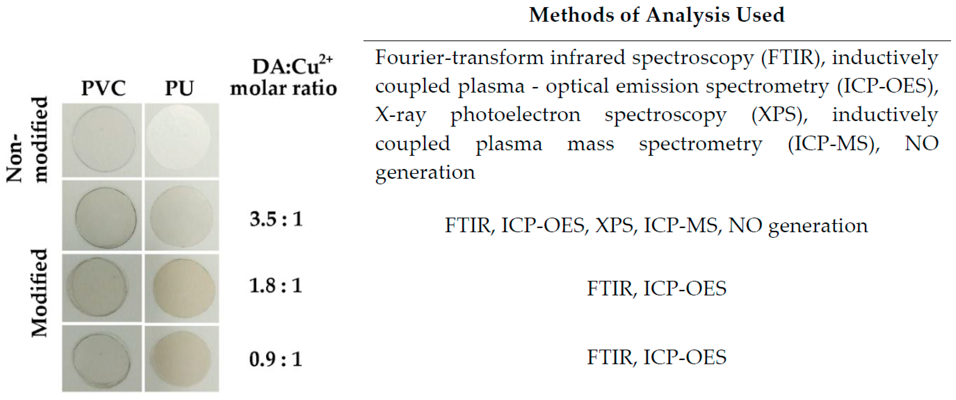
A series of copper-modified polymers with different DA: Cu 2 + molar ratios were prepared (Figure 1). This was done to find the optimal concentration of copper on the surface of Cu-modified polymers. Previously it was established that catecholamines, and in particular pDA, form stable metal-pDA complexes [32] and the molar ratio of dopamine to metal salt in the solution should be between 2:1 and 3:1 to ensure full incorporation of metal into the pDA [33]. A higher ratio of copper ions in the incubation solution leads to aggregation of pDa in solution. The polymer samples with different DA: Cu 2 + molar ratios were prepared (Figure 1).
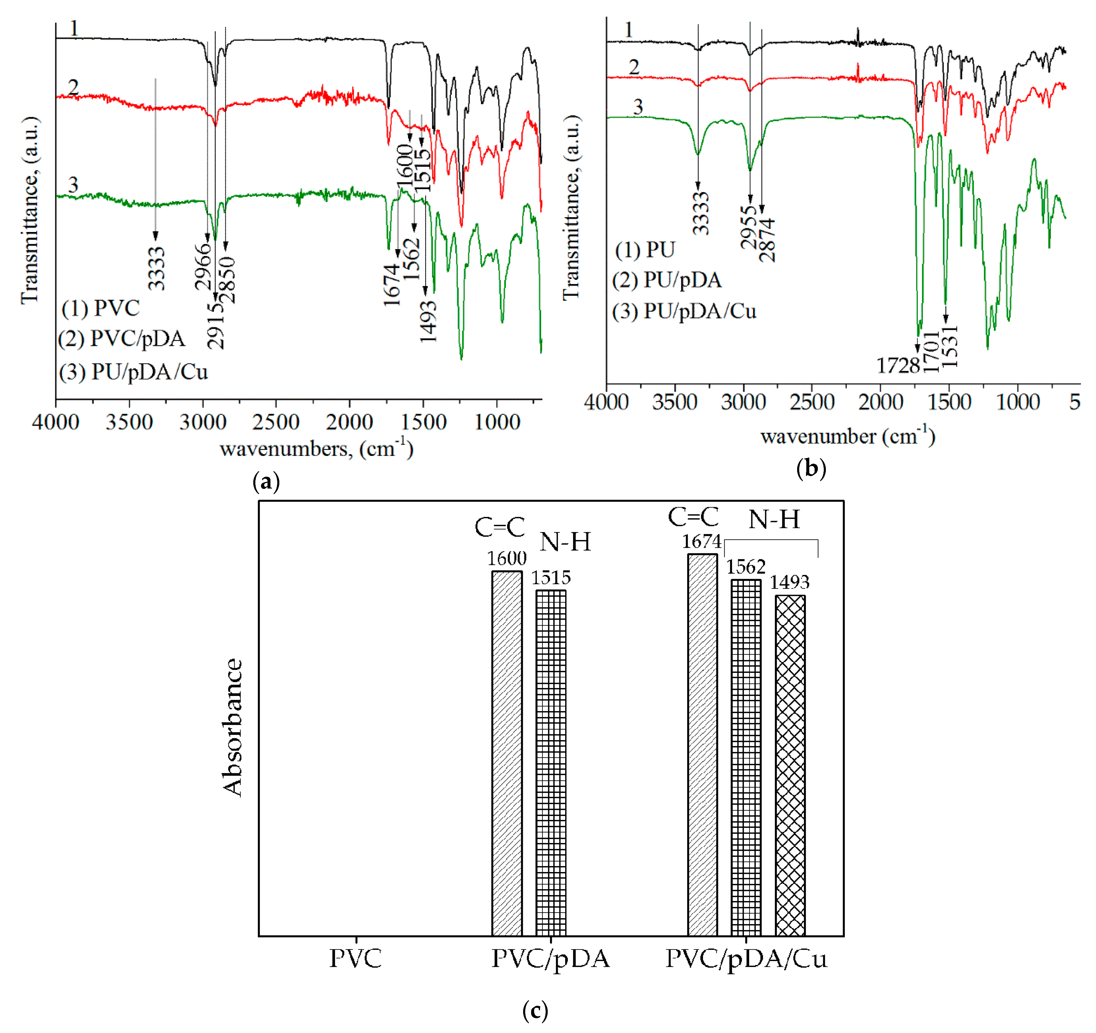
The results of FTIR analysis of Cu-modified samples are shown in Figure 2. Spectra of unmodified PVC and PU are typical of these polymers as reported in literature. Their assignation is given in the legend to Figure 2. In the spectrum of PVC/pDA at 3333 cm−1 and in the region between 1500–1600 cm−1 new peaks at 1600 cm−1 and 1515 cm−1 appeared, as shown in Figure 2a. These peaks can be attributed to pDA as reported in [31]. Although the chemical structure of pDA is still under discussion, it is generally accepted that it contains indole and/or indoline units [29]. Accordingly, these peaks were assigned to the overlapping C=C stretching vibrations from indole ring and, by different authors, to N–H scissoring vibrations or C=N vibrations thus presenting evidence of pDA formation [34]. A peak at 333 cm−1 in the spectrum of Cu-modified PVC was attributed to N–H vibrations. The peaks at 2867 cm−1, 2850 cm−1 and 2817 cm−1 in the FTIR spectrum of non-modified PVC were assigned to C–H stretching vibrations (Figure 2a, line 1).
The appearance of these peaks confirmed the presence of pDA film on the PVC surface. In the spectrum of Cu-modified PVC (Figure 2a) the band at 1600 cm−1 shifted to higher wavenumbers appearing at 1674 cm−1 and its intensity was decreased. The band at 1515 cm−1 split into two bands at 1493 cm−1 and 1562 cm−1. Such split indicates metal binding with polydopamine [35]. The FTIR spectra of Cu-modified PU are shown in Figure 2b. The peaks centred at 3333 cm−1, 2955 cm−1, 2874 cm−1, 1728 cm−1, 1701 cm−1, 1531 cm−1 are peaks originating from the PU. Peaks at 3333 cm−1, 2954 cm−1, 2874 cm−1 are from N–H and C–H groups; the band at 1728 cm−1 is due to the carbonyl groups in urethane bonds (C=O); the band centred at 1701 cm−1 is attributed to carbonyl groups in urea bond; the 1531 cm−1 band was assigned to secondary amide (RCONHR’). The FTIR spectra of PU modified by PDA and Cu did not provide clear evidence of the presence of PDA and copper on the modified PU surface because PU matrix bands overlapped with PDA peaks which have absorbance in the same region.
The characteristic peaks of pDA appeared in the FTIR spectra after modification confirms the successful modification by pDA. The shift peak at 1600 cm−1 to higher wavenumbers and split of the band at 1515 cm−1 into two bands at 1491 cm−1 and 1562 cm−1 indicates the formation of a Cu-PDA complex as observed previously for Fe-PDA and Zn-PDA complexes [8,36].
It was reported that the amount of 13 nmol of Cu 2 + immobilized on a modified PU was sufficient to produce physiologically relevant levels of NO in 2 mL of 10 µM solution of GSNO/GSH in PBS [15]. The amount of copper in the coatings was estimated using ICP-OES. The results are shown in Table 1. The highest copper content was found in the samples obtained at DA: Cu 2 + molar ratio 3.5:1, being 3.86 nmol cm−2 and 6.04 nmol cm−2 for Cu-modified PVC and PU, respectively. Subsequently, the samples obtained at DA: Cu 2 + molar ratio 3.5:1 were chosen for further investigation.
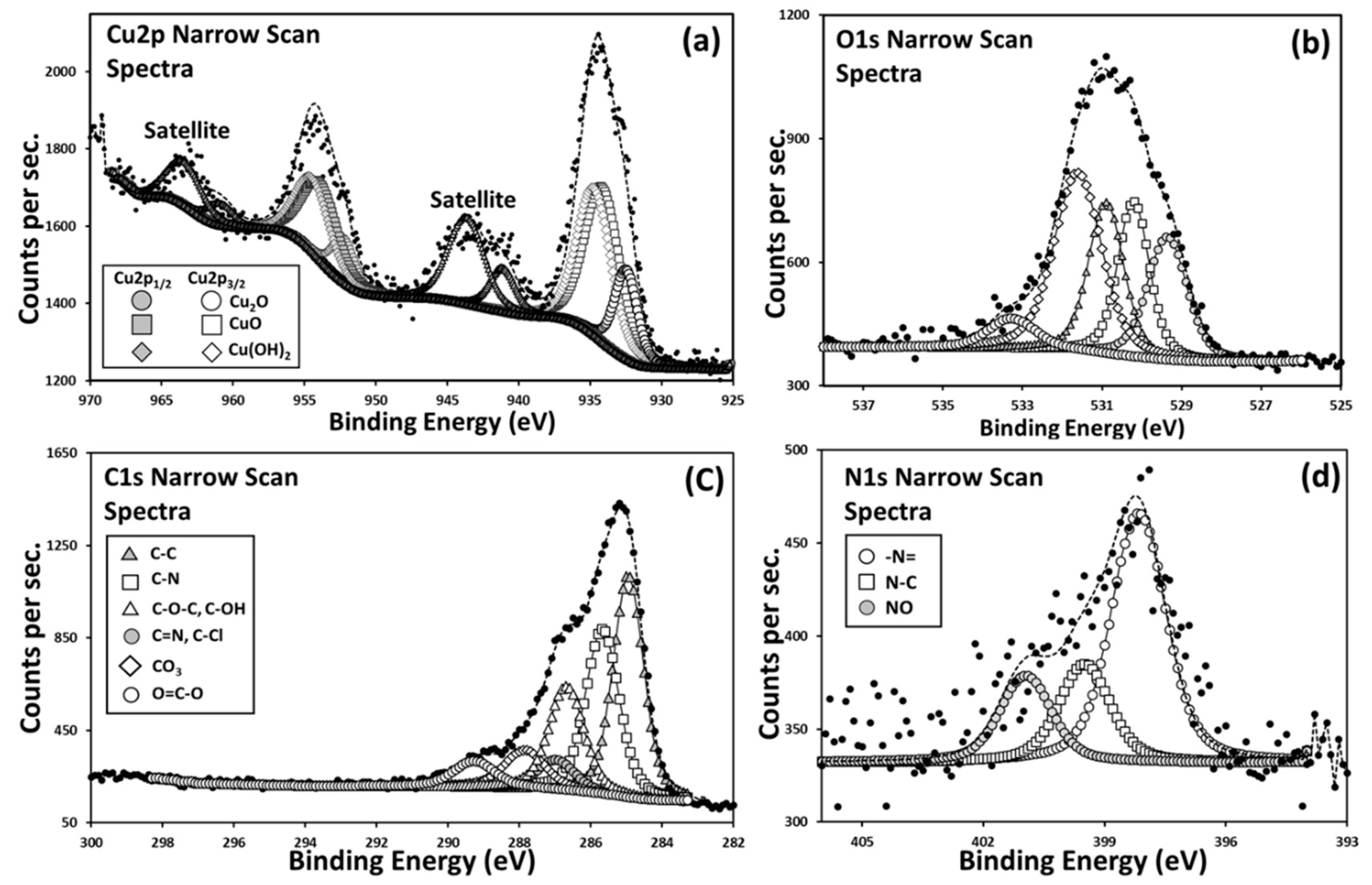
XPS analysis was performed to detect copper in the coating along with finding the valence states of individual elements to better understand about their chemical surroundings and the mechanism of attachment to the polymer surface. In Figure 3, the narrow scan spectra of Cu2p, O1s, C1s and N1s of Cu-modified PVC surface are shown. The two peaks observed for the binding energy (BE) of copper with maxima at 955 eV and 935 eV can be attributed to Cu 2p1/2 orbital and Cu 2p3/2 orbitals respectively, and peaks around 943 and 964 eV are the satellite peaks, which are common to Cu2p XPS data. The position of the main Cu 2p3/2 peak ca. 933–934 eV and the satellite peak ca. 942–943 eV is typical of Cu(II) catecholate complexes [37]. Deconvoluted Cu2p3/2 narrow scan data for four representative systems are shown in Figure S1. Cu is present in both of its chemical states of Cu+ and Cu+2, with Cu2O, CuO and Cu(OH)2. All peaks are assigned with references under NIST database. The XPS data suggest that Cu-pDA was successfully immobilized on the surface of PVC and PU, which are consistent with the results of FTIR and ICP-OES studies. XPS of C1s and N1s also confirm the presence of C–N bonds, i.e., pDA and XPS of O1s indicate the presence of oxygen atoms in the state similar to that in Cu–O bonds. The presence of Cu(I) species in the surface layer is likely to be the result of a redox reaction between pDA and Cu(II).
The atomic fraction of copper element in the surface coating of Cu-modified PVC is 1.5% (Table 2), which is close to the reported 1.4% on polysulfone and 1.36% on polyvinydienefluoride ultrafiltration membranes [38]. In case of Cu modified PU, there is much less Cu detected than expected from other experimental results. It could be due to various reasons, but most possible reason could be that majority of the Cu is located below 10 nm of top surface layer, making it relatively unquantifiable using XPS and the other reason could be possible adventitious organic layer on the top.
Using XPS, we evaluated the leaching of copper from Cu-modified polymers to 100 µM GSNO/GSH solution during 1 h (Table 2). Copper content of PVC/pDA/Cu and PU/pDA/Cu decreased from 1.5 to 1.1 atomic % and from 0.54 to 0.44 atomic %, respectively, after incubation in GSNO/GSH solution. A decrease in surface carbon, nitrogen and oxygen contents should be observed in case of the detachment of PDA. The XPS results show that the carbon content, O/C and N/C atomic ratio of initial PVC/pDA/Cu are 72.4, 0.22 and 0.08, respectively, whereas the carbon content after incubation in the 100 µM GSNO/GSH solution reduced to 67.8 atomic %, and the atomic ratios of O/C and N/C of PVC/pDA/Cu increased to 0.27 and 0.10, which indicated that chemical reactions took place during incubation of the sample in GSNO/GSH. The same trend similar to PVC/pDA/Cu is observed for PU/pDA/Cu. Despite the widespread application of pDA coatings for biomaterials development, its structure is still under discussion. It is known that GSNO exhibits oxidizing properties, so the increase in the O/C ratio could be explained by the surface oxidation, but it is difficult to determine which groups were oxidized. Similarly, the reduction in carbon content could be explained by the surface oxidation, however it requires further investigation. On the other hand, the pDA-coated material used in the catalytic oxidation in the reaction medium containing 30% aqueous hydrogen peroxide exhibited good stability [39]. Moreover, the Michael addition reaction between pDA and glutathione (GSH) can occur due to the ability of pDA to react with amine- and thiol-containing molecules, which could also explain the increase in O/C and N/C ratios.
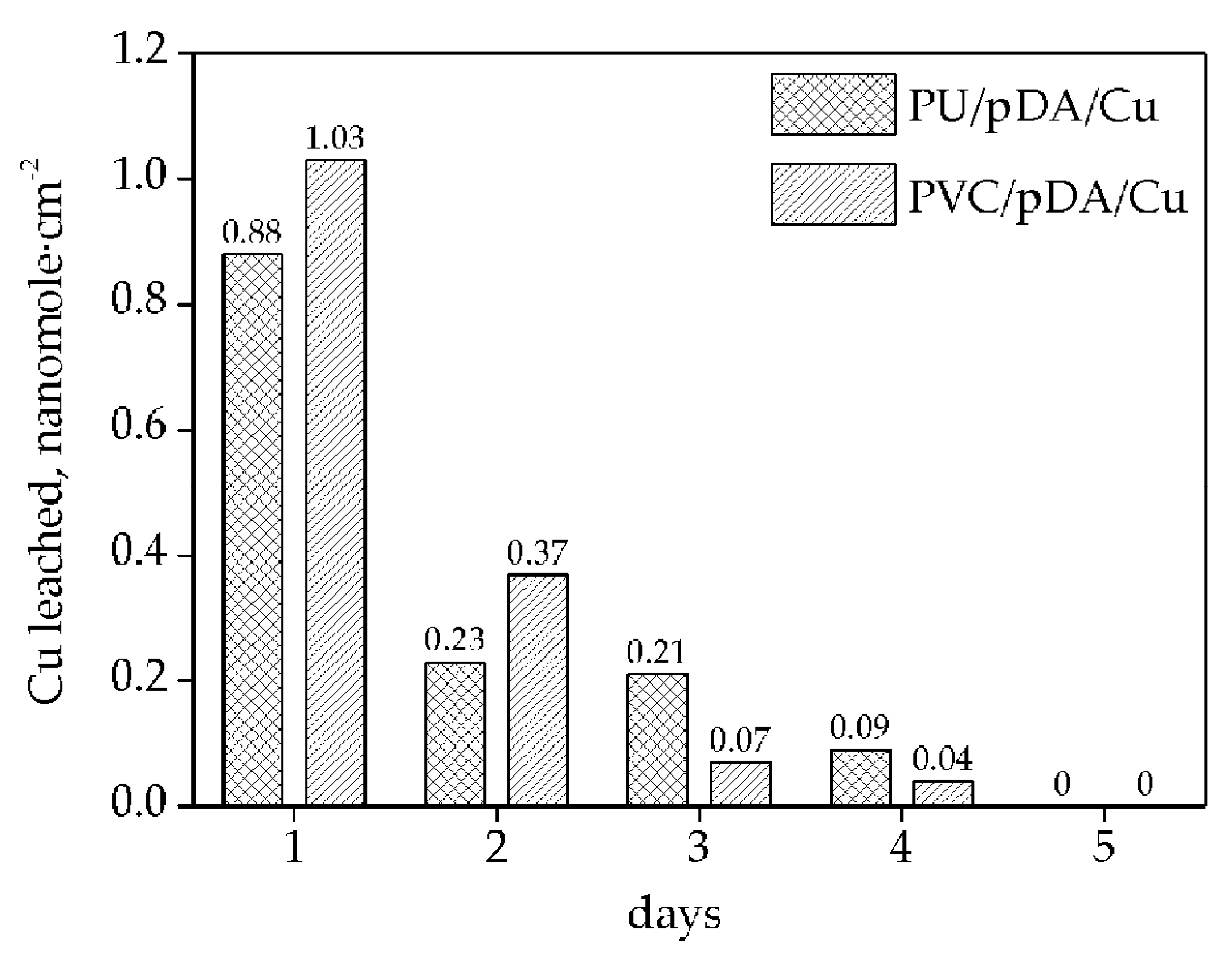
Leaching of Cu from Cu-modified samples to PBS during 5 days was evaluated. Cumulative leaching from Cu-modified PVC for 5 days was 1.51 nmol·cm−2, for Cu-modified PU was equal to 1.41 nmol·cm−2 (Figure 4). That is about 39% of Cu content on Cu-modified PVC and 23% on Cu-modified PU.
The normal level of free copper in the human blood serum is 1.6–2.4 μM or 100–150 ppb [40]. The highest measured Cu leaching level from samples was recorded at 50.7 ppb in five days (Table 3), which is well below cytotoxic concentrations for mammalian cells. It was reported that the viability of mammalian cells was not affected until the copper concentration reached 1000 ppb [41]. Our results suggest that copper leaching from the samples should pose no risk to health.

3.2. Measurement of NO Generation Catalyzed by Modified Materials

According to XPS data copper on the surface of Cu-modified polymers exists in the form of Cu 2 + and Cu + . The actual catalyst of GSNO decomposition is Cu + . However, the catalytic mechanism includes Cu redox-cycle between the Cu 2 + and Cu + . The NO generating mechanism involves the decomposition of GSNO by Cu + ions, which partially exist in the sample surface and also form by reduction of Cu 2 + ions by glutathione (GSH) (Figure 5) [35]. The traces amounts of Cu + affect the catalysis. These reactions proceeds until GSNO depletion and its conversion to disulfide.
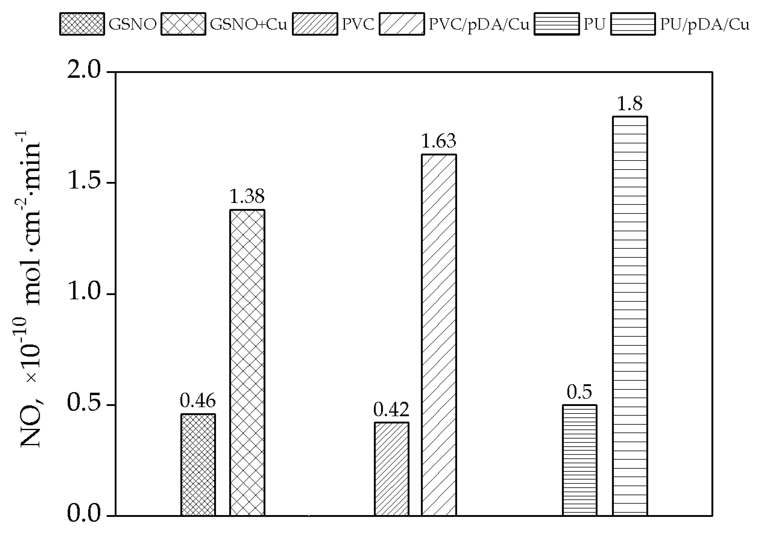
NO generation ability of Cu-containing samples was studied using GSNO as the nitric oxide donor, because GSNO is a compound relatively stable to self-decomposition and has long half-life. The NO release from S-nitrosoglutathione catalyzed by Cu-polymer samples was found to be within the range (0.43 − 1.4) × 10−10 mol · cm 2 · min 1 (Figure 6). These data are within the physiological level of NO release by endothelial cells, which is (0.5 − 4) × 10−10 mol · cm 2 · min 1 [4].
It was reported that GSNO is also able to release NO in the presence of GSH without Cu [42], but both endogenous and synthetic RSNOs are prone to catalytic decomposition by copper ions [17,32]. We have shown that Cu-pDA modified PVC and PU can catalyse NO generation in the presence of GSNO. The highest NO generation rate was exhibited by the Cu-modified PU which correlated with higher copper content in the polymer in comparison with Cu-modified PVC according to ICP-OES analysis.
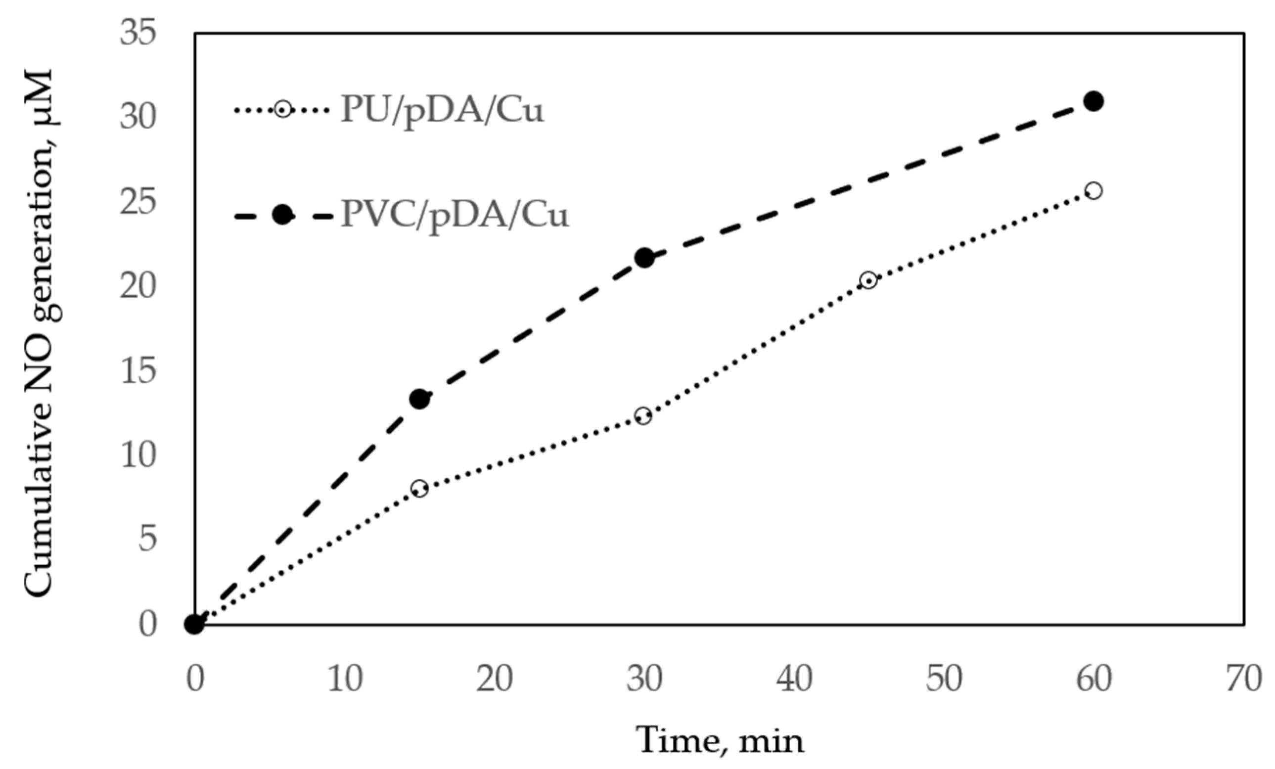
According to the kinetics of NO generation, about 35% of GSNO was catalytically decomposed by PVC/pDA/Cu and 25% by PU/pDA/Cu within the first hour (Figure 7).

4. Conclusions

Cu-modified PVC and PU polymers for catalytic NO generation were produced using a one-step preparation method. The presence of Cu on both modified polymers was proved by ICP-OES and XPS data. According to ICP-OES measurements, the amount of Cu species on the surface of Cu-modified PU was higher than on the Cu-modified PVC. FTIR spectra showed the characteristic peaks of pDA and Cu complexes with pDA on the modified PVC polymer surface. The FTIR data did not give clear evidence of polydopamine presence and Cu complex formation on Cu-modified PU surface because of overlapping of the characteristic absorbance bands of the coatings with strong bands from PU matrix. It was found that a small amount of copper can leach from the modified polymer surfaces during five days, however it is within its physiological level in blood. The NO release test showed that Cu-modified PVC and PU polymers are capable of providing the physiological level of NO at physiological pH. The obtained data suggest that Cu-modified polymers potentially can produce nitric oxide in the blood from endogenous S-nitrosothiols. Taking into account the catalytic mechanism of NO generation, this process could continue long enough while in contact with blood to secure the long-term haemocompatibility of medical implants.

Supplementary Materials

The following are available online at https://www.mdpi.com/2504-5377/3/1/33/s1, Figure S1: Cu2p3/2 narrow scan XPS data of four samples, Cu modified PVC and PU with PDA, before and after incubation. Corresponding peaks are assigned following NIST database.

Author Contributions

Conceptualization, L.M. and S.M.; Methodology, L.A. and L.M.; Investigation, L.A., S.R. and L.M.; Writing—Original Draft Preparation, L.A.; Writing—Review & Editing, L.M. and S.M.; Visualization, L.A. and S.R.; Supervision, L.M.; Project Administration, L.A.; Funding Acquisition, L.M. and S.M.

Funding

This research was funded by the Marie Skłodowska-Curie Action of the European Union H2020-MSCA-IF-2016, grant number 749207.

Acknowledgments

This work is supported by the Marie Skłodowska-Curie Action of the European Union (H2020-MSCA-IF-2016, grant agreement No. 749207). The authors acknowledge the DelStar Company for donating PU films and Peter Lyons and the Magdalena Grove at School of Environment and Technology, University of Brighton, for ICP-OES and ICP-MS measurements.

Conflicts of Interest

The authors declare no conflict of interest.

References

  1. Dumitriu, S. Polymeric Biomaterials, Revised and Expanded; CRC Press: Boca Raton, FL, USA, 2001. [Google Scholar]
  2. Bajpai, A.K.; Bajpai, J.; Saini, R.K.; Agrawal, P.; Tiwari, A. Smart Biomaterial Devices: Polymers in Biomedical Sciences; CRC Press: Boca Raton, FL, USA, 2016. [Google Scholar]
  3. Yang, Y.; Qi, P.K.; Yang, Z.L.; Huang, N. Nitric oxide based strategies for applications of biomedical devices. Biosurf. Biotribol. 2015, 1, 177–201. [Google Scholar] [CrossRef] [Green Version]
  4. de Mel, A.; Murad, F.; Seifalian, A.M. Nitric oxide: A guardian for vascular grafts? Chem. Rev. 2011, 111, 5742–5767. [Google Scholar] [CrossRef] [PubMed]
  5. Naghavi, N.; Mel, A.D.; Alavijeh, O.S.; Cousins, B.G.; Seifalian, A.M. Nitric oxide donors for cardiovascular implant applications. Small 2013, 9, 22–35. [Google Scholar] [CrossRef] [PubMed]
  6. Pant, J.; Goudie, M.J.; Hopkins, S.P.; Brisbois, E.J.; Handa, H. Tunable nitric oxide release from s-nitroso-n-acetylpenicillamine via catalytic copper nanoparticles for biomedical applications. ACS Appl. Mater. Interfaces 2017, 9, 15254–15264. [Google Scholar] [CrossRef]
  7. Wo, Y.; Brisbois, E.J.; Bartlett, R.H.; Meyerhoff, M.E. Recent advances in thromboresistant and antimicrobial polymers for biomedical applications: Just say yes to nitric oxide (no). Biomater. Sci. 2016, 4, 1161–1183. [Google Scholar] [CrossRef] [PubMed]
  8. Wu, H.; Ang, J.M.; Kong, J.; Zhao, C.; Du, Y.; Lu, X. One-pot synthesis of polydopamine–zn complex antifouling coatings on membranes for ultrafiltration under harsh conditions. RSC Advances 2016, 6, 103390–103398. [Google Scholar] [CrossRef]
  9. Damodaran, V.B.; Joslin, J.M.; Wold, K.A.; Lantvit, S.M.; Reynolds, M.M. S-nitrosated biodegradable polymers for biomedical applications: Synthesis, characterization and impact of thiol structure on the physicochemical properties. J. Mater. Chem. 2012, 22, 5990–6001. [Google Scholar] [CrossRef]
  10. Riccio, D.A.; Schoenfisch, M.H. Nitric oxide release: Part I. Macromolecular scaffolds. Chem. Soc. Rev. 2012, 41, 3731–3741. [Google Scholar] [CrossRef]
  11. Seabra, A.B.; Marcato, P.D.; de Paula, L.B.; Durán, N. New strategy for controlled release of nitric oxide. J. Nano Res. 2012, 20, 61–67. [Google Scholar] [CrossRef]
  12. Kim, J.; Saravanakumar, G.; Choi, H.W.; Park, D.; Kim, W.J. A platform for nitric oxide delivery. J. Mater. Chem. B 2014, 2, 341–356. [Google Scholar] [CrossRef]
  13. Dicks, A.P.; Swift, H.R.; Williams, D.L.H.; Butler, A.R.; Al-Sa’doni, H.H.; Cox, B.G. Identification of cu+ as the effective reagent in nitric oxide formation from s-nitrosothiols (rsno). J. Chem. Soc. Perkin Trans. 2 1996, 481–487. [Google Scholar] [CrossRef]
  14. Puiu, S.C.; Zhou, Z.; White, C.C.; Neubauer, L.J.; Zhang, Z.; Lange, L.E.; Mansfield, J.A.; Meyerhoff , M.E.; Reynolds, M.M. Metal ion-mediated nitric oxide generation from polyurethanes via covalently linked copper(ii)-cyclen moieties. J. Biomed. Mater. Res. Part B Appl. Biomater. 2009, 91, 203–212. [Google Scholar] [CrossRef] [PubMed]
  15. Hwang, S.; Meyerhoff, M.E. Polyurethane with tethered copper(ii)–cyclen complex: Preparation, characterization and catalytic generation of nitric oxide from s-nitrosothiols. Biomaterials 2008, 29, 2443–2452. [Google Scholar] [CrossRef] [PubMed]
  16. Luo, R.; Liu, Y.; Yao, H.; Jiang, L.; Wang, J.; Weng, Y.; Zhao, A.; Huang, N. Copper-incorporated collagen/catechol film for in situ generation of nitric oxide. ACS Biomater. Sci. Eng. 2015, 1, 771–779. [Google Scholar] [CrossRef]
  17. Neufeld, M.J.; Lutzke, A.; Jones, W.M.; Reynolds, M.M. Nitric oxide generation from endogenous substrates using metal–organic frameworks: Inclusion within poly(vinyl alcohol) membranes to investigate reactivity and therapeutic potential. ACS Appl. Mater. Interfaces 2017, 9, 35628–35641. [Google Scholar] [CrossRef] [PubMed]
  18. Oh, B.K.; Meyerhoff, M.E. Spontaneous catalytic generation of nitric oxide from s-nitrosothiols at the surface of polymer films doped with lipophilic copper(ii) complex. J. Am. Chem. Soc. 2003, 125, 9552–9553. [Google Scholar] [CrossRef]
  19. Nagababu, E.; Rifkind, J.M. Routes for formation of s-nitrosothiols in blood. Cell Biochem. Biophys. 2013, 67, 385–398. [Google Scholar] [CrossRef]
  20. Zhang, Y.; Hogg, N. S-nitrosothiols: Cellular formation and transport. Free Radic. Biol. Med. 2005, 38, 831–838. [Google Scholar] [CrossRef]
  21. Vanin, A.F.; Papina, A.A.; Serezhenkov, V.A.; Koppenol, W.H. The mechanisms of s-nitrosothiol decomposition catalyzed by iron. Nitric Oxide 2004, 10, 60–73. [Google Scholar] [CrossRef]
  22. Peaston, R.T.; Weinkove, C. Measurement of catecholamines and their metabolites. Ann. Clin. Biochem. 2004, 41, 17–38. [Google Scholar] [CrossRef]
  23. Liu, Y.; Ai, K.; Lu, L. Polydopamine and its derivative materials: Synthesis and promising applications in energy, environmental, and biomedical fields. Chem. Rev. 2014, 114, 5057–5115. [Google Scholar] [CrossRef] [PubMed]
  24. Zhang, C.; Gong, L.; Xiang, L.; Du, Y.; Hu, W.; Zeng, H.; Xu, Z.-K. Deposition and adhesion of polydopamine on the surfaces of varying wettability. ACS Appl. Mater. Interfaces 2017, 9, 30943–30950. [Google Scholar] [CrossRef] [PubMed]
  25. Mrówczyński, R.; Markiewicz, R.; Liebscher, J. Chemistry of polydopamine analogues. Polym. Int. 2016, 65, 1288–1299. [Google Scholar] [CrossRef]
  26. Saiz-Poseu, J.; Mancebo-Aracil, J.; Nador, F.; Busqué, F.; Ruiz-Molina, D. The chemistry behind catechol-based adhesion. Angew. Chem. Int. Ed. 2019, 58, 696–714. [Google Scholar] [CrossRef]
  27. Ding, Y.H.; Floren, M.; Tan, W. Mussel-inspired polydopamine for bio-surface functionalization. Biosurf. Biotribol. 2016, 2, 121–136. [Google Scholar] [CrossRef]
  28. Huang, N.; Zhang, S.; Yang, L.; Liu, M.; Li, H.; Zhang, Y.; Yao, S. Multifunctional electrochemical platforms based on the michael addition/schiff base reaction of polydopamine modified reduced graphene oxide: Construction and application. ACS Appl. Mater. Interfaces 2015, 7, 17935–17946. [Google Scholar] [CrossRef]
  29. Liebscher, J.; Mrówczyński, R.; Scheidt, H.A.; Filip, C.; Hădade, N.D.; Turcu, R.; Bende, A.; Beck, S. Structure of polydopamine: A never-ending story? Langmuir 2013, 29, 10539–10548. [Google Scholar] [CrossRef]
  30. Ryu, J.H.; Messersmith, P.B.; Lee, H. Polydopamine surface chemistry: A decade of discovery. ACS Appl. Mater. Interfaces 2018, 10, 7523–7540. [Google Scholar] [CrossRef]
  31. Dreyer, D.R.; Miller, D.J.; Freeman, B.D.; Paul, D.R.; Bielawski, C.W. Elucidating the structure of poly(dopamine). Langmuir 2012, 28, 6428–6435. [Google Scholar] [CrossRef]
  32. Szpoganicz, B.; Gidanian, S.; Kong, P.; Farmer, P. Metal binding by melanins: Studies of colloidal dihydroxyindole-melanin, and its complexation by cu(ii) and zn(ii) ions. J. Inorg. Biochem. 2002, 89, 45–53. [Google Scholar] [CrossRef]
  33. Wang, Z.; Xie, Y.; Li, Y.; Huang, Y.; Parent, L.R.; Ditri, T.; Zang, N.; Rinehart, J.D.; Gianneschi, N.C. Tunable, metal-loaded polydopamine nanoparticles analyzed by magnetometry. Chem. Mater. 2017, 29, 8195–8201. [Google Scholar] [CrossRef]
  34. Zangmeister, R.A.; Morris, T.A.; Tarlov, M.J. Characterization of polydopamine thin films deposited at short times by autoxidation of dopamine. Langmuir 2013, 29, 8619–8628. [Google Scholar] [CrossRef]
  35. Zhang, C.; Biggs, T.D.; Devarie-Baez, N.O.; Shuang, S.; Dong, C.; Xian, M. S-nitrosothiols: Chemistry and reactions. Chem. Commun. 2017, 53, 11266–11277. [Google Scholar] [CrossRef] [PubMed]
  36. Sever, M.J.; Weisser, J.T.; Monahan, J.; Srinivasan, S.; Wilker, J.J. Metal-mediated cross-linking in the generation of a marine-mussel adhesive. Angew. Chem. Int. Ed. 2004, 43, 448–450. [Google Scholar] [CrossRef] [PubMed]
  37. Brown, D.G.; Hughes, W.J.; Knerr, G. Electronic and x-ray photoelectron spectra of copper catecholate complexes. Inorg. Chim. Acta 1980, 46, 123–126. [Google Scholar] [CrossRef]
  38. Liu, Z.; Hu, Y.; Liu, C.; Zhou, Z. Surface-independent one-pot chelation of copper ions onto filtration membranes to provide antibacterial properties. Chem. Commun. 2016, 52, 12245–12248. [Google Scholar] [CrossRef] [PubMed] [Green Version]
  39. Ye, W.; Shi, X.; Su, J.; Chen, Y.; Fu, J.; Zhao, X.; Zhou, F.; Wang, C.; Xue, D. One-step reduction and functionalization protocol to synthesize polydopamine wrapping ag/graphene hybrid for efficient oxidation of hydroquinone to benzoquinone. Appl. Catal. B Environ. 2014, 160–161, 400–407. [Google Scholar] [CrossRef]
  40. Brewer, G.J. Wilson’s disease. In Harrison’s Principles of Internal Medicine, 18th ed.; Longo, D., Fauci, A., Kasper, D., Hauser, S., Jameson, J., Loscalzo, J., Eds.; McGraw-Hill Education: New York, NY, USA, 2011; pp. 3188–3190. [Google Scholar]
  41. Valodkar, M.; Rathore, P.S.; Jadeja, R.N.; Thounaojam, M.; Devkar, R.V.; Thakore, S. Cytotoxicity evaluation and antimicrobial studies of starch capped water soluble copper nanoparticles. J. Hazard. Mater. 2012, 201–202, 244–249. [Google Scholar] [CrossRef]
  42. Singh, S.P.; Wishnok, J.S.; Keshive, M.; Deen, W.M.; Tannenbaum, S.R. The chemistry of the s-nitrosoglutathione/glutathione system. Proc. Natl. Acad. Sci. USA 1996, 93, 14428–14433. [Google Scholar] [CrossRef]
Scheme 1. Scheme of S-nitrosoglutathione (GSNO) structure, reaction of GSNO decomposition by copper.
Colloids 03 00033 sch001
Scheme 2. Scheme of dopamine and polydopamine (pDA) structure [26,29,30].
Colloids 03 00033 sch002
Figure 1. (Left)—Photographs of non-modified poly(vinyl chloride) (PVC) and polyurethane (PU) (upper row) and Cu-modified polymers (lower rows) obtained via incubation of polymers in in Tris-HCl buffer with different DA: Cu 2 + molar ratios; (Right)—methods of analysis used to determine copper content and NO generation by the polymers.
Colloids 03 00033 g001
Figure 2. (a) Infrared spectra of PVC discs: (1) non-modified; (2) modified with PDA and (3) modified with Cu-PDA; (b) Infrared spectra of PU discs: (1) non-modified; (2) modified with PDA and (3) modified with Cu-PDA; (c) A diagram of appearing and shifting absorbance bands in Fourier-transform infrared spectroscopy (FTIR) spectra of PVC, PVC/PDA and PVC/PDA/Cu.
Colloids 03 00033 g002
Figure 3. XPS narrow spectra of PVC/pDA/Cu disc surface; (a) Cu2p, (b) O1s, (c) C1s and (d) N1s. References to individual peak assignments are shown in supporting information table SI-T1.
Colloids 03 00033 g003
Figure 4. ICP-MS data of copper leached from the surface of Cu-modified polymers during 5 days.
Colloids 03 00033 g004
Figure 5. The schematic presentation of the mechanism of nitric oxide (NO) generation from GSNO in the presence of GSH in phosphate-buffered saline (PBS).
Colloids 03 00033 g005
Figure 6. NO generation after incubation of Cu-modified polymers with 100 µM GSNO/GSH in the PBS during 1 h.
Colloids 03 00033 g006
Figure 7. NO generation after incubation of Cu-modified polymers with 100 µM GSNO/GSH in the PBS.
Colloids 03 00033 g007
Table 1. Copper content on the surface of PVC and PU modified by dopamine from solutions with different DA: Cu 2 + molar ratios according to ICP-OES.
SampleCu, nmol cm−2DA: Cu2+ Molar Ratio
PVC/pDA/Cu3.86 ± 0.303.5:1
PVC/pDA/Cu1.35 ± 0.281.8:1
PVC/pDA/Cu0.96 ± 0.300.9:1
PU/pDA/Cu6.04 ± 0.173.5:1
PU/pDA/Cu1.97 ± 0.421.8:1
PU/pDA/Cu1.4 ± 0.700.9:1
Table 2. Atomic composition of the Cu-modified PVC before and after incubation with 100 µM GSNO/l-glutathione reduced (GSH) solution during 1 h obtained by XPS.
PVC/pDA/CuPU/pDA/Cu
ElementsBeforeAfterBeforeAfter
N1s5.66.93.233.82
C1s72.467.875.6166.26
O1s15.718.520.1425.08
Cl2p4.44.70.111.75
Na1s0.10.70.372.65
Cu2p31.51.10.540.44
N/C ratio0.080.10.040.06
O/C ratio0.220.270.270.38
Table 3. Cumulative Cu leaching from the samples during 5 days determined by ICP-MS data.
Sample5 Days, ppb5 Days, % from Original
PVC/pDA/Cu (3.5:1)50.7 ± 14.539
PU/pDA/Cu (3.5:1)48.1 ± 11.623