Article

Rational Design of Lecithin–Cholesterol Liposomes for Encapsulation and Sustained Release of Diclofenac

by
Ángela Sánchez-García
1,
Francisco Ortega
1,2,
Ramón G. Rubio
1 and
Eduardo Guzmán
1,2,*
1
Departamento de Química Física, Facultad de Ciencias Químicas, Universidad Complutense de Madrid, Plaza de las Ciencias s/n, Ciudad Universitaria, 28040 Madrid, Spain
2
Instituto Pluridisciplinar, Universidad Complutense de Madrid, Paseo Juan XXIII 1, 28040 Madrid, Spain
*
Author to whom correspondence should be addressed.
Colloids Interfaces 2026, 10(2), 25; https://doi.org/10.3390/colloids10020025
Submission received: 26 December 2025 / Revised: 3 February 2026 / Accepted: 3 March 2026 / Published: 4 March 2026
(This article belongs to the Special Issue Biocolloids and Biointerfaces: 3rd Edition)

Abstract

Liposomes are widely recognized as versatile nanocarriers in drug delivery due to their biocompatibility, tunable physicochemical properties, and ability to incorporate both hydrophilic and hydrophobic compounds. In this study, the encapsulation and release of diclofenac, a nonsteroidal anti-inflammatory drug (NSAID), using lecithin–cholesterol liposomes are explored. Encapsulation parameters were first optimized with calcein as a model fluorophore, confirming that cholesterol addition enhances encapsulation efficiency by reducing membrane permeability. Guided by these results, liposomes containing equal weight fractions of lecithin and cholesterol were selected as an optimized formulation, providing calcein and diclofenac encapsulation efficiencies up to approximately 35% while maintaining hydrodynamic diameters below 300 nm with low polydispersity (PdI < 0.2), optimal for intravenous administration and prolonged systemic circulation. Release studies demonstrated sustained drug release over 15 days, with cumulative release exceeding 80%. Weibull modeling yielded θ ≈ 1 and β values up to ~1.6 at higher loadings, with β > 1 indicating a complex, sigmoidal (non-Fickian) release mechanism. These findings support the potential of liposomes as delivery platforms for NSAIDs with solubility and bioavailability limitations.

1. Introduction

Personalized medicine has become a central objective of modern pharmaceutical research, yet significant challenges remain, particularly for drugs with poor aqueous solubility, limited stability in biological fluids, and narrow therapeutic window [1,2]. For many small-molecule drugs, especially hydrophobic ones, it is difficult to achieve and maintain therapeutic concentrations in the bloodstream without inducing systemic toxicity, which has driven intense interest in controlled drug delivery systems capable of improving solubility, protecting labile compounds, and sustaining plasma levels over time [3,4,5,6,7,8,9,10]. Among these systems, colloidal carriers such as polymeric micro- and nanoparticles, as well as lipid-based formulations, have been extensively developed to tune drug loading, release kinetics, and biodistribution, with several products reaching the market [11,12].
Within this landscape, liposomes/vesicles occupy a particularly prominent position because their lipid bilayers mimic cell membranes, providing biocompatibility, an amphiphilic architecture that can simultaneously host hydrophilic and hydrophobic molecules, and tunable physicochemical properties that allow modulation of release behavior [4,5]. Initially introduced as simplified models of biological membranes, liposomes rapidly emerged as versatile nanocarriers capable of encapsulating proteins, enzymes, antibiotics, and small-molecule drugs, leading to systematic studies on how liposome size, lipid composition, and surface properties affect stability and pharmacokinetics [13,14,15,16,17,18]. The clinical success of liposomal doxorubicin and subsequent generations of PEGylated and stimulus-responsive liposomes further consolidated their role in oncology and beyond, and recent advances have extended their application to nucleic acids and vaccines, as exemplified by mRNA-based COVID-19 formulations [19,20,21,22,23,24,25].
Among the diverse therapeutic classes explored for liposomal delivery, nonsteroidal anti-inflammatory drugs (NSAIDs) are particularly attractive candidates because they often exhibit poor aqueous solubility, limited oral bioavailability, and dose-dependent gastrointestinal and cardiovascular side effects [26,27,28]. Encapsulation of NSAIDs in liposomes can mitigate these limitations by improving apparent solubility, shielding the drug from premature degradation, prolonging residence time at the site of administration, and enabling sustained or targeted release [5,29]. Diclofenac, a widely prescribed NSAID with potent analgesic and anti-inflammatory activity, is a paradigmatic example: conventional formulations are effective but suffer from rapid metabolism, short half-life, and significant adverse effects upon chronic use, motivating the development of liposomal diclofenac systems that show improved permeation, reduced local irritation, and prolonged anti-inflammatory responses [30,31,32,33].
In this context, lecithin-based liposomes are particularly appealing because lecithin is abundant in natural cellular membranes, conferring biocompatibility, and is inexpensive and readily available, which facilitates translation of encapsulation platforms to large-scale applications. Lecithin also provides the fundamental bilayer matrix of the liposomes, with an amphiphilic structure that creates an aqueous inner core for hydrophilic cargo and a hydrophobic domain suitable for incorporating lipophilic drugs such as diclofenac. To further modulate the physicochemical properties of these liposomes, cholesterol is introduced as a native membrane constituent that intercalates between lecithin acyl chains, placing its hydroxyl group near the phospholipid headgroups and its rigid steroid ring in the hydrophobic core, thereby tightening bilayer packing and reducing free volume in the membrane. This ordering effect lowers passive permeability and stabilizes the liposomes against fusion and leakage, which is expected to enhance encapsulation efficiency and prolong retention of the encapsulated drug [34,35,36,37,38,39,40,41]. Indeed, previous studies have shown that moderate cholesterol contents in lecithin-based liposomes can significantly increase encapsulation efficiency for various bioactives, including β-carotene, ursolic acid, vitamins, and peptides, highlighting the potential of cholesterol–lecithin mixtures as tunable encapsulation matrices [39,40,41]. Therefore, the incorporation of cholesterol, which enhances membrane ordering, may create an effective diffusion barrier that enables multi-week sustained drug release, rendering lecithin–cholesterol-based formulations suitable for chronic anti-inflammatory therapy with diclofenac.
Given that diclofenac is amphiphilic and poorly soluble in water, a more ordered lecithin–cholesterol bilayer is anticipated to favor its partition into the membrane and reduce back-diffusion into the aqueous phase, providing a rational basis for selecting these mixtures as carriers for this drug. However, despite numerous reports on liposomal diclofenac formulations, there is still a lack of systematic studies that explicitly correlate lecithin–cholesterol composition with encapsulation efficiency, liposome size, and long-term release kinetics for this drug. The present work addresses this gap by establishing a quantitative link between the composition of lecithin–cholesterol bilayers, encapsulation efficiency, liposome size, and release profiles for diclofenac. First, calcein is used as a hydrophilic model compound to optimize the cholesterol content in soybean lecithin liposomes, allowing identification of an optimized membrane composition. This composition is then applied to diclofenac, and the resulting liposomes are characterized in terms of encapsulation efficiency, colloidal size distribution, and sustained release over extended periods, with the release data analyzed using empirical kinetic models (Weibull-type) to gain insight into how initial diclofenac loading affects the rate and apparent mechanism of drug release from these liposomes. It should be noted that the present study does not compare different liposome fabrication techniques; instead, a single reverse-phase evaporation protocol is used as a controlled platform to elucidate how lecithin–cholesterol composition and diclofenac loading affect encapsulation and long-term release.
Multiple diclofenac nanocarriers, including conventional liposomes, niosomes, and hybrid vesicles, have been shown to improve topical and transdermal delivery relative to commercial formulations, generally combining high encapsulation efficiencies with rapid drug release to promote skin permeation within 24–48 h [29,42,43,44]. Unlike topical diclofenac liposomes (Encapsulation efficiency > 50%, burst release [29,42,43,44]), this work quantifies lecithin–cholesterol composition effects on sub-300 nm size, moderate EE (~35%), and multi-week release for potential intravenous NSAID delivery, addressing a gap in mechanistic bilayer-permeability studies. The present work addresses this gap by focusing on the rational design and mechanistic characterization of lecithin–cholesterol liposomes that exhibit moderate encapsulation efficiencies, narrow size distributions, and controlled multi-day release under well-defined sink conditions. Our aim is to establish quantitative relationships between membrane composition, diclofenac loading, encapsulation efficiency, liposome size, and long-term in vitro release kinetics for a single, pharmaceutically relevant lecithin–cholesterol formulation prepared by reverse-phase evaporation, rather than to compare its pharmacological performance with free diclofenac or other nanocarriers.

2. Materials and Methods

2.1. Chemicals

Liposomes for this study were prepared using mixtures with different weight ratio of soybean lecithin (2-linoleoyl-1-palmitoyl-sn-glycero-3-phosphatidylcholine, purity > 90%; impurities consist primarily of neutral lipids, with moisture and non-lipidic compounds contributing less than 2% by weight), supplied by Alfa Aesar (Haverhill, KS, USA), and cholesterol was purchased from Merck (Darmstadt, Germany). All lipids were stored at −20 °C until further use. For lipid dissolution and liposome preparation, chloroform (ethanol-stabilized) and methanol were employed. Both solvents were CHROMASOLV grade, suitable for HPLC, with a purity > 99.9%, and were obtained from Merck (Darmstadt, Germany).
The liposomes prepared in this study were employed to encapsulate different molecules of biomedical interest. Two compounds were selected: calcein (bis[N,N-bis(carboxymethyl)aminomethyl]fluorescein) and diclofenac (purity > 98%). Both compounds were purchased from Sigma-Aldrich (Darmstadt, Germany).
Ultrapure deionized water of Milli-Q quality with a resistivity greater than 18 MΩ·cm and a total organic carbon content below 6 ppm was used for all the experiments. This water was obtained from a AquaMAX™-Ultra 370 Series multicartridge purification system (Young Lin Instrument Co., Ltd., Anyang, Republic of Korea).

2.2. Reverse-Phase Evaporation Method for Liposome Preparation

Liposomes were prepared using the reverse-phase evaporation method [45], a versatile multistep technique that allows the production of both empty and drug-loaded liposomes. In brief, a defined volume of lipid solution in chloroform (1 mL, 10 g/L), containing lecithin (PC) or a PC–cholesterol (CHOL) mixture at a fixed weight ratio, was combined with 1.5 mL of water or an aqueous solution containing the hydrophilic molecule to be encapsulated. The mixture was homogenized by mild shaking, and methanol (2.1 mL) was then added. The system was incubated for 30 min to facilitate the formation of inverse micelles in the organic phase. Next, 1 mL of water and 1 mL of chloroform were added, and the mixture was centrifuged at 15,000 rpm for 30 min to separate the organic phase, containing inverse micelles, from the aqueous fraction, which contained unencapsulated drug or pure water. After the initial centrifugation step, the aqueous fraction, containing the fraction of drug that did not partition into the inverse micelles, was collected and retained for encapsulation efficiency analysis. This means that the quantified non-encapsulated drug corresponds to the material that never entered the micellar structures that subsequently generate the liposomes.
To the organic phase, 1.5 mL of water and 1 mL of lipid solution in chloroform were added, and the mixture was homogenized using a vortex mixer followed by 5 min of sonication in an ultrasound bath. The organic solvents were then removed under reduced pressure using a rotary evaporator, resulting in the formation of liposomes dispersed in the aqueous phase. The liposome dispersion was further diluted with 4 mL of water and homogenized again in an ultrasound bath for 5 min to ensure uniformity. The final liposome suspension was either characterized immediately or transferred into a dialysis bag for subsequent release studies.
The reverse-phase evaporation method was selected for liposome preparation in this study because it enables the production of liposomes with mean diameters of approximately 300 nm and a narrow size distribution using soybean lecithin–cholesterol mixtures, which is advantageous for intravenous administration, where particle size and low polydispersity contribute to prolonged blood circulation. In addition, reverse-phase evaporation is known to provide a relatively large internal aqueous volume and higher encapsulation efficiencies for hydrophilic solutes compared with conventional thin-film hydration [46,47]. In this work, we therefore focused on a single, well-controlled preparation route to isolate the influence of lecithin–cholesterol composition and diclofenac loading on liposome size, encapsulation efficiency, and release kinetics, leaving the evaluation of more scalable methods such as ethanol injection or thin-film hydration to future studies.

2.3. Techniques

The average hydrodynamic diameter ( d h a p p ) of the liposomes dispersed in water was determined by dynamic light scattering (DLS) using a Zetasizer Nano ZS (Malvern Instruments Ltd., Malvern, UK) equipped with a 632 nm He–Ne laser, operating in a quasi-backscattering configuration (scattering angle of 173°). Prior to analysis, the samples were filtered through a 0.45 µm cellulose acetate membrane (Fisher Scientific, Hampton, NH, USA) and loaded into the measurement cell. The intensity autocorrelation function obtained from the DLS experiments was analyzed using the CONTIN algorithm, which provides the size distribution of the liposomes. Detailed information on the DLS measurement procedure and data treatment is available elsewhere [48,49].
The absorbance spectra of aqueous solutions of the drugs in the UV–visible region (190–800 nm) were measured by using a spectrophotometer V-730 (Jasco Corp., Tokyo, Japan). On the other hand, the emission spectra of aqueous solutions of the drugs were evaluated by fluorescence spectroscopy, using a FP-6500 fluorescence spectrophotometer (Jasco Corp., Tokyo, Japan).

2.4. Encapsulation Efficiency (EE) Evaluation

The encapsulation efficiency of hydrophilic drugs in the prepared liposomes was assessed using spectroscopic methods. Calibration curves were first established by measuring either the UV–visible absorbance or the fluorescence intensity of aqueous solutions of the respective drugs at known concentrations. Encapsulation efficiency was determined from the aqueous fraction collected after the centrifugation step described in Section 2.2. At this point in the protocol, the drug remaining in the aqueous phase represents the non-encapsulated fraction, as only the drug incorporated into the inverse micelles proceeds to form liposomes during solvent evaporation. No leakage was detected during the initial steps of the dialysis experiments, confirming that the EE values reflect the amount of drug retained in the final, fully formed liposomes. Thus, the concentration of the free drug in the separated aqueous fraction was used to calculate encapsulation efficiency (EE) according to the following equation,
E E ( % ) = c d r u g 0 c d r u g f c d r u g 0   · 100 ,
where c d r u g 0 is the concentration of drug initially introduced in the system, and c d r u g f represents the concentration of drug that remains unencapsulated determined by using spectroscopic techniques. Potential losses of drug due to adsorption on glassware or filters were considered negligible compared with the fraction remaining in the aqueous phase.

2.5. Evaluation of the Release Profile

The release kinetics of the encapsulated molecules were evaluated using a dialysis method. Briefly, 5 mL of liposome dispersion was placed into a dialysis bag (Spectra/Por™ 3 RC dialysis membrane tubing, molecular weight cut-off 3.5 kDa; Fisher Scientific, Waltham, MA, USA), which was then immersed in a beaker containing a 100 mL water as the release medium, ensuring a volume ratio ≥ 10, and the system was kept at 25 °C under gentle magnetic stirring to maintain homogeneity in the external phase. At predetermined time intervals (every 24 h), aliquots of the external aqueous medium were withdrawn, and diclofenac absorbance was monitored at λ max = 275 nm using calibration curves constructed in the concentration range 5 × 10⁻³–5 × 10⁻² g/L (see Appendix B). After each measurement, the aliquot was returned to the beaker to maintain the volume constant, thereby enabling the determination of the cumulative release profile. Ultrapure water was selected as the release medium to provide a simple, well-defined sink environment with minimal specific solute–medium interactions, thereby allowing us to focus on the influence of liposome composition and initial diclofenac loading on the release kinetics; under these conditions, the diclofenac concentration in the external phase remains far below its aqueous solubility limit and sink conditions are maintained throughout the experiment. The present release experiments are thus intended as mechanistic measurements of bilayer-controlled release rather than biorelevant simulations of in vivo conditions.

3. Results

3.1. Optimizing the Membrane Composition to Maximize Encapsulation Efficiency

A critical first step in the development of liposomal encapsulation platforms for bioactive compounds is the optimization of encapsulation conditions. Such optimization requires a systematic evaluation of the physicochemical properties of the carrier system, including lipid composition and encapsulation efficiency. However, due to the high cost and limited availability of therapeutic agents, preliminary optimization is commonly performed using model molecules rather than the target bioactive substances. In this study, calcein was selected as a model compound to investigate encapsulation processes. Calcein is a well-established fluorophore widely used in viability assays and cytotoxicity studies, which exhibits strong fluorescence with an emission maximum around 525 nm upon excitation at 460 nm [50]. Its fluorescent properties provide a convenient and sensitive means to quantify the encapsulation efficiency of liposomes, making it an ideal probe for method development. Based on the above considerations, a solution of calcein with a fixed concentration was encapsulated in liposomes with compositions spanning from pure lecithin (100%) to binary mixtures containing equal weight fractions (50%) of lecithin and cholesterol. This compositional range allowed us to assess how cholesterol content influences encapsulation efficiency.
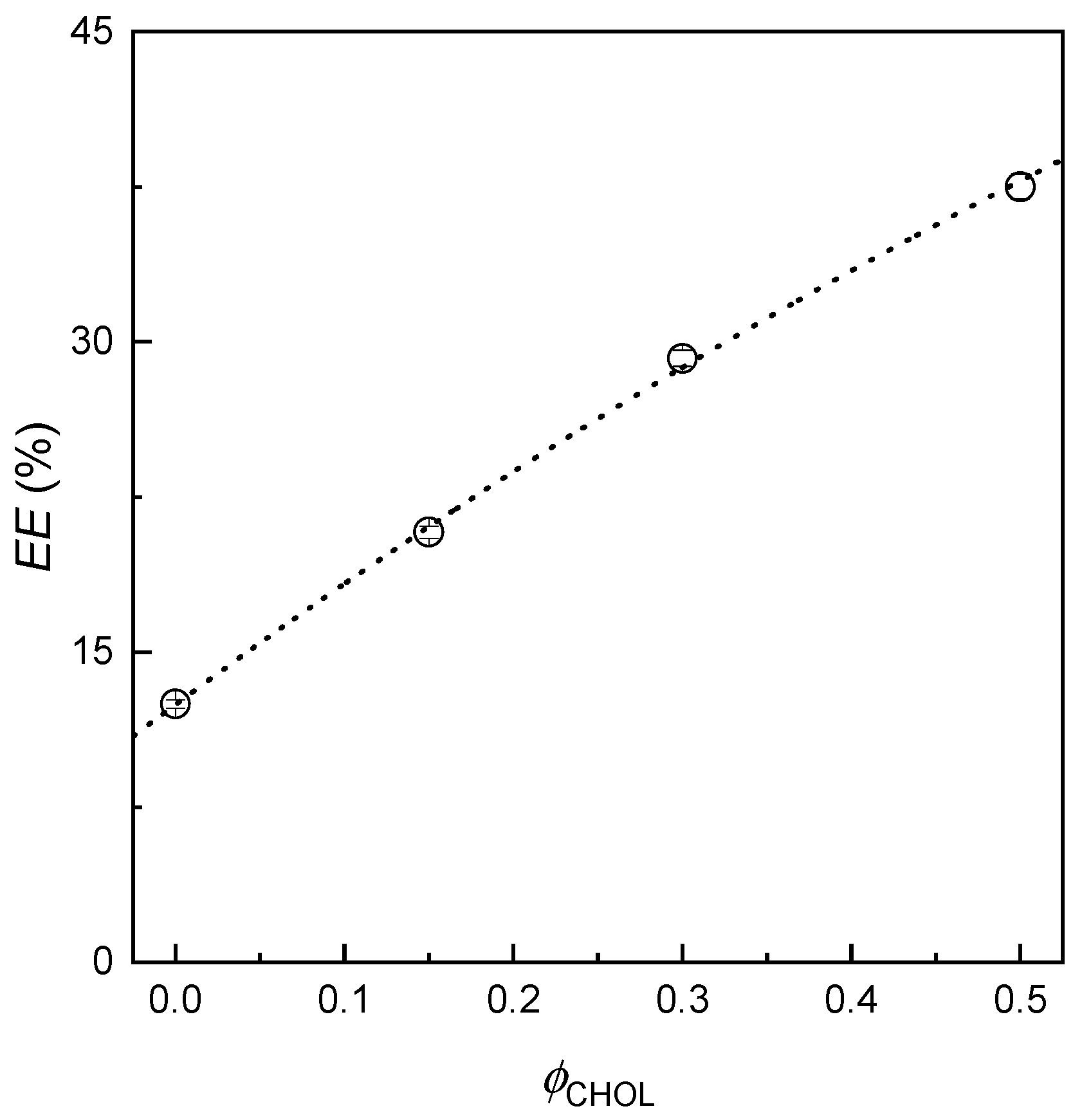
To determine the encapsulation efficiency (EE) of the liposomal systems, the amount of non-encapsulated calcein ( c C A L f ) present in the aqueous supernatant collected after the centrifugation step of the liposome preparation was quantified by fluorescence spectroscopy using the previously established calibration. In all experiments, the initial calcein concentration was fixed at 0.04 mM, a value selected to remain, in the case of negligible encapsulation efficiency, below the threshold at which self-quenching effects significantly interfere with accurate fluorescence detection of the non-encapsulated fraction (see Appendix A). Figure 1 shows the dependence of the encapsulation efficiency (EE) of calcein in liposomes with different compositions as a function of the cholesterol weight fraction (ϕCHOL) included in the lipid mixture.
The results indicate that the encapsulated fraction of calcein is relatively low, ranging from approximately 10% to 40% of the initial concentration, which could be considered modest in terms of encapsulation efficiency. Nevertheless, the main objective of this part of the work was to successfully encapsulate calcein within the liposomes and optimize the composition of these liposomes to maximize the EE, which was achieved, establishing a baseline for the development of liposomal platforms for bioactive compound delivery.
A more detailed analysis of the data reveals that increasing the cholesterol content in the lipid bilayer up to the 50% of the total weight of the liposome membrane significantly enhances the encapsulated fraction. Specifically, the encapsulation efficiency rises by approximately a factor of three, from around 12 ± 2% in liposomes composed solely of lecithin to over 37 ± 3% for membranes containing 50 wt% cholesterol, representing a marked improvement. This pronounced increase indicates that packing defects and water-filled voids in the disordered lecithin-only bilayers constitute efficient leakage pathways for calcein [51]. The incorporation of cholesterol constrains the rotational freedom of the lecithin acyl chains and promotes tighter lateral packing, thereby reducing the free volume available for solute diffusion across the membrane. As a consequence, the permeability of the bilayer decreases and a larger fraction of calcein remains trapped within the liposomes [52,53].
Based on the above findings, it is expected that incorporating moderate amounts of cholesterol improves the performance of liposomes as encapsulation vehicles. This is a result of cholesterol ability to promote the formation of a liquid-ordered phase in phospholipid bilayers, where the sterol acts as a molecular spacer that enhances bilayer thickness and mechanical rigidity, and reduces the effective area per lipid, while maintaining lateral mobility, i.e., fluidity, which collectively minimizes passive leakage of hydrophilic solutes [54,55]. This “tight packing” mechanism, rather than drug-specific binding, explains the ~3-fold EE enhancement at 50 wt% cholesterol [37,56]. Accordingly, in the following our work will focus on the use of liposomes for encapsulation with the same weight fraction of lecithin and cholesterol in the membrane. It is important to note, however, that for biomedical applications, a balance must be achieved between encapsulation efficiency and the ability to release the active compound. Excessive membrane rigidity, while beneficial for retention, could limit the release of the payload, potentially reducing therapeutic efficacy.

3.2. Encapsulation of Diclofenac in Liposomes

Previous investigations of calcein encapsulation in lecithin–cholesterol liposomes showed that membranes containing equal weight fractions of the two components yielded the highest encapsulation efficiencies. On this basis, the same membrane composition was adopted for diclofenac loading, providing a balance between high encapsulation efficiency and adequate membrane flexibility, as excessive rigidification may impair drug release in biomedical applications. At this point, it should be noted that although calcein and diclofenac may occupy different regions of the liposomes (aqueous core vs. interfacial bilayer), the 50:50 lecithin–cholesterol ratio remains appropriate because cholesterol modulates global bilayer packing and permeability rather than interacting with a specific solute. Accordingly, liposomes with equal weight fractions of the two components were prepared using different initial diclofenac concentrations ( c D I C 0 ). It is worth noting that the initial diclofenac concentrations used for loading (0.01–0.05 g/L, i.e., 10–50 mg/L) were selected to span the region where encapsulation efficiency increases and then approaches saturation for the lecithin–cholesterol composition, while remaining within the linear range of the UV–vis calibration curve. These values fall within the same order of magnitude as systemic diclofenac concentrations achieved after clinically used doses, which are typically reported in the low mg/L range, but the present experiments should be regarded as mechanistic in vitro studies rather than direct simulations of a particular dosing regimen [57,58].
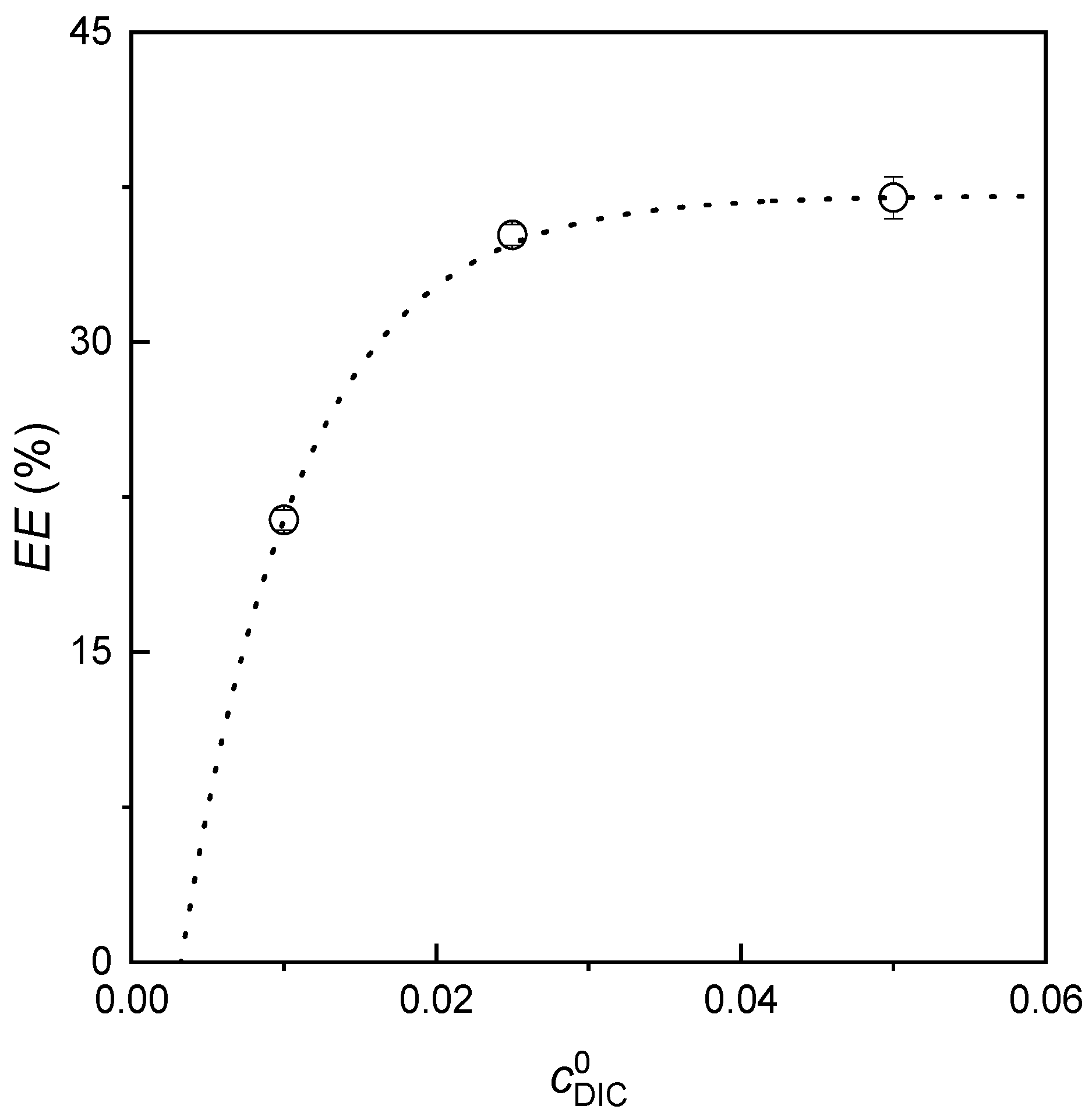
Encapsulation efficiency was quantified by UV–visible spectroscopy through analysis of the aqueous phase remaining after separation of the organic and aqueous phases during liposome preparation (see Appendix B). Figure 2 reports the dependence of diclofenac encapsulation efficiency (EE) in lecithin–cholesterol liposomes on the initial drug concentration ( c D I C 0 ) for this membrane composition. It is worth noting that translating the optimized lecithin–cholesterol composition from calcein to diclofenac assumes that the same reduction in bilayer permeability and increase in structural stability will also benefit an amphiphilic NSAID, which can reside both in the aqueous core and within the hydrophobic region of the membrane.
A detailed analysis of the encapsulation efficiency data indicates that increasing the initial diclofenac concentration during liposome preparation leads to higher encapsulation efficiency, consistent with the observations reported by Lopes et al. [59]. This behavior can be rationalized by considering that a partition equilibrium is established during the first steps of the liposome preparation between diclofenac trapped within the inverse micelles and diclofenac remaining free in the aqueous medium, with the equilibrium shifting toward encapsulation as the concentration increases. Once liposomes are obtained, the amphiphilic nature of diclofenac, with its hydrophobic aromatic system and ionizable carboxylate, enables the molecule to partition simultaneously between the aqueous core of the liposomes (accessible during reverse-phase evaporation via inverse micelles) and the hydrophobic interior of the lecithin–cholesterol bilayer. Previous studies of diclofenac–phospholipid interactions confirm that the drug associates strongly with phosphatidylcholine membranes, localizing primarily in the upper and lower leaflets with concentration-dependent perturbation of bilayer structure [60,61,62].
The enhancement in encapsulation efficiency with concentration is particularly evident at the two lowest diclofenac concentrations. However, doubling the concentration from 0.025 g/L to 0.050 g/L produces only a modest increase in the encapsulated fraction, suggesting that encapsulation approaches saturation for the used liposomes at approximately 35%. Notably, this value is similar to the encapsulation fraction previously observed for calcein under comparable conditions, which appears as confirmation of the initial working hypothesis. In fact, according to the working hypothesis, the reduced bilayer permeability and enhanced stability of lecithin–cholesterol liposomes also benefit the encapsulation of amphiphilic NSAIDs. This can be rationalized considering that in a condensed mechanically stabilized membrane, the passive permeability to both amphiphilic and hydrophilic solutes is diminished, which favors their retention within the liposomal carrier. The similar encapsulation fractions obtained for calcein and diclofenac (both close to 35%) under the same lecithin–cholesterol composition further support the view that the cholesterol-induced modulation of bilayer structure, rather than specific solute–lipid interactions, governs the encapsulation performance in these systems [63]. This interpretation is consistent with previous studies reporting that cholesterol contents in the range of 40–50 mol% provide an optimal compromise between mechanical robustness and membrane flexibility in phospholipid bilayers, a range that closely matches the 1:1 weight ratio employed in the present work [54,55,64,65].
It is important to note that although the encapsulation fraction increases, the absolute amount of diclofenac encapsulated may not change substantially with higher initial concentrations. This behavior is consistent with previous reports, where encapsulation efficiency generally rises with the concentration of the encapsulated substance until a maximum is reached at a threshold concentration. Beyond this threshold, the efficiency may plateau or even decrease, depending on factors such as the physicochemical properties of the molecule, the characteristics of the encapsulation platform, and the preparation method [66,67].

3.3. Effect of Diclofenac Encapsulation in Liposome Size

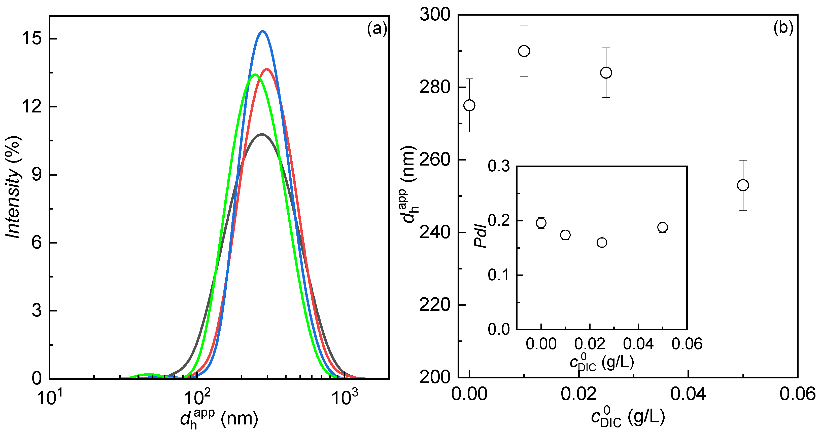
The size of encapsulation platforms plays a critical role in the transport of active compounds through the bloodstream, as well as their penetration into cells and tissues [68]. In this context, the apparent hydrodynamic diameter of the liposomes was determined using dynamic light scattering (DLS). Figure 3a presents the apparent hydrodynamic diameter distributions obtained by DLS for the liposomes employed to encapsulate varying concentrations of diclofenac.
Dynamic light scattering measurements examining the influence of diclofenac concentration on liposome size reveal that, regardless of the initial diclofenac content, the liposomes display a size distribution characteristic of a single, well-defined population. Furthermore, only minor variations are observed in the hydrodynamic diameter across the range of concentrations studied, indicating that diclofenac encapsulation does not substantially alter the overall structural characteristics of the liposomes. Notably, at the highest diclofenac concentration examined, a slight shift in the size distribution toward smaller diameters is detected, suggesting a possible contraction of the liposomal structure with increasing drug loading. This slight reduction in apparent hydrodynamic diameter (~5–10%) at the highest drug loading suggests an interaction between diclofenac molecules and the lipid bilayer, consistent with diclofenac’s known affinity for lipid membranes [62,69]. This interaction induces local bilayer condensation, leading to subtle structural rearrangements within the membrane [59]. This is in line with previous studies on the interactions of amphiphilic drugs with phospholipid membranes, which reported a reduction in the size of the resulting assemblies [60,70,71]. Indeed, surface-active drugs, including NSAIDs, can self-associate and interact with lipid bilayers, promoting membrane perturbation and partial solubilization in a manner reminiscent of detergent-like effects [70,72,73]. Previous studies of diclofenac–liposome interactions have documented 25–28% size reductions in cholesterol-free formulations, whereas our more modest reduction reflects cholesterol’s stabilizing effect, which moderates drug-induced bilayer perturbation [62,69]. The corresponding average apparent hydrodynamic diameters supporting the above discussed findings are shown in Figure 3b.
It is noteworthy that the observed size reduction is accompanied by consistently low polydispersity (PdI < 0.2, see inset in Figure 3b) across all formulations, indicating that bilayer condensation does not compromise colloidal stability. This reflects a homogeneous and stable liposomal population, and, together with particle sizes below 300 nm, supports the suitability of these carriers for diclofenac delivery despite relatively low encapsulation efficiency. The narrow size distribution and low PdI across all drug loadings also suggest that the chosen lecithin–cholesterol ratio not only enables effective encapsulation but also produces mechanically robust liposomes resistant to aggregation and fusion, a critical requirement for systemic administration. In particular, this type of diclofenac loaded liposomes may be useful for intravenous injection, where the size plays a central role in guaranteeing longevity in systemic circulation by minimizing uptake by the reticuloendothelial system while remaining small enough for potential extravasation at inflamed sites [74,75]. It is worth noting that the observed size reduction provides direct evidence of diclofenac localization within the bilayer, as only bilayer-penetrating amphiphiles can induce measurable liposome contraction. The effect is loading-dependent and moderated by cholesterol, consistent with diclofenac occupying both the bilayer hydrophobic core and polar headgroup region [62].

3.4. Release Kinetics of Encapsulated Diclofenac from Liposomes

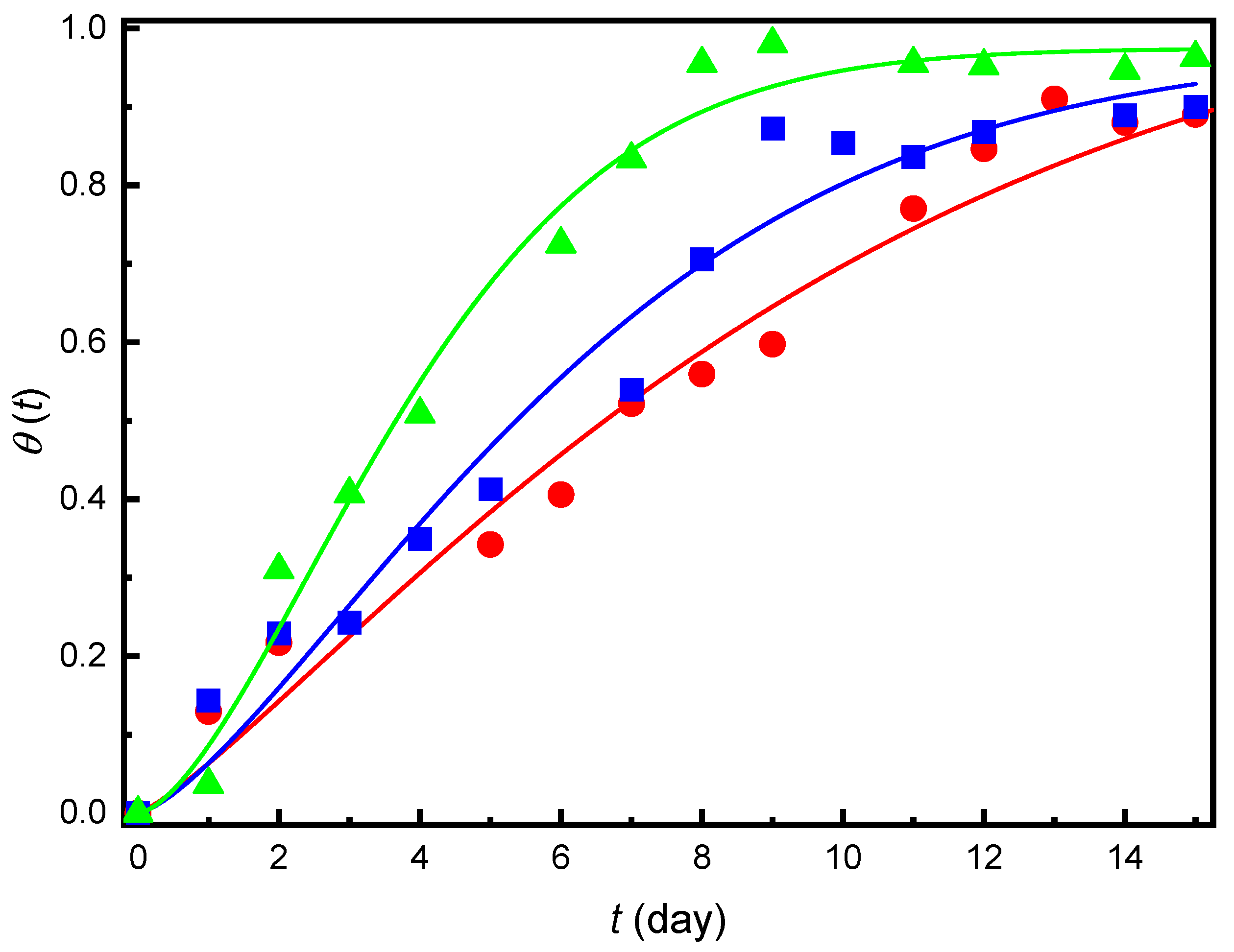
For the potential application of liposomal carriers, it is essential not only to determine their encapsulation efficiency but also to evaluate the release behavior of the encapsulated compound. Figure 4 shows the release kinetics of diclofenac from liposomes obtained encapsulating initially different quantities of diclofenac, expressed in terms of the cumulative release fraction (θ(t) = n(t)/n0), where n(t) is the molar amount of diclofenac released at time t and n0 is the initial molar amount encapsulated within the liposomes.
The results clearly indicate that, regardless of the initial diclofenac concentration or the encapsulation efficiency, the release follows a sustained profile over several days, reaching more than 80% of the encapsulated amount after 15 days. Despite these common features, a strong dependence of both the release kinetics and the maximum released fraction on the initial diclofenac concentration is observed. This behavior is expected, since release is inherently a kinetic process influenced by concentration. Specifically, higher initial drug concentrations lead to greater released amounts and faster release rates. Considering that the release is passive, with liposome dispersions placed in contact with an aqueous volume approximately ten times larger than that of the dispersion, it can be assumed that an osmotic pressure gradient develops between the interior of the liposomes and the surrounding medium. This osmotic imbalance is more pronounced when the initial encapsulated concentration is higher, thereby promoting both an increased and faster release under these conditions.
To gain further insight into the drug release process from the liposomal systems, it is useful to analyze the release kinetics using an appropriate mathematical model [76]. Among the models commonly applied to describe sustained release, the Weibull model is one of the most widely used [77]. It is important to note that the Weibull function is an empirical model, and its parameters provide a compact description of the release profile rather than a unique mechanistic assignment. The Weibull model assumes that certain factors influencing drug release depend solely on the remaining drug mass. According to the Weibull equation, the cumulative release can be expressed as follows,
θ t = θ ( 1 e ( t / α ) β ) ,
where θ represents the asymptotic fraction of drug released at infinite time, α is a scale parameter with dimensions of time that reflects the release rate, and β is a shape parameter that provides information on the release mechanism. Values of β close to unity are consistent with approximately first-order release, whereas β > 1 indicates a departure from simple first-order kinetics, often associated with multi-stage or structurally evolving systems. Table 1 summarizes the fitting parameters obtained for the release of diclofenac from the used liposomes (see Figure 4).
The results indicate that, regardless of the initial diclofenac concentration, the release tends toward 100% at infinite time, as can be clearly observed for the highest concentration evaluated. In contrast, for the lower concentrations, complete release is not reached within the experimental timeframe, highlighting a slower release process. The scale parameter α agrees with the experimental observations: higher initial concentrations yield smaller α values, consistent with faster release kinetics. Equally informative are the values of the shape parameter β obtained from the Weibull fits, which provide additional insight into the release mechanism. Its variation is consistent with a transition from nearly first-order release at low concentration ( β 1 ) to a more complex, apparently biphasic process at higher concentration ( β > 1 ). In particular, when 1.0 < β < 2.0 the Weibull function describes a release profile in which the rate initially accelerates and subsequently decelerates, a behavior that has been associated with complex, biphasic and anomalous transport mechanisms in a variety of drug-delivery systems. The increase in β from values close to 1.3 at the lowest diclofenac loading to approximately 1.6 at the highest loading therefore indicates that the contribution of a fast-release fraction, becomes more pronounced as the liposomes approach saturation, consistent with the coexistence of a weakly bound, rapidly released diclofenac population (surface drug + osmotic swelling) and a more strongly retained, core-encapsulated fraction responsible for the sustained release at longer times. This may reflect structural rearrangements of the liposomes or changes in drug–lipid interactions during release, although the Weibull analysis alone cannot unambiguously distinguish between these scenarios [78,79]. This kinetic dichotomy directly reflects the dual aqueous core–bilayer localization established during liposome formation. It is worth noting that the release behavior observed for the present liposomes differs markedly from that of many previously described diclofenac formulations, where drug release is essentially complete within 2–24 h, either from simple solutions or from fast-releasing vesicular carriers [43,80,81]. In our case, the optimized lecithin–cholesterol bilayer acts as an effective diffusion barrier, enabling long-term delivery of diclofenac, in contrast to kinetically faster, permeability-enhancing liposomes developed for topical use. The sustained release profile observed here is specifically tailored for intravenous administration, where liposomes serve as circulating drug reservoirs that gradually release diclofenac into plasma, maintaining therapeutic levels over extended periods without the gastrointestinal toxicity and short half-life limitations of oral dosing. This design philosophy differs fundamentally from topical diclofenac liposomes, which emphasize deformable structures and rapid release (<24 h) for skin permeation rather than monodisperse populations optimized for blood compatibility and long-term stability. Thus, the present work should be regarded as a formulation-level and mechanistic study that defines how a specific lecithin–cholesterol composition and diclofenac loading window control liposome size, encapsulation fraction, and release kinetics under sink conditions, rather than a comprehensive pharmacological comparison with free diclofenac or other nanocarrier platforms.
The comparison of the results obtained in this study with previously reported diclofenac liposomal-like systems points out that the present formulation was designed to favor structural robustness and long-term release rather than maximizing encapsulation efficiency at all costs. However, it should be noted that these comparisons are qualitative and based on independent reports, as no head-to-head in vitro experiments versus free diclofenac or alternative liposomal formulations were performed in this work [29]. Typical diclofenac liposomes intended for topical or transdermal delivery, including transfersomes, ethosomes or modified phosphatidylcholine vesicles, frequently exhibit encapsulation efficiencies in the range of 40–70% but are often characterized by broader or multi-modal size distributions and, in some cases, by pronounced initial burst release leading to nearly complete drug release within a few hours. For instance, conventional small unilamellar vesicles prepared by thin-film hydration typically achieve encapsulation efficiencies (EE) of 40–60% with sizes of 80–150 nm but exhibit near-complete drug release within 24 h, often dominated by a pronounced initial burst. Deformable vesicles such as cerosomes and transfersomes designed for transdermal delivery attain higher EE values (50–75%) with sizes ranging from 100 to 400 nm and polydispersity indices (PdI) of 0.2–0.4, releasing most drug within 2–24 h to enhance skin permeation [42,43,82]. In contrast, the lecithin–cholesterol liposomes investigated here display more modest encapsulation efficiencies (≈35%) but maintain hydrodynamic diameters below 300 nm with low polydispersity (PdI < 0.2) and provide sustained diclofenac release over 15 days, with cumulative release exceeding 80%, demonstrating a substantially different balance between loading capacity, colloidal stability and release kinetics. These performance differences arise from fundamentally different design priorities. Transdermal systems prioritize maximum drug loading and enhanced membrane permeability (achieved through edge activators, solvents, or deformable structures), accepting broader size distributions and faster release kinetics. Our lecithin–cholesterol composition, optimized for systemic delivery, trades moderate EE for superior colloidal control (monodisperse population < 300 nm, PdI < 0.2) and extended plasma-level maintenance (weeks rather than hours), with cholesterol providing bilayer stabilization that resists drug-induced perturbation while maintaining mechanical robustness against aggregation and fusion. The primary shortcoming, lower EE compared to specialized topical formulations, is thus a deliberate trade-off for pharmaceutical characteristics essential for reproducible systemic pharmacokinetics and long-term stability.

4. Conclusions

This study demonstrates that lecithin–cholesterol liposomes constitute a rationally designed platform for diclofenac delivery, in which lecithin provides a biocompatible bilayer scaffold, and cholesterol modulates membrane packing, rigidity, and permeability. Optimization experiments with calcein showed that increasing the cholesterol fraction up to 50 wt% in the membrane enhances encapsulation efficiency by approximately a factor of three, consistent with the formation of tighter, less permeable bilayers that better retain both hydrophilic and amphiphilic molecules. Diclofenac encapsulation in liposomes containing equal weight fractions of lecithin and cholesterol showed encapsulation efficiency values up to ~35%, comparable to calcein, with liposomes maintaining a uniform colloidal population of sub-300 nm diameter. Release experiments revealed sustained release over two weeks, with cumulative release exceeding 80%. Weibull modeling indicated faster release at higher drug concentrations and suggested biphasic kinetics, likely related to drug–lipid interactions or membrane rearrangements. Altogether, the similar maximum encapsulation efficiencies found for calcein and diclofenac, the slight size reduction observed at the highest diclofenac loading, and the increase in the Weibull shape parameter β with drug concentration point to a scenario in which cholesterol-induced ordering of the lecithin bilayer reduces passive permeability, while high diclofenac loadings introduce localized perturbations that give rise to distinct drug populations and an overall biphasic release profile. It is important to note that from an application perspective, the obtained encapsulation efficiency is relatively modest. However, it reflects a formulation deliberately tuned to combine sub-300 nm size, low polydispersity, and multi-day release with a simple lecithin–cholesterol composition, rather than to maximize drug loading. In future work, higher diclofenac loadings may be pursued by varying the lecithin:cholesterol ratio and total lipid concentration, or modifying the liposome composition to promote drug partitioning into the bilayer, and optimizing the drug-to-lipid ratio and phase volumes during reverse-phase evaporation, or by adopting active loading strategies developed for other ionizable drugs.
It should be emphasized that the release experiments were performed in water, a medium deliberately chosen to minimize specific solute–medium interactions and to isolate the contribution of bilayer structure to diclofenac release. While this simplified environment is appropriate for mechanistic analysis, it does not fully reproduce physiological conditions, where pH 7.4 buffers, salts and proteins can alter diclofenac ionization state, binding, and partitioning between phases. Future studies will therefore extend the present approach to buffered and serum-containing media at 37 °C to evaluate how physiological pH, ionic strength, and protein binding modulate the Weibull parameters and the balance between the fast- and slow-release diclofenac populations identified here.
The results demonstrate the promise of these lecithin–cholesterol liposomes as delivery systems for diclofenac and, more broadly, for other NSAIDs with solubility or bioavailability limitations. The study establishes a clear relationship between lipid composition, diclofenac loading, and the resulting encapsulation efficiency, size distribution, and long-term release behavior. However, several formulation parameters relevant to translational development were not examined. In particular, the ζ-potential, which provides insight into colloidal stability and interactions with biological interfaces, and the storage stability of the liposomes under different environmental conditions (temperature, pH, ionic strength) were not assessed. These measurements would help determine long-term physical robustness, aggregation resistance, and suitability for pharmaceutical manufacturing and in vivo use. Another important limitation is the absence of direct in vitro comparisons with free diclofenac or with liposomes formulated using alternative phospholipids or auxiliary excipients. Such comparisons are necessary to demonstrate any pharmacological or biopharmaceutical advantage beyond the mechanistic trends described here. Future work should therefore include ζ-potential characterization, systematic storage-stability studies, and serum-stability assays. It will also be essential to perform comparative in vitro release experiments, cell-based evaluations, and in vivo pharmacokinetic studies to determine how the sustained release observed in aqueous media translates into therapeutic performance relative to free drug and other nanocarrier architectures. Beyond extending the release studies to biorelevant media and in vivo models, future formulation optimization will focus on increasing diclofenac loading while preserving the long-term release characteristics demonstrated here. Finally, the present study relied exclusively on the reverse-phase evaporation method for liposome preparation. Although well suited for mechanistic laboratory investigations, this technique is not readily scalable. To enable translation of the lecithin–cholesterol formulation toward manufacturing-relevant processes, alternative methods such as ethanol injection or thin-film hydration followed by size-reduction steps should be explored.

Author Contributions

Conceptualization, R.G.R. and E.G.; methodology, Á.S.-G. and E.G.; software, Á.S.-G. and E.G.; validation, E.G.; formal analysis, Á.S.-G. and E.G.; investigation, Á.S.-G., F.O., R.G.R. and E.G.; resources, F.O., R.G.R. and E.G.; data curation, Á.S.-G. and E.G.; writing—original draft preparation, Á.S.-G. and E.G.; writing—review and editing, F.O., R.G.R. and E.G.; visualization, Á.S.-G. and E.G.; supervision, R.G.R. and E.G.; project administration, F.O., R.G.R. and E.G.; funding acquisition, F.O. and E.G. All authors have read and agreed to the published version of the manuscript.

Funding

The work was supported under the grant PID2023-147156NB-I00 funded by MCIN/AEI/10.13039/501100011033 (Spain).

Data Availability Statement

Data are available upon request.

Acknowledgments

The authors express their gratitude to the Unidad de Espectroscopía y Correlación (CAI de Técnicas Químicas) at Universidad Complutense de Madrid for granting access to their facilities.

Conflicts of Interest

The authors declare no conflicts of interest. The funders had no role in the design of the study; in the collection, analyses, or interpretation of data; in the writing of the manuscript; or in the decision to publish the results.

Appendix A. Characterization of Calcein Aqueous Solutions

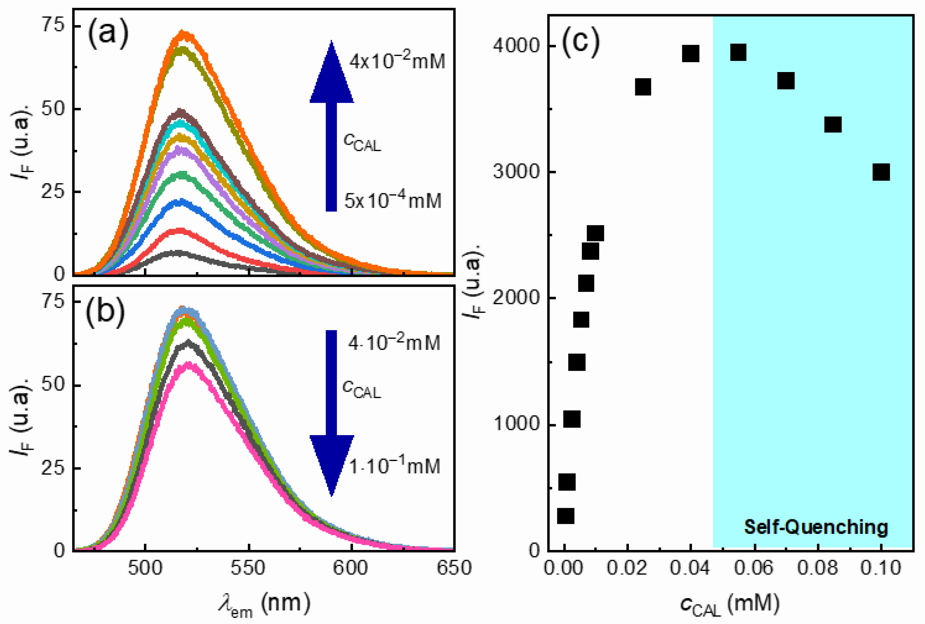
To evaluate the encapsulation efficiency of the prepared liposomal vectors, it was first necessary to analyze the photophysical behavior of calcein in aqueous solution. This calibration provides the reference required to determine the fraction of encapsulated molecules relative to the initial amount introduced. Given the electronic properties of calcein and the high intensity of its fluorescence emission, its behavior in solution was characterized by fluorescence emission spectroscopy. The excitation wavelength (λexc) was set at 460 nm, in agreement with literature [83], and the evolution of the fluorescence emission intensity (IF) for solutions with different calcein concentrations was monitored as a function of the emission wavelength (λem), as illustrated in Figure A1a,b for calcein solutions of different concentrations.
As expected from the Lambert–Beer law, the emission intensity increased with concentration up to a threshold of approximately 0.04 mM (Figure A1a). Beyond this concentration, however, the fluorescence intensity decreased with increasing calcein concentration (cCAL), a phenomenon attributable to self-quenching (Figure A1b). This self-quenching effect has been previously reported and is commonly associated with a shift in the equilibrium toward the formation of calcein dimers at higher concentrations, leading to reduced emission efficiency [84].
To perform a detailed analysis of the fluorescence intensity as a function of calcein concentration in solution, it is important to consider that the intensity corresponding to a spectroscopic transition is not limited to the emission maximum, but rather to the integrated area under the entire emission band. This parameter can be extracted by numerical integration using the OriginPro 2023 software (OriginLab Corporation, Northampton, MA, USA). Using this approach, it was possible to plot the integrated intensity values obtained for all studied concentrations against calcein concentration (Figure A1c). As expected, two distinct regions can be observed: at low concentrations, the fluorescence intensity increases with cCAL, while at higher cCAL it decreases due to self-quenching. Furthermore, the analysis shows that the linear region described by the Lambert–Beer law is relatively narrow.
Figure A1. Fluorescence spectroscopy results for aqueous calcein solutions in the concentration range of 5 × 10⁻⁴ to 1 × 10⁻¹ mM. (a) Emission spectra of calcein solutions in the range 5 × 10⁻⁴ to 4 × 10⁻² mM, showing the expected Lambert–Beer dependence. (b) Emission spectra of calcein solutions in the range 4 × 10⁻² to 1 × 10⁻¹ mM, highlighting self-quenching effects. (c) Dependence of fluorescence intensity on calcein concentration across the studied range.
Colloids 10 00025 g0a1

Appendix B. Characterization of Diclofenac Aqueous Solutions

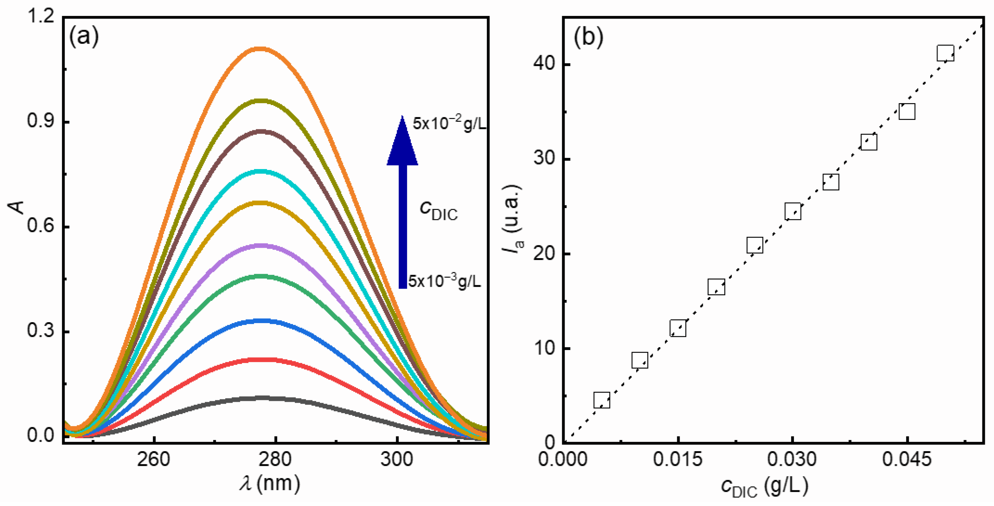
As discussed for calcein, the first step before assessing the ability of liposomes to encapsulate a given molecule is to study its behavior in solution, to determine the concentration dependence of a property that can be readily measured. For diclofenac, unlike calcein, whose UV–visible absorption is relatively high even at very low concentrations, its optical behavior in solution can be reliably evaluated using UV–visible spectroscopy. Figure A2a shows the evolution of the UV–visible spectrum of diclofenac, represented as absorbance (A) versus wavelength (λ) for different diclofenac concentrations (cDIC).
The UV spectrum of diclofenac exhibits a relatively broad absorption band centered around 275 nm. The absorbance of this band increases with concentration within the explored range, in agreement with Lambert–Beer’s law. A detailed analysis of the absorption bands, performed via an integration procedure similar to that applied to the calcein fluorescence data, allows extraction of information related to the transition intensity. The variation in the absorption band intensity (Ia) as a function of diclofenac concentration is shown in Figure A2b, revealing a linear relationship that can be interpreted in terms of the Lambert–Beer law ( I a = ε m l c D I C , with ε m being the mass absorption coefficient of diclofenac for the considered transition and l the optical path length, which assume a value of 1 cm in this study). Fitting the experimental data using this relation provides a mass absorptivity for diclofenac of (805 ± 7) × 103 cm2/g, which was subsequently used to calculate concentrations in the aqueous phase.
Figure A2. UV absorption spectroscopy results for aqueous diclofenac solutions with concentrations ranging from 5 × 10⁻³ to 5 × 10⁻² g/L. (a) Absorbance spectra of diclofenac solutions. (b) Dependence of absorbed intensity on diclofenac concentration. Symbols represent experimental data, and the dotted line corresponds to the fit according to the Lambert–Beer law.
Colloids 10 00025 g0a2

References

  1. Haleem, A.; Javaid, M.; Singh, R.P.; Rab, S.; Suman, R. Applications of Nanotechnology in Medical Field: A Brief Review. Glob. Health J. 2023, 7, 70–77. [Google Scholar] [CrossRef] [Scilit]
  2. Abrahams, E.; Downing, G.J. On the Modern Evolution of Personalized Medicine. In The New Era of Precision Medicine; Bydon, M., Ed.; Academic Press: Cambridge, MA, USA, 2024; pp. 1–25. [Google Scholar]
  3. Nasrallah, S.; Alhadid, A.; Minceva, M. Revealing the Solubility Enhancement of Active Pharmaceutical Ingredients through Eutectic Mixtures Formation: A Parameter Study. Cryst. Growth Des. 2024, 24, 6364–6372. [Google Scholar] [CrossRef] [Scilit]
  4. Singh Raman, A.P.; Jain, P.; Kumar, A.; Singh, P.; Kumari, K.; Bahadur, I.; Mohammad, F.; Kaushik, N.K. Eutectic Mixtures to Enhance the Solubility of Active Pharmaceutical Ingredients: Density Functional Theory and Infrared Spectroscopy Approaches. ChemistrySelect 2023, 8, e202300824. [Google Scholar] [CrossRef] [Scilit]
  5. Gatto, M.S.; Johnson, M.P.; Najahi-Missaoui, W. Targeted Liposomal Drug Delivery: Overview of the Current Applications and Challenges. Life 2024, 14, 672. [Google Scholar] [CrossRef] [Scilit]
  6. Rezagholizade-shirvan, A.; Soltani, M.; Shokri, S.; Radfar, R.; Arab, M.; Shamloo, E. Bioactive Compound Encapsulation: Characteristics, Applications in Food Systems, and Implications for Human Health. Food Chem. X 2024, 24, 101953. [Google Scholar] [CrossRef] [Scilit]
  7. Pedder, J.H.; Sonabend, A.M.; Cearns, M.D.; Michael, B.D.; Zakaria, R.; Heimberger, A.B.; Jenkinson, M.D.; Dickens, D. Crossing the Blood–Brain Barrier: Emerging Therapeutic Strategies for Neurological Disease. Lancet Neurol. 2025, 24, 246–260. [Google Scholar] [CrossRef] [Scilit]
  8. Reddy, K.T.K.; Reddy, A.S. Recent Breakthroughs in Drug Delivery Systems for Targeted Cancer Therapy: An Overview. Cell. Mol. Biomed. Rep. 2025, 5, 13–27. [Google Scholar] [CrossRef] [Scilit]
  9. Hu, Y.; Song, J.; Feng, A.; Li, J.; Li, M.; Shi, Y.; Sun, W.; Li, L. Recent Advances in Nanotechnology-Based Targeted Delivery Systems of Active Constituents in Natural Medicines for Cancer Treatment. Molecules 2023, 28, 7767. [Google Scholar] [CrossRef] [Scilit]
  10. Li, J.; Wang, Q.; Xia, G.; Adilijiang, N.; Li, Y.; Hou, Z.; Fan, Z.; Li, J. Recent Advances in Targeted Drug Delivery Strategy for Enhancing Oncotherapy. Pharmaceutics 2023, 15, 2233. [Google Scholar] [CrossRef] [Scilit] [PubMed]
  11. Beach, M.A.; Nayanathara, U.; Gao, Y.; Zhang, C.; Xiong, Y.; Wang, Y.; Such, G.K. Polymeric Nanoparticles for Drug Delivery. Chem. Rev. 2024, 124, 5505–5616. [Google Scholar] [CrossRef] [Scilit]
  12. Karimi, S.; Namazi, H.; Aghazadeh, M. Simultaneous Release of Hydrophilic and Hydrophobic Drugs by a PH-Sensitive Bio-Carrier Based on Layered Double Hydroxides Decorated with l-Serine-Chitosan. J. Polym. Environ. 2025, 33, 2758–2775. [Google Scholar] [CrossRef] [Scilit]
  13. Bangham, A.D.; Hill, M.W.; Miller, N.G.A. Preparation and Use of Liposomes as Models of Biological Membranes. In Methods in Membrane Biology; Korn, E.D., Ed.; Springer: Boston, MA, USA, 1974; Volume 1, pp. 1–68. [Google Scholar]
  14. Gregoriadis, G. The Carrier Potential of Liposomes in Biology and Medicine. N. Engl. J. Med. 1976, 295, 704–710. [Google Scholar] [CrossRef] [Scilit]
  15. Juliano, R.L.; Stamp, D. The Effect of Particle Size and Charge on the Clearance Rates of Liposomes and Liposome Encapsulated Drugs. Biochem. Biophys. Res. Comm. 1975, 63, 651–658. [Google Scholar] [CrossRef] [Scilit]
  16. Nsairat, H.; Khater, D.; Sayed, U.; Odeh, F.; Al Bawab, A.; Alshaer, W. Liposomes: Structure, Composition, Types, and Clinical Applications. Heliyon 2022, 8, e09394. [Google Scholar] [CrossRef] [Scilit]
  17. van der Koog, L.; Gandek, T.B.; Nagelkerke, A. Liposomes and Extracellular Vesicles as Drug Delivery Systems: A Comparison of Composition, Pharmacokinetics, and Functionalization. Adv. Healthc. Mater. 2022, 11, 2100639. [Google Scholar] [CrossRef] [Scilit]
  18. Hamad, I.; Harb, A.A.; Bustanji, Y. Liposome-Based Drug Delivery Systems in Cancer Research: An Analysis of Global Landscape Efforts and Achievements. Pharmaceutics 2024, 16, 400. [Google Scholar] [CrossRef] [Scilit]
  19. Gabizon, A.A.; Gabizon-Peretz, S.; Modaresahmadi, S.; La-Beck, N.M. Thirty Years from FDA Approval of Pegylated Liposomal Doxorubicin (Doxil/Caelyx): An Updated Analysis and Future Perspective. BMJ Oncol. 2025, 4, e000573. [Google Scholar] [CrossRef] [Scilit]
  20. Aloss, K.; Hamar, P. Recent Preclinical and Clinical Progress in Liposomal Doxorubicin. Pharmaceutics 2023, 15, 893. [Google Scholar] [CrossRef] [Scilit]
  21. Agrawal, S.S.; Baliga, V.; Londhe, V.Y. Liposomal Formulations: A Recent Update. Pharmaceutics 2024, 17, 36. [Google Scholar] [CrossRef] [Scilit]
  22. Nikolova, M.P.; Kumar, E.M.; Chavali, M.S. Updates on Responsive Drug Delivery Based on Liposome Vehicles for Cancer Treatment. Pharmaceutics 2022, 14, 2195. [Google Scholar] [CrossRef] [Scilit] [PubMed]
  23. Tenchov, R.; Bird, R.; Curtze, A.E.; Zhou, Q. Lipid Nanoparticles—From Liposomes to MRNA Vaccine Delivery, a Landscape of Research Diversity and Advancement. ACS Nano 2021, 15, 16982–17015. [Google Scholar] [CrossRef] [Scilit]
  24. Hou, X.; Zaks, T.; Langer, R.; Dong, Y. Lipid Nanoparticles for MRNA Delivery. Nat. Rev. Mat. 2021, 6, 1078–1094. [Google Scholar] [CrossRef] [Scilit]
  25. Musielak, E.; Krajka-Kuźniak, V. Liposomes and Ethosomes: Comparative Potential in Enhancing Skin Permeability for Therapeutic and Cosmetic Applications. Cosmetics 2024, 11, 191. [Google Scholar] [CrossRef] [Scilit]
  26. Drosopoulou, K.; Kosheleva, R.I.; Ofrydopoulou, A.; Tsoupras, A.; Mitropoulos, A. Topical and Transdermal Delivery of Nonsteroidal Anti-Inflammatory Drugs (NSAIDs) for Inflammation and Pain: Current Trends and Future Directions in Delivery Systems. Processes 2025, 13, 907. [Google Scholar] [CrossRef] [Scilit]
  27. Sankar, S.; Kalidass, B.; Indrakumar, J.; Kodiveri Muthukaliannan, G. NSAID-Encapsulated Nanoparticles as a Targeted Therapeutic Platform for Modulating Chronic Inflammation and Inhibiting Cancer Progression: A Review. Inflammopharmacology 2025, 33, 2493–2522. [Google Scholar] [CrossRef] [Scilit] [PubMed]
  28. Wirth, T.; Lafforgue, P.; Pham, T. NSAID: Current Limits to Prescription. Jt. Bone Spine 2024, 91, 105685. [Google Scholar] [CrossRef] [Scilit]
  29. Kuznetsova, D.A.; Vasilieva, E.A.; Kuznetsov, D.M.; Lenina, O.A.; Filippov, S.K.; Petrov, K.A.; Zakharova, L.Y.; Sinyashin, O.G. Enhancement of the Transdermal Delivery of Nonsteroidal Anti-Inflammatory Drugs Using Liposomes Containing Cationic Surfactants. ACS Omega 2022, 7, 25741–25750. [Google Scholar] [CrossRef] [Scilit] [PubMed]
  30. Placha, D.; Jampilek, J. Chronic Inflammatory Diseases, Anti-Inflammatory Agents and Their Delivery Nanosystems. Pharmaceutics 2021, 13, 64. [Google Scholar] [CrossRef] [Scilit] [PubMed]
  31. Pauna, A.-M.R.; Tartau, L.M.; Vasilescu, A.M.; Abu Koush, A.; Stan, R.T.; Moraru, M.C.; Popa, C.G.; Gavril, L.C.; Florentina Gavril, R.; Crauciuc, D.V.; et al. Assessment of the Anti-Inflammatory Effectiveness of Diclofenac Encapsulated in Chitosan-Coated Lipid Microvesicles in Rats. Pharmaceutics 2025, 17, 607. [Google Scholar] [CrossRef] [Scilit]
  32. Kvolik, S. Diclofenac: The Impact of Different Routes of Administration on the Efficiency of the Drug. Galen. Med. J. 2024, 3, 36–40. [Google Scholar] [CrossRef] [Scilit]
  33. Sikorska, D.; Chlabicz, S.; Rydzewska, G.; Samborski, W.; Tykarski, A.; Woroń, J. Nonsteroidal Anti-Inflammatory Drugs in Clinical Practice—Are There Any New Reports? Rheumatol. Forum 2024, 10, 26–37. [Google Scholar] [CrossRef] [Scilit]
  34. Bhatia, A.; Goni, V.; Chopra, S.; Singh, B.; Katare, O.P. Evaluation of Efficacy and Safety of a Novel Lipogel Containing Diclofenac: A Randomized, Placebo Controlled, Double-Blind Clinical Trial in Patients with Signs and Symptoms of Osteoarthritis. Contemp. Clin. Trials Commun. 2020, 20, 100664. [Google Scholar] [CrossRef] [Scilit] [PubMed]
  35. Basak, S.; Das, T.K. Liposome-Based Drug Delivery Systems: From Laboratory Research to Industrial Production—Instruments and Challenges. ChemEngineering 2025, 9, 56. [Google Scholar] [CrossRef] [Scilit]
  36. Zafar, A.; Alruwaili, N.K.; Imam, S.S.; Yasir, M.; Alsaidan, O.A.; Alquraini, A.; Rawaf, A.; Alsuwayt, B.; Anwer, M.d.K.; Alshehri, S.; et al. Development and Optimization of Nanolipid-Based Formulation of Diclofenac Sodium: In Vitro Characterization and Preclinical Evaluation. Pharmaceutics 2022, 14, 507. [Google Scholar] [CrossRef] [Scilit] [PubMed]
  37. Bernardino de la Serna, J.; Perez-Gil, J.; Simonsen, A.C.; Bagatolli, L.A. Cholesterol Rules. J. Biol. Chem. 2004, 279, 40715–40722. [Google Scholar] [CrossRef] [Scilit]
  38. Guzmán, E.; Ferrari, M.; Santini, E.; Liggieri, L.; Ravera, F. Effect of Silica Nanoparticles on the Interfacial Properties of a Canonical Lipid Mixture. Colloids Surf. B Biointerfaces 2015, 136, 971–980. [Google Scholar] [CrossRef] [Scilit]
  39. Bot, F.; Cossuta, D.; O’Mahony, J.A. Inter-Relationships between Composition, Physicochemical Properties and Functionality of Lecithin Ingredients. Trends Food Sci. Technol. 2021, 111, 261–270. [Google Scholar] [CrossRef] [Scilit]
  40. Namvar, S.; Fathi-Achachlouei, B.; Shaddel, R. Effect of Different Ratios of Lecithin-Cholesterol on the Encapsulation Stability of Beta- Carotene-Loaded Nanoliposomes. Food Res. J. 2024, 33, 131–149. [Google Scholar]
  41. Joshi, B.; Shyaula, S.L.; Joshi, T.P.; Bhandari, N.L.; Budhathoki, U.; Gyawali, R.; Thapa, P. Optimization of Liposomes Encapsulated with Ursolic Acid Using Response Surface Method. Indian J. Pharm. Sci. 2025, 87, 94–101. [Google Scholar] [CrossRef] [Scilit]
  42. Fathi-Azarbayjani, A.; Ng, K.X.; Chan, Y.W.; Chan, S.Y. Lipid Vesicles for the Skin Delivery of Diclofenac: Cerosomes vs. Other Lipid Suspensions. Adv. Pharm. Bull. 2015, 5, 25–33. [Google Scholar] [CrossRef] [Scilit]
  43. Ghanbarzadeh, S.; Arami, S. Enhanced Transdermal Delivery of Diclofenac Sodium via Conventional Liposomes, Ethosomes, and Transfersomes. BioMed Res. Int. 2013, 2013, 616810. [Google Scholar] [CrossRef] [Scilit] [PubMed]
  44. Sacha, M.; Faucon, L.; Hamon, E.; Ly, I.; Haltner-Ukomadu, E. Ex Vivo Transdermal Absorption of a Liposome Formulation of Diclofenac. Biomed. Pharmacother. 2019, 111, 785–790. [Google Scholar] [CrossRef] [Scilit]
  45. Mertins, O.; Sebben, M.; Pohlmann, A.R.; da Silveira, N.P. Production of Soybean Phosphatidylcholine–Chitosan Nanovesicles by Reverse Phase Evaporation: A Step by Step Study. Chem. Phys. Lipids 2005, 138, 29–37. [Google Scholar] [CrossRef] [Scilit] [PubMed]
  46. Szoka, F.; Papahadjopoulos, D. Procedure for Preparation of Liposomes with Large Internal Aqueous Space and High Capture by Reverse-Phase Evaporation. Proc. Nat. Acad. Sci. USA 1978, 75, 4194–4198. [Google Scholar] [CrossRef] [Scilit]
  47. Szoka, F.; Papahadjopoulos, D. Comparative Properties and Methods of Preparation of Lipid Vesicles (Liposomes). Annu. Rev. Biophys. Bioeng. 1980, 9, 467–508. [Google Scholar] [CrossRef] [Scilit]
  48. Berne, B.J.; Pecora, R. Dynamic Light Scattering: With Applications to Chemistry, Biology, and Physics; John Wiley & Sons: New York, NY, USA, 2003. [Google Scholar]
  49. Fernández-Peña, L.; Gutiérrez-Muro, S.; Guzmán, E.; Lucia, A.; Ortega, F.; G. Rubio, R. Oil-In-Water Microemulsions for Thymol Solubilization. Colloids Interfaces 2019, 3, 64. [Google Scholar] [CrossRef] [Scilit]
  50. Moran, A.L. Calcein as a Marker in Experimental Studies Newly-Hatched Gastropods. Mar. Biol. 2000, 137, 893–898. [Google Scholar] [CrossRef] [Scilit]
  51. Guzmán, E.; Liggieri, L.; Santini, E.; Ferrari, M.; Ravera, F. DPPC-DOPC Langmuir Monolayers Modified by Hydrophilic Silica Nanoparticles: Phase Behaviour, Structure and Rheology. Colloids Surf. A Physicochem. Eng. Asp. 2012, 413, 174–183. [Google Scholar] [CrossRef] [Scilit]
  52. Manojlovic, V.; Winkler, K.; Bunjes, V.; Neub, A.; Schubert, R.; Bugarski, B.; Leneweit, G. Membrane Interactions of Ternary Phospholipid/Cholesterol Bilayers and Encapsulation Efficiencies of a RIP II Protein. Colloids Surf. B 2008, 64, 284–296. [Google Scholar] [CrossRef] [Scilit]
  53. Taylor, K.M.G.; Taylor, G.; Kellaway, I.W.; Stevens, J. Drug Entrapment and Release from Multilamellar and Reverse-Phase Evaporation Liposomes. Int. J. Pharm. 1990, 58, 49–55. [Google Scholar] [CrossRef] [Scilit]
  54. Khodadadi, E.; Khodadadi, E.; Chaturvedi, P.; Moradi, M. Comprehensive Insights into the Cholesterol-Mediated Modulation of Membrane Function Through Molecular Dynamics Simulations. Membranes 2025, 15, 173. [Google Scholar] [CrossRef] [Scilit]
  55. Raffy, S.; Teissié, J. Control of Lipid Membrane Stability by Cholesterol Content. Biophys. J. 1999, 76, 2072–2080. [Google Scholar] [CrossRef] [Scilit]
  56. Canepa, E.; Bochicchio, D.; Gasbarri, M.; Odino, D.; Canale, C.; Ferrando, R.; Canepa, F.; Stellacci, F.; Rossi, G.; Dante, S.; et al. Cholesterol Hinders the Passive Uptake of Amphiphilic Nanoparticles into Fluid Lipid Membranes. J. Phys. Chem. Lett. 2021, 12, 8583–8590. [Google Scholar] [CrossRef] [Scilit] [PubMed]
  57. Pacifici, G.M. Clinical Pharmacology of Diclofenac. Biomed. J. Sci. Tech. Res. 2024, 56, 48609–48621. [Google Scholar] [CrossRef] [Scilit]
  58. Davies, N.M.; Anderson, K.E. Clinical Pharmacokinetics of Diclofenac. Clin. Pharmacokinet. 1997, 33, 184–213. [Google Scholar] [CrossRef] [Scilit] [PubMed]
  59. Lopes, L.B.; Scarpa, M.V.; Silva, G.V.J.; Rodrigues, D.C.; Santilli, C.V.; Oliveira, A.G. Studies on the Encapsulation of Diclofenac in Small Unilamellar Liposomes of Soya Phosphatidylcholine. Colloids Surf. B 2004, 39, 151–158. [Google Scholar] [CrossRef] [Scilit]
  60. Lichtenberger, L.M.; Zhou, Y.; Jayaraman, V.; Doyen, J.R.; O’Neil, R.G.; Dial, E.J.; Volk, D.E.; Gorenstein, D.G.; Boggara, M.B.; Krishnamoorti, R. Insight into NSAID-Induced Membrane Alterations, Pathogenesis and Therapeutics: Characterization of Interaction of NSAIDs with Phosphatidylcholine. BIochim. Biophys. Acta-Mol. Cell Biol. Lipids 2012, 1821, 994–1002. [Google Scholar] [CrossRef] [Scilit]
  61. Atnyukova, A.N.; Kashnik, A.S.; Selyutina, O.Y.; Baranov, D.S.; Polyakov, N.E.; Dzuba, S.A. Location of the Diclofenac Molecule in Model Lipid Membranes from Spin-Label-Enhanced NMR Relaxation. Langmuir 2025, 41, 27272–27278. [Google Scholar] [CrossRef] [Scilit]
  62. Lopes, L.B.; Scarpa, M.V.; Pereira, N.L.; Oliveira, L.C.d.; Oliveira, A.G. Interaction of Sodium Diclofenac with Freeze-Dried Soya Phosphatidylcholine and Unilamellar Liposomes. Rev. Bras. Cienc. Farm. 2006, 42, 497–504. [Google Scholar] [CrossRef] [Scilit]
  63. Maswadeh, H.; Abdulhalim, A.; Demetzos, C. Improvement of Encapsulation Efficiency of Diclofenac Sodium in to Uncoated and Chitosan-Coated Liposomes. Ind. J. Pharm. Sci. 2004, 66, 607–612. [Google Scholar]
  64. Wu, H.; Yu, M.; Miao, Y.; He, S.; Dai, Z.; Song, W.; Liu, Y.; Song, S.; Ahmad, E.; Wang, D.; et al. Cholesterol-Tuned Liposomal Membrane Rigidity Directs Tumor Penetration and Anti-Tumor Effect. Acta Pharm. Sin. B 2019, 9, 858–870. [Google Scholar] [CrossRef] [Scilit] [PubMed]
  65. Sullan, R.M.A.; Li, J.K.; Hao, C.; Walker, G.C.; Zou, S. Cholesterol-Dependent Nanomechanical Stability of Phase-Segregated Multicomponent Lipid Bilayers. Biophys. J. 2010, 99, 507–516. [Google Scholar] [CrossRef] [Scilit]
  66. Li, C.; Guan, H.; Li, Z.; Wang, F.; Wu, J.; Zhang, B. Study on Different Particle Sizes of DOX-Loaded Mixed Micelles for Cancer Therapy. Colloids Surf. B 2020, 196, 111303. [Google Scholar] [CrossRef] [Scilit]
  67. Press, A.T.; Ramoji, A.; vd Lühe, M.; Rinkenauer, A.C.; Hoff, J.; Butans, M.; Rössel, C.; Pietsch, C.; Neugebauer, U.; Schacher, F.H.; et al. Cargo–Carrier Interactions Significantly Contribute to Micellar Conformation and Biodistribution. NPG Asia Mater. 2017, 9, e444. [Google Scholar] [CrossRef] [Scilit]
  68. Yan, L.; Amirshaghaghi, A.; Huang, D.; Miller, J.; Stein, J.M.; Busch, T.M.; Cheng, Z.; Tsourkas, A. Protoporphyrin IX (PpIX)-Coated Superparamagnetic Iron Oxide Nanoparticle (SPION) Nanoclusters for Magnetic Resonance Imaging and Photodynamic Therapy. Adv. Funct. Mat. 2018, 28, 1707030. [Google Scholar] [CrossRef] [Scilit] [PubMed]
  69. Taylor, K.M.P.; Roseman, M.A. Effect of Cholesterol on the Tight Insertion of Cytochrome B5 into Large Unilamellar Vesicles. Biochim. Biophys. Acta 1996, 1278, 35–40. [Google Scholar] [CrossRef] [Scilit]
  70. Schreier, S.; Malheiros, S.V.P.; de Paula, E. Surface Active Drugs: Self-Association and Interaction with Membranes and Surfactants. Physicochemical and Biological Aspects. Biochim. Biophys. Acta-Biomembr. 2000, 1508, 210–234. [Google Scholar] [CrossRef] [Scilit]
  71. Stoye, I.; Schröder, K.; Müller-Goymann, C.C. Transformation of a Liposomal Dispersion Containing Ibuprofen Lysinate and Phospholipids into Mixed Micelles—Physico-Chemical Characterization and Influence on Drug Permeation through Excised Human Stratum Corneum. Eur. J. Pharm. Biopharm. 1998, 46, 191–200. [Google Scholar] [CrossRef] [Scilit] [PubMed]
  72. Schütze, W.; Müller-Goymann, C.C. Phase Transformation of a Liposomal Dispersion into a Micellar Solution Induced by Drug-Loading. Pharm. Res. 1998, 15, 538–543. [Google Scholar] [CrossRef] [Scilit]
  73. Rades, T.; Müller-Goymann, C.C. Investigations on the Micellisation Behaviour of Fenoprofen Sodium. Int. J. Pharm. 1997, 159, 215–222. [Google Scholar] [CrossRef] [Scilit]
  74. Bulbake, U.; Doppalapudi, S.; Kommineni, N.; Khan, W. Liposomal Formulations in Clinical Use: An Updated Review. Pharmaceutics 2017, 9, 12. [Google Scholar] [CrossRef] [Scilit] [PubMed]
  75. Deshpande, P.P.; Biswas, S.; Torchilin, V.P. Current Trends in The Use of Liposomes for Tumor Targeting. Nanomedicine 2013, 8, 1509–1528. [Google Scholar] [CrossRef] [Scilit]
  76. Jain, A.; Jain, S.K. In Vitro Release Kinetics Model Fitting of Liposomes: An Insight. Chem. Phys. Lipids 2016, 201, 28–40. [Google Scholar] [CrossRef] [Scilit]
  77. Papadopoulou, V.; Kosmidis, K.; Vlachou, M.; Macheras, P. On the Use of the Weibull Function for the Discernment of Drug Release Mechanisms. Int. J. Pharm. 2006, 309, 44–50. [Google Scholar] [CrossRef] [Scilit]
  78. Corsaro, C.; Neri, G.; Mezzasalma, A.M.; Fazio, E. Weibull Modeling of Controlled Drug Release from Ag-PMA Nanosystems. Polymers 2021, 13, 2897. [Google Scholar] [CrossRef] [Scilit]
  79. Martín-Camacho, U.d.J.; Rodríguez-Barajas, N.; Sánchez-Burgos, J.A.; Pérez-Larios, A. Weibull β Value for the Discernment of Drug Release Mechanism of PLGA Particles. Int. J. Pharm. 2023, 640, 123017. [Google Scholar] [CrossRef] [Scilit]
  80. Camino, A.; Taly, A.; Rodriguez, C.; Inatti, A.; Pena, E.; Serrano, X. Liposomes Carrying Diclofenac Diethylammonium: A Penetrability and Permeation Study. J. Biosci. Med. 2024, 12, 206–218. [Google Scholar] [CrossRef]
  81. Chopra, L.; Thakur, K.K.; Chohan, J.S.; Sharma, S.; Ilyas, R.A.; Asyraf, M.R.M.; Zakaria, S.Z.S. Comparative Drug Release Investigations for Diclofenac Sodium Drug (DS) by Chitosan-Based Grafted and Crosslinked Copolymers. Materials 2022, 15, 2404. [Google Scholar] [CrossRef] [Scilit] [PubMed]
  82. Manconi, M.; Caddeo, C.; Sinico, C.; Valenti, D.; Mostallino, M.C.; Biggio, G.; Fadda, A.M. Ex Vivo Skin Delivery of Diclofenac by Transcutol Containing Liposomes and Suggested Mechanism of Vesicle–Skin Interaction. Eur. J. Pharm. Biopharm. 2011, 78, 27–35. [Google Scholar] [CrossRef] [Scilit] [PubMed]
  83. Memoli, A.; Palermiti, L.G.; Travagli, V.; Alhaique, F. Effects of Surfactants on the Spectral Behaviour of Calcein (II): A Method of Evaluation. J. Pharm. Biomed. Anal. 1999, 19, 627–632. [Google Scholar] [CrossRef] [Scilit]
  84. Chen, R.F.; Knutson, J.R. Mechanism of Fluorescence Concentration Quenching of Carboxyfluorescein in Liposomes: Energy Transfer to Nonfluorescent Dimers. Anal. Biochem. 1988, 172, 61–77. [Google Scholar] [CrossRef] [Scilit] [PubMed]
Figure 1. Dependence of calcein encapsulation efficiency on the cholesterol content in liposomes composed of lecithin and cholesterol. The cholesterol fraction represents its weight percentage in the lipid membrane. The symbols represent experimental data, and the line is a guide for the eyes. The reported values represent the mean of three independent experiments. The corresponding error bars indicate the standard deviation, although their size is smaller than the symbols used in the plots.
Colloids 10 00025 g001
Figure 2. Dependence of diclofenac encapsulation efficiency in liposomes composed of lecithin and cholesterol with equal weight fractions of both components on the initial concentration of encapsulated diclofenac. The symbols represent experimental data, and the line is a guide for the eyes. The reported values represent the mean of three independent experiments. The corresponding error bars indicate the standard deviation, although their size is smaller than the symbols used in the plots.
Colloids 10 00025 g002
Figure 3. Dynamic light scattering (DLS) results for the encapsulation of different diclofenac concentrations using liposomes. (a) Apparent hydrodynamic diameter distributions derived from the analysis of DLS data: () 0 g/L, () 0.01 g/L, () 0.025 g/L, and () 0.05 g/L. The reported distribution represents the mean of five independent experiments. (b) Average hydrodynamic diameters ( d h a p p ) of liposomes used for the encapsulation of different initial concentrations of diclofenac ( c D I C 0 ). The inset represents the PdI values of liposomes used for the encapsulation of different initial concentrations of diclofenac ( c D I C 0 ). The reported values represent the mean of five independent experiments. The corresponding error bars indicate the standard deviation.
Colloids 10 00025 g003
Figure 4. Time-dependent cumulative release profiles of diclofenac from liposomes for different initial diclofenac concentrations used. Symbols represent experimental data, while solid lines correspond to fits using the Weibull release model. Different colors indicate the initial diclofenac concentrations used for encapsulation: (, ) 0.01 g/L, (, ) 0.025 g/L, and (, ) 0.05 g/L. The reported values represent the mean of three independent experiments. The corresponding error bars indicate the standard deviation, although their size is smaller than the symbols used in the plots.
Colloids 10 00025 g004
Table 1. Weibull model parameters for diclofenac release from liposomes.
c D I C 0 (g/L) θ α (s)β
0.011.1 ± 0.110 ± 21.2 ± 0.2
0.0250.97 ± 0.097 ± 11.4 ± 0.2
0.050.97 ± 0.034.5 ± 0.31.6 ± 0.2
Disclaimer/Publisher’s Note: The statements, opinions and data contained in all publications are solely those of the individual author(s) and contributor(s) and not of MDPI and/or the editor(s). MDPI and/or the editor(s) disclaim responsibility for any injury to people or property resulting from any ideas, methods, instructions or products referred to in the content.