Article

Flexible Piezoresistive Sensors Based on PANI/rGO@PDA/PVDF Nanofiber for Wearable Biomonitoring

1
School of Materials and Energy, University of Electronic Science and Technology of China, Chengdu 611731, China
2
State Key Laboratory of Electronic Thin Films and Integrated Devices, School of Optoelectronic Science and Engineering, University of Electronic Science and Technology of China, Chengdu 611731, China
3
School of Materials Science and Engineering, Southwest Jiaotong University, Chengdu 610031, China
*
Authors to whom correspondence should be addressed.
J. Compos. Sci. 2025, 9(7), 339; https://doi.org/10.3390/jcs9070339
Submission received: 24 May 2025 / Revised: 23 June 2025 / Accepted: 24 June 2025 / Published: 30 June 2025
(This article belongs to the Special Issue Polymer Composites and Fibers, 3rd Edition)

Abstract

Fibrous structure is a promising building block for developing high-performance wearable piezoresistive sensors. However, the inherent non-conductivity of the fibrous polymer remains a bottleneck for highly sensitive and fast-responsive piezoresistive sensors. Herein, we reported a polyaniline/reduced graphene oxide @ polydopamine/poly (vinylidene fluoride) (PANI/rGO@PDA/PVDF) nanofiber piezoresistive sensor (PNPS) capable of versatile wearable biomonitoring. The PNPS was fabricated by integrating rGO sheets and PANI particles into a PDA-modified PVDF nanofiber network, where PDA was implemented to boost the interaction between the nanofiber networks and functional materials, PANI particles were deposited on a nanofiber substrate to construct electroactive nanofibers, and rGO sheets were utilized to interconnect nanofibers to strengthen in-plane charge carrier transport. Benefitting from the synergistic effect of multi-dimensional electroactive materials in piezoresistive membranes, the as-fabricated PNPS exhibits a high sensitivity of 13.43 kPa−1 and a fast response time of 9 ms, which are significantly superior to those without an rGO sheet. Additionally, a wide pressure detection range from 0 to 30 kPa and great mechanical reliability over 12,000 cycles were attained. Furthermore, the as-prepared PNPS demonstrated the capability to detect radial arterial pulses, subtle limb motions, and diverse respiratory patterns, highlighting its potential for wearable biomonitoring and healthcare assessment.

1. Introduction

Flexible and wearable pressure sensors have gained widespread attention for potential applications in health monitoring [1,2], human–machine interfaces [3,4], and artificial intelligence [5,6]. Various pressure sensors have been developed based on different working mechanisms, including capacitive [7,8], piezoresistive [9,10], triboelectric [11], piezoelectric [12], and magnetoelastic [13,14] effects. Among them, piezoresistive nanocomposites composed of conformable substrates and a conductive filler network have become an ideal choice for next-generation pressure sensors due to the intrinsic good reproducibility, low manufacturing cost, simple device structure, and relatively low power consumption. To boost the electromechanical conversion efficiency, a variety of morphological and topological optimization strategies have been developed [15]. Specifically, micro-textured surfaces have been meticulously designed and fabricated with versatile, distinct microstructures, including micro-pyramid [16], micro-cone [17,18], and other configurations [19]. Although such micro-textured surfaces improve the compressibility of functional layers and sensitivity, the limited deformation space incurs rapid saturation of conducting paths and a narrow pressure detection range. In contrast, the internal microstructures, such as porous [20,21], hollow sphere [22,23], and fibrous structures [24,25,26,27], have been proven to be an efficient means to promote the sensitivity and the detection range. Among them, the fibrous configuration possesses the merits of abundant contact sites, low hysteresis, and skin-conformal interface, making it a promising candidate for a building block for high-performance flexible piezoresistive sensors. Although previous research has made considerable progress on fibrous piezoresistive sensors, the simultaneous accomplishment of high sensitivity, fast response, reliable stability, and wide detection range remains a challenge for full-scale physiological activity monitoring.
To strengthen the sensing performance of piezoresistive sensors, multifarious conductive materials combined with a fibrous framework have been developed, including carbon nanotubes [28,29,30], silver nanowires (Ag NWs) [31,32], reduced graphene oxide (rGO) [33,34,35], conducting polymer [36], and so on. For instance, Pan et al. [37] employed in situ polymerization to deposit conductive polypyrrole (PPy) particles onto poly (vinylidene fluoride) (PVDF) nanofibers, yielding electroactive fibrous composites featuring exceptional mechanical flexibility and high sensitivity (120 kPa−1). However, the ultrathin and low-modulus fibrous materials suffer from rapid deformation saturation and a constrained detection range. The increase of the fibrous material thickness can moderately broaden the detection range but substantially augment the response time. To tackle this issue, integrating multi-dimensional electroactive materials establishes multi-channeled carrier transport pathways and boosts pressure-sensitive properties [20,38,39]. Gao et al. [40] designed a woven functional fiber by integrating one-dimensional (1D) Ag NWs and two-dimensional (2D) rGO, which dramatically promotes sensitivity (ΔR/R0 = 2.5 kPa−1 by more than 70-fold) and shortens the response time from 1.1 s to 0.22 s. The interfacial synergistic effect among multi-dimensional sensitive materials offers a promising route to construct multi-property optimized piezoresistive sensors.
Herein, an electroactive electrospun nanofiber film was fabricated by integrating rGO sheets and polyaniline (PANI) particles into PDA-modified PVDF nanofiber network architecture. The polydopamine (PDA) serves as an intermediate layer to boost the interaction between the nanofiber substrates and conductive materials. The PANI particles were utilized to construct electroactive nanofibers, and the rGO sheets were utilized to interconnect electroactive nanofibers. Benefitting from the synergistic effect of the PANI particles and the rGO sheets providing a multi-channel charge carrier transport pathway, the as-developed PANI/rGO@PDA/PVDF nanofiber piezoresistive sensors (PNPS) exhibit high sensitivity (13.43 kPa−1), a broader pressure range (up to 30 kPa), a fast response time (9 ms), and cyclic stability (12,000 cycles). With the superior pressure-sensing performance, the fabricated piezoresistive sensors demonstrate excellent feasibility for limb motion detection, artery pulsation identification, and respiratory monitoring. This work not only offers insights into the synergistic effect of multi-dimensional electroactive coupling mechanisms but also paves the way for designing high-performance wearable bioelectronics for individual healthcare management.

2. Materials and Methods

2.1. Materials

Polyvinylidene fluoride (PVDF, 5.34 × 10⁶ g mol−1), dopamine-HCl (DA), tris-(hydroxymethyl)aminomethane (Tris), and aniline monomer were purchased from Sigma-Aldrich. N, N-dimethylacetamide (DMAC, ≥99.5%), acetone (analytically pure), hydrochloric acid (analytically pure), ammonium persulfate, anhydrous ethanol (≥99.7%), polydimethylsiloxane (PDMS) silicone elastomer (Sylgard 184), and ascorbic acid were obtained from Chengdu Kelong Chemical Reagent Co., Ltd. (Chengdu, China). All the materials were used as received without further purification.

2.2. Surface Modification of Electrospinning Nanofibers Using Dopamine

First, 3.404 g PVDF and 1.721 g DA were dissolved in the mixture solvent (10 mL) of DMAC and acetone with a volume ratio of 6:4, followed by magnetic stirring for 2.5 h under a 50 °C water bath to form a DA/PVDF homogeneous solution. The solution was transferred into a 10 mL BD plastic syringe that connected to a flat-tipped needle with an inner diameter of 0.61 mm. The DA/PVDF nanofiber films were prepared by electrospinning for 8 min at an applied voltage of 18 kV and a feeding rate of 10 μL min−1. The 50 mg Tris was dissolved into 400 mL of deionized (DI) water to form a Tris buffer solution (pH: 8.5, 10 mM). Subsequently, the obtained DA/PVDF film was fixed on the rectangular acrylic frame and then immersed in the Tris buffer solution for 8 h. The PDA was wrapped on the surface of the PVDF nanofiber because of the self-polymerization of DA. Subsequently, the composite films were carefully washed with DI water several times and dried in a drying oven for 4 h to obtain the PDA/PVDF composite nanofiber film.

2.3. Preparation of Sensing Nanofiber Materials

The obtained PDA/PVDF nanofiber films were immersed in an aqueous solution of graphene oxide (GO) at 2 mg/mL for 12 h and then dried for 4 h. Next, 0.5 g of ascorbic acid powder was added to 50 mL of DI water and ultrasonicated for 10 min to form the ascorbic acid-based reduced solution. Then, GO@PDA/PVDF composite nanofiber films were soaked in the ascorbic acid solution and heated in a water bath at 80 °C for 2 h. Then, the as-fabricated nanofiber films were washed with DI water and anhydrous ethanol several times. Finally, the composite films were dried in an oven at 60 °C for 24 h to obtain the rGO@PDA/PVDF composite nanofiber films.
To prepare the PANI/rGO@PDA/PVDF nanofiber films, a certain amount of ammonium persulphate (APS) was added to 20 mL of dilute the hydrochloric acid solution (2 mol/L), and 78 mL aniline monomer was added to 200 mL of dilute hydrochloric acid solution (2 mol/L), where the mass ratio of the aniline monomer to APS was 0.45. The rGO@PDA/PVDF composite nanofiber film was immersed in an acid solution with aniline monomers, and the acid solution with APS was added dropwise under ice bath conditions. The color of the mixed solution transitions from light green to dark blue, indicating the self-polymerization of aniline monomers into polyaniline. The rGO/PANI@PDA/PVDF composite nanofiber films were obtained by drying for 4 h at 60 °C in a drying oven.

2.4. Fabrication of PANI/rGO@PDA/PVDF Nanofiber Piezoresistive Sensors

The preparation of the flexible electrode is described in detail as follows: The PDMS elastomer base and curing agent were mixed at a 10:1 weight ratio, spin-coated onto an acrylic substrate at 800 rpm, and cured at 60 °C for 10 h in an oven, followed by peeling the PDMS film from the substrate. Subsequently, a Ni/Au electrode (5 nm/100 nm thickness) was deposited onto the PDMS film to form a piece of a flexible electrode. The PANI/rGO@PDA/PVDF nanofibrous composite film was sectioned into 1 cm × 1 cm squares, sandwiched between two pieces of flexible electrodes, and connected with copper wires adhered to the top and bottom of the electrodes using silver paste. Finally, the assembled piezoresistive sensor was encapsulated with polyurethane (PU) medical tape to complete the device fabrication.

2.5. Characterization and Measurement

The morphologies of composite nanofiber films were characterized using a field emission scanning electron microscope (SEM, S-4800, HITACHI, Chiyoda, Japan,) operated at 5 kV. Fourier transform infrared spectroscopy (FTIR) was used to measure the spectra of composite films using an IRAffinity-1S spectrometer(SHIMADZU, Kyoto, Japan). The electromechanical properties of the piezoresistive sensor were characterized using a home-made system comprising three components, where an electric vertical test stand (SJS-500, SHANDU, Wenzhou, China) was utilized to load the external pressures on the devices, a digital force gauge (SH-50, Wenzhou Sundoo Instruments Co., Ltd., Wenzhou, China) was applied to measure the value of the applied pressures, and a Keithley 4200 SCS was utilized to record the output current of piezoresistive sensors.

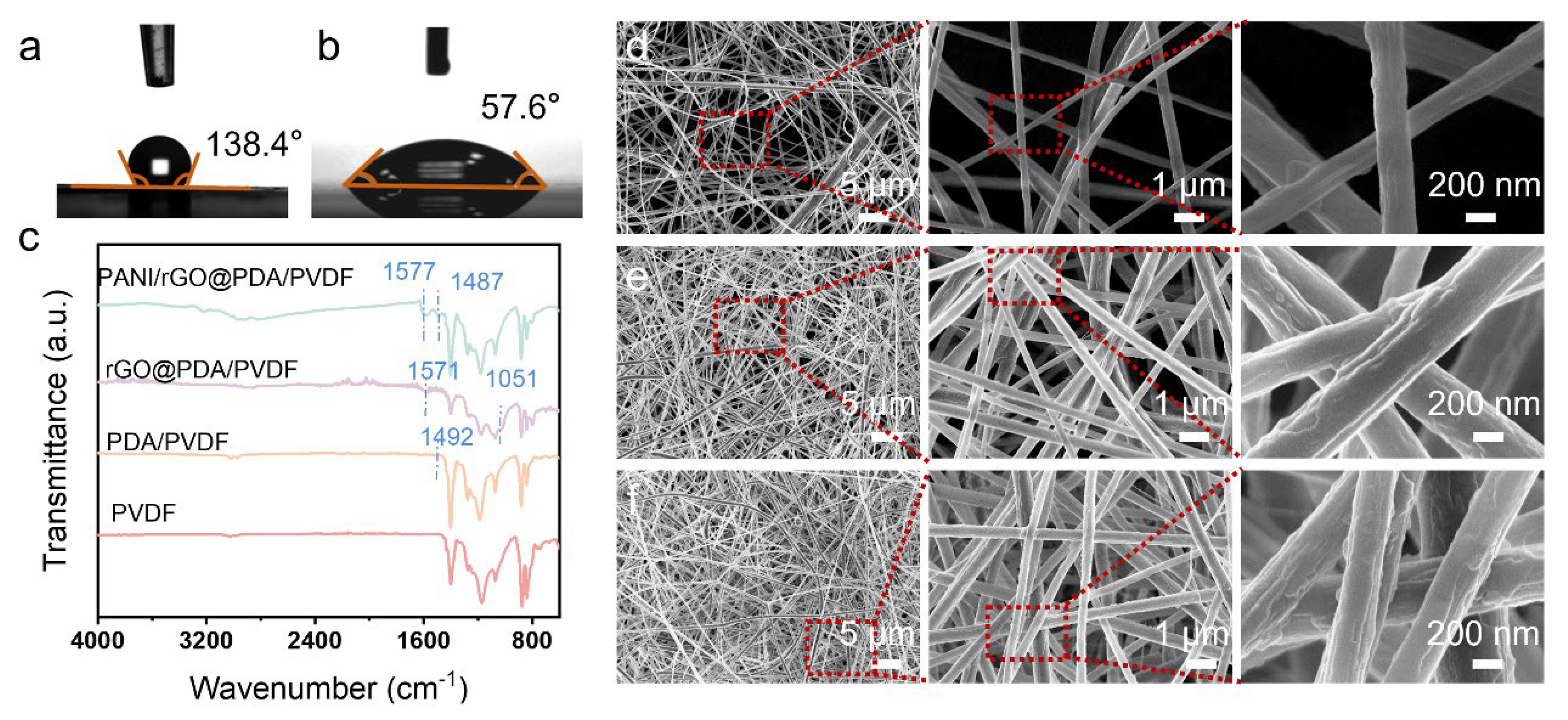
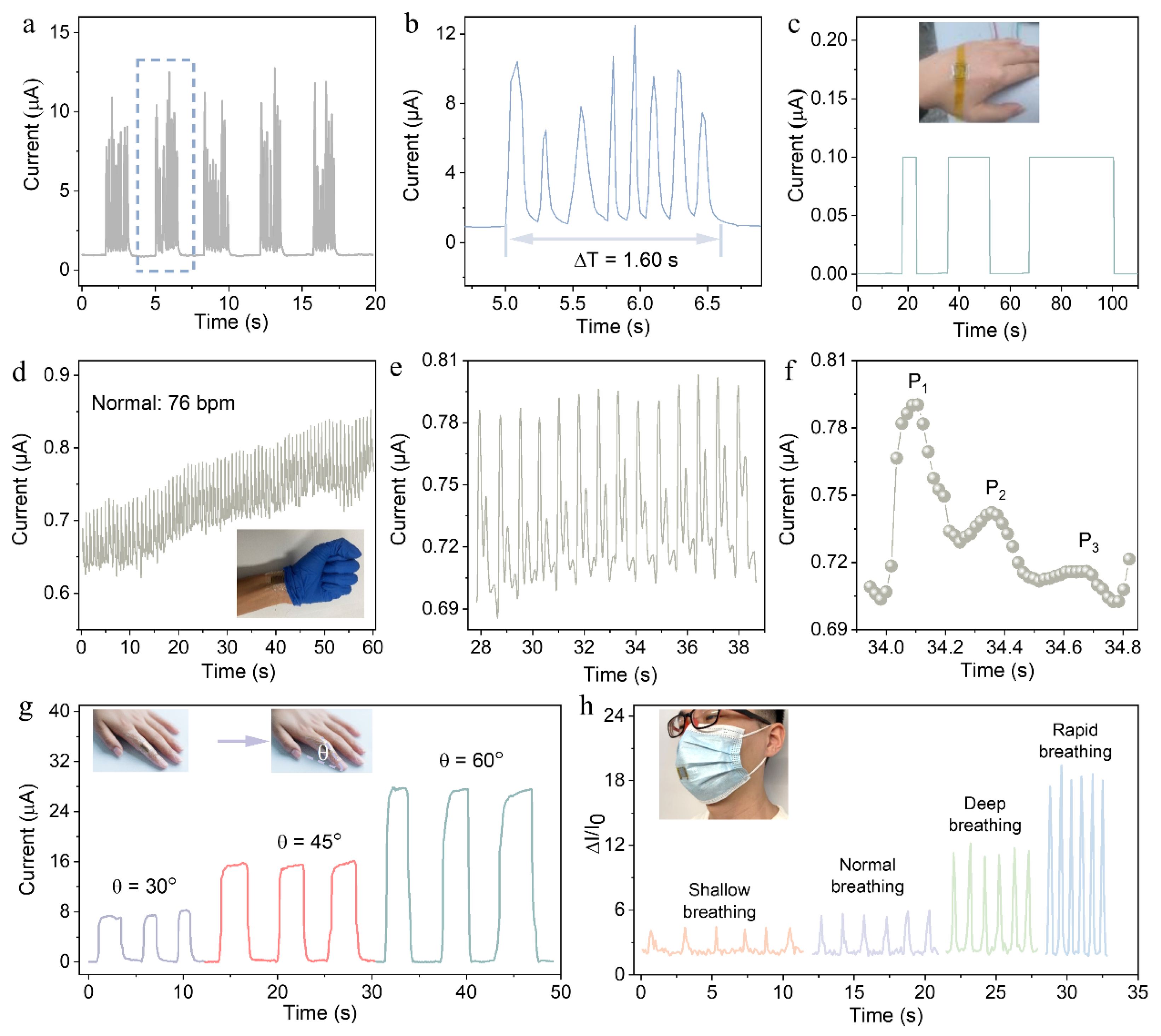
3. Results and Discussion

Figure 1a illustrates the PANI/rGO@PDA/PVDF nanofibrous piezoresistive sensors (PNPS) fabrication procedure. The PVDF nanofiber films were fabricated via electrospinning and served as the mechanical scaffold to support fibrous sensing materials. The high adhesion and surface group of PDA realized the strong interaction between the PVDF nanofibers and electroactive materials. The PANI particles were deposited onto the PVDF nanofiber surface via in situ polymerization to form a piezoresistive sensing shell, and the rGO sheets were implemented to interconnect electroactive nanofibers to boost in-plane conductive networks. Subsequently, the fibrous piezoresistive sensors were fabricated by sandwiching PANI/rGO@PDA/PVDF nanofibers active films between two pieces of flexible Au electrodes, followed by PU medical tape encapsulation. To characterize the morphology of electrospun fibrous composite films, scanning electron microscopy (SEM) was performed to analyze the topological properties of the as-prepared DA/PVDF, PDA/PVDF, rGO/PDA/PVDF, and rGO/PANI@PDA/PVDF nanofiber films, as shown in Figure 1b–i. The diameter of the DA/PVDF nanofibers was ca. 278 nm (Figure 1b,c), while the diameter of the PDA/PVDF nanofibers was ca. 364 nm (Figure 1d,e). The slight increase in nanofiber diameter implies the deposition of PDA on the PVDF nanofibers. Clearly, 2D rGO sheets stack on the nanofibers (Figure 1f) and interconnect with nanofibers, building up hierarchical conductive networks among the nanofibers (Figure 1g). According to Figure 1h,i, a number of PANI particles can be clearly observed on the surface of the composite nanofibers, indicative of the successful preparation of the PANI/rGO@PDA/PVDF nanofibers.
Figure 2a,b depicts the contact angle analysis of the electrospun PDA-modified and unmodified PVDF films. Evidently, the contact angles decrease from 138.4° to 57.6° after the PDA surficial modification. This is attributed to the fact that the hydrophilicity of electrospun PVDF nanofiber films has been largely promoted by introducing PDA with plentiful hydrophilic groups such as hydroxy and amino groups. FTIR spectroscopy was used to analyze the chemical composition of the as-synthesized nanofibers (Figure 2c). Compared to the spectrum of pure electrospun PVDF nanofiber films, the absorption peak emerging at ~1487.91 cm−1 in the PDA/PVDF films is attributed to the C=C stretching vibration of the PDA [41]. Meanwhile, characteristic absorption peaks at 1571.68 cm−1 and 1055.91 cm−1 in the spectrum of the rGO@PDA/PVDF nanofiber films correspond to C-H bending vibration in the oxygen-depleted carbon lattice and C=O stretching vibration of the rGO sheet, respectively [42,43]. In addition, there are two new absorption peaks at 1577.68 cm−1 and 1487.91 cm−1 in the spectrum of PANI/rGO@PDA/PVDF nanofiber films (PNFs), which are attributed to the C=C tensile vibration of the benzene ring and quinone ring of the PANI molecules, respectively [44]. The topological properties, like fibrous distribution density and porosity, dramatically affect the deposition of electroactive materials. To uncover the morphological characteristics, PNF electrospun nanofibers with various electrospinning durations (3 min, 6 min, and 9 min) were characterized and are presented in Figure 2d–f. Notably, the fibrous distribution density is positively proportional to the electrospinning time. The sparse fibrous distribution density fabricated within 3 min fails to construct sufficient conducting paths, especially under subtle pressures, while an excessively thick membrane synthesized within 9 min hinders the deposition of electroactive materials and reduces the mechano-electric conversion efficiency. To balance the performance metrics of sensitivity and pressure detection range, the PNF fabricated with a 6 min electrospinning time was utilized in the following.
To explore the pressure-sensing performance of the as-received PNPS, a force-induced electrical signal detection system was built consisting of a computer, a stepping motor (vertical test stand), and a constant current source (Keithley 4200), as shown in Figure 3a. A set of current–voltage (I–V) curves was recorded under various external stimuli with applied voltages ranging from −1 V to 1 V. As shown in Figure 3b, the good linearity of all I–V curves confirms the ohmic contact between the electroactive nanofiber films and the electrodes. Meanwhile, the slope of the I–V curves increases with rising applied pressure from 0 to 600 Pa (in 100 Pa increments), attributable to the increase in the device’s contact resistance. Figure 3c reveals the sensitivity of the electroactive nanofiber films with or without rGO sheets. The sensitivity (S) is calculated as S = (ΔI/I0)/ΔP, where the value (S) is obtained from the slope of linear regression between the relative current change (ΔI/I0) and applied pressures (P). With increasing applied pressures from 0 to 30 kPa, the relative current rapidly increases due to the enhancement of the conductive pathway between the nanofibers and electrodes. The sensitivities of the electroactive nanofiber films comprise two sections: the Sr1 and S1 in the low-pressure range from 0 to 10 kPa and the Sr2 and S2 in the high-pressure range from 10 to 30 kPa. The corresponding sensitivities of Sr1, Sr2, S1, and S2 are 13.41, 2.31, 8.22, and 0.92, respectively. Furthermore, the electroactive nanofiber films without rGO sheets have a relatively slow response and recovery time of 0.32 and 1.52 s (Figure 3d), while fast response and recovery times of 9 ms and 45 ms were achieved by the electroactive nanofiber films with rGO sheets (Figure 3e). Notably, the higher sensitivity and faster response and recovery rate of rGO-doped films stemmed from the interconnection between the rGO sheets and the electroactive nanofiber, which boosted the in-plane conductive pathway and provided a rapid charge transfer channel.
To illustrate the dynamic sensing response of the PNPS, various dynamic pressures of 0.5 kPa, 2 kPa, and 5 kPa are cyclically loaded and unloaded on the devices. As presented in Figure 3f, the stable and regular output waveforms without obvious signal decline or baseline drift verify the good reproducibility and repeatability of the sensors, indicating that they have excellent capability to monitor external pressure in real-time. To evaluate the ability of as-prepared PNPS to distinguish subtle pressure, a 116 mg sponge (equivalent to 12 Pa) was repeatedly loaded onto and removed from the PNPS (Figure 3g). Evidently, the distinct current output waveforms demonstrate an external pressure detection limit as low as 12 Pa.
Additionally, Figure 3h shows the real-time output current in response to the continuous dripping of water droplets (approximately 38 mg) onto the surface of the piezoresistive sensors. The stepped increase in current as the water droplets increase confirms the feasibility of the PNPS for monitoring minimal stimuli. After 12,000 compressive loading/unloading cycles under a pressure of 400 Pa, the current response of the devices was clearly retained (Figure 3i). Notably, the amplitude of the signal waveform (the inset in Figure 3i) in different periods did not obviously alter, indicating the potential of PNPS for durable and stable human motion biomonitoring.
To demonstrate the sensing capability of the PNPS for wearable biomonitoring, the as-fabricated devices were attached to different sites on the human body. Figure 4a presents the dynamic signal fluctuation in response to the finger pressing when the flexible sensor is attached to the finger pulp. The successive finger touch on the devices at a relatively fast frequency of 4–6 Hz can be implemented to simulate early-stage Parkinson’s disease with static tremors. Figure 4b presents the enlarged view of a set of imitated static tremors at 5 Hz, implying the potential of the PNPS in early screening of diseases. Additionally, the PGPP piezoresistive sensor was worn on the dorsal hand surface to monitor the palmar motion during repetitive fist-clenching actions. Figure 4c demonstrates that the output current elevates during fist clenching and returns to baseline levels upon hand relaxation, indicating the potential of the PNPS for assisting palmar grasp rehabilitation training.
The subtle radial arterial signal of a 24-year-old male was recorded by the output current waveform. As plotted in Figure 4d, the detected heartbeat of 76 beats per minute (bpm) matches well with the test results from a commercial Huawei watch. The magnified view in Figure 4e presents 14 representative pulse waveforms derived from Figure 4d, where uniform oscillation patterns and waveform congruence are observed. Additionally, three characteristic peaks of a radial arterial pulse [45,46], including the early systolic pressure (P1), late systolic shoulder enhancement (P2), and diastolic pulse waveform (P3), can be clearly observed in the single pulse waveform (Figure 4f). These experimental results verify the reliability and accuracy of the as-developed PNPS for real-time arterial physiological monitoring.
To assess the capability to monitor limb motion, the as-fabricated PNPS was mounted on the index finger (Figure 4g). The output-relative current proportional to the bending angles (30°, 45°, 60°) confirms a prominent durability and sensitivity to body movement. This phenomenon could be attributed to increased compressive deformation of the PNPS at larger finger bending angles, which generate higher output currents. Additionally, the as-fabricated sensor was fixed on the mask to monitor respiration. As plotted in Figure 4h, the respiratory patterns of shallow, normal, deep, and rapid breathing can be explicitly quantified, with the signal frequency and amplitude respectively corresponding to the breathing rate and depth, allowing for the real-time assessment of respiratory status. These results demonstrate its significant potential for applications in disease diagnosis and rehabilitation training. Table 1 compares the sensing performance of our device with that of previously reported pressure sensors. Notably, the as-fabricated PNPS exhibits high sensitivity across a wide pressure range, a fast response time, and good stability [4,24,47,48,49].

4. Conclusions

In this work, we developed an electroactive electrospun nanofiber-based piezoresistive sensor by integrating rGO sheets and PANI particles into PDA-modified PVDF nanofiber network architecture. The PANI particles were deposited onto a nanofiber substrate to construct electroactive nanofibers, and the rGO sheets were utilized to interconnect nanofibers to strengthen in-plane charge carrier transport. Benefitting from the integration of the multi-dimensional conductive channel, optimal pressure-sensing behaviors were attained with a high sensitivity of 13.43 kPa−1, a fast response/recovery time of 9 ms/45 ms, a wide pressure detection range from 0 Pa to 30 kPa, and good reproducibility. Additionally, the excellent sensing capability of the electroactive nanofiber piezoresistive sensors could be utilized to monitor human activities from subtle vital signs (e.g., radial arterial pulse, respiration) to large-scale movements (e.g., limb bending and finger touch). This work not only strengthens the fundamental mechanism of the charge transport between the multi-dimensional conductive materials for piezoresistive sensors, but also establishes a novel route for the development of high-performance wearable electronics.

Author Contributions

Conceptualization, C.C., Y.S., and G.X.; methodology, F.W., and Y.S.; formal analysis, H.P.; investigation, H.L., Y.W., and H.P.; data curation, Y.W., and H.P.; writing—original draft preparation, H.P.; writing—review and editing, H.P. and Y.S.; visualization, H.P.; supervision, C.C., Y.S., and F.W.; funding acquisition, Y.S., and H.P. All authors have read and agreed to the published version of the manuscript.

Funding

This work was supported by the National Natural Science Foundation of China (Nos. 62074027, 61671115) and the Postdoctoral Fellowship Program (Grade B) of the China Postdoctoral Science Foundation (No. GZB20230106).

Data Availability Statement

The raw data supporting the conclusions of this article will be made available by the authors on request.

Conflicts of Interest

The authors declare no conflicts of interest.

References

  1. Zhou, Q.; Ding, Q.; Geng, Z.; Hu, C.; Yang, L.; Kan, Z.; Dong, B.; Won, M.; Song, H.; Xu, L.; et al. A Flexible Smart Healthcare Platform Conjugated with Artificial Epidermis Assembled by Three-Dimensionally Conductive MOF Network for Gas and Pressure Sensing. Nano-Micro Lett. 2024, 17, 50–69. [Google Scholar] [CrossRef] [PubMed]
  2. Guo, R.; Fang, Y.; Wang, Z.; Libanori, A.; Xiao, X.; Wan, D.; Cui, X.; Sang, S.; Zhang, W.; Zhang, H.; et al. Deep Learning Assisted Body Area Triboelectric Hydrogel Sensor Network for Infant Care. Adv. Funct. Mater. 2022, 32, 2204803. [Google Scholar] [CrossRef]
  3. Yang, J.S.; Chung, M.K.; Yoo, J.Y.; Kim, M.U.; Kim, B.J.; Jo, M.S.; Kim, S.H.; Yoon, J.B. Interference-free nanogap pressure sensor array with high spatial resolution for wireless human-machine interfaces applications. Nat. Commun. 2025, 16, 2024–2034. [Google Scholar] [CrossRef] [PubMed]
  4. Y Guo, Y.; Zhong, M.; Fang, Z.; Wan, P.; Yu, G. A Wearable Transient Pressure Sensor Made with MXene Nanosheets for Sensitive Broad-Range Human-Machine Interfacing. Nano Lett. 2019, 19, 1143–1150.ok. [Google Scholar] [CrossRef]
  5. Wei, X.; Li, H.; Yue, W.; Gao, S.; Chen, Z.; Li, Y.; Shen, G. A high-accuracy, real-time, intelligent material perception system with a machine-learning-motivated pressure-sensitive electronic skin. Matter 2022, 5, 1481–1501. [Google Scholar] [CrossRef]
  6. Kim, Y.; Chortos, A.; Xu, W.; Liu, Y.; Oh, J.Y.; Son, D.; Kang, J.; Foudeh, A.M.; Zhu, C.; Lee, Y.; et al. A bioinspired flexible organic artificial afferent nerve. Science 2018, 360, 998–1003. [Google Scholar] [CrossRef]
  7. Zhang, Y.; Yang, J.; Hou, X.; Li, G.; Wang, L.; Bai, N.; Cai, M.; Zhao, L.; Wang, Y.; Zhang, J.; et al. Highly stable flexible pressure sensors with a quasi-homogeneous composition and interlinked interfaces. Nat. Commun. 2022, 13, 1317–1328. [Google Scholar] [CrossRef]
  8. Niu, H.; Li, H.; Gao, S.; Li, Y.; Wei, X.; Chen, Y.; Yue, W.; Zhou, W.; Shen, G. Perception-to-cognition tactile sensing based on artificial-intelligence-motivated human full-Skin bionic electronic skin. Adv. Mater. 2022, 34, 2202622. [Google Scholar] [CrossRef]
  9. Pan, H.; Chen, G.; Chen, Y.; Di Carlo, A.; Mayer, M.A.; Shen, S.; Chen, C.; Li, W.; Subramaniam, S.; Huang, H.; et al. Biodegradable cotton fiber-based piezoresistive textiles for wearable biomonitoring. Biosens. Bioelectron. 2023, 222, 114999. [Google Scholar] [CrossRef]
  10. Zheng, X.; Cao, W.; Hong, X.; Zou, L.; Liu, Z.; Wang, P.; Li, C. Versatile Electronic Textile Enabled by a Mixed-Dimensional Assembly Strategy. Small 2023, 19, 2208134. [Google Scholar] [CrossRef]
  11. Wang, J.; Liu, Y.; Li, X.; Zeng, W.; Zhao, T.; Luo, B.; Liu, T.; Chi, M.; Cai, C.; Zhang, S.; et al. Graded Nanotexturing Architectural Wearable Triboelectric Sensor for Programmable Haptic Exploration. Nano Lett. 2024, 24, 13542–13550. [Google Scholar] [CrossRef]
  12. Su, Y.; Li, W.; Cheng, X.; Zhou, Y.; Yang, S.; Zhang, X.; Chen, C.; Yang, T.; Pan, H.; Xie, G.; et al. High-performance piezoelectric composites via beta phase programming. Nat. Commun. 2022, 13, 4867–4878. [Google Scholar] [CrossRef]
  13. Zhao, X.; Zhou, Y.; Li, A.; Xu, J.; Karjagi, S.; Hahm, E.; Rulloda, L.; Li, J.; Hollister, J.; Kavehpour, P.; et al. A self-filtering liquid acoustic sensor for voice recognition. Nat. Electron. 2024, 7, 924–932. [Google Scholar] [CrossRef]
  14. Zhou, Y.; Zhao, X.; Xu, J.; Fang, Y.; Chen, G.; Song, Y.; Li, S.; Chen, J. Giant magnetoelastic effect in soft systems for bioelectronics. Nat. Mater. 2021, 20, 1670–1676. [Google Scholar] [CrossRef] [PubMed]
  15. Ruth, S.R.A.; Feig, V.R.; Tran, H.; Bao, Z. Microengineering Pressure Sensor Active Layers for Improved Performance. Adv. Funct. Mater. 2020, 30, 2003491. [Google Scholar] [CrossRef]
  16. Yang, R.; Dutta, A.; Li, B.; Tiwari, N.; Zhang, W.; Niu, Z.; Gao, Y.; Erdely, D.; Xin, X.; Li, T.; et al. Iontronic pressure sensor with high sensitivity over ultra-broad linear range enabled by laser-induced gradient micro-pyramids. Nat. Commun. 2023, 14, 2907–2918. [Google Scholar] [CrossRef]
  17. Bai, N.; Wang, L.; Xue, Y.; Wang, Y.; Hou, X.; Li, G.; Zhang, Y.; Cai, M.; Zhao, L.; Guan, F.; et al. Graded interlocks for iontronic pressure sensors with high sensitivity and high linearity over a broad range. ACS Nano 2022, 16, 4338–4347. [Google Scholar] [CrossRef]
  18. Yang, J.; Liu, L.; Zhang, D.; Zhang, H.; Ma, J.; Zheng, J.; Wang, C. Dual-stage surficial microstructure to enhance the sensitivity of MXene pressure sensors for human physiological signal acquisition. ACS Appl. Mater. Interfaces 2024, 16, 1096–1106. [Google Scholar] [CrossRef]
  19. Wang, S.; Deng, W.; Yang, T.; Ao, Y.; Zhang, H.; Tian, G.; Deng, L.; Huang, H.; Huang, J.; Lan, B.; et al. Bioinspired MXene-based piezoresistive sensor with two-stage enhancement for motion capture. Adv. Funct. Mater. 2023, 33, 2214503. [Google Scholar] [CrossRef]
  20. Li, L.; Cheng, Y.; Cao, H.; Liang, Z.; Liu, Z.; Yan, S.; Li, L.; Jia, S.; Wang, J.; Gao, Y. MXene/rGO/PS spheres multiple physical networks as high-performance pressure sensor. Nano Energy 2022, 95, 106986. [Google Scholar] [CrossRef]
  21. Wang, L.; Li, H.; Zhao, C.; Zhang, L.; Li, J.; Din, S.U.; Wang, Z.; Sun, J.; Torres, S.A.G.; Fan, Z.; et al. Aluminium surface work hardening enables multi-scale 3D lithography. Nat. Mater. 2025, 24, 39–47. [Google Scholar] [CrossRef]
  22. Pan, L.; Chortos, A.; Yu, G.; Wang, Y.; Isaacson, S.; Allen, R.; Shi, Y.; Dauskardt, R.; Bao, Z. An ultra-sensitive resistive pressure sensor based on hollow-sphere microstructure induced elasticity in conducting polymer film. Nat. Commun. 2014, 5, 3002–3009. [Google Scholar] [CrossRef] [PubMed]
  23. Xiong, W.; Zhang, F.; Qu, S.; Yin, L.; Li, K.; Huang, Y. Marangoni-driven deterministic formation of softer, hollow microstructures for sensitivity-enhanced tactile system. Nat. Commun. 2024, 15, 5596–5608. [Google Scholar] [CrossRef]
  24. Wang, Q.; Jian, M.; Wang, C.; Zhang, Y. Carbonized silk nanofiber membrane for transparent and sensitive electronic skin. Adv. Funct. Mater. 2017, 27, 1605657. [Google Scholar] [CrossRef]
  25. Yin, R.; Yang, S.; Li, Q.; Zhang, S.; Liu, H.; Han, J.; Liu, C.; Shen, C. Flexible conductive Ag nanowire/cellulose nanofibril hybrid nanopaper for strain and temperature sensing applications. Sci. Bull. 2020, 65, 899–908. [Google Scholar] [CrossRef] [PubMed]
  26. Chen, G.; Xiao, X.; Zhao, X.; Tat, T.; Bick, M.; Chen, J. Electronic textiles for wearable point-of-care systems. Chem. Rev. 2022, 122, 3259–3291. [Google Scholar] [CrossRef]
  27. Dai, J.; Xie, G.; Huo, X.; Li, J.; Deng, S.; Su, Y. Implantable and Biodegradable Smart Textiles for Continuous Limb and Gastrointestinal Motility Monitoring. Small 2025, 21, 2407773. [Google Scholar] [CrossRef]
  28. Park, J.; Lee, Y.; Hong, J.; Ha, M.; Jung, Y.-D.; Lim, H.; Kim, S.Y.; Ko, H. Giant tunneling piezoresistance of composite elastomers with interlocked microdome arrays for ultrasensitive and multimodal electronic skins. ACS Nano J. 2014, 8, 4689–4697. [Google Scholar] [CrossRef]
  29. Cai, Y.; Shen, J.; Dai, Z.; Zang, X.; Dong, Q.; Guan, G.; Li, L.J.; Huang, W.; Dong, X. Extraordinarily stretchable all-carbon collaborative nanoarchitectures for epidermal sensors. Adv. Mater. 2017, 29, 1606411. [Google Scholar] [CrossRef]
  30. Zhang, S.; Wen, L.; Wang, H.; Zhu, K.; Zhang, M. Vertical CNT–Ecoflex nanofins for highly linear broad-range-detection wearable strain sensors. J. Mater. Chem. C 2018, 6, 5132–5139. [Google Scholar] [CrossRef]
  31. Cheng, R.; Zeng, J.; Wang, B.; Li, J.; Cheng, Z.; Xu, J.; Gao, W.; Chen, K. Ultralight, flexible and conductive silver nanowire/nanofibrillated cellulose aerogel for multifunctional strain sensor. Chem. Eng. J. 2021, 424, 130565. [Google Scholar] [CrossRef]
  32. Guan, F.; Xie, Y.; Wu, H.; Meng, Y.; Shi, Y.; Gao, M.; Zhang, Z.; Chen, S.; Chen, Y.; Wang, H.; et al. Silver Nanowire-Bacterial Cellulose Composite Fiber-Based Sensor for Highly Sensitive Detection of Pressure and Proximity. ACS Nano J. 2020, 14, 15428–15439. [Google Scholar] [CrossRef] [PubMed]
  33. Chen, S.; Jiang, K.; Lou, Z.; Chen, D.; Shen, G. Recent Developments in Graphene-Based Tactile Sensors and E-Skins. Adv. Mater. Technol. 2018, 3, 1700248. [Google Scholar] [CrossRef]
  34. Tao, L.Q.; Zhang, K.N.; Tian, H.; Liu, Y.; Wang, D.Y.; Chen, Y.Q.; Yang, Y.; Ren, T.L. Graphene-paper pressure sensor for detecting human motions. ACS Nano J. 2017, 11, 8790–8795. [Google Scholar] [CrossRef] [PubMed]
  35. Bosque, A.d.; Sánchez-Romate, X.F.; Sánchez, M.; Ureña, A. Ultrasensitive and highly stretchable sensors for human motion monitoring made of graphene reinforced polydimethylsiloxane: Electromechanical and complex impedance sensing performance. Carbon 2022, 192, 234–248. [Google Scholar] [CrossRef]
  36. Lou, Z.; Chen, S.; Wang, L.; Shi, R.; Li, L.; Jiang, K.; Chen, D.; Shen, G. Ultrasensitive and ultraflexible e-skins with dual functionalities for wearable electronics. Nano Energy 2017, 38, 28–35. [Google Scholar] [CrossRef]
  37. Pan, H.; Xie, G.; Pang, W.; Wang, S.; Wang, Y.; Jiang, Z.; Du, X.; Tai, H. Surface engineering of a 3D topological network for ultrasensitive piezoresistive pressure sensors. ACS Appl. Mater. Interfaces 2020, 12, 38805. [Google Scholar] [CrossRef]
  38. Bi, L.; Yang, Z.; Chen, L.; Wu, Z.; Ye, C. Compressible AgNWs/Ti3C2Tx MXene aerogel-based highly sensitive piezoresistive pressure sensor as versatile electronic skins. J. Mater. Chem. A 2020, 8, 20030–20036. [Google Scholar] [CrossRef]
  39. Zhi, H.; Zhang, X.; Wang, F.; Wan, P.; Feng, L. Flexible Ti3C2Tx MXene/PANI/Bacterial Cellulose Aerogel for e-Skins and Gas Sensing. ACS Appl. Mater. Interfaces 2021, 13, 45987–45994. [Google Scholar] [CrossRef]
  40. Cao, M.; Wang, M.; Li, L.; Qiu, H.; Padhiar, M.A.; Yang, Z. Wearable rGO-Ag NW@cotton fiber piezoresistive sensor based on the fast charge transport channel provided by Ag nanowire. Nano Energy 2018, 50, 528–535. [Google Scholar] [CrossRef]
  41. Wardani, A.K.; Ariono, D.; Subagjo, S.; Wenten, I.G. Fouling tendency of PDA/PVP surface modified PP membrane. Surf. Interfaces. 2020, 19, 100464. [Google Scholar] [CrossRef]
  42. Zhu, T.; Chang, S.; Song, Y.-F.; Lahoubi, M.; Wang, W. PVP-encapsulated CoFe2O4/rGO composites with controllable electromagnetic wave absorption performance. Chem. Eng. J. 2019, 373, 755–766. [Google Scholar] [CrossRef]
  43. Wang, D.; Giannakis, S.; Tang, J.; Luo, K.; Tang, J.; He, Z.; Song, S.; Wang, L. Effect of rGO content on enhanced Photo-Fenton degradation of Venlafaxine using rGO encapsulated magnetic hexagonal FeTiO3 nanosheets. Chem. Eng. J. 2023, 478, 147319. [Google Scholar] [CrossRef]
  44. Luo, W.; Wei, Y.; Zhuang, Z.; Lin, Z.; Li, X.; Hou, C.; Li, T.; Ma, Y. Fabrication of Ti3C2Tx MXene/polyaniline composite films with adjustable thickness for high-performance flexible all-solid-state symmetric supercapacitors. Electrochim. Acta 2022, 406, 139871. [Google Scholar] [CrossRef]
  45. Su, Y.; Chen, C.; Pan, H.; Yang, Y.; Chen, G.; Zhao, X.; Li, W.; Gong, Q.; Xie, G.; Zhou, Y.; et al. Muscle fibers inspired high-performance piezoelectric textiles for wearable physiological monitoring. Adv. Funct. Mater. 2021, 31, 2010962. [Google Scholar] [CrossRef]
  46. Meng, K.; Xiao, X.; Wei, W.; Chen, G.; Nashalian, A.; Shen, S.; Xiao, X.; Chen, J. Wearable pressure sensors for pulse wave monitoring. Adv. Mater. 2022, 34, 2109357. [Google Scholar] [CrossRef]
  47. Chen, S.; Chen, Y.; Li, D.; Xu, Y.; Xu, F. Flexible and Sensitivity-Adjustable Pressure Sensors Based on Carbonized Bacterial Nanocellulose/Wood-Derived Cellulose Nanofibril Composite Aerogels. ACS Appl. Mater. Interfaces 2021, 13, 8754–8763. [Google Scholar] [CrossRef]
  48. Wang, B.; Shi, T.; Zhang, Y.; Chen, C.; Li, Q.; Fan, Y. Lignin-based highly sensitive flexible pressure sensor for wearable electronics. J. Mater. Chem. C 2018, 6, 6423–6428. [Google Scholar] [CrossRef]
  49. Zheng, Y.; Yin, R.; Zhao, Y.; Liu, H.; Zhang, D.; Shi, X.; Zhang, B.; Liu, C.; Shen, C. Conductive MXene/cotton fabric based pressure sensor with both high sensitivity and wide sensing range for human motion detection and E-skin. Chem. Eng. J. 2021, 420, 127720. [Google Scholar] [CrossRef]
Figure 1. Structural design and characterization of PANI/rGO@PDA/PVDF nanofiber piezoresistive sensors. (a) Schematic illustration of the procedure of the rGO/PANI@PDA/PVDF piezoresistive sensor. Scanning electron microscope (SEM) images of the as-prepared DA/PVDF (b), PDA/PVDF (d), rGO/PDA/PVDF (f), and rGO/PANI@PDA/PVDF (h) composite nanofibers, respectively. Scale bars, 5 µm. High-resolution SEM image of DA/PVDF (c), PDA/PVDF (e), rGO/PDA/PVDF (g), and PANI/rGO@PDA/PVDF (i) composite nanofibers. Scale bars, 500 nm.
Jcs 09 00339 g001
Figure 2. The characterization of PANI/rGO@PDA/PVDF nanofiber films. (a) Static contact angles of the distilled water on the surfaces of PVDF nanofiber film. (b) Static contact angles of the distilled water on the surfaces of PDA/PVDF nanofiber film. (c) FTIR spectrum of PDA/PVDF and PANI/rGO@PDA/PVDF. Scanning electron microscope (SEM) images and their partial enlarged drawing of the as-prepared PDA/PVDF with different electrospinning durations: (d) 3 min, (e) 6 min, (f) 9 min.
Jcs 09 00339 g002
Figure 3. The sensing performance of the PANI/rGO@PDA/PVDF nanofiber piezoresistive sensors (PNPS). (a) Schematic illustration of measurement platform for pressure-sensing performance testing. (b) Current–voltage (I–V) curves of PNPS with various pressures from 0 Pa to 600 Pa. (c) The relative current change versus applied pressure curve for PNPS. (d) The response time and recovery time of the sensor based on PANI@PDA/PVDF nanofiber piezoresistive sensors. (e) The response and recovery times of the PNPS piezoresistive sensors. (f) The real-time current change of the piezoresistive sensors in response to dynamic pressures from 0.5 to 5 kPa. (g) The transient response of piezoresistive sensors to the loading and removal of a block of sponge (about 116 mg), corresponding to a pressure of 12 Pa. (h) The real-time output current profile in response to a water droplet continuously dripping on the PNPS (one water droplet is about 38 mg). (i) The durability experiment was conducted under a pressure of 400 Pa with 12,000 loading/unloading cycles. The insets show an enlarged view of the relative current variation curve at different cycling stages.
Jcs 09 00339 g003
Figure 4. The on-body application of the prepared PANI/rGO@PDA/PVDF nanofiber piezoresistive sensors (PNPS) for wearable biomonitoring. (a) The real-time current change of the sensor under imitated knocking of early-stage Parkinson’s disease featuring a static tremor frequency of 5 Hz. (b) Enlarged view of one cycle of the electrical signal highlighted in (a). (c) Real-time current profile in response to palmar motion. (d) Real-time current variation curve of a 23-year-old male’s wrist pulses in static status. Inset is the photograph of the devices attached to the volunteer’s wrist. (e) The partial, enlarged drawing in the real-time signals of (d). (f) Enlarged view of the single pulse wave with characteristic peaks. (g) Dynamic output current waveforms of finger bending at various angles. (h) Respiratory monitoring enabled by the prepared PNPS. Inset is a photograph of the PNPS mounted on the mask.
Jcs 09 00339 g004
Table 1. Summary of the sensing performance of fibrous piezoresistive sensors.
Sensing MaterialSensitivity (kPa−1)/Pressure RangeResponse Time (ms)Stability(Cycles)Reference
Ti3C2Tx-paper3.81/0.982–10 kPa1110,000[4]
Carbonized cellulose0.2/0–3 kPa
0.15/3–10 kPa
N/AN/A[47]
Graphene/paper17.2/0–2 kPa
0.012/2–20 kPa
120300[48]
Carbonized silk nanofiber34.71/0.8–400 Pa
1.16/0.4–5 kPa
16.510,000[24]
MXene/cotton5.3/0–1.3 kPa50900[49]
PANI/rGO@PDA/PVDF13.43/0–10 kPa912,000This work
N/A: Not detected/not applicable under the present experimental conditions.
Disclaimer/Publisher’s Note: The statements, opinions and data contained in all publications are solely those of the individual author(s) and contributor(s) and not of MDPI and/or the editor(s). MDPI and/or the editor(s) disclaim responsibility for any injury to people or property resulting from any ideas, methods, instructions or products referred to in the content.

