Article

Collagen/Polypyrrole Biomimetic Electroactive Composite Coating with Fiber Network Structure on Titanium Surface for Bone Tissue Engineering

1
School of Materials Science and Engineering, National Key Laboratory of Silicon and Advanced Semiconductor Materials, Zhejiang University, Hangzhou 310027, China
2
Institute of Wenzhou, Zhejiang University, Wenzhou 325006, China
*
Authors to whom correspondence should be addressed.
These authors contributed equally to this work.
J. Compos. Sci. 2025, 9(7), 325; https://doi.org/10.3390/jcs9070325
Submission received: 7 May 2025 / Revised: 17 June 2025 / Accepted: 19 June 2025 / Published: 24 June 2025
(This article belongs to the Special Issue Biomedical Composite Applications)

Abstract

Both biochemical cues and the electrophysiological microenvironment play a pivotal role in influencing cell behaviors. In this study, collagen/polypyrrole biomimetic electroactive composite coatings with a fiber network structure were constructed on the surface of titanium substrates by hot alkali treatment and stepwise electrochemical deposition. Materialistic characterization and electrochemical performance tests demonstrated that the titanium electrodes modified with collagen/polypyrrole composite coatings exhibited the surface morphology of a collagen film layer, and their electroactivity was significantly enhanced. Cellular experiments demonstrated that the collagen in the composite coatings could provide good biomimetic biochemical cues as a main extracellular matrix component, which have a substantial effect in promoting cell adhesion, proliferation, and osteogenic differentiation. Furthermore, under exogenous electrical signals, the polypyrrole coating has the capacity to facilitate an appropriate electrophysiological microenvironment, thereby promoting osteogenic differentiation. The collagen/polypyrrole composite coating exhibited a better effect in promoting osteogenic differentiation among all samples by simultaneously providing the appropriate biochemical cues and electrophysiological microenvironments. This work demonstrates the feasibility of synergistic pro-osteogenesis by biochemical cues and an electrophysiological microenvironment, which is instructive for the field of bone tissue engineering.

1. Introduction

Bone tissue possesses a natural regenerative capacity sufficient to heal minor injuries [1,2]. However, bone defects exceeding a critical size threshold will not heal spontaneously, necessitating clinical intervention [3,4]. Bone tissue engineering has emerged as a potential new therapy for the direct repair of bone defects or the tissue engineering of bone tissue for transplantation [5,6,7]. In this therapy, biomaterials play a key role in providing scaffolds and extracellular environments to support regenerative cells and promote tissue regeneration [5,8,9,10,11].
Certain biomaterials have been shown to simulate the specific microenvironment of bone tissue, which is crucial for promoting the repair and regeneration of bone defects [12,13,14,15]. Biomimetic materials and electroactive materials have been demonstrated to provide extracellular matrix (ECM) microenvironments [16] and electrical microenvironments [17], respectively. These microenvironments have been shown to regulate various cellular behaviors [18,19], thus attracting extensive research attention.
In light of the critical role of the ECM in the cellular microenvironment, various ECM-mimetic biomaterials have been utilized in the field of bone tissue engineering [20,21,22]. The selection of specific components is of paramount importance when ECM components are used to synthesize artificial ECM materials. Among these components, collagen (Col), as the most prevalent structural protein of the ECM [23,24,25], is highly favored [26,27]. Researchers have developed various forms of collagen-based ECM-mimetic materials, including collagen hydrogels [28], collagen microspheres, collagen coatings, and collagen scaffolds [29]. Que et al. [30] employed a bottom-up strategy to modify and reassemble natural collagen, resulting in collagen hydrogels with distinct degradation properties, cell adhesion characteristics, and mechanical properties. Matsunaga et al. [31] prepared mono-dispersed collagen gel beads with a diameter smaller than 300 μm using microfluidic technology and performed cell seeding and culture on them. Ma et al. [32] extracted rat tail collagen gels followed by freeze-drying and cross-linking, and successfully prepared heparinized collagens scaffolds, which were made into nerve conduits and significantly promoted nerve growth and functional recovery of nerve defects in rats. Collagen, a prevalent ECM-based material, is characterized by its exceptional biocompatibility and the presence of abundant biochemical cues [33]. However, it is important to note that collagen’s electrical conductivity is limited [34]. While it possesses piezoelectric properties [35], these properties are weak and require strict structural conditions for their expression. Consequently, it is difficult to apply the weak piezoelectricity of collagen, for the structure of collagen undergoes changes during material preparation.
Electroactive materials have the capacity to regulate the electrophysiological microenvironment by controlling the surface potential of the material. This process enables the delivery of electrical stimuli to the cells, thereby influencing cellular physiological activities [36,37,38]. Due to the precise and controllable nature of the electrical signals they apply, these materials are considered the next generation of smart biomaterials [39]. Polypyrrole (Ppy), as a representative conductive polymer [40], exhibits excellent conductivity and electroactivity [41,42]. When exposed to external electrical signals, it generates a stable and adjustable electrical microenvironment for cells [43]. It is noteworthy that this material possesses a certain biocompatibility and has been demonstrated to support the adhesion and growth of various kinds of cells [44]. However, the majority of existing electroactive materials primarily focus on providing electrical signals, often neglecting the biochemical cues that cells require.
Consequently, a more promising biomaterial design strategy could entail the construction of biomimetic electroactive materials, which are capable of providing both the extracellular matrix (ECM) microenvironment and the electrical microenvironment. This would result in enhanced regulatory effects on cell behavior.
This study proposes a method for constructing a fibrous network pore structure on the surface of a titanium substrate through hot alkali treatment. Stepwise electrochemical deposition was employed to prepare a Col/Ppy composite film layer on a porous-surfaced titanium substrate. The porous surface structure of the titanium substrate enabled a higher deposition of polypyrrole (Ppy) and facilitated better adhesion of the collagen (Col) film layer. The materialistic and electrochemical properties of the Col/Ppy composite film-modified porous-surfaced titanium substrates were characterized. Furthermore, the biological evaluation of the composite films was conducted through the assessment of BMSC adhesion, proliferation, and osteogenic differentiation.

2. Materials and Methods

2.1. Preparation of Col/Ppy Composite Films

First, the titanium (Ti, 99.7%; Xi’an Tishan Metal Technology Co., Ltd., Xi’an, China) substrate (20 mm × 10 mm × 0.5 mm) underwent a series of ultrasonic cleanings in deionized water, anhydrous ethanol (≥99.7%; Sinopharm Chemical Reagent Co., Ltd.), and acetone (≥99.7%; Sinopharm Chemical Reagent Co., Ltd.), in that order, for 15 min, followed by drying at 60 °C for subsequent use. Sodium hydroxide (≥96.0%; Sinopharm Chemical Reagent Co., Ltd.) was dissolved in deionized water to prepare a 0.5 M sodium hydroxide solution. The cleaned Ti substrate was then placed into the prepared sodium hydroxide solution and submerged in the solution. And they were subjected to hot alkali treatment in an 80 °C oven for 24 h. Afterward, the alkali-treated Ti sheet was rinsed with deionized water. Then, the sheet was subjected to a sequence of ultrasonic treatments with deionized water, anhydrous ethanol, and acetone, in that order, for a duration of 15 min each, followed by drying. This process resulted in a porous-surfaced titanium substrate with a nanofiber-like porous morphology on the surface. Prior to electrochemical deposition, the porous-surfaced titanium substrate was spray-coated with Pt for 120 s to enhance the conductivity of the substrate. The resulting porous-surfaced titanium substrate was designated as PTi.
Next, the preparation of collagen (Col, type I collagen; Beijing Yierkang Biotechnology Co., Ltd., Beijing, China) and polypyrrole (Ppy, Aladdin) electrochemical deposition solutions was carried out. Pyrrole electrochemical deposition solution was obtained by adding 510 μL of concentrated hydrochloric acid (36.0%~38.0%; Sinopharm Chemical Reagent Co., Ltd., Shanghai, China) and 416 μL of pyrrole monomer to 30 mL of deionized water, followed by stirring for 3 h. Additionally, 510 μL of concentrated hydrochloric acid and 1.07 g of type I collagen were added to 30 mL of deionized water. The mixture was stirred for 5 h to ensure complete dissolution of the collagen. Afterward, 0.1 M sodium hydroxide solution was added to adjust the pH of the solution to 4.1–4.7, obtaining the collagen electrochemical deposition solution.
Finally, the electrochemical deposition was carried out by using a three-electrode system, with a Pt electrode acting as the counter electrode, an Ag/AgCl electrode serving as the reference electrode, and the Pt-treated porous-surfaced titanium electrode functioning as the working electrode. The pyrrole electrochemical deposition solution, which had been prepared earlier, was used as the electrolyte. A constant voltage of 0.9 V was applied for 120 s to perform the deposition, resulting in a polypyrrole (Ppy) film layer on the surface of the PTi substrate. This modified PTi was designated PTi-Ppy. The collagen electrochemical deposition solution that had been prepared earlier was utilized as the electrolyte, and a constant voltage of −2 V was applied for 120 s to achieve electrochemical deposition, resulting in a collagen (Col) film layer on the surface of the PTi substrate, which was named PTi-Col. Following the above parameters, the Ppy film layer was first electrochemically deposited, and then the electrolyte was replaced to electrochemically deposit the Col film layer, resulting in a Col/Ppy composite film on the surface of the PTi substrate, which was named PTi-Col/Ppy.
In the course of all electrochemical deposition processes, it was ensured that only half of the PTi substrate (10 mm × 10 mm) was immersed in the electrolyte. Subsequent to the electrochemical deposition, the half of the substrate containing the deposited film was excised, thereby obtaining a sample with dimensions of 10 mm × 10 mm. The obtained samples, namely, PTi, PTi-Col, PTi-Ppy, and PTi-Col/Ppy, were cleaned with deionized water, followed by drying. These samples were later employed for the subsequent experimental procedures.

2.2. Characterization of the Composite Films

The surface morphology of each film was observed at multiple magnifications using a scanning electron microscope (FE-SEM; FEI SU-70, Eindhoven, The Netherlands). The overall morphology of each sample was characterized using the Atomic Force Microscopy (AFM) functionality of a near-field optical microscope (NSOM; NTEGRA Spectra C, NTMDT, Moscow, Russia). The surface composition of each sample was analyzed using Energy-Dispersive X-ray Spectroscopy (EDS), Fourier Transform Infrared Spectroscopy (FTIR; Nicolet iS50, Thermo Fisher, Waltham, MA, USA), and the micro-Raman functionality of the near-field optical microscope (NSOM; NTEGRA Spectra C, NTMDT, Moscow, Russia). Additionally, the surface composition of the samples was further analyzed using the Attenuated Total Reflectance mode of the Fourier Transform Infrared Spectrometer (ATR-FTIR). Based on the morphology characterization and compositional analysis, the water contact angle of each sample was measured using a contact angle goniometer (CAI; OCA 20, Dataphysics, Stuttgart, Germany) to evaluate their hydrophilicity.
The electrochemical tests were conducted using an electrochemical workstation (CHI660E, Chenhua Instrument, Shanghai, China), with Phosphate-Buffered Saline (PBS, Hyclone) solution as the electrolyte. A three-electrode system was employed, with a Pt electrode serving as the counter electrode, an Ag/AgCl electrode functioning as the reference electrode, and the sample electrode designated as the working electrode. Electrochemical Impedance Spectroscopy (EIS) tests were conducted at open-circuit potential. The cyclic voltammetry (CV) curve was used to characterize the potential redox reactions of each sample and to calculate the charge capacity. The charge injection ability of each sample was characterized by Qinj. This test simulated the amount of charge transferred to the cells and their culture environment under a specific voltage during electrical stimulation.

2.3. Cell Culture

Bone marrow mesenchymal stem cells (BMSCs) were extracted from healthy 3-week-old male Sprague Dawley (SD) rats. After euthanizing the rats, BMSCs were flushed from the tibias and femurs using α-MEM medium (Hangzhou Geno BioTech Co., Ltd., Hangzhou, China) which had been supplemented with 10% fetal bovine serum (FBS) (Cellmax Beijing Co., Ltd., Beijing, China) and 1% penicillin–streptomycin. The isolated primary BMSCs were then cultured for 24 h, after which the medium was changed. And then the cells were gently rinsed with fresh medium to remove any residual rat tissue. The culture medium was changed every 2 days to remove non-adherent cells. When the percentage of the adherent primary cells reached approximately 80–90%, the medium was removed, and the cells were gently rinsed with PBS solution. Subsequently, 0.25% trypsin was added, and the cells were incubated at 37 °C for 2 min for digestion. Following this, an equal volume of culture medium was added to neutralize the trypsin and halt the digestion process. Finally, the cells were detached from the culture dish surface and suspended uniformly in liquid by gentle pipetting using a pipette. Liquid in the culture flask was then collected and subjected to centrifugation at 1000 rpm for 3 min. After discarding the supernatant, the cell pellet was resuspended by adding culture medium and pipetting. The cells were then passaged at a 1:3 ratio. Once the percentage of cells adhering to the bottom of the bottle had reached 80–90% again, the passaging procedure was repeated. Following the third passage, the resulting BMSCs were used for subsequent cellular experiments. The culture medium employed for these experiments comprised low-glucose BMEM supplemented with 10% fetal bovine serum (FBS) and 1% antibiotics (penicillin–streptomycin). The method for digestion and acquisition of cell suspension was consistent with the passaging procedure described above. Three days after cell seeding, osteogenic induction factors (10 mM ascorbic acid, 1 mM dexamethasone, and 1 mM β-glycerophosphate) were added to the culture medium to promote BMSC differentiation toward osteogenesis. The animal experiments in this study were approved by the Institutional Animal Care and Use Committee of Zhejiang University, Hangzhou, China.

2.4. Cell Viability Assay and Alkaline Phosphatase (ALP) Assay

BMSCs were cultured on each sample for 1 day or 3 days, after which the cell viability was assessed using the Cell Counting Kit-8 (CCK-8, Dojindo, Kyushu, Japan). The cytoskeleton of BMSCs cultured on each sample for 1 day was stained with iFluor594 Conjugate-Phalloidin (ATT Bioquest, Sunnyvale, CA, USA), and the stained cells were observed under a laser scanning confocal microscope (CLSM).
ALP activity of BMSCs was characterized using the ALP Quantification Kit (Wako). After culturing BMSCs for 7 days, the culture medium was removed, and the samples were washed three times with PBS. Thereafter, the samples were transferred to a new 24-well plate. Cell lysis buffer (Sigma, St. Louis, MO, USA) was then added, and the cells were lysed on ice for 15 min with occasional pipetting. The obtained cell lysates were then subjected to centrifugation at 12,000 rpm for 15 min at 4 °C. During the testing phase, the absorbance of each sample was measured at a wavelength of 405 nm using a microplate reader (Infinite F50, Tecan, Switzerland), and the ALP content of each sample was calculated based on the standard curve of absorbance versus concentration. Additionally, the total protein content was measured using a BCA assay kit (Thermo Scientific, Waltham, MA, USA). The absorbance of each sample was measured at a wavelength of 562 nm using the same microplate reader (Infinite F50, Tecan, Männedorf, Switzerland), and the BCA content was calculated based on the standard curve of absorbance versus concentration. The ALP content of each sample was normalized based on the BCA content, thus yielding the ALP activity of BMSCs for each sample. The ALP activity of BMSCs after 7 days of culture was used to evaluate the effect of the cellular microenvironment provided by each sample on the osteogenic differentiation behavior of the cells. Simultaneously, biphasic pulse electrical stimulation with a frequency of 1 Hz, a voltage of 0.5 V, and a duty cycle of 20% was applied to each sample. The ALP activity of BMSCs on each sample after electrical stimulation was also characterized.

2.5. Statistical Analysis

All experiments and related data were statistically analyzed using one-way analysis of variance (ANOVA) and Scheffe’s post hoc test in SPSS software (version 22.0). Quantitative data are presented as the mean ± standard deviation (S.D.). The t-test revealed significant differences between groups, and these differences were considered statistically significant when p < 0.05 (* p < 0.05, ** p < 0.01, *** p < 0.001).

3. Results and Discussion

3.1. Materialistic Characterization of the Composite Films

The porous-surfaced titanium substrate (PTi) with a fibrous network-like pore structure was prepared through hot alkali treatment. The porous surface structure of the substrate could facilitate the deposition of more Ppy and enhance the attachment of the Col film layer. Pt spraying on the substrate was applied to improve the surface conductivity, thereby facilitating subsequent electroplating. In the following step, a Col/Ppy composite film (PTi-Col/Ppy) was prepared on the PTi surface through stepwise electrochemical deposition, as shown in Figure 1a. Samples with only Col or Ppy deposited, named PTi-Col and PTi-Ppy, respectively, were used as controls.
Based on the SEM images of the composite films (Figure 1b), at the cellular scale (around tens of micrometers), all the samples exhibited relatively flat morphologies. These morphologies are determined by the overall morphology of the Ti sheet substrate itself. At a smaller scale, it can be observed that the surfaces of PTi and PTi-Ppy display a distinct fibrous network-like pore structure, indicating that hot alkali treatment successfully etched the surface of the Ti sheet and created a structure with a larger specific surface area for the deposition of Ppy and Col. The PTi-Ppy also presents a porous-surfaced structure, indicating that the Ppy film deposited under the electroplating parameters of 0.9 V and 120 s does not completely obscure the fibrous network structure. The retained rough porous-surfaced structure facilitated the adhesion of the Col film layer to the sample during stepwise electrochemical deposition. In contrast, the fibrous network structure on the surface of the PTi-Col and PTi-Col/Ppy samples is completely absent. At the micron and submicron scales, a collagen fiber morphology can be observed on the surfaces of PTi-Col and PTi-Col/Ppy. At this magnification level, it is evident that the pores on the surface of PTi-Ppy are substantially filled compared to PTi, and the fibrous structure has undergone significant thickening. This phenomenon can be attributed to the electrochemical deposition of Ppy on the surface of PTi, which was treated as a scaffold. However, this finding requires further validation through subsequent compositional characterization.
The AFM morphology images (Figure 1c) show results similar to the results of low-magnification SEM images: all samples exhibit relatively flat surfaces. The roughness results for each sample (Figure 1d) indicate that the roughness was approximately 150 nm for all samples, suggesting that, in terms of overall roughness, the surface features of all samples were similar. It is worth noting that the nanopores in the fibrous network structure on the surfaces of PTi and PTi-Col/Ppy are smaller and shallower, which hindered the detection of these pores by the AFM probe. As a result, the microscopic roughness induced by this porous-surfaced structure is not reflected in the AFM data.
The EDS results (Table 1) indicate that the O content of the surface of all samples was relatively high. This is attributed to the presence of O in the titanium dioxide (TiO2) in PTi, which was detected under 20 kV during the EDS measurement, in addition to the O in the electrodeposited film layer. However, Col contains C, N, and O elements, whereas Ppy contains only C and N elements. Consequently, the content of O in PTi-Col is higher than that in PTi-Ppy, while the content of O in PTi-Col/Ppy is intermediate. Correspondingly, the content of C exhibits a decrease in the following order: PTi-Ppy, PTi-Col/Ppy, and PTi-Col.
The ATR-FTIR results (Figure 2a) show that both PTi-Col and PTi-Col/Ppy exhibit distinct characteristic peaks in the amide I and amide II bands, thereby confirming the presence of the collagen film layer. Two points are noteworthy: Firstly, PTi-Ppy displays a prominent peak near 1550 cm−1, which is in close proximity to the amide II band, corresponding to the C=C stretching vibration characteristic peak in the pyrrole ring [45]. Secondly, a peak can also be observed for PTi around 1640 cm−1, which is speculated to be the bending vibration characteristic peak of residual -OH groups on the porous-surfaced titanium structure formed during hot alkali treatment.
The Raman spectra results for each sample are shown in Figure 2b, with peak intensity analysis provided in Table 2. It is evident that both PTi-Ppy and PTi-Col/Ppy samples manifest prominent Raman characteristic peaks of Ppy, while PTi and PTi-Col show an absence of Ppy peaks. Further Raman peak intensity analysis reveals that the Ppy conjugated linkages in PTi-Ppy and PTi-Col/Ppy [46,47,48] are similar, indicating that the deposition of collagen does not significantly affect the Ppy initially deposited on the substrate. Additionally, the peak intensity ratios of the two conductive structures are approximate to 1, suggesting that both free cationic and divalent cationic structures contribute to the conductivity of the Ppy conjugated chains in this experiment.
According to the water contact angle test results (Figure 2c), PTi exhibits a relatively hydrophilic property. However, after the deposition of Col, Ppy, or composite films, the hydrophilicity of the samples decreases, although their WCA results remained at around 60°, which indicates that they were still in a relatively hydrophilic state. The reduction in hydrophilicity after the modification of the three types of films is attributed not only to the poorer hydrophilicity of Col and Ppy compared to Ti or TiO2 but also to the change in the porous-surfaced microstructure of the substrate caused by the coatings. The porous-surfaced microstructure of the fiber network in PTi allows for water absorption, which, in combination with its inherently good hydrophilicity, results in excellent hydrophilicity. However, the Ppy film partially bridges the pores, and the Col film layer completely covers the pores. Ultimately, the combination of both compositional and microstructural factors led to a decrease in the hydrophilicity of the samples.

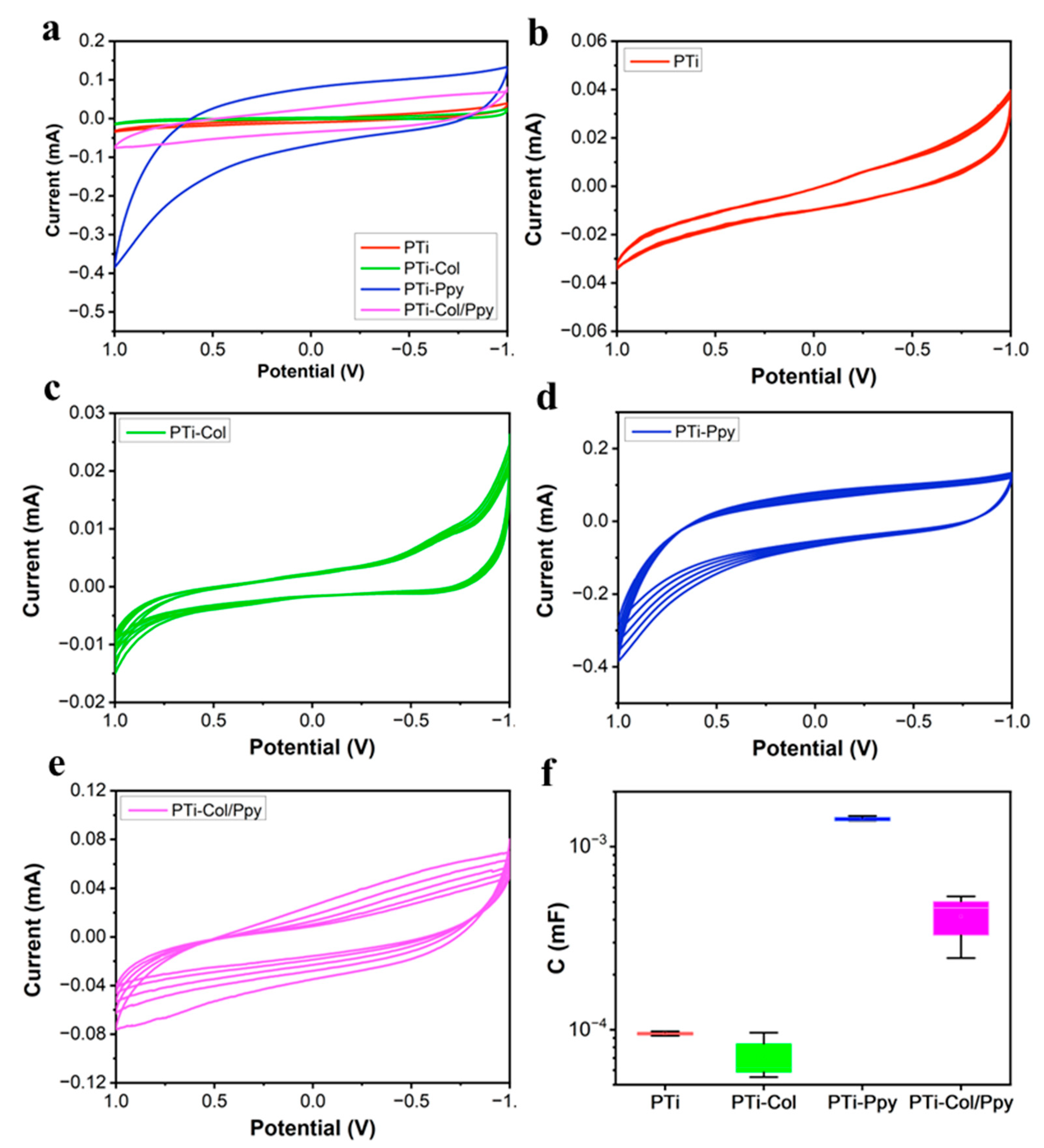
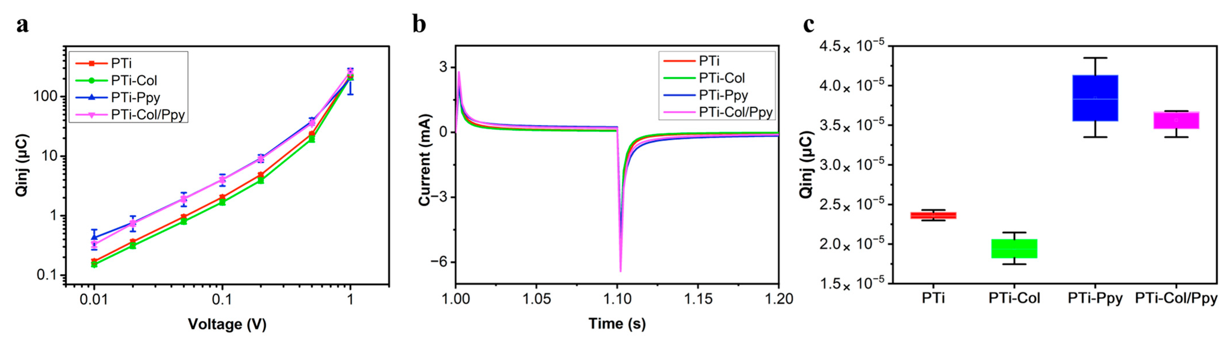
3.2. Electrochemical Performance of the Composite Films

Electrochemical Impedance Spectroscopy (EIS) is commonly used to characterize the electrochemical performance of materials [49]. The Bode and Nyquist plot results for the EIS tests for each sample are shown in Figure 3. According to the Nyquist plot results in Figure 3c–d, all samples exhibited a capacitive semicircular arc at a high frequency and a straight line at a low frequency, indicating that electronic transfer (high frequency) and mass diffusion (low frequency) processes could occur on the surface of the samples. Furthermore, the Bode plots in Figure 3a–b demonstrate that PTi-Ppy and PTi-Col/Ppy have lower electrochemical impedance and smaller phase angles in the low-frequency region below 100 Hz. This suggests that Ppy, as an electrochemically active conductive material with excellent performance [50], significantly enhances the electrochemical properties of porous-surfaced titanium. At 1 Hz, the impedances of PTi-Ppy and PTi-Col/Ppy were 541.7 Ω and 994.6 Ω, respectively, representing a decrease of 90.6% and 82.8% compared to the impedance of PTi (5786 Ω). The excellent electrochemical performance of Ppy is often attributed to its pseudocapacitance characteristics and reversible redox reactions [51]. It is important to note that, since EIS is conducted at open-circuit potential, the latter effect has minimal influence. The primary reason for the reduced electrochemical impedance of PTi is the pseudocapacitive characteristics of Ppy. Another noteworthy observation is that the electrochemical impedance of PTi-Col and PTi-Col/Ppy is higher than that of PTi and PTi-Ppy, indicating that the Col film layer reduces the electrochemical performance of the material to some extent.
According to the CV curves (Figure 4), the potential redox reactions of each sample were characterized and their charge capacities were calculated [52]. According to Figure 4a, the CV curves of the samples exhibited no significant redox peaks, indicating that the potential window is safe and that the Ppy deposition is minimal, insufficient to elicit noticeable reversible oxidation–reduction of Ppy chains [53]. Based on Figure 4b–e, the CV stability of the samples was generally good, while PTi-Col/Ppy exhibited slight performance fluctuations, which may be related to minor degradation or shedding of collagen during the testing process. The calculated values of the charge capacities for the samples, as depicted in Figure 4f, indicate that both PTi-Ppy (1.416 ± 0.049 mF) and PTi-Col/Ppy (0.416 ± 0.151 mF) demonstrate superior charge capacities. The charge capacities of PTi-Ppy and PTi-Col/Ppy are approximately 14.9 and 4.4 times the charge capacity of PTi (0.095 ± 0.003 mF), respectively. Meanwhile, the charge capacity of PTi-Col was recorded as 0.071 ± 0.022 mF. Similarly, when comparing PTi with PTi-Col or comparing PTi-Ppy with PTi-Col/Ppy, a decrease in charge capacity can be observed after the electrochemical deposition of collagen. These results are consistent with the EIS results, indicating that the Ppy film layer significantly enhances the electrochemical performance of the material, while the electrochemical deposition of the collagen film layer reduces the charge capacity. However, the charge capacity of PTi-Col/Ppy is still higher than that of PTi.
The charge injection capacity of each sample was characterized by Qinj, and this test result can simulate the amount of charge transferred to the cells and their culture environment under actual electrical stimulation at a certain voltage. The results are shown in Figure 5. The Qinj results are consistent with the EIS and CV results. The Qinj–voltage curves indicate that PTi-Ppy and PTi-Col/Ppy samples generally exhibit significantly higher charge injection ability. Figure 5c presents a more intuitive comparison of the Qinj of each sample at a voltage of 0.5 V. The Qinj of PTi-Ppy (38.44 ± 5.02 μC) and the Qinj of PTi-Col/Ppy (35.65 ± 1.85 μC) are 1.63 times and 1.51 times that of PTi (23.62 ± 0.66 μC), respectively, while the Qinj of PTi-Col (19.43 ± 2.01 μC) is only 0.82 times that of PTi. It can be concluded from the results for EIS, CV, and Qinj that the composite film group PTi-Col/Ppy and the Ppy control group PTi-Ppy exhibit superior electroactivity.

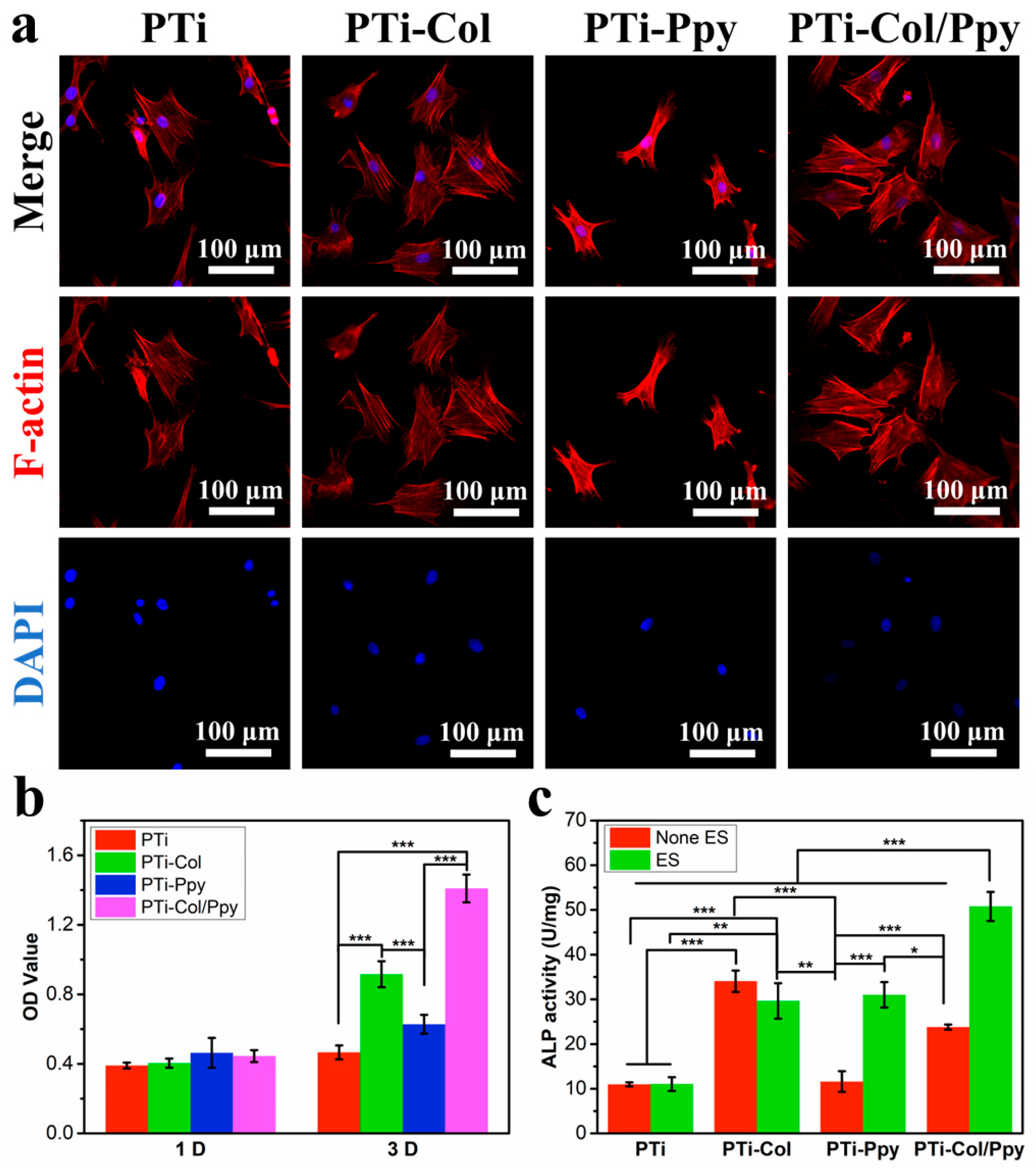
3.3. Biological Effects of the Composite Films

The cell cytoskeleton of BMSCs cultured for one day on each sample was stained by iFluor594 Conjugate-Phalloidin. And their adhesion status was observed, as shown in Figure 6a. BMSCs on PTi, PTi-Col, and PTi-Col/Ppy exhibited a larger spread area and more prominent actin filaments, indicating good adhesion, while BMSCs on PTi-Ppy showed a smaller spread area and a more contracted state. The combination of CCK-8 assay results and cytoskeleton staining results demonstrates that the electrochemical-deposited collagen film layer provides a better biomimetic ECM microenvironment, which is more conducive to the adhesion and proliferation of BMSCs.
BMSCs were cultured on each sample for 1 day or 3 days, followed by CCK-8 testing, and the absorbance results are shown in Figure 6b. The samples on which BMSCs were cultured for 1 day exhibited similar values in absorbance, indicating that the BMSCs were able to adhere well to the surface of all the samples and that the number of adhered BMSCs was approximately similar. After 3 days of culture, BMSCs on PTi-Col, PTi-Ppy, and PTi-Col/Ppy showed significant proliferation, with a marked increase in absorbance. Additionally, the samples with electrochemical-deposited collagen film layers exhibited better proliferation effects, indicating that the electrochemical-deposited collagen film layers have excellent biocompatibility, simulating the biomimetic ECM microenvironment effectively on the sample surface, which is beneficial for the growth and proliferation of BMSCs.
The ALP activity of BMSCs which were cultured for 7 days was evaluated to assess the impact of the cellular microenvironment on osteogenic differentiation behavior. In addition, a biphasic pulse electrical stimulation with a frequency of 1 Hz, a voltage of 0.5 V, and a duty cycle of 20% was applied to each sample for an hour per day over the 7-day period, and the ALP activity of BMSCs on the samples after electrical stimulation was also measured, as shown in Figure 6c. When comparing the ALP activity of each sample without electrical stimulation, PTi-Col and PTi-Col/Ppy, which contain collagen, exhibited higher osteogenic activity compared to PTi and PTi-Ppy. This finding indicates that the biomimetic ECM microenvironment provided by collagen not only promotes cell adhesion and proliferation but also facilitates osteogenic differentiation. After the application of electrical stimulation, no significant change in osteogenic activity was observed for PTi or PTi-Col, while a significant increase in osteogenic activity was observed for PTi-Ppy and PTi-Col/Ppy. This result indicates that PTi-Ppy and PTi-Col/Ppy, under exogenous electrical signals, provided a suitable electrical microenvironment to induce osteogenic differentiation of BMSCs, which is clearly attributed to the improved electroactivity of these samples after Ppy modification. Furthermore, the osteogenic activity of PTi-Ppy after electrical stimulation was comparable to that of PTi-Col, suggesting that the electrical microenvironment provided by PTi-Ppy under electrical stimulation had an osteogenic differentiation-inducing effect similar to that of the biomimetic ECM microenvironment provided by the collagen film layer. And PTi-Col/Ppy, which simultaneously provides favorable ECM biochemical cues and electrical signal cues, constructed a well-designed biomimetic electrical microenvironment, achieving the best osteogenic differentiation promotion effect among all groups.
Cell experiments demonstrate that the collagen film layer obtained by electrochemical deposition can provide a good biomimetic ECM microenvironment, thereby promoting the adhesion, proliferation, and osteogenic differentiation of BMSCs. Furthermore, the surface morphology and compositional characteristics of the collagen film layer on the surface of PTi-Col/Ppy are the factors contributing to its good biological activity. Additionally, under exogenous electrical signals, PTi-Col/Ppy, like the Ppy control group (PTi-Ppy), can provide an electrical microenvironment that helps promote osteogenic differentiation. Thanks to the effect of the biomimetic electrical microenvironment, PTi-Col/Ppy exhibits the best osteogenic differentiation-promoting effect. This finding demonstrates that PTi-Col/Ppy, as a biomimetic electroactive material, can achieve superior cell behavior regulation results by constructing a biomimetic electrical microenvironment.
This study successfully prepared Col/Ppy composite films by stepwise electrochemical deposition, thereby demonstrating that the biochemical cues provided by collagen and the electrophysiological microenvironment induced by electrical stimulation can synergistically promote osteogenic differentiation. This finding is of considerable significance for the fields of bone repair and tissue engineering. The material in this study is suitable for use in in vitro cell cultures, primarily due to the limitations of external electrical stimulation sources, which constrain the in vivo application of this material. Given the prevalence of titanium as a bone implant material [54], Ti substrates do not negatively affect the in vivo application of the material. And polypyrrole has demonstrated cell and tissue compatibility in vivo [55]. If this material can be further added to a sheet made of piezoelectric materials that can provide exogenous electrical stimulation, it is expected that in vivo implantation will be realized by this material, and its application value will be greatly enriched [56].
Moreover, other smart systems can promote osteogenic differentiation [57]. Liu et al. [58] fabricated a composite piezoelectric fibrous membrane with titanium dioxide nanoparticles and polyvinylidene fluoride, which effectively induced osteogenic differentiation at the early stage by stimulation of electrical and mechanical signals through piezoelectric fibers. Additionally, different magnetic field strengths have various effects on cells, while medium-strength magnetic fields are the most widely used, promoting osteogenic differentiation [59]. The material described in this paper can also be supplemented with additional materials, allowing it to further promote osteogenesis differentiation and the in vivo application of the material under the control of different smart systems.

4. Conclusions

This study successfully obtained Col/Ppy composite film-modified porous-surfaced titanium electrodes through hot alkali treatment and stepwise electrochemical deposition. Material characterization confirmed the presence of collagen and Ppy on the porous-surfaced titanium substrate, with the two materials modifying the substrate in a layered structure. Electrochemical tests showed that the modification of Ppy significantly enhanced the electrochemical performance of the material, imparting good electroactivity. However, the collagen layer, positioned at the uppermost layer, might hinder the electrochemical performance of Ppy to a certain extent, leading to a slight reduction in the electroactivity of this material. Cell experiments on BMSCs indicated that the collagen layer in the composite film provided a favorable biomimetic ECM microenvironment, significantly promoting cell adhesion, proliferation, and osteogenic differentiation. The Ppy layer in the composite film, when stimulated by external electrical signals, provided an appropriate electrical microenvironment for the cells and effectively promoted osteogenic differentiation. This study confirms that the selection of collagen and Ppy components can provide a better biomimetic electrical microenvironment and an improved interface, promoting osteogenic differentiation. When combined with titanium metal, this kind of material holds promise for application in bone tissue engineering.

Author Contributions

Methodology, Y.L. and X.X.; investigation, Y.L. and X.X.; data curation, Y.L. and X.X.; writing—original draft preparation, Y.L. and X.X.; visualization, X.H.; conceptualization, K.C., C.W., and Y.L.; writing—review and editing, K.C., W.W., and C.W.; funding acquisition, K.C. and C.W. All authors have read and agreed to the published version of the manuscript.

Funding

This work was financially supported by the National Natural Science Foundation of China (Grant Nos. 52271252, 32271373, and U24A20762) and the Postdoctoral Fellowship Program of the China Postdoctoral Science Foundation (GZC20232243).

Institutional Review Board Statement

The animal experiments in this study were approved by the Institutional Animal Care and Use Committee of Zhejiang University, Hangzhou, China (Project Approval No. ZJU202202735).

Informed Consent Statement

Not applicable.

Data Availability Statement

Dataset available on request from the authors.

Conflicts of Interest

The authors declare no conflicts of interest.

References

  1. Xue, N.; Ding, X.; Huang, R.; Jiang, R.; Huang, H.; Pan, X.; Min, W.; Chen, J.; Duan, J.; Liu, P.; et al. Bone Tissue Engineering in the Treatment of Bone Defects. Pharm.-Base 2022, 15, 879. [Google Scholar] [CrossRef] [PubMed]
  2. Zha, K.; Tian, Y.; Panayi, A.C.; Mi, B.; Liu, G. Recent Advances in Enhancement Strategies for Osteogenic Differentiation of Mesenchymal Stem Cells in Bone Tissue Engineering. Front. Cell Dev. Biol. 2022, 10, 824812. [Google Scholar] [CrossRef] [PubMed]
  3. Koons, G.L.; Diba, M.; Mikos, A.G. Materials Design for Bone-Tissue Engineering. Nat. Rev. Mater. 2020, 5, 584–603. [Google Scholar] [CrossRef]
  4. Zhu, G.; Zhang, T.; Chen, M.; Yao, K.; Huang, X.; Zhang, B.; Li, Y.; Liu, J.; Wang, Y.; Zhao, Z. Bone Physiological Microenvironment and Healing Mechanism: Basis for Future Bone-Tissue Engineering Scaffolds. Bioact. Mater. 2021, 6, 4110–4140. [Google Scholar] [CrossRef]
  5. Dang, M.; Saunders, L.; Niu, X.; Fan, Y.; Ma, P.X. Biomimetic Delivery of Signals for Bone Tissue Engineering. Bone Res. 2018, 6, 25. [Google Scholar] [CrossRef] [PubMed]
  6. Bai, Y.; Wang, Z.; He, X.; Zhu, Y.; Xu, X.; Yang, H.; Mei, G.; Chen, S.; Ma, B.; Zhu, R. Application of Bioactive Materials for Osteogenic Function in Bone Tissue Engineering. Small Methods 2024, 8, 2301283. [Google Scholar] [CrossRef]
  7. Sivakumar, P.M.; Yetisgin, A.A.; Sahin, S.B.; Demir, E.; Cetinel, S. Bone Tissue Engineering: Anionic Polysaccharides as Promising Scaffolds. Carbohydr. Polym. 2022, 283, 119142. [Google Scholar] [CrossRef]
  8. Cao, Z.; Bian, Y.; Hu, T.; Yang, Y.; Cui, Z.; Wang, T.; Yang, S.; Weng, X.; Liang, R.; Tan, C. Recent Advances in Two-Dimensional Nanomaterials for Bone Tissue Engineering. J. Mater. 2023, 9, 930–958. [Google Scholar] [CrossRef]
  9. Kazimierczak, P.; Przekora, A. Osteoconductive and Osteoinductive Surface Modifications of Biomaterials for Bone Regeneration: A Concise Review. Coatings 2020, 10, 917. [Google Scholar] [CrossRef]
  10. Aykora, D.; Uzun, M. Bone Tissue Engineering for Osteointegration: Where are we Now? Polym. Bull. 2024, 81, 8595–8605. [Google Scholar] [CrossRef]
  11. Gholami, K.; Ege, F.; Barzegar, R. Prediction of Composite Mechanical Properties: Integration of Deep Neural Network Methods and Finite Element Analysis. J. Compos. Sci. 2023, 7, 54. [Google Scholar] [CrossRef]
  12. Cui, J.; Yu, B.; Li, D.; Fu, Z.; Yang, X.; Jiang, L.; Wang, X.; Lin, K. Remodeling Electrophysiological Microenvironment for Promoting Bone Defect Repair Via Electret Hybrid Electrospun Fibrous Mat. Adv. Fiber Mater. 2024, 6, 1855–1873. [Google Scholar] [CrossRef]
  13. Shen, N.; Zhang, Z.; Wu, F.; Zhu, Z.; Liu, J.; Qi, X.; Li, Q.; Wang, X.; Ma, Q.; Xiang, H. Advances in Self-Powered Biomaterials for Bone Defect Repair. Adv. Compos. Hybrid Mater. 2024, 8, 38. [Google Scholar] [CrossRef]
  14. Lv, Z.; Ji, Y.; Wen, G.; Liang, X.; Zhang, K.; Zhang, W. Structure-Optimized and Microenvironment-Inspired Nanocomposite Biomaterials in Bone Tissue Engineering. Burn. Trauma 2024, 12, tkae036. [Google Scholar] [CrossRef]
  15. Shi, S.; Xu, X.; He, X.; Fan, S.; Liu, P.; Wu, C.; Cheng, K. Pvtf Nanoparticles/Pla Electroactive Degradable Membrane for Bone Tissue Regeneration. Coatings 2024, 14, 115. [Google Scholar] [CrossRef]
  16. Xu, X.; Wang, H.; Zhang, S.; Mei, X.; Ying, B.; Li, R.; Qin, Y. Ecm-Inspired 3D Printed Polyetherimide Scaffold with Arg-Gly-Asp Peptides for the Improvement of Bioactivity and Osteogenic Differentiation of Osteoblasts. Mater. Today Commun. 2022, 30, 103166. [Google Scholar] [CrossRef]
  17. Jin, Z.; Lu, B.; And Xu, Y. Constructing an Electrical Microenvironment Based on Electroactive Polymers in the Field of Bone Tissue Engineering. Int. J. Polym. Mater. Polym. Biomater. 2023, 72, 937–967. [Google Scholar] [CrossRef]
  18. Chen, Z.; Kang, L.; Wang, Z.; Xu, F.; Gu, G.; Cui, F.; Guo, Z. Recent Progress in the Research of Biomaterials Regulating Cell Behavior. Rsc Adv. 2014, 4, 63807–63816. [Google Scholar] [CrossRef]
  19. Huang, G.; Li, F.; Zhao, X.; Ma, Y.; Li, Y.; Lin, M.; Jin, G.; Lu, T.J.; Genin, G.M.; Xu, F. Functional and Biomimetic Materials for Engineering of the Three-Dimensional Cell Microenvironment. Chem. Rev. 2017, 117, 12764–12850. [Google Scholar] [CrossRef]
  20. Hussey, G.S.; Dziki, J.L.; Badylak, S.F. Extracellular Matrix-Based Materials for Regenerative Medicine. Nat. Rev. Mater. 2018, 3, 159–173. [Google Scholar] [CrossRef]
  21. Liu, M.; Zeng, X.; Ma, C.; Yi, H.; Ali, Z.; Mou, X.; Li, S.; Deng, Y.; He, N. Injectable Hydrogels for Cartilage and Bone Tissue Engineering. Bone Res. 2017, 5, 17014. [Google Scholar] [CrossRef]
  22. Oliver-Cervelló, L.; Martin-Gómez, H.; Gonzalez-Garcia, C.; Salmeron-Sanchez, M.; Ginebra, M.; Mas-Moruno, C. Protease-Degradable Hydrogels with Multifunctional Biomimetic Peptides for Bone Tissue Engineering. Front. Bioeng. Biotech. 2023, 11, 1192436. [Google Scholar] [CrossRef]
  23. Zhou, H.; Li, W.; Pan, L.; Zhu, T.; Zhou, T.; Xiao, E.; Wei, Q. Human Extracellular Matrix (Ecm)-like Collagen and its Bioactivity. Regen. Biomater. 2024, 11, rbae008. [Google Scholar] [CrossRef] [PubMed]
  24. McColgan-Bannon, K.I.S.; Upson, S.; Gentile, P.; Tausif, M.; Russell, S.; Dalgarno, K.; Ferreira, A.M. Biomimetic Properties of Force-Spun Phbv Membranes Functionalised with Collagen as Substrates for Biomedical Application. Coatings 2019, 9, 350. [Google Scholar] [CrossRef]
  25. Li, W.; Chi, N.; Clutter, E.D.; Zhu, B.; Wang, R.R. Aligned Collagen-Cnt Nanofibrils and the Modulation Effect on Ovarian Cancer Cells. J. Compos. Sci. 2021, 5, 148. [Google Scholar] [CrossRef] [PubMed]
  26. Li, Y.; Liu, Y.; Li, R.; Bai, H.; Zhu, Z.; Zhu, L.; Zhu, C.; Che, Z.; Liu, H.; Wang, J.; et al. Collagen-Based Biomaterials for Bone Tissue Engineering. Mater. Des. 2021, 210, 110049. [Google Scholar] [CrossRef]
  27. Lin, Z.; Tao, Y.; Huang, Y.; Xu, T.; Niu, W. Applications of Marine Collagens in Bone Tissue Engineering. Biomed. Mater. 2021, 16, 042007. [Google Scholar] [CrossRef]
  28. Tanrikulu, I.C.; Dang, L.; Nelavelli, L.; Ellison, A.J.; Olsen, B.D.; Jin, S.; Raines, R.T. Synthetic Collagen Hydrogels through Symmetric Self-Assembly of Small Peptides. Adv. Sci. 2024, 11, 2303228. [Google Scholar] [CrossRef]
  29. Lin, K.; Zhang, D.; Macedo, M.H.; Cui, W.; Sarmento, B.; Shen, G. Advanced Collagen-Based Biomaterials for Regenerative Biomedicine. Adv. Funct. Mater. 2019, 29, 1804943. [Google Scholar] [CrossRef]
  30. Que, R.A.; Crakes, D.R.; Abdulhadi, F.; Niu, C.; Da Silva, N.A.; Wang, S. Tailoring Collagen to Engineer the Cellular Microenvironment. Biotechnol. J. 2018, 13, 1800140. [Google Scholar] [CrossRef]
  31. Matsunaga, Y.T.; Morimoto, Y.; Takeuchi, S. Molding Cell Beads for Rapid Construction of Macroscopic 3D Tissue Architecture. Adv. Mater. 2011, 23, H90–H94. [Google Scholar] [CrossRef] [PubMed]
  32. Ma, F.; Zhu, T.; Xu, F.; Wang, Z.; Zheng, Y.; Tang, Q.; Chen, L.; Shen, Y.; Zhu, J. Neural Stem/Progenitor Cells on Collagen with Anchored Basic Fibroblast Growth Factor as Potential Natural Nerve Conduits for Facial Nerve Regeneration. Acta Biomater. 2017, 50, 188–197. [Google Scholar] [CrossRef]
  33. Mohammad, F.M.B.; Yogeswaran, L. Recent Development in the Fabrication of Collagen Scaffolds for Tissue Engineering Applications: A Review. Curr. Pharm. Biotechnol. 2019, 20, 992–1003. [Google Scholar]
  34. Bonfrate, V.; Manno, D.; Serra, A.; Salvatore, L.; Sannino, A.; Buccolieri, A.; Serra, T.; Giancane, G. Enhanced Electrical Conductivity of Collagen Films through Long-Range Aligned Iron Oxide Nanoparticles. J. Colloid. Interf. Sci. 2017, 501, 185–191. [Google Scholar] [CrossRef] [PubMed]
  35. Cartwright, V.F.; Brown, C.P. Hierarchical Piezoresponse in Collagen. Adv. Mater. Technol. 2022, 7, 2101166. [Google Scholar] [CrossRef]
  36. Liu, Z.; Wan, X.; Wang, Z.L.; Li, L. Electroactive Biomaterials and Systems for Cell Fate Determination and Tissue Regeneration: Design and Applications. Adv. Mater. 2021, 33, 2007429. [Google Scholar] [CrossRef] [PubMed]
  37. Chen, C.; Bai, X.; Ding, Y.; Lee, I. Electrical Stimulation as a Novel Tool for Regulating Cell Behavior in Tissue Engineering. Biomater. Res. 2019, 23, 25. [Google Scholar] [CrossRef]
  38. Liu, Y.; Lu, J. Research Trends of Piezoelectric Biomaterials in Osteochondral Tissue Engineering. Mater. Today Commun. 2024, 41, 110264. [Google Scholar] [CrossRef]
  39. Tandon, B.; Magaz, A.; Balint, R.; Blaker, J.J.; Cartmell, S.H. Electroactive Biomaterials: Vehicles for Controlled Delivery of Therapeutic Agents for Drug Delivery and Tissue Regeneration. Adv. Drug Deliv. Rev. 2018, 129, 148–168. [Google Scholar] [CrossRef]
  40. Zhu, Y.; Yao, L.; Liu, Z.; Weng, W.; Cheng, K. Electrical Potential Specified Release of Bsa/Hep/Polypyrrole Composite Film and its Cellular Responses. Acs Appl. Mater. Inter. 2019, 11, 25457–25464. [Google Scholar] [CrossRef]
  41. Kim, S.; Jang, L.K.; Jang, M.; Lee, S.; Hardy, J.G.; Lee, J.Y. Electrically Conductive Polydopamine–Polypyrrole as High Performance Biomaterials for Cell Stimulation In Vitro and Electrical Signal Recording In Vivo. Acs Appl. Mater. Inter. 2018, 10, 33032–33042. [Google Scholar] [CrossRef]
  42. Elyashevich, G.; Rosova, E.; Zoolshoev, Z.; Saprykina, N.; Kuryndin, I. Reversibility of Swelling, Ph Sensitivity, Electroconductivity, and Mechanical Properties of Composites Based on Polyacrylic Acid Hydrogels and Conducting Polymers. J. Compos. Sci. 2023, 7, 261. [Google Scholar] [CrossRef]
  43. Wu, C.; He, X.; Weng, W.; Zhang, T.; Huang, D.; Cheng, K.; Chen, Z. Electroactive Extracellular Matrix/Polypyrrole Composite Films and their Microenvironmental Effects on Osteogenic Differentiation of Bmscs. Chem. Eng. J. 2022, 443, 136508. [Google Scholar] [CrossRef]
  44. Kim, D.H.; Richardson-Burns, S.M.; Hendricks, J.L.; Sequera, C.; Martin, D.C. Effect of Immobilized Nerve Growth Factor On Conductive Polymers: Electrical Properties and Cellular Response. Adv. Funct. Mater. 2007, 17, 79–86. [Google Scholar] [CrossRef]
  45. Da Silva, F.A.G.; Alcaraz-Espinoza, J.J.; Da Costa, M.M.; de Oliveira, H.P. Synthesis and Characterization of Highly Conductive Polypyrrole-Coated Electrospun Fibers as Antibacterial Agents. Compos. Part. B Eng. 2017, 129, 143–151. [Google Scholar] [CrossRef]
  46. Duchet, J.; Legras, R.; Demoustier-Champagne, S. Chemical Synthesis of Polypyrrole: Structure–Properties Relationship. Synth. Met. 1998, 98, 113–122. [Google Scholar] [CrossRef]
  47. Liu, Y.; Hwang, B.; Jian, W.; Santhanam, R. In Situ Cyclic Voltammetry-Surface-Enhanced Raman Spectroscopy: Studies on the Doping–Undoping of Polypyrrole Film. Thin Solid. Film. 2000, 374, 85–91. [Google Scholar] [CrossRef]
  48. Liu, Y.C.; Hwang, B.J. Interaction of Copper(I)-Polypyrrole Complexes Prepared by Depositing–Dissolving Copper onto and from Polypyrroles. Thin Solid. Film. 1999, 339, 233–239. [Google Scholar] [CrossRef]
  49. Duan, Y.Y.; Clark, G.M.; Cowan, R.S.C. A Study of Intra-Cochlear Electrodes and Tissue Interface by Electrochemical Impedance Methods in Vivo. Biomaterials 2004, 25, 3813–3828. [Google Scholar] [CrossRef]
  50. Zare, E.N.; Agarwal, T.; Zarepour, A.; Pinelli, F.; Zarrabi, A.; Rossi, F.; Ashrafizadeh, M.; Maleki, A.; Shahbazi, M.; Maiti, T.K.; et al. Electroconductive Multi-Functional Polypyrrole Composites for Biomedical Applications. Appl. Mater. Today 2021, 24, 101117. [Google Scholar] [CrossRef]
  51. Xu, Y.; Xiang, S.; Zhang, X.; Zhou, H.; Zhang, H. High-Performance Pseudocapacitive Removal of Cadmium Via Synergistic Valence Conversion in Perovskite-Type Femno3. J. Hazard. Mater. 2022, 439, 129575. [Google Scholar] [CrossRef]
  52. Deng, M.; Yang, X.; Silke, M.; Qiu, W.; Xu, M.; Borghs, G.; Chen, H. Electrochemical Deposition of Polypyrrole/Graphene Oxide Composite on Microelectrodes Towards Tuning the Electrochemical Properties of Neural Probes. Sens. Actuators B Chem. 2011, 158, 176–184. [Google Scholar] [CrossRef]
  53. Antony, N.; Mohanty, S.; Nayak, S.K. Electrochemical Inspection of Polypyrrole/Chitosan/Zinc Oxide Hybrid Composites. J. Appl. Polym. Sci. 2020, 137, 49561. [Google Scholar] [CrossRef]
  54. Lin, W.; He, X.; Guo, X.; Xu, D.; Cheng, K. Biocompatible Pvtf Coatings on Ti with Improved Bonding Strength. Coatings 2023, 13, 1224. [Google Scholar] [CrossRef]
  55. Yow, S.; Lim, T.H.; Yim, E.K.F.; Lim, C.T.; Leong, K.W. A 3D Electroactive Polypyrrole-Collagen Fibrous Scaffold for Tissue Engineering. Polymers 2011, 3, 527–544. [Google Scholar] [CrossRef]
  56. Lin, W.; Zhou, Z.; Chen, Z.; Xu, K.; Wu, C.; Duan, X.; Dong, L.; Chen, Z.; Weng, W.; Cheng, K. Accelerated Bone Regeneration on the Metal Surface through Controllable Surface Potential. Acs Appl. Mater. Interfaces 2023, 15, 46493–46503. [Google Scholar] [CrossRef] [PubMed]
  57. Liu, S.; Zhang, L.; Li, Z.; Gao, F.; Zhang, Q.; Bianco, A.; Liu, H.; Ge, S.; Ma, B. Materials-Mediated in Situ Physical Cues for Bone Regeneration. Adv. Funct. Mater. 2024, 34, 2306534. [Google Scholar] [CrossRef]
  58. Liu, J.; Cheng, Y.; Wang, H.; Yang, D.; Liu, C.; Dou, W.; Jiang, X.; Deng, H.; Yang, R. Regulation of Tio2 @Pvdf Piezoelectric Nanofiber Membranes on Osteogenic Differentiation of Mesenchymal Stem Cells. Nano Energy 2023, 115, 108742. [Google Scholar] [CrossRef]
  59. Xia, Y.; Sun, J.; Zhao, L.; Zhang, F.; Liang, X.; Guo, Y.; Weir, M.D.; Reynolds, M.A.; Gu, N.; Xu, H.H.K. Magnetic Field and Nano-Scaffolds with Stem Cells to Enhance Bone Regeneration. Biomaterials 2018, 183, 151–170. [Google Scholar] [CrossRef]
Figure 1. (a) Schematic diagram of the fabrication of PTi-Col/Ppy. (b) SEM morphology images of PTi-Col/Ppy and control groups. (c) AFM images of PTi-Col/Ppy and control groups. (d) Surface roughness of PTi-Col/Ppy and control groups.
Jcs 09 00325 g001
Figure 2. (a) FTIR results for PTi-Col/Ppy and control groups. (b) Raman results for PTi-Col/Ppy and control groups. (c) WCA results for PTi-Col/Ppy and control groups. (*** p < 0.001).
Jcs 09 00325 g002
Figure 3. EIS curves of PTi-Col/Ppy and control groups: (a) impedance vs. frequency; (b) phase vs. frequency; (c) Nyquist curves; (d) partial enlargement of Nyquist curves.
Jcs 09 00325 g003
Figure 4. CV results for PTi-Col/Ppy and control groups: (a) summary of CV curves; (be) stability of CV curves; (f) calculated values of charge capacities of PTi-Col/Ppy and control groups.
Jcs 09 00325 g004
Figure 5. Qinj analysis of PTi-Col/Ppy and control groups: (a) Qinj vs. voltage plots; (b) current vs. time plots at a voltage of 0.5 V; (c) Qinj of PTi-Col/Ppy and control groups at a voltage of 0.5 V.
Jcs 09 00325 g005
Figure 6. (a) Cytoskeleton staining of BMSCs cultured for 1 day on PTi-Col/Ppy and control groups. (b) CCK-8 results for BMSCs on PTi-Col/Ppy and control groups. (c) ALP activity of BMSCs cultured for 7 days on PTi-Col/Ppy and control groups. (* p < 0.05, ** p < 0.01, *** p < 0.001).
Jcs 09 00325 g006
Table 1. Content of elements on the surface of PTi-Col/Ppy and control groups.
Atomic Percent (%)CNO
PTi---------
PTi-Col12.926.560.6
PTi-Ppy33.923.942.2
PTi-Col/Ppy22.623.853.6
Table 2. Analysis of Raman results for PTi-Col/Ppy and control groups.
I1575/I1500(I1045 + I970)/(I1080 + I930)
PTi1.002 ± 0.004---
PTi-Col1.003 ± 0.010---
PTi-Ppy1.086 ± 0.0151.028 ± 0.008
PTi-Col/Ppy1.069 ± 0.0201.041 ± 0.014
Disclaimer/Publisher’s Note: The statements, opinions and data contained in all publications are solely those of the individual author(s) and contributor(s) and not of MDPI and/or the editor(s). MDPI and/or the editor(s) disclaim responsibility for any injury to people or property resulting from any ideas, methods, instructions or products referred to in the content.

