Article

Enhanced Photoelectrochemical Water Splitting Using a NiFe2O4/NG@MIL-100(Fe)/TiO2 Composite Photoanode: Synthesis, Characterization, and Performance

1
School of Environmental Science and Engineering, Tianjin University, Tianjin 300072, China
2
Tianjin Key Laboratory of Molecular Optoelectronic Science, Department of Chemistry, School of Science, Tianjin University, Tianjin 300072, China
3
Tianjin Key Laboratory of Low-Dimensional Materials Physics and Preparing Technology, School of Science, Tianjin University, Tianjin 300072, China
4
Center of Excellence on Catalysis and Catalytic Reaction Engineering, Department of Chemical Engineering, Faculty of Engineering, Chulalongkorn University, Bangkok 10330, Thailand
*
Author to whom correspondence should be addressed.
J. Compos. Sci. 2025, 9(5), 250; https://doi.org/10.3390/jcs9050250
Submission received: 15 March 2025 / Revised: 7 May 2025 / Accepted: 13 May 2025 / Published: 17 May 2025
(This article belongs to the Special Issue Advancements in Composite Materials for Energy Storage Applications)

Abstract

:
NiFe2O4 and TiO2 are widely studied for photoelectrochemical (PEC) applications due to their unique properties. Nitrogen-doped graphene (NG) and metal–organic frameworks (MOFs), such as MIL-100(Fe) (where MIL stands for Materials of Lavoisier Institute), are commonly incorporated to enhance PEC performance by offering a high surface area and facilitating efficient charge transport. Composite systems are commonly employed to overcome the limitations of individual PEC catalysts. In this study, a highly efficient NiFe2O4/NG@MIL-100(Fe)/TiO2 photoanode was developed to enhance photoelectrochemical water-splitting performance. The composite was synthesized via a hydrothermal method with a two-step heating process. X-ray diffraction confirmed the expected crystal structures, with peak broadening in NiFe2O4 indicating reduced crystallite size and increased lattice strain. X-ray photoelectron spectroscopy of the Ni 2p and Fe 2p regions validated the successful integration of NiFe2O4 into the composite. Electrochemical analysis demonstrated excellent performance, with linear sweep voltammetry achieving a peak photocurrent density of 3.5 mA cm−2 at 1.23 V (vs RHE). Electrochemical impedance spectroscopy revealed a reduced charge-transfer resistance of 50 Ω, indicating improved charge transport. Optical and electronic properties were evaluated using UV-Vis spectroscopy and Tauc plots, revealing a direct bandgap of 2.1 eV. The composite exhibited stable photocurrent under amperometric J-t testing for 2000 s, demonstrating its durability. These findings underscore the potential of NiFe2O4/NG@MIL-100(Fe)/TiO2 as a promising material for renewable energy applications, particularly in photoelectrochemical water splitting.

1. Introduction

Currently, researchers are primarily focused on converting solar energy into green energy storage solutions to address the ongoing energy crisis [1,2,3,4,5,6,7,8]. One promising approach is the production of hydrogen fuel through the conversion of solar energy into sustainable energy. In this context, photoelectrochemical (PEC) water splitting plays a pivotal role in harnessing sunlight to generate clean and renewable hydrogen, thereby reducing dependence on fossil fuels and helping to meet global energy demands [8,9,10,11,12]. The development of novel photocatalysts is crucial for enhancing the efficiency of hydrogen fuel production [13,14]. Among various semiconductors, metal oxide-based nano materials have developed as promising photocatalysts due to their unique structural features and favorable optoelectronic properties [15,16]. For example, NiFe2O4 is a spinel ferrite that is inexpensive, widely available, and exhibits favorable electrical properties that facilitate efficient charge transfer [17]. These characteristics make NiFe2O4 an attractive candidate for PEC applications, as it offers both the stability and catalytic activity essential for effective hydrogen production [18]. However, metal oxide-based photocatalysts also face challenges in PEC applications, such as wide bandgaps, fast charge recombination, low surface area, poor conductivity, and limited stability. These issues hinder their efficiency in visible light absorption and hydrogen production [19].
Recently, metal–organic frameworks (MOFs) have attracted significant attention as visible-light-absorbing materials and co-catalysts for semiconductor photoanodes, offering advantages over conventional water-splitting co-catalysts such as CoOOH and NiOOH. Visible-light-absorbing MOFs can enhance optical harvesting efficiency, while their porous structures do not hinder light penetration to the underlying semiconductor, facilitating accelerated charge transfer. Additionally, the high surface area and porosity of MOFs support effective diffusion of ions, electrolyte solutions, and gas bubbles, contributing to structural stability and sustained PEC performance [20,21,22]. Moreover, negatively charged coordinating groups and nonmetal atoms in MOFs can modulate the coordination environment of metal centers, thereby optimizing intermediate binding energies through d-band center tuning [23]. The well-ordered architecture of MOFs also provides remarkable synthetic tunability due to their flexible organic linkers and adjustable metal nodes, enabling tailored integration into photoelectrodes for enhanced PEC performance and transformation of traditional photoelectronic materials [24,25].
Nanocomposite catalysts significantly enhance PEC water-splitting performance by offering synergistic effects, enhanced light harvesting, improved charge separation and transport, increased active sites, optimized band alignment, and superior structural stability. For example, integrating the high surface area and excellent charge-transfer properties of carbon materials with the strong light absorption and catalytic capabilities of MOFs results in highly efficient photocatalysts [26,27]. This unique combination enhances solar energy harvesting and effectively drives the redox reactions required for hydrogen production [28]. MOFs offer solutions through their tunable bandgaps, high porosity, and large surface area, which enhance light harvesting and catalytic activity. MOFs also facilitate better charge separation and transport, reducing electron–hole recombination. When hybridized with metal oxides, MOFs improve conductivity and stability, making them promising materials for enhancing photoelectrochemical water-splitting efficiency. Their modular structure allows further optimization via functionalization or doping. Heterostructured composites play a crucial role in enhancing PEC water splitting by forming interfaces between semiconductors with different band structures [29,30,31,32]. These interfaces generate internal electric fields that promote efficient charge separation and directional transport of electrons and holes, minimizing recombination losses [33,34,35]. Additionally, heterojunctions allow for tailored band alignment, facilitating redox reactions and improving overall photocatalytic efficiency. By combining materials with varying bandgaps, heterostructures also broaden the light absorption range and improve solar energy utilization. Moreover, the interfacial regions often provide more active sites, further boosting the hydrogen and oxygen evolution reactions.
In this study, we developed an innovative NiFe2O4/NG@MIL-100(Fe)/TiO2 nanocomposite using a hydrothermal method with a two-step heating process. The integration of NiFe2O4 with nitrogen-doped graphene (NG), MIL-100(Fe), and TiO2 aims to enhance three key aspects of PEC catalysts: charge separation, light absorption, and electronic transport. MIL-100(Fe), a MOF composed of Fe(III) ions and 1,3,5-benzenetricarboxylic acid linkers, offers a high surface area, mesoporosity, and structural stability, making it ideal for photocatalysis and PEC applications. Its iron centers facilitate light absorption, charge mediation, and efficient electron transport, improving catalytic performance. The combination of MIL-100(Fe) with the other components enhances visible light absorption and charge transport, optimizing water-splitting processes. The hydrothermal synthesis ensures proper integration of these components into a single structure, resulting in enhanced PEC efficiency and increased photocurrent density. Together, these elements offer a promising approach for sustainable hydrogen production through a breakthrough in photocatalytic materials.

2. Materials and Methods

2.1. Materials

The following materials were required for the hydrothermal synthesis of NiFe2O4/NG@MIL-100(Fe)/TiO2: The nickel source is Ni(II) Nitrate Hexahydrate (Ni(NO3)2·6H2O); the iron source is Ferric Nitrate Nonahydrate (Fe(NO3)3·9H2O); the carbonaceous support is Graphene Oxide (GO); and the extra component is Titanium Dioxide (TiO2). Most of the chemicals used in this study were purchased from Aladdin Scientific (Shanghai, China). Deionized water was used as solvent, and solutions of sodium hydroxide (NaOH) and hydrochloric acid were used for pH adjustment.

2.2. Synthesis of NiFe2O4

A one-pot hydrothermal method was used for synthesizing NiFe2O4 nanoparticles by reacting sodium hydroxide (NaOH) with deionized water as the solvent, and nickel nitrate hexahydrate Ni(NO3)2·6H2O, ferric nitrate nonahydrate Fe(NO3)3·9H2O (both with 99.8% purity, Sigma-Aldrich, St. Louis, MO, USA), and sodium hydroxide (NaOH) were used as precursors. The ratio of nitrates (grams) to water (milliliters) was kept at 1:3, and the nitrates were dissolved in distilled water. Until nanoparticle production was seen, this solution was vigorously swirled. To get pure spinel phases, NaOH was then progressively added to the mixture at a ratio of 1:4 (NaOH: nitrates). The pH was then maintained at 11. After two hours of vigorous stirring, the resultant liquid was put into a 300-mL Teflon-lined stainless-steel autoclave. After being sealed, the autoclave was hydrothermally treated for 24 h at 180 °C. Following the procedure, the autoclave was allowed to gradually drop down to ambient temperature. The precursor material was filtered and repeatedly cleaned with distilled water and ethanol. The nickel ferrite nanoparticles were then separated and allowed to dry for 6 h at 60 °C [36].

2.3. Synthesis of Nitrogen-Doped Graphene

Graphene oxide (GO) was created from graphite powder to create nitrogen-doped graphene (NG). Generally, concentrated sulfuric acid was present when 1 g of graphite powder and 0.5 g of sodium nitrate (NaNO3) were combined. After the mixture was frozen for t = 24 h, 3 g of KMnO4 were added. The mixture was heated to 40 °C for 10 min. Then, 46 mL of water was added, and the temperature was gradually raised to 90 °C. After applying hydrogen peroxide to the whole mixture, the resultant product, or GO, was filtered, cleaned with a 5% HCl solution, and dried at 60 °C for 24 h. The nitrogen source (e.g., typically around urea 0.5 g) was dissolved in the graphene oxide dispersion. Stir the mixture using a sonicator (Tianjin Automatic Science Instrument Co., Tianjin, China) for 2 h and a magnetic stirrer for about 30 min to ensure complete dissolution, and then transfer the solution into a Teflon-lined autoclave (Tefic biotech, Xian, China) and keep it in the oven at 200 °C for 15 h. After that, it was cooled at room temperature, washed with water and ethanol, and dried at 60 °C for 8 h [37,38].

2.4. Synthesis of NiFe2O4/NG

The synthesis of the NiFe2O4/NG nanocomposite was carried out hydrothermally. In deionized water, nitrogen-doped graphene (NG) was continuously stirred while NiFe2O4 was dissolved in a 1:2 molar ratio. After bringing the mixture’s pH down to around 10–11, NG was added, and metal hydroxides started to develop. After that, sodium hydroxide, or NaOH, was progressively added. The liquid was stirred for 2 h before being put in a Teflon-lined stainless-steel autoclave (Tefic biotech, Xian, China). The sealed autoclave was heated to 180 °C for 24 h to facilitate the crystallization of NiFe2O4 and the incorporation of NG. After cooling to room temperature, the product was filtered and cleaned using ethanol and deionized water [39].

2.5. Synthesis of MIL-100(Fe)

The synthesis procedure was adapted from the work of Guesh et al. [32]. First, 2 g of Fe(NO3)3⋅9H2O was dissolved in 30 mL of deionized water and stirred for 30 min to ensure complete dissolution. Separately, benzene-1,3,5-tricarboxylic acid was dissolved in 20 mL of ethanol and also stirred for 30 min. The two solutions were then gradually combined to form a homogeneous mixture. To control particle size and morphology, the TMA suspension was added dropwise to the mixture. The resulting solution was transferred into a Teflon-lined stainless steel autoclave and placed in a preheated oven at 180 °C for 16 h. After the reaction, the vessel was carefully removed and allowed to cool to room temperature. The product was collected by centrifugation and washed with ethanol and water to adjust the pH and remove any unreacted residues. Finally, the sample was dried in an oven at 60 °C for 8 h [40,41].

2.6. Synthesis of NiFe2O4/NG@MIL-100(Fe) Nanocomposite

The NiFe2O4/NG@MIL-100(Fe) nanocomposite was made by hydrothermal methods utilizing a mixture of metal nitrates and MIL-100(Fe). NiFe2O4/NG was dissolved in deionized water with the incorporation of nitrogen-doped graphene (NG) into the solution under continuous agitation. To confirm the precipitation of metal hydroxides and their deposition onto NG, sodium hydroxide (NaOH) was gradually added to the mixture, raising the pH to about 10–11. Before the final mixture was amalgamated with MIL-100(Fe), it was stirred for 2 h.
Following the addition of MIL-100(Fe) and the NiFe2O4/NG precursor, the entire mixture was placed in a stainless-steel autoclave lined with Teflon. To ensure proper crystallization of NiFeO4 and uniform inclusion of NG and MIL-100(Fe), the autoclave was shut and heated to 180 °C for 24 h. After the product naturally cooled to room temperature, it was filtered and thoroughly cleaned with ethanol and deionized water to get rid of impurities. To improve crystallinity and ensure stable integration of the composite components, the NiFe2O4/NG@MIL-100(Fe) nanocomposite was optionally calcined at 400 °C for 2 h after being dried at 60 °C for 6–8 h.

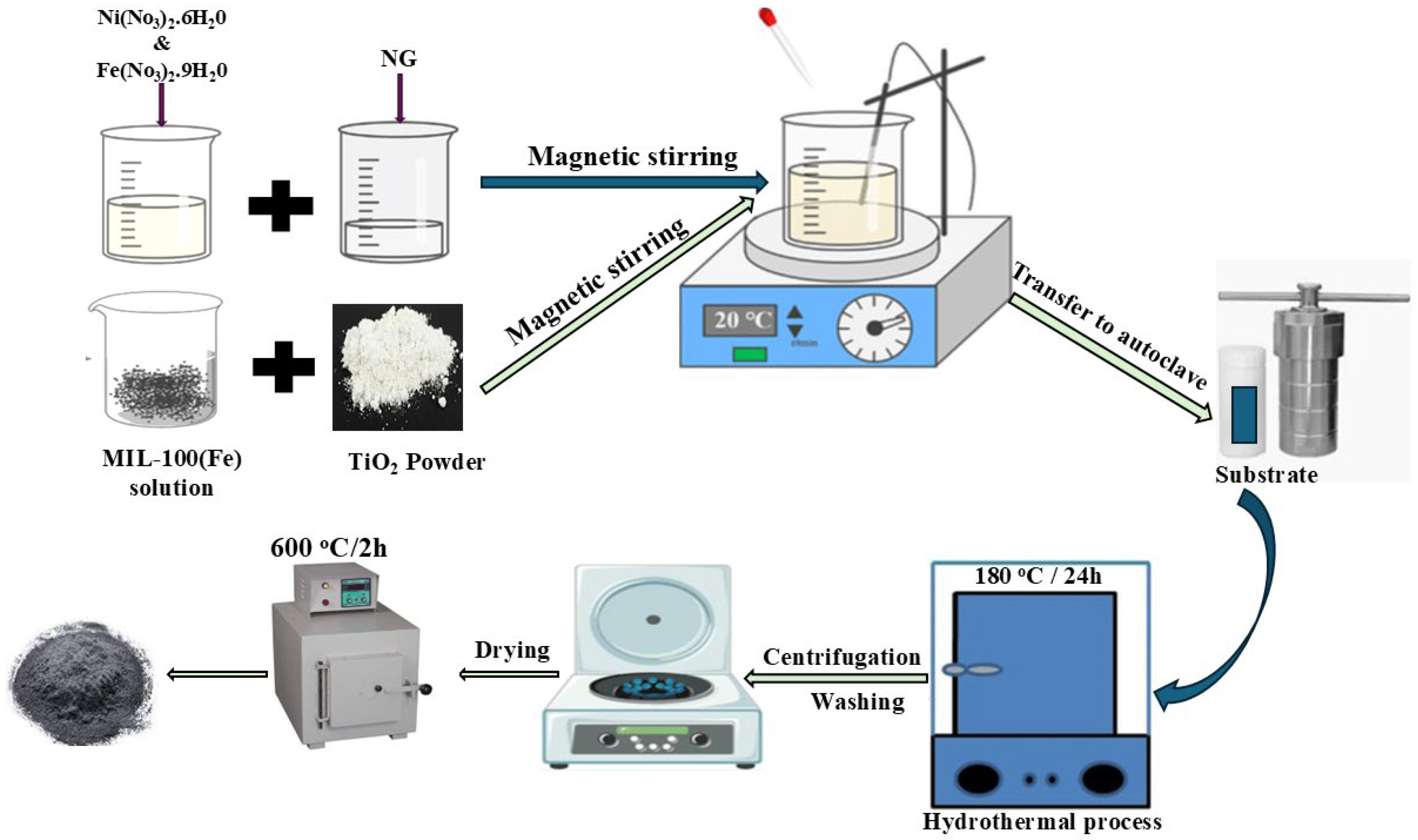
2.7. Synthesis and Fabrication of NiFe2O4/NG@MIL-100(Fe)/TiO2 Nanocomposite

A combination of metal nitrates, MIL-100(Fe), and TiO2 was used in the hydrothermal production of the NiFe2O4/NG@MIL-100(Fe)/TiO2 nanocomposite (Figure 1). Deionized water was used to dissolve ferric nitrate nonahydrate (Fe(NO3)3·9H2O) and nickel nitrate hexahydrate (Ni(NO3)2·6H2O) in a 1:2 molar ratio. The solution was continuously stirred while nitrogen-doped graphene (NG) was added. Sodium hydroxide (NaOH) was incrementally added to modify the pH to around 10–11, promoting the precipitation of metal hydroxides and their accumulation on NG. The resultant mixture was agitated for 2 h. MIL-100(Fe) was manufactured by dissolving 2 g of Fe(NO3)3·9H2O in 30 mL of deionized water and combining it with benzene-1,3,5-tricarboxylic acid diluted in ethanol. The two solutions were combined to create a homogeneous suspension. Upon preparation of the NiFe2O4/NG precursor, it was amalgamated with the synthesized MIL-100(Fe) and commercially sourced TiO2. The complete mixture was placed into a Teflon-lined stainless-steel autoclave, sealed, and subjected to heating at 180 °C for 24 h to facilitate the crystallization of NiFe2O4 and the uniform incorporation of NG, MIL-100(Fe), and TiO2. Upon naturally cooling to ambient temperature, the product was subjected to filtration and extensively cleaned with deionized water and ethanol to eliminate contaminants. The NiFe2O4/NG@MIL-100(Fe)/TiO2 nanocomposite was dried at 60 °C for 24 h and optionally calcined at 600 °C for 2 h to enhance crystallinity and ensure stable integration of all components. To prepare the NiFe2O4/NG@MIL-100(Fe)/TiO2 photoanode, 0.05 g of the nanocomposite was dispersed in distilled water and stirred for 30 min. The resulting suspension was then applied onto a 1 cm × 1 cm fluorine-doped tin oxide (FTO) glass substrate using the carbon paint method. The coated substrate was pre-dried on a hotplate at 60 °C for 10 min, followed by overnight drying at room temperature.

2.8. Characterization

D/MAX-2500 X-ray diffractometer (Ricagu, Tokyo, Japan) was used to confirm the X-ray diffraction (XRD) analysis with Cu Kα radiation while maintaining a scanning rate of 10 °C min−1; the X-ray current was kept for 40 mA, and an X-ray voltage was conducted at 40 kV. For analyzing bond formations and functional groups, the Fourier transform infrared (FTIR) was conducted, and the spectra of the samples were recorded at 25 °C, covering the wavenumber range of 4000 to 400 cm−1 (Perkin Elmer Spectrum GX FTIR Spectrometer, Waltham, MA, USA). The analysis of oxidation state and electron movement of the photoanode was conducted by using X-ray photoelectron spectroscopy (XPS) (ULVAC-PHI, Kanagawa, Japan). JEOL JSM-6701F (Tokyo, Japan) scanning electron microscope (SEM) was used to confirm surface morphology and structural analysis, and the morphological structures of the photoanodes was confirmed by transmission electron microscope (TEM, 2100F JEM, JEOL Ltd., Akishima, Japan). UV–vis diffuse reflectance spectra (UV–vis) were conducted to measure the bandgap of photoanodes at room temperature in the 200–800 nm range (Perkin Elmer, Waltham, MA, USA).
Using a conventional three-electrode configuration, the electrochemical performance was evaluated using linear sweep voltammetry (LSV). The working electrodes employed in the assessment were NiFe2O4, NiFe2O4/NG, NiFe2O4/NG@MIL-100(Fe), and NiFe2O4/NG@MIL-100(Fe)/TiO2. An Ag/AgCl electrode was used as the reference electrode, and a platinum electrode served as the counter electrode in this experiment. The electrodes were immersed in a 1 M NaOH solution. The LSV measurements were performed using a potentiostat (PGSTAT128N, Metrohm Autolab, Utrecht, The Netherlands), a piece of equipment frequently utilized in electrochemical research. The optical absorption spectra were obtained using a Perkin-Elmer Lambda 950 UV-vis spectrometer (PerkinElmer, Waltham, MA, USA), as previously mentioned. Electrochemical impedance spectroscopy (EIS) analysis was performed in the dark, with an amplitude of 10 mV, and in the frequency range of 1 MHz to 1 Hz.
One method frequently used in scientific study to ascertain the flat-band potential of photoelectrodes is the Mott–Schottky plot. Equation (1) can be used to compute the x-axis intercept and derive the flat-band potentials (Vfb):
1 C 2 = 2 e 0 ε ε 0 N d V V f b k T e 0
A semiconductor is made up of the following parts: In this study, the variable of interest is the applied voltage, as V. One of the most important parameters in this investigation is the charge carrier density, or Nd. An indicator of the capacitance connected to the space charge in a system is the space charge capacitance, as C. T stands for temperature. One of the fundamental constants in physics is the Boltzmann constant, represented by the letter k.

2.9. Photoelectrochemical Water Splitting

For the PEC water-splitting activity, 0.1 M Na2SO4 solution was used with a three-electrode configuration. This included Ag/AgCl reference electrode, a synthesized photoanode as the working electrode, and platinum as the counter electrode. The measurements of both dark and illuminated currents were taken using the PGSTAT128N potentiostat (Metrohm AG, Herisau, Switzerland) equipped with a 350 W xenon lamp and 1.5 AM filter. Calculations were performed to determine the charge carrier densities and conduction band using a Mott–Schottky analysis at 1 kHz.

3. Results and Discussions

3.1. Physiochemical Characterizations of NiFe2O4/NG@MIL-100(Fe)/TiO2

3.1.1. XRD Analysis

The XRD pattern of the NiFe2O4/NG@MIL-100(Fe)/TiO2 nanocomposite was obtained and is presented in Figure 2. The diffraction peaks observed at 2θ values of 30.3°, 35.6°, 37.3°, 43.3°, 53.7°, 57.2°, and 62.9° correspond to the cubic spinel structure of NiFe2O4 and match well with (JCPDS No. 01-074-2081) [42]. These peaks are indexed to the (220), (311), (222), (400), (105), (511), and (440) planes, confirming the presence of NiFe2O4 in the nanocomposite as shown in Figure S1. Peaks attributed to the anatase phase of TiO2 were also identified at 2θ values of 25.3°, 37.8°, 48.0°, and 55.1°, which corresponded to match with JCPDS No. 01-070-7348 [43]. Additionally, the diffraction peaks at 2θ values of 15.9°, 19.6°, and 26.9° indicate the presence of the MIL-100(Fe) framework (COD: 7102029) [44]. The broad peaks around 26.0°, 43.5° and 45.0° are attributed to the graphitic (002), (400), and (101) planes of nitrogen-doped graphene (NG), indicating its exfoliated state in the composite. The combined analysis confirms the successful synthesis of the NiFe2O4/NG@MIL-100(Fe)/TiO2 nanocomposite with all constituent materials incorporated into the final structure.
The XRD pattern for the NiFe2O4/NG@MIL-100(Fe) nanocomposite shows characteristic peaks at 2θ values of 30.3°, 35.6°, 37.3°, 43.3°, 53.7°, 57.2°, and 62.9°, corresponding to the (220), (311), (222), (400), (105), (511), and (440) planes of cubic spinel NiFe2O4. These peaks match well with the standard JCPDS No. 01-074-2081 [42] confirming the formation of the NiFe2O4 phase in the composite. Additionally, peaks attributed to the MIL-100(Fe) framework (COD: 7102029) [44] are observed at 2θ values of 15.9°, 19.6°, and 26.9°, indicating the presence of MIL-100(Fe) consistent (COD: 7102029) [44] with the material shown in Figure S1. The XRD pattern for NiFe2O4/NG exhibits peaks at similar 2θ values to those of the NiFe2O4 phase, including notable peaks at 30.3°, 35.6°, 43.3°, and 57.2°, corresponding to the (220), (311), (400), and (511) planes, respectively. The broad peak around 26° can be attributed to the (002) plane of nitrogen-doped graphene (NG), indicating its graphitic structure. However, the NG signature may appear less intense due to the dominance of NiFe2O4 peaks.
Finally, the XRD pattern of pure NiFe2O4 reveals distinct peaks at 2θ values of 30.3°, 35.6°, 37.3°, 43.3°, 53.7°, and 62.9°, corresponding to the (220), (311), (222), (400), (511), and (440) planes. These peaks align well with the standard JCPDS No. 01-074-2081 [42] confirming the formation of pure cubic spinel NiFe2O4 shown in Figure S1.

3.1.2. Morphology Studies

Advanced characterization techniques were utilized to gain insights into the structural, morphological, and optoelectronic properties of the NiFe2O4/NG@MIL-100(Fe)/TiO2 nanocomposite. Moreover, transmission electron microscopy (TEM) and field emission scanning electron microscopy (FESEM) images were used to analyze the samples’ surface morphology and microstructures. At a lower magnification of 100 nm, TEM images reveal distinct features and particles with irregular, somewhat rounded shapes (Figure 3a). While the individual particles become more distinct at a greater magnification (50 nm, Figure 3b), the particles seem crowded together with different diameters, suggesting some degree of agglomeration. Although the general forms and edges are still rounded, the particle surface’s finer features are visible.
The HRTEM image in Figure 3c at 5 nm shows the information about the material’s internal crystal structure and matches the NiFe2O4 XRD crystals (311) by displaying a high-resolution lattice image with visible atomic-scale lattice fringes of 0.49 nm, confirming crystallinity and enhancing the structural stability of the composite. Figure 3d shows the selected area electron diffraction (SAED) pattern of the sample, proving the polycrystalline nature and well-distributed TiO2 coating on the surface of NiFe2O4/NG@MIL-100(Fe). SEM images in Figure 3e–g display the NiFe2O4/NG@MIL-100(Fe)/TiO2 nanocomposite. NiFe2O4 nanoparticles were uniformly distributed on the surface of the NG@MIL-100(Fe) matrix, as seen by the overall morphology of the nanocomposite at 500 µm in Figure 3e. A homogeneous structure is produced by the uniform distribution of TiO2 nanoparticles on the composite, as shown by SEM. Figure 3f shows the surface morphology of the composite at a high magnification of 200 µm. It highlights the even distribution of TiO2 particles on the NiFe2O4/NG@MIL-100(Fe) surface. This distribution is essential for improving photocatalytic performance.
Figure 3g at 1 µm shows the cross-section of the composite, illustrating the layered structure of the nanocomposite and demonstrating the uniform distribution of the various components (NiFe2O4,NG, MIL-100(Fe), and TiO2), thereby confirming the successful synthesis of the composite material.
A scanning transmission electron microscope (STEM) was used to further probe the minute structural elements. A high-resolution view of the NiFe2O4/NG@MIL-100(Fe)/TiO2 nanocomposite at the atomic level is shown by the STEM picture in Figure 3h. The efficient integration of the NiFe2O4,NG, MIL-100(Fe), and TiO2 components is demonstrated by the STEM investigation without any apparent phase separation noted, which guarantees the homogeneity and uniformity of the composite material at the nanoscale.
The nanocomposite’s elemental composition was ascertained by elemental mapping combined with energy dispersion X-ray spectroscopy (EDS). Figure 3i–n show the EDX mapping analysis results of NiFe2O4/NG@MIL-100(Fe)/TiO2, revealing the uniform dispersion of Ni, Fe, O, N, C, and Ti. The percentage distribution of these elements is presented in Table S1. Figure 4 shows the EDX spectrum of the sample, providing further evidence of the existence of NiFe2O4/NG@MIL-100(Fe)/TiO2.

3.1.3. FTIR Analysis

The presence of NiFe2O4/NG@MIL-100(Fe)/TiO2, NiFe2O4/NG@MIL-100(Fe), NiFe2O4/NG, and NiFe2O4 was confirmed and is presented by FTIR analysis as shown in Figure 5. The broad peaks around 3419 cm−1 in all samples are attributed to the O–H stretching vibrations, indicating the presence of adsorbed water molecules on the surface of the materials. The peaks at 1650 cm−1 in the NiFe2O4/NG and NiFe2O4/NG@MIL-100(Fe) spectra correspond to the bending vibrations of adsorbed water molecules. In the NiFe2O4 spectrum, distinct peaks are observed at 1697 cm−1 and 1339 cm−1, which are associated with the stretching vibrations of C=O and C–N bonds, respectively. The stretching vibrations of the organic linker in the MIL-100(Fe) structure are responsible for the bands at 1716 cm−1 in the NiFe2O4/NG@MIL-100(Fe) spectra. The peaks observed in the range of 1069–1071 cm−1 in the spectra of NiFe2O4, NiFe2O4/NG, and NiFe2O4/NG@MIL-100(Fe) indicate C–O stretching vibrations. Additionally, peaks at 790–769 cm−1 in NiFe2O4/NG@MIL-100(Fe) are assigned to the Ti–O–Ti stretching vibrations, confirming the presence of TiO2 in the composite. Lastly, the strong absorption bands at 737 cm−1 and 1069 cm−1 in the NiFe2O4/NG@MIL-100(Fe)/TiO2 composite confirm the presence of both MIL-100(Fe) and TiO2, suggesting a successful incorporation of both components in the final nanocomposite structure.

3.1.4. XPS Analysis

Figure 6 shows the existence of different components, chemical nature, and bonding in NiFe2O4/NG@MIL-100(Fe)/TiO2, which was investigated using XPS. The figure illustrates the complexity of the composite by clearly characterizing the presence of each of the constituent elements, including Ni, Fe, N, C, Ti, and O. The high-resolution Ni XPS spectrum reveals two distinct Ni-related peaks (Figure 6a). The peaks at 855.95 eV and 873.51 eV correspond to Ni2+ 2p3/2 and Ni2+ 2p1/2, respectively, confirming the presence of Ni2+ oxidation states. Additionally, a satellite peak is observed, further validating the Ni2+ configuration in the composite structure [45,46]. XPS data for Fe 2p present two main Fe3+ peaks at 711.54 eV (Fe3+ 2p3/2) and 724.84 eV (Fe3+ 2p1/2) supported by satellite peaks at 719.85 eV and 733.92 eV, indicating the presence of Fe3+ oxidation states (Figure 6b). The spectrum displays two peaks at 709.5 eV (Fe2+ 2p3/2) and 723.2 eV (Fe2+ 2p1/2) that indicate trace amounts of divalent iron. The fitted spectral data reveal both Fe2+ and Fe3+ species in the material, yet Fe3+ predominates, as observed in NiFe2O4 samples. The observed satellite peaks, together with binding energies, validate that Fe3+ represents the main species in the sample moving forward, although weak Fe2+ signals could stem from synthesis-related reduction processes or fitting errors [47,48,49,50,51]. The N 1s XPS spectrum is deconvoluted into three individual peaks, each representing a different nitrogen species (Figure 6c). The peaks are attributed to pyridinic N at 398.72 eV, pyrrolic N at 399.92 eV, and graphitic N at 401.54 eV. This demonstrates the distribution of nitrogen species within the material [52,53]. Several peaks that correspond to different carbon bonds can be seen in the C 1s spectrum (Figure 6d). C-C at 284.8 eV, C-N at 285.9 eV, C-O at 286.32 eV, and C=O at 288.12 eV are among them; they verify that several carbon species are present in the composite and are interacting with other elements [54,55,56,57]. Two notable peaks in the Ti 2p spectrum are seen at 458.51 eV (Ti4+ 2p3/2) and 464.52 eV (Ti4+ 2p1/2) (Figure 6e). This validates titanium’s oxidation state in TiO2, which is essential to the composition and characteristics of the material [58,59,60]. Several peaks that represent various oxygen species can be seen in the O 1s spectrum (Figure 6f). While additional peaks show the presence of metal–oxide (M-O) and metal–carbonate interactions, a prominent peak at 530.54 eV is linked to C=O bonds. This spectrum offers important information on the oxygen-bonding environment in the composite material. [61,62].

3.1.5. BET Measurement

BET curve for NiFe2O4/NG@MIL-100(Fe)/TiO2 is shown in Figure S2, and the inset shows the pore diameter distribution. Table S2 displays the BET surface area values of all the samples. NiFe2O4/NG@MIL-100(Fe)/TiO2 has a surface area value of 661 m2/g, which is significantly higher than that of pristine NiFe2O4 (60.5 m2/g). The increased surface area of the composite allows for better electron transport which results in improved PEC performance.

3.1.6. Williamson–Hall Plot

The Williamson–Hall (W-H) plot demonstrates how the lattice strain affects the nanocomposites (Figure 7). According to the W-H analysis, the slope of regression lines provides data about the lattice strain magnitude. According to the analysis, the three samples (NiFe2O4/NG@MIL-100(Fe)/TiO2, NiFe2O4/NG@MIL-100(Fe), and NiFe2O4/NG) demonstrated tensile strain through positive slopes, but the NiFe2O4/NG@MIL-100(Fe)/TiO2 nanocomposite showed the maximum tensile strain. NiFe2O4 without modifications undergoes compressive strain caused by its lower number of crystal defects. These experimental results demonstrate that introducing NG alongside MIL-100(Fe) and TiO2 to the composites generates tensile strain effects. The tensile strain noted in these materials results in superior optoelectronic and photoelectrochemical properties that positively affect water-splitting performance.

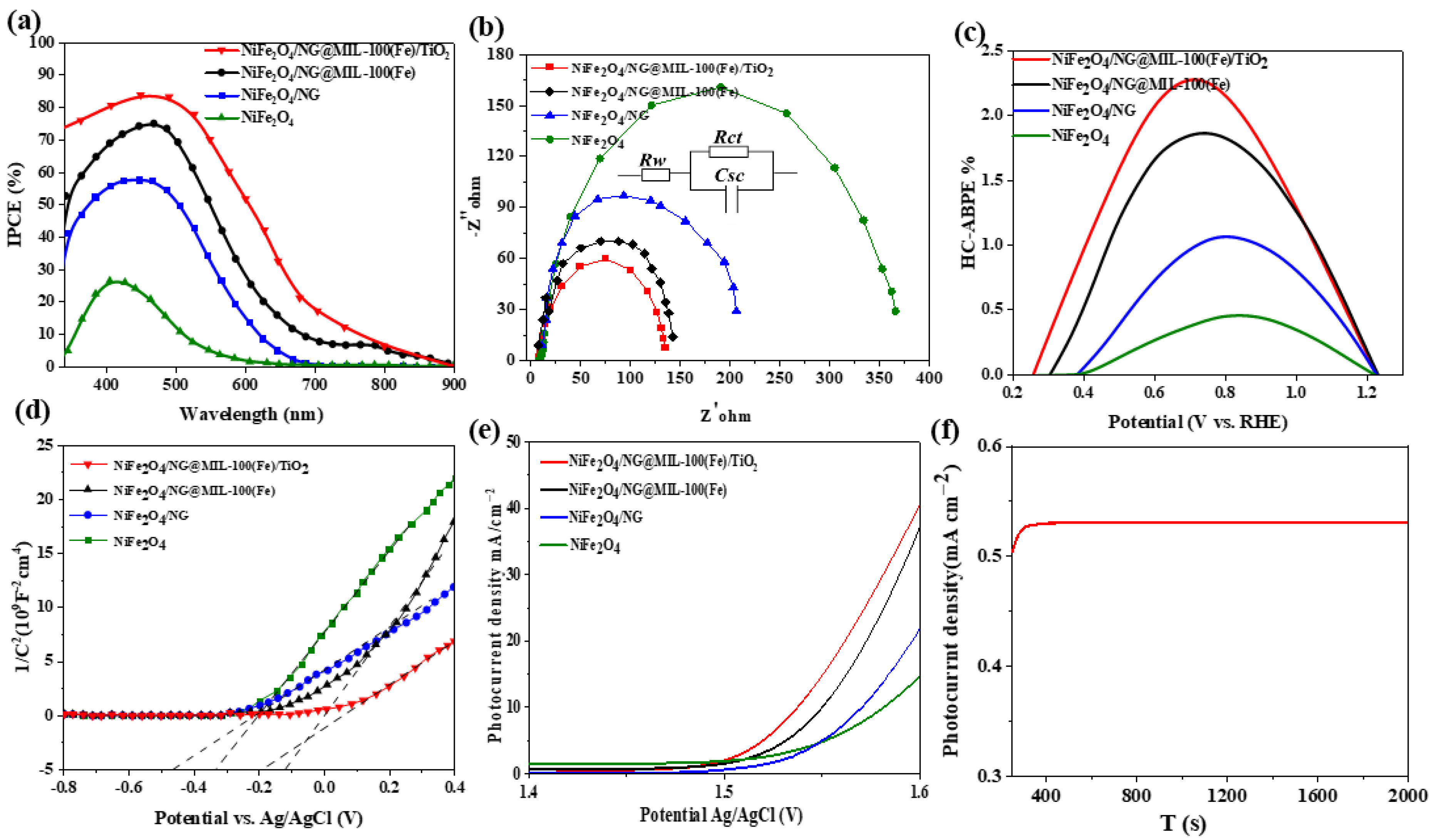
3.2. Optoelectronic Analysis

The light absorption patterns together with Tauc plot data for NiFe2O4, NiFe2O4/NG, NiFe2O4/NG@MIL-100(Fe), and NiFe2O4/NG@MIL-100(Fe)/TiO2 nanocomposites are shown in Figure 8a. All samples exhibit significant absorbance throughout both visible and ultraviolet spectrums during measurements between 200–800 nm. Light-harvesting capabilities of NiFe2O4/NG@MIL-100(Fe)/TiO2 reach their peak due to its highest absorbance within the visible range of the spectrum. The strong light absorption capability is vital for improving photocatalytic and photoelectrochemical efficiency during water-splitting reactions. The research shows that the structural integration of NG and MIL-100(Fe) together with TiO2 generates substantial light absorption ratio in the visible spectrum of the composite material. NiFe2O4/NG@MIL-100(Fe)/TiO2 demonstrates maximum light absorption all throughout the visible wavelength region, making it highly efficient in terms of light harvesting. Two factors work together to boost photonic activity through nitrogen-doped graphene (NG) and MIL-100(Fe) and TiO2 synergies. The optical properties of NiFe2O4 have improved after structural modifications and doping because the undoped NiFe2O4 sample demonstrates the lowest visual light absorbance ratio. The applied doping method shifts the absorption edge toward shorter wavelengths, resulting in a reduction of optical band gap energy, thus potentially enhancing photocatalytic efficiency under visible light conditions. These nanocomposites’ band gap energy (Eg) was analyzed using the Tauc plot, as illustrated in Figure 8b. The absorption coefficient α was calculated using the following equation:
α = 1 d ln 1 T
where α is the optical absorption coefficient, d is the sample thickness, and T is the transmission. The Tauc plot relates the absorption coefficient to the incident photon energy by the equation
α h v = A h v E g n
where A is a constant, Eg is the band gap energy, and n is ½ for direct band gap transitions. From the Tauc plot, the band gap energy for the synthesized samples was found to decrease in the doped nanocomposites compared to pristine NiFe2O4, with the values approximately as follows: NiFe2O4 ~2.4 eV, NiFe2O4/NG ~2.3eV, NiFe2O4/NG@MIL-100(Fe) ~2.2 eV, and NiFe2O4/NG@MIL-100(Fe)/TiO2 ~2.1 eV. Among the samples, NiFe2O4/NG@MIL-100(Fe)/TiO2 shows the highest absorbance in the visible region and the lowest band gap of ~2.1 eV, which enhances its light-harvesting capability. The reduction in the band gap energy across the doped samples, particularly NiFe2O4/NG@MIL-100(Fe)/TiO2, confirms their superior light absorption. These improvements in light absorption and reduced band gap make the nanocomposite an efficient candidate for photoelectrochemical water-splitting applications.

3.3. Electrochemical Characterizations and Photoelectrochemical Water Splitting

Photocurrent densities were evaluated under illumination to assess the photoelectrochemical water-splitting performance of NiFe2O4, NiFe2O4/NG, NiFe2O4/NG@MIL-100(Fe), and NiFe2O4/NG@MIL-100(Fe)/TiO2. The NiFe2O4/NG@ MIL-100(Fe)/TiO2 nanocomposite exhibits the greatest IPCE, peaking at around 80% in the visible spectrum, according to Figure 9a, which shows the incident photon-to-current efficiency (IPCE) (%) as a function of wavelength (nm). This high efficiency highlights its exceptional optoelectronic properties and enhanced ability to harvest light across a broad wavelength range. The remarkable performance is attributed to the synergistic interactions between the components of the nanocomposite, which facilitate efficient charge separation and transfer. These characteristics make NiFe2O4/NG@MIL-100(Fe)/TiO2 a promising candidate for water-splitting applications [63].
TRPL (Time-resolved photoluminescence) analysis was employed to analyze NiFe2O4/NG@MIL-100(Fe)/TiO2 in order to acquire valuable knowledge about carrier behavior, focusing on their recombination mechanisms (Figure S3). The TRPL spectra demonstrate time-dependent photoluminescence intensity decay behavior whose rates differ between samples, resulting in color-coded curves. Faster decay behavior in the TRPL spectra indicates elevated recombination rates for charge carriers, thus minimizing their efficient separation that usually affects photocatalytic performance negatively. The speed of decay in photoluminescence intensity provides insight into charge carrier separation capabilities because rapid decay signifies efficient charge separation. The observed outcomes carry substantial importance for water-splitting technology because extended charge-carrier durations both enhance photoelectrochemical reaction efficiency and decrease energy waste through improved charge separation.
The photoelectrochemical water-splitting performance of different samples was characterized by measuring the photocurrent densities under AM 1.5 G simulated sunlight (100 mW/cm2). Electrochemical impedance spectroscopy (EIS) was conducted to understand the charge-transfer kinetics at the electrode–electrolyte interface. The Nyquist plots, shown in the provided Figure 9b, reveal the electrochemical impedance of the pristine and doped samples. The charge-transfer resistance (RCT) is inferred from the radius of the arcs in the Nyquist plots; smaller arc radii indicate lower charge-transfer resistance, which facilitates more efficient electron transfer during the water-splitting process [64]. Among the nanocomposites, NiFe2O4/NG@MIL-100(Fe)/TiO2 shows the lowest charge-transfer resistance, both under light and dark conditions, indicating its superior ability to facilitate charge transfer at the semiconductor–electrolyte interface presented. A commonly used fitting model consisting of a resistive–capacitive (RC) circuit with two equivalent resistances, RS and RCT, and a constant phase element (CPE1) was employed for the doped samples to model the electrochemical behavior. The fitted values for charge-transfer resistance agree with the arc radii observed in the Nyquist plots. The significantly lower RCT for NiFe2O4/NG@MIL-100(Fe)/TiO2 confirms its superior charge-transfer capability, making it highly promising for water-splitting applications presented in Figure 9b.
Figure 9c shows the the half-cell applied bias photon-to-current efficiency (HC-ABPE%) of NiFe2O4, NiFe2O4/NG, NiFe2O4/NG@MIL-100(Fe), and NiFe2O4/NG@MIL-100(Fe)/TiO2. According to the data, the NiFe2O4/NG@MIL-100(Fe)/TiO2 nanocomposite has the greatest HC-ABPE%, peaking at around 2.5% at about 0.7 V. NiFe2O4/NG@MIL-100(Fe) and NiFe2O4/NG come in second and third, respectively, with NiFe2O4 demonstrating the lowest efficiency. The structural synergy between NiFe2O4, NG, MIL-100(Fe), and TiO2 is responsible for the NiFe2O4/NG@MIL-100(Fe)/TiO2 nanocomposite’s better HC-ABPE%, which highlights its improved optoelectronic capabilities and efficient charge separation. This improvement in performance highlights the promise of NiFe2O4/NG@MIL-100(Fe)/TiO2 as an effective photoelectrochemical material for water-splitting applications, which is consistent with the structural and optoelectronic characteristics of the nanocomposite that were developed for the best possible solar energy consumption.
Mott–Schottky (MS) analysis was performed to study the effect of the composite structure on carrier density (Nd) and flat-band potential (Vfb) for the nanocomposites. As shown in the Mott–Schottky plots in Figure 9d, the positive slopes of all samples confirm the n-type semiconductor behavior, with electrons as the majority of charge carriers [65]. Among the samples, NiFe2O4/NG@MIL-100(Fe)/TiO2 demonstrated the highest carrier density, which was nearly double that of the pristine NiFe2O4 sample. The increased carrier density is attributed to the introduction of MIL-100(Fe) and TiO2, which enhance the conductivity and mobility of charge carriers [66,67]. The flat-band potential (Vfb) for NiFe2O4, NiFe2O4/NG, NiFe2O4/NG@MIL-100(Fe), and NiFe2O4/NG@MIL-100(Fe)/TiO2 was found to be 0.45 V, 0.35 V, 0.50 V, and 0.58 V (vs. RHE), respectively. A lower flat-band potential implies easier charge separation under an applied potential, which is crucial for efficient water-splitting applications [68].
The impact of the composite structure on photocurrent density was examined in a 1 M NaOH solution using linear sweep voltammetry (LSV). Of the materials analyzed, NiFe2O4/NG@MIL-100(Fe)/TiO2) had the highest photocurrent density, measuring over 3.5 mA cm−2 at 1 V (vs. Ag/AgCl) (Figure 9e). Nitrogen-doped graphene (NG), MIL-100(Fe), and TiO2 work in concert to enhance conductivity, light absorption, and charge separation, all of which lead to improved performance results. The enhanced photocurrent density highlights the potential applications of this nanocomposite in efficient water-splitting systems.
An amperometric current density versus time curve was used to evaluate the stability of the NiFe2O4/NG@MIL-100(Fe/TiO2 nanocomposite), showing a constant photocurrent density of 0.5 mA cm−2 maintained for 2000 s at 1 V (vs. Ag/AgCl). As illustrated in Figure 9f, this validates the endurance of the nanocomposite and emphasizes its potential for the long term. In order to evaluate the reusability and stability of NiFe2O4/NG@MIL-100(Fe)/TiO2 composite during photocatalytic cycles, XRD measurements were conducted before and after its use for photocatalytic operations (Figure S4). XRD data of the NiFe2O4/NG@MIL-100(Fe)/TiO2 composite reveals that its crystalline structure persists unchanged following photocatalytic usage in the reaction system. The material kept its structural integrity along with phase stability throughout the photocatalytic process because no major changes or phase transitions occurred in XRD peak positions.
A detailed comparison of PEC performance parameters with similar heterostructured photoanodes is provided in Table S2. The NiFe₂O₄/NG@MIL-100(Fe)/TiO₂ composite exhibits competitive performance, indicating its potential for practical PEC applications.

3.4. Photoelectrochemical Mechanism for Water Splitting

The schematic diagram of the NiFe2O4/NG@MIL-100(Fe)/TiO2 composite in a photoelectrochemical (PEC) water-splitting system is shown in Figure 10, highlighting its crucial role in sunlight-driven hydrogen and oxygen production. This composite, integrating NiFe2O4, nitrogen-doped graphene (NG), MIL-100(Fe), and TiO2, combines unique properties that significantly enhance photocatalytic performance.
NiFe2O4 acts as the primary light absorber, generating electron–hole pairs upon sunlight exposure. The excited electrons transfer through NG, which serves as an excellent conductive pathway, and subsequently move from MIL-100(Fe) to TiO2 (3.2 eV), where TiO2 facilitates charge separation and provides additional photocatalytic sites. By reducing electron–hole recombination, the photogenerated electrons are able to travel through the external circuit to the Pt cathode, where they accumulate and reduce hydrogen ions (H+) to hydrogen atoms, which subsequently combine to form hydrogen gas (H2). Meanwhile, the holes in the valence band are directed to the photoanode surface, promoting the oxidation of water molecules and producing O2 gas, thus completing the water-splitting process.
The NiFe2O4/NG@MIL-100(Fe)/TiO2 composite extends light absorption into the visible range, whereas TiO2 alone primarily responds to ultraviolet light. The composite architecture efficiently integrates NG with MIL-100(Fe) and NiFe2O4, enhancing visible-light harvesting and charge separation, and reducing electron–hole recombination. Optimized band alignment among the components further elevates photocatalytic activity. Additionally, the nanocomposite exhibits superior charge mobility and lower charge transfer resistance, optimizing its PEC water-splitting performance. The synergistic roles of NG (electron transfer), TiO2 (structural stability), and NiFe2O4 and MIL-100(Fe) (light absorption) collectively contribute to the high photoactivity of the system.

4. Conclusions

In this study, we synthesized and characterized NiFe2O4/NG@MIL-100(Fe)/TiO2 nanocomposites using a hydrothermal method. The structural, optoelectronic, and photoelectrochemical properties were optimized for water-splitting applications. XRD and microscopy confirmed well-defined crystal structures, while UV-visible absorption spectra and Tauc plot analysis revealed enhanced light-harvesting and reduced band gap energy. The NiFe2O4/NG@MIL-100(Fe)/TiO2 composite demonstrated the highest photocurrent density and improved charge separation efficiency, as indicated by linear sweep voltammetry (LSV) and electrochemical impedance spectroscopy (EIS). Mott–Schottky analysis further showed an increase in carrier density, confirming the material’s suitability for long-term photoelectrochemical performance. These results highlight the potential of NiFe2O4/NG@MIL-100(Fe)/TiO2 as a promising photocatalyst for efficient water-splitting applications.

Supplementary Materials

The following supporting information can be downloaded at https://www.mdpi.com/article/10.3390/jcs9050250/s1. Figure S1: XRD pattern of the NiFe2O4/NG@MIL-100(Fe), NiFe2O4/NG, and NiFe2O4 samples, and corresponding standard cards of MIL-100(Fe)(COD: 7102029) and NiFe2O4 (JCPDS card No. 01-074-2081); Figure S2: BET curve for NiFe2O4/NG@MIL-100(Fe)/TiO2 composite (inset shows the pore diameter distribution); Figure S3: TRPL for NiFe2O4/NG@MIL-100(Fe)/TiO2 composite. Figure S4: XRD patterns of the NiFe2O4/NG@MIL-100(Fe)/TiO2 before and after photocatalytic reaction; Table S1: Elemental percentage distribution table for NiFe2O4/NG@MIL-100(Fe)/TiO2; Table S2: Comparison of different pore size distribution of BET in NiFe2O4/NG MIL-100(Fe)/TiO2,NiFe2O4, NG, MIL-100(Fe), and TiO2; Table S3: Comparative analysis of performance of NiFe2O4/NG@MIL-100(Fe)/TiO2 against similar catalysts for photoelectrochemical water splitting [16,69,70,71,72,73,74,75,76].

Author Contributions

Conceptualization, X.L.; Methodology, W.R. and B.M.; Formal analysis, F.S; Investigation, W.R., S.A., and M.U.; Resources, B.M. and X.L.; Data curation, W.R., F.S., S.A. and M.U.; Writing—original draft, W.R.; Writing—review & editing, X.L.; Funding acquisition, X.L. All authors have read and agreed to the published version of the manuscript.

Funding

This work was partially financially supported by the Independent Innovation Fund of Tianjin University (Grant No. 2024XSU-0006) and the National Key R&D Program of China (Grant No. 2019YFC1407800).

Data Availability Statement

Data are contained within the article or Supplementary Materials.

Conflicts of Interest

The authors declare no conflicts of interest.

References

  1. Qi, Y.; Zhang, B.; Zhang, G.; Zheng, Z.; Xie, T.; Chen, S.; Ma, G.; Li, C.; Domen, K.; Zhang, F. Efficient overall water splitting of a suspended photocatalyst boosted by metal-support interaction. Joule 2024, 8, 193–203. [Google Scholar] [CrossRef]
  2. Ling, L.; Liu, L.; Feng, Y.; Zhu, J.; Bian, Z. Synthesis of TiO2 mesocrystal film with enhanced photocatalytic activity. Chin. J. Catal. 2018, 39, 639–645. [Google Scholar] [CrossRef]
  3. Abarca González, J.A.; Díaz Sainz, G.; Merino García, I.; Irabien Gulías, Á.; Albo Sánchez, J. Photoelectrochemical CO2 electrolyzers: From photoelectrode fabrication to reactor configuration. J. Energy Chem. 2023, 85, 455–480. [Google Scholar] [CrossRef]
  4. Zhang, Q.; Liu, G.; Liu, T. Controllable Synthesis Heterojunction of g-C3N4 and BiVO4 to Enhance the Photocatalytic Oxygen Evolution Activity. ACS Sustain. Chem. Eng. 2024, 12, 5675–5684. [Google Scholar] [CrossRef]
  5. She, H.; Zhou, H.; Li, L.; Zhao, Z.; Jiang, M.; Huang, J.; Wang, L.; Wang, Q. Construction of a two-dimensional composite derived from TiO2 and SnS2 for enhanced photocatalytic reduction of CO2 into CH4. ACS Sustain. Chem. Eng. 2018, 7, 650–659. [Google Scholar] [CrossRef]
  6. Yin, D.; Ning, X.; Zhang, Q.; Du, P.; Lu, X. Dual modification of BiVO4 photoanode for synergistically boosting photoelectrochemical water splitting. J. Colloid Interface Sci. 2023, 646, 238–244. [Google Scholar] [CrossRef]
  7. Feng, C.; Liu, L.; Fu, H.; Zhan, F.; Jia, H.; Cheng, X.; Yu, F.; Zhou, Q.; Bian, Z.; Zhang, B. Structural tuning of BiVO4/MnFe-MOF photoanodes boosts hole extraction for photoelectrochemical water splitting. Catal. Sci. Technol. 2024, 14, 4860–4868. [Google Scholar] [CrossRef]
  8. Li, R.; Zhang, C.; You, K.; Li, B.; Bu, W.; Meng, X.; Ma, B.; Ding, Y. Molecular confined synthesis of magnetic CoOx/Co/C hybrid catalyst for photocatalytic water oxidation and CO2 reduction. Chin. Chem. Lett. 2023, 34, 108801. [Google Scholar] [CrossRef]
  9. Wang, H.; Li, X.; Zhao, X.; Li, C.; Song, X.; Zhang, P.; Huo, P. A review on heterogeneous photocatalysis for environmental remediation: From semiconductors to modification strategies. Chin. J. Catal. 2022, 43, 178–214. [Google Scholar] [CrossRef]
  10. Richards, B.S.; Hudry, D.; Busko, D.; Turshatov, A.; Howard, I.A. Photon upconversion for photovoltaics and photocatalysis: A critical review: Focus review. Chem. Rev. 2021, 121, 9165–9195. [Google Scholar] [CrossRef]
  11. Mary, A.S.; Murugan, C.; Mahendiran, D.; Murugan, P.; Pandikumar, A. Investigation of Mn incorporation into NiOOH electrocatalyst loaded on BiVO4 photoanode for enhanced photoelectrochemical water splitting: Experimental and theoretical approach. Mater. Today Energy 2024, 41, 101541. [Google Scholar] [CrossRef]
  12. Shao, J.; Liu, H.; Jiang, X.; Wang, Y.; Luo, L.; Li, Y.; Zhang, Z.; Lu, B. High Performance Ferroelectric-modulated Photoelectrodes by Using Grid Charge Collector. Adv. Funct. Mater. 2024, 34, 2316409. [Google Scholar] [CrossRef]
  13. Bie, C.; Yu, H.; Cheng, B.; Ho, W.; Fan, J.; Yu, J. Design, fabrication, and mechanism of nitrogen-doped graphene-based photocatalyst. Adv. Mater. 2021, 33, 2003521. [Google Scholar] [CrossRef] [PubMed]
  14. Chen, Z.; Yao, D.; Chu, C.; Mao, S. Photocatalytic H2O2 production systems: Design strategies and environmental applications. Chem. Eng. J. 2023, 451, 138489. [Google Scholar] [CrossRef]
  15. Al-Asbahi, B.A.; Haji Jumali, M.H.; Yap, C.C.; Mat Salleh, M. Influence of TiO2 Nanoparticles on Enhancement of Optoelectronic Properties of PFO-Based Light Emitting Diode. J. Nanomater. 2013, 2013, 561534. [Google Scholar] [CrossRef]
  16. Wang, T.; Liu, X.; Liu, M.; Liao, R.; Zhan, H.; Qi, X.; Wang, Y.; Huang, Y. The enhanced photocatalytic activity of TiO2 (B)/MIL-100 (Fe) composite via Fe–O clusters. New J. Chem. 2022, 46, 739–746. [Google Scholar] [CrossRef]
  17. Rashid, T.; Raza, A.; Saleh, H.A.; Khan, S.; Rahaman, S.; Pandey, K.; AlDamen, M.A.; Sama, F.; Ahmad, A.; Shahid, M. Green Synthesis of Ni/Fe3O4/rGO Nanocomposites for Desulfurization of Fuel. ACS Appl. Nano Mater. 2023, 6, 18905–18917. [Google Scholar] [CrossRef]
  18. Yeh, Y.-H.; Chang, C.-L.; Tseng, Z.-C.; Hsiao, V.K.; Huang, C.-Y. Enhancing the Photoelectrochemical Performance of a Nanoporous Silicon Photocathode through Electroless Nickel Deposition. Nanomaterials 2023, 13, 2552. [Google Scholar] [CrossRef]
  19. Lakhera, S.K.; Rajan, A.; Rugma, T.; Bernaurdshaw, N. A review on particulate photocatalytic hydrogen production system: Progress made in achieving high energy conversion efficiency and key challenges ahead. Renew. Sustain. Energy Rev. 2021, 152, 111694. [Google Scholar] [CrossRef]
  20. Dou, Y.; Zhou, J.; Zhou, A.; Li, J.-R.; Nie, Z. Visible-light responsive MOF encapsulation of noble-metal-sensitized semiconductors for high-performance photoelectrochemical water splitting. J. Mater. Chem. A 2017, 5, 19491–19498. [Google Scholar] [CrossRef]
  21. Yoon, J.W.; Kim, D.H.; Kim, J.-H.; Jang, H.W.; Lee, J.-H. NH2-MIL-125(Ti)/TiO2 nanorod heterojunction photoanodes for efficient photoelectrochemical water splitting. Appl. Catal. B Environ. 2019, 244, 511–518. [Google Scholar] [CrossRef]
  22. Ali, M.; Pervaiz, E.; Noor, T.; Rabi, O.; Zahra, R.; Yang, M. Recent advancements in MOF-based catalysts for applications in electrochemical and photoelectrochemical water splitting: A review. Int. J. Energy Res. 2021, 45, 1190–1226. [Google Scholar] [CrossRef]
  23. Li, C.F.; Zhao, J.W.; Xie, L.J.; Wu, J.Q.; Ren, Q.; Wang, Y.; Li, G.R. Surface-adsorbed carboxylate ligands on layered double hydroxides/metal–organic frameworks promote the electrocatalytic oxygen evolution reaction. Angew. Chem. 2021, 133, 18277–18285. [Google Scholar] [CrossRef]
  24. Jia, M.; Xiong, W.; Yang, Z.; Cao, J.; Zhang, Y.; Xiang, Y.; Xu, H.; Song, P.; Xu, Z. Metal-organic frameworks and their derivatives-modified photoelectrodes for photoelectrochemical applications. Coord. Chem. Rev. 2021, 434, 213780. [Google Scholar] [CrossRef]
  25. Jia, L.; Arain, M.; Ahmed, A.; Yikai, F.; Zhenda, C.; Hussain, I.; Ashraf, G.A.; Ahmed, S.B.; Dai, H. Emerging trends in metal-organic framework (MOFs) photocatalysts for hydrogen energy using water splitting: A state-of-the-art review. J. Ind. Eng. Chem. 2024, 131, 54–135. [Google Scholar]
  26. Shateran, F.; Ghasemzadeh, M.A.; Aghaei, S.S. Preparation of NiFe2O4@MIL-101 (Fe)/GO as a novel nanocarrier and investigation of its antimicrobial properties. RSC Adv. 2022, 12, 7092–7102. [Google Scholar] [CrossRef]
  27. Alves, R.C.; Schulte, Z.M.; Luiz, M.T.; Bento da Silva, P.c.; Frem, R.C.; Rosi, N.L.; Chorilli, M. Breast cancer targeting of a drug delivery system through postsynthetic modification of curcumin@ N3-bio-MOF-100 via click chemistry. Inorg. Chem. 2021, 60, 11739–11744. [Google Scholar] [CrossRef]
  28. Liu, Y.; Zhu, Q.; Zhang, T.; Yan, X.; Duan, R. Analysis of chemical-looping hydrogen production and power generation system driven by solar energy. Renew. Energy 2020, 154, 863–874. [Google Scholar] [CrossRef]
  29. Alhabradi, M.; Yang, X.; Alruwaili, M.; Tahir, A.A. Nano multi-layered HfO2/α-Fe2O3 nanocomposite photoelectrodes for photoelectrochemical water splitting. Heliyon 2024, 10, e27078. [Google Scholar] [CrossRef]
  30. Chnani, A.; Strehle, S. Hematite nanowire and nanoflake-decorated photoelectrodes: Implications for photoelectrochemical water splitting. ACS Appl. Nano Mater. 2021, 5, 1016–1022. [Google Scholar] [CrossRef]
  31. Kavitha, S.; Ranjith, R.; Jayamani, N.; Vignesh, S.; Palanivel, B.; Djellabi, R.; Bianchi, C.; Alharthi, F.A. Fabrication of visible-light-responsive TiO2/α-Fe 2O3-heterostructured composite for rapid photo-oxidation of organic pollutants in water. J. Mater. Sci. Mater. Electron. 2022, 33, 8906–8919. [Google Scholar] [CrossRef]
  32. Guesh, K.; Caiuby, C.A.; Mayoral, A.l.; Díaz-García, M.; Díaz, I.; Sanchez-Sanchez, M. Sustainable preparation of MIL-100 (Fe) and its photocatalytic behavior in the degradation of methyl orange in water. Cryst. Growth Des. 2017, 17, 1806–1813. [Google Scholar] [CrossRef]
  33. Majumder, S.; Yadav, A.A.; Hunge, Y.M.; Jeffery, A.A.; Thomas, N.; Alshgari, R.A.; Chicardi, E.; Mushab, M.; Kim, K.H. Exploring FeVO4/ZnCo2O4 np heterojunctions for superior photoelectrochemical performance. J. Alloys Compd. 2025, 1010, 177281. [Google Scholar] [CrossRef]
  34. Majumder, S.; Yadav, A.A.; Palanisamy, A.K.; Hunge, Y.M.; KN, S.; Jeffery, A.A.; Tighezza, A.M.; Srinivasan, R.; Somu, P.; Kim, K.H. Design and optimization of FeVO4/Fe2TiO5 heterojunction photoanode for efficient photoelectrochemical water splitting. Ceram. Int. 2025, 51, 2536–2546. [Google Scholar] [CrossRef]
  35. Yadav, A.; Hunge, Y.; Kang, S.-W. Ultrasound assisted synthesis of highly active nanoflower-like CoMoS4 electrocatalyst for oxygen and hydrogen evolution reactions. Ultrason. Sonochemistry 2021, 72, 105454. [Google Scholar] [CrossRef] [PubMed]
  36. Babu Naidu, K.C.; Madhuri, W. Hydrothermal synthesis of NiFe2O4 nano-particles: Structural, morphological, optical, electrical and magnetic properties. Bull. Mater. Sci. 2017, 40, 417–425. [Google Scholar] [CrossRef]
  37. Yang, J.; Guo, D.; Zhao, S.; Lin, Y.; Yang, R.; Xu, D.; Shi, N.; Zhang, X.; Lu, L.; Lan, Y.Q. Cobalt phosphides nanocrystals encapsulated by P-doped carbon and married with P-doped graphene for overall water splitting. Small 2019, 15, 1804546. [Google Scholar] [CrossRef]
  38. Soysal, F. Nitrogen-doped reduced graphene oxide decorated with silver nanoparticles for photoelectrochemical water splitting. J. Mater. Sci. Mater. Electron. 2023, 34, 259. [Google Scholar] [CrossRef]
  39. Chandel, N.; Sharma, S.; Dutta, V.; Raizada, P.; Hosseini-Bandegharaei, A.; Kumar, R.; Gupta, V.K.; Agarwal, S.; Singh, P. Adsorption and photocatalysis compiled toxic dyes mineralization using graphitic carbon nitride modified ZnFe2O4 and CoFe2O4 photocatalysts supported onto N-doped graphene. Desalination Water Treat. 2020, 191, 381–399. [Google Scholar] [CrossRef]
  40. He, X.; Fang, H.; Gosztola, D.J.; Jiang, Z.; Jena, P.; Wang, W.-N. Mechanistic insight into photocatalytic pathways of MIL-100 (Fe)/TiO2 composites. ACS Appl. Mater. Interfaces 2019, 11, 12516–12524. [Google Scholar] [CrossRef]
  41. Simon, M.A.; Anggraeni, E.; Soetaredjo, F.E.; Santoso, S.P.; Irawaty, W.; Thanh, T.C.; Hartono, S.B.; Yuliana, M.; Ismadji, S. Hydrothermal synthesize of HF-free MIL-100 (Fe) for isoniazid-drug delivery. Sci. Rep. 2019, 9, 16907. [Google Scholar] [CrossRef]
  42. Sivakumar, P.; Ramesh, R.; Ramanand, A.; Ponnusamy, S.; Muthamizhchelvan, C. Synthesis, Studies and Growth Mechanism of Ferromagnetic NiFe2O4 Nanosheet. Appl. Surf. Sci. 2012, 258, 6648–6652. [Google Scholar] [CrossRef]
  43. Sanadi, P.D.; Chougale, R.K.; Malavekar, D.B.; Kim, J.H.; Masimukku, S.; Chang-Chien, G.P.; Kamble, G.S. Efficient hydrogen evolution via neutral water electrolysis using nanocrystalline TiO2 electrocatalyst. Scientific Reports 2025, 15, 16074. [Google Scholar] [CrossRef]
  44. Hamedi, A.; Trotta, F.; Borhani Zarandi, M.; Zanetti, M.; Caldera, F.; Anceschi, A.; Nateghi, M.R. In Situ Synthesis of MIL-100(Fe) at the Surface of Fe(3)O(4)@AC as Highly Efficient Dye Adsorbing Nanocomposite. Int. J. Mol. Sci. 2019, 20, 5612. [Google Scholar] [CrossRef]
  45. Wu, X.-L.; Jiang, L.-Y.; Cao, F.-F.; Guo, Y.-G.; Wan, L.-J. LiFePO4 nanoparticles embedded in a nanoporous carbon matrix: Superior cathode material for electrochemical energy-storage devices. Adv. Mater. 2009, 21, 2710–2714. [Google Scholar] [CrossRef] [PubMed]
  46. Aijaz, A.; Masa, J.; Rösler, C.; Xia, W.; Weide, P.; Botz, A.J.; Fischer, R.A.; Schuhmann, W.; Muhler, M. Co@Co3O4 encapsulated in carbon nanotube-grafted nitrogen-doped carbon polyhedra as an advanced bifunctional oxygen electrode. Angew. Chem. Int. Ed. 2016, 55, 4087–4091. [Google Scholar] [CrossRef] [PubMed]
  47. Yamashita, T.; Hayes, P. Analysis of XPS spectra of Fe2+ and Fe3+ ions in oxide materials. Appl. Surf. Sci. 2008, 254, 2441–2449. [Google Scholar] [CrossRef]
  48. Descostes, M.; Mercier, F.; Thromat, N.; Beaucaire, C.; Gautier-Soyer, M. Use of XPS in the determination of chemical environment and oxidation state of iron and sulfur samples: Constitution of a data basis in binding energies for Fe and S reference compounds and applications to the evidence of surface species of an oxidized pyrite in a carbonate medium. Appl. Surf. Sci. 2000, 165, 288–302. [Google Scholar]
  49. Kang, B.K.; Im, S.Y.; Lee, J.; Kwag, S.H.; Kwon, S.B.; Tiruneh, S.; Kim, M.-J.; Kim, J.H.; Yang, W.S.; Lim, B. In-situ formation of MOF derived mesoporous Co3N/amorphous N-doped carbon nanocubes as an efficient electrocatalytic oxygen evolution reaction. Nano Res. 2019, 12, 1605–1611. [Google Scholar] [CrossRef]
  50. Zhang, Y.; Rui, K.; Ma, Z.; Sun, W.; Wang, Q.; Wu, P.; Zhang, Q.; Li, D.; Du, M.; Zhang, W. Cost-effective vertical carbon nanosheets/iron-based composites as efficient electrocatalysts for water splitting reaction. Chem. Mater. 2018, 30, 4762–4769. [Google Scholar] [CrossRef]
  51. Saeed, F.; Ahmad, M.; Zada, A.; Qi, D.; Wang, Y. Phosphorus-doped CoFe2O4 nanoparticles decorated nitrogen-doped graphene for efficient and stable electrocatalytic water splitting. Int. J. Hydrogen Energy 2024, 59, 1196–1204. [Google Scholar] [CrossRef]
  52. Li, X.; Niu, Z.; Jiang, J.; Ai, L. Cobalt nanoparticles embedded in porous N-rich carbon as an efficient bifunctional electrocatalyst for water splitting. J. Mater. Chem. A 2016, 4, 3204–3209. [Google Scholar] [CrossRef]
  53. Saeed, F.; Samia; Ahmad, M.; Rehman, W.; Sana, Y.; Ameen, S.; Altowyan, A.S.; Zada, A. Sulfur-Doped Zinc Oxide-Nikel Oxide as Efficient Bifunctional Electrocatalyst for Overall Water Splitting. Electrocatalysis 2024, 16, 15–27. [Google Scholar] [CrossRef]
  54. Feng, H.; Yanan, D.; Ting, X.; Yongzhi, L.; Xin, Z.; Jianyi, X.; Guofang, Z.; Ying, C.; Yanghuan, Z. Influence of adding graphene on the hydrogen storage thermodynamics and kinetics of as-milled CeMg12–Ni alloy. Int. J. Hydrogen Energy 2023, 48, 13213–13226. [Google Scholar] [CrossRef]
  55. Li, R.; Xu, J.; Ba, J.; Li, Y.; Liang, C.; Tang, T. Facile synthesis of nanometer-sized NiFe layered double hydroxide/nitrogen-doped graphite foam hybrids for enhanced electrocatalytic oxygen evolution reactions. Int. J. Hydrogen Energy 2018, 43, 7956–7963. [Google Scholar] [CrossRef]
  56. Long, Y.; Zhao, G.; Yang, L.; An, Y.; Xu, Y.; Xu, C. Ultralow Ru loaded CoNi bimetallic phosphides nanorods as efficient electrocatalyst for overall water splitting. Int. J. Hydrogen Energy 2024, 51, 336–348. [Google Scholar] [CrossRef]
  57. Wang, H.; Lee, H.-W.; Deng, Y.; Lu, Z.; Hsu, P.-C.; Liu, Y.; Lin, D.; Cui, Y. Bifunctional non-noble metal oxide nanoparticle electrocatalysts through lithium-induced conversion for overall water splitting. Nat. Commun. 2015, 6, 7261. [Google Scholar] [CrossRef]
  58. Dionigi, F.; Strasser, P. NiFe-based (oxy) hydroxide catalysts for oxygen evolution reaction in non-acidic electrolytes. Adv. Energy Mater. 2016, 6, 1600621. [Google Scholar] [CrossRef]
  59. Görlin, M.; Chernev, P.; Ferreira de Araújo, J.; Reier, T.; Dresp, S.r.; Paul, B.; Krähnert, R.; Dau, H.; Strasser, P. Oxygen evolution reaction dynamics, faradaic charge efficiency, and the active metal redox states of Ni–Fe oxide water splitting electrocatalysts. J. Am. Chem. Soc. 2016, 138, 5603–5614. [Google Scholar] [CrossRef]
  60. Peng, P.; Lin, X.-M.; Liu, Y.; Filatov, A.S.; Li, D.; Stamenkovic, V.R.; Yang, D.; Prakapenka, V.B.; Lei, A.; Shevchenko, E.V. Binary transition-metal oxide hollow nanoparticles for oxygen evolution reaction. ACS Appl. Mater. Interfaces 2018, 10, 24715–24724. [Google Scholar] [CrossRef]
  61. Zhang, H.; Li, X.; Hähnel, A.; Naumann, V.; Lin, C.; Azimi, S.; Schweizer, S.L.; Maijenburg, A.W.; Wehrspohn, R.B. Bifunctional heterostructure assembly of NiFe LDH nanosheets on NiCoP nanowires for highly efficient and stable overall water splitting. Adv. Funct. Mater. 2018, 28, 1706847. [Google Scholar] [CrossRef]
  62. Rehman, W.; Saeed, F.; Zhao, Y.; Maryam, B.; Arain, S.; Ayaz, M.; Jamil, A.; Liu, X. Boosted Photoelectrochemical Water Oxidation Performance with a Quaternary Heterostructure: CoFe2O4/MWCNT-Doped MIL-100 (Fe)/TiO2. Catalysts 2024, 14, 901. [Google Scholar] [CrossRef]
  63. Wang, Y.; Chen, X.; Xiu, H.; Zhuang, H.; Li, J.; Zhou, Y.; Liu, D.; Kuang, Y. General in situ photoactivation route with IPCE over 80% toward CdS photoanodes for photoelectrochemical applications. Small 2021, 17, 2104307. [Google Scholar] [CrossRef] [PubMed]
  64. Hahn, N.T.; Mullins, C.B. Photoelectrochemical performance of nanostructured Ti-and Sn-doped α-Fe2O3 photoanodes. Chem. Mater. 2010, 22, 6474–6482. [Google Scholar] [CrossRef]
  65. Tamgadge, R.M.; Shukla, A. Fluorine-doped anatase for improved supercapacitor electrode. Electrochim. Acta 2018, 289, 342–353. [Google Scholar] [CrossRef]
  66. Moto, K.; Yoshimine, R.; Suemasu, T.; Toko, K. Improving carrier mobility of polycrystalline Ge by Sn doping. Sci. Rep. 2018, 8, 14832. [Google Scholar] [CrossRef]
  67. Paul Inbaraj, C.R.; Gudelli, V.K.; Mathew, R.J.; Ulaganathan, R.K.; Sankar, R.; Lin, H.Y.; Lin, H.-I.; Liao, Y.-M.; Cheng, H.-Y.; Lin, K.-H. Sn-Doping Enhanced Ultrahigh Mobility In1–x Sn x Se Phototransistor. ACS Appl. Mater. Interfaces 2019, 11, 24269–24278. [Google Scholar] [CrossRef]
  68. Kang, S.H.; Kim, J.-Y.; Kim, Y.; Kim, H.S.; Sung, Y.-E. Surface Modification of Stretched TiO2 Nanotubes for Solid-State Dye-Sensitized Solar Cells. J. Phys. Chem. C 2007, 111, 9614–9623. [Google Scholar] [CrossRef]
  69. Ziylan-Yavas, A.; Mizukoshi, Y.; Maeda, Y.; Ince, N.H. Supporting of Pristine TiO2 with Noble Metals to Enhance the Oxidation and Mineralization of Paracetamol by Sonolysis and Sonophotolysis. Appl. Catal. B Environ. 2015, 172–173, 7–17. [Google Scholar] [CrossRef]
  70. Malara, F.; Minguzzi, A.; Marelli, M.; Morandi, S.; Psaro, R.; Dal Santo, V.; Naldoni, A. α-Fe2O3/NiOOH: An Effective Heterostructure for Photoelectrochemical Water Oxidation. ACS Cat. 2015, 5, 5292–5300. [Google Scholar] [CrossRef]
  71. Wang, Y.; Zhang, Y.; Zhao, G.; Tian, H.; Shi, H.; Zhou, T. Design of a Novel Cu2O/TiO2/Carbon Aerogel Electrode and Its Efficient Electrosorption-Assisted Visible Light Photocatalytic Degradation of 2,4,6-Trichlorophenol. ACS Appl. Mat. Inter. 2012, 4, 3965–3972. [Google Scholar] [CrossRef] [PubMed]
  72. Jia, P.-Y.; Guo, R.-T.; Pan, W.-G.; Huang, C.-Y.; Tang, J.-Y.; Liu, X.-Y.; Qin, H.; Xu, Q.-Y. The MoS2/TiO2 heterojunction composites with enhanced activity for CO2 photocatalytic reduction under visible light irradiation. Colloids Surf. A Physicochem. Eng. Asp. 2019, 570, 306–316. [Google Scholar] [CrossRef]
  73. Nayak, S.; Mohapatra, L.; Parida, K. Visible light-driven novel g-C3N4/NiFe-LDH composite photocatalyst with enhanced photocatalytic activity towards water oxidation and reduction reaction. J. Mat. Chem. 2015, 3, 18622–18635. [Google Scholar] [CrossRef]
  74. Guo, J.; Yang, X.; Bai, S.; Xiang, X.; Luo, R.; He, J.; Chen, A. Effect of Mo doping and NiFe-LDH cocatalyst on PEC water oxidation efficiency. J. Colloid Interface Sci. 2019, 540, 9–19. [Google Scholar] [CrossRef]
  75. Liu, G.; Li, Y.; Xiao, Y.; Jia, D.; Li, C.; Zheng, J.; Liu, X. Nanoporous Fe-doped BiVO4 Modified with MIL-53 (Fe) for Enhanced Photoelectrochemical Stability and Water Splitting Perfromances. Catal. Lett. 2018, 149, 870–875. [Google Scholar] [CrossRef]
  76. Tian, K.; Wu, L.; Han, T.; Gao, L.; Wang, P.; Chai, H.; Jin, J. Dual modification of BiVO4 photoanode by rare earth element neodymium doping and further NiFe2O4 co-catalyst deposition for efficient photoelectrochemical water oxidation. J. Alloy. Compd. 2022, 923, 166352. [Google Scholar] [CrossRef]
Figure 1. Schematic representation of hydrothermal synthesis process for NiFe2O4/NG@ MIL-100(Fe)/TiO2.
Jcs 09 00250 g001
Figure 2. XRD pattern of NiFe2O4/NG@MIL-100(Fe)/TiO2 samples and corresponding standard JCPDS cards of No. 01-070-7348 (TiO2), COD: 7102029 (MIL-100(Fe)), and No. 01-074-2081 (NiFe2O4).
Jcs 09 00250 g002
Figure 3. TEM (a,b), HRTEM (c), SAED (d), SEM (eg), and EDX mapping (hn) images of NiFe2O4/NG@MIL-100(Fe)/TiO2 at different magnifications.
Jcs 09 00250 g003
Figure 4. EDX spectrum of NiFe2O4/NG@MIL-100(Fe)/TiO2.
Jcs 09 00250 g004
Figure 5. FTIR spectra for NiFe2O4/NG@MIL-100(Fe)/TiO2, NiFe2O4/NG@MIL-100(Fe), NiFe2O4/NG, and NiFe2O4.
Jcs 09 00250 g005
Figure 6. XPS of NiFe2O4/NG@MIL-100(Fe)/TiO2: (a) Ni 2p; (b) Fe 2p; (c) N 1s; (d) c 1s; (e) Ti 2p; and (f) O 1s.
Jcs 09 00250 g006
Figure 7. The W-H analysis of NiFe2O4/NG@MIL-100(Fe)/TiO2, NiFe2O4/NG @MIL-100(Fe), NiFe2O4/NG, and NiFe2O4.
Jcs 09 00250 g007
Figure 8. UV-VIS spectra (a) and Tauc plot (b) of NiFe2O4/NG@MIL-100(Fe)/TiO2, NiFe2O4/NG@MIL-100(Fe), NiFe2O4/NG, and NiFe2O4.
Jcs 09 00250 g008
Figure 9. NiFe2O4/NG@MIL-100(Fe)/TiO2: (a) IPCE. (b) Nyquist plots inset: Equivalent circuit for EIS. (c) ABPE. (d) Mott–Schottky plots. (e) LSV plots. (f) Amperometric J-t Curve of NiFe2O4/NG@MIL-100(Fe)/TiO2, for 2000 s of operation.
Jcs 09 00250 g009
Figure 10. Schematic representation Diagrammatic illustration of NiFe2O4/NG@MIL-100(Fe)/TiO2 PEC water splitting.
Jcs 09 00250 g010
Disclaimer/Publisher’s Note: The statements, opinions and data contained in all publications are solely those of the individual author(s) and contributor(s) and not of MDPI and/or the editor(s). MDPI and/or the editor(s) disclaim responsibility for any injury to people or property resulting from any ideas, methods, instructions or products referred to in the content.

