Article

Response Surface Methodology-Based Optimization for Enhancing the Viability of Microencapsulated Lactobacillus plantarum in Composite Materials

by
Rafael González-Cuello
1,*,
Joaquín Hernández-Fernández
2,3,4 and
Rodrigo Ortega-Toro
1,*
1
Food Packaging and Shelf-Life Research Group (FP&SL), Food Engineering Program, University of Cartagena, Cartagena de Indias 130015, Colombia
2
Chemistry Program, Department of Natural and Exact Sciences, San Pablo Campus, University of Cartagena, Cartagena de Indias 130015, Colombia
3
Chemical Engineering Program, School of Engineering, Universidad Tecnológica de Bolívar, Parque Industrial y Tecnológico Carlos Vélez Pombo Km 1 Vía Turbaco, Cartagena 130001, Colombia
4
Department of Natural and Exact Science, Universidad de la Costa, Barranquilla 080002, Colombia
*
Authors to whom correspondence should be addressed.
J. Compos. Sci. 2025, 9(4), 189; https://doi.org/10.3390/jcs9040189
Submission received: 19 March 2025 / Revised: 9 April 2025 / Accepted: 12 April 2025 / Published: 15 April 2025

Abstract

:
Probiotics have gained prominence and consumer appreciation due to their potential health benefits. However, maintaining their viability and stability during gastric transit remains a challenge. This study aims to enhance the viability of microencapsulated Lactobacillus plantarum in composite microcapsules exposed to simulated gastric juice. The independent variables investigated were low-acyl gellan gum (LAG), bacterial cellulose (BC), and calcium concentrations. The microcapsules were prepared using the internal ionic gelation method. The resulting microcapsules exhibited a uniform size distribution, with a diameter of approximately between 15 to 120 μm, making them suitable for food applications. Response surface methodology (RSM) based on the Box–Behnken design was successfully employed to optimize the concentrations of LAG, BC, and calcium. Under optimal conditions—0.63% w/v LAG, 17.91% w/v BC, and 25.12 mM Ca—the highest L. plantarum viability reached 94.28% after exposure to simulated gastric juice, with an R2 value of 99.64%. These findings demonstrate the feasibility of developing multicomponent microcapsules that effectively protect probiotic bacteria against gastric fluids, offering a promising alternative for the food industry in designing probiotic-enriched food systems.

1. Introduction

The global population increasingly demands high-quality products with both nutritional and health-promoting properties, commonly referred to as functional foods [1,2]. These foods are defined as those that, in addition to providing essential nutrients, offer various health benefits. When consumed regularly, they contribute to metabolic and physiological wellbeing, aiding in the prevention of chronic, degenerative, and cardiovascular diseases, among others [3,4]. Their functional properties are attributed to bioactive components such as probiotics, prebiotics, vitamins, and other beneficial substances that improve their nutritional profile [5,6].
According to the Food and Agriculture Organization (FAO) and the World Health Organization (WHO), probiotics are defined in the Codex Alimentarius as live microorganisms that provide health benefits when administered in adequate and effective amounts [7]. Probiotic bacteria are commonly available on the market in a lyophilized form, which may, in turn, reduce their viability. Lactobacillus plantarum, a probiotic, has been shown to support immune regulation. Additionally, L. plantarum has demonstrated various clinical benefits, such as lowering serum cholesterol levels, inhibiting cancer cell proliferation, suppressing pathogen growth, and preventing or treating conditions such as irritable bowel syndrome, ulcerative colitis, and diarrhea [8]. This microorganism has been widely used in fermented foods, the pharmaceutical industry, and as a starter culture in fruits and meats [9,10,11]. Several studies have indicated that a minimum initial dose of 10⁶ CFU/g is required to maximize the health benefits of probiotics [12,13]. However, probiotics often encounter adverse conditions during processing, storage, and passage through the gastrointestinal tract, where exposure to gastric fluids, bile salts, and digestive enzymes can significantly reduce their viability [14].
Microencapsulation has emerged as an effective technique to enhance probiotic stability and minimize viability losses [15]. Various methods can be used for probiotic microencapsulation, including emulsion, extrusion, spray drying, and freeze drying [16]. However, probiotics encapsulated through spray drying and freeze drying often experience reduced viability due to factors such as high temperatures, crystallization, elevated pressure, and osmotic stress [17]. Among the available microencapsulation techniques, ionic gelation is one of the most widely employed, as it provides simple and accessible conditions for the formation of microparticles without requiring high temperatures or organic solvents [18]. Therefore, ionic gelation represents a promising approach for protecting and improving the viability of probiotic bacteria. Nevertheless, microcapsules composed of certain polymers tend to be highly porous, which can lead to a decrease in the activity of the encapsulated bioactive compound. To address this issue, polysaccharide blends are often used to reduce wall porosity [19].
Gellan gum is an extracellular anionic heteropolysaccharide produced by the bacterium Sphingomonas elodea, consisting of repeating tetra saccharide units (1,3-β-d-glucose, 1,4-β-d-glucuronic acid, 1,4-β-d-glucose, and 1,4-α-l-rhamnose) [20]. It is available in two forms: high acyl (HAG) and low acyl (LAG). The low-acylated variant is obtained by hydrolyzing native gellan under alkaline conditions at high temperatures, thereby reducing its acyl groups [21]. LAG forms stable gels that withstand high temperatures and low pH conditions, and it is resistant to enzymatic degradation [22]. Bacterial cellulose (BC), also known as biocellulose, is an innovative natural polymer synthesized through microbial fermentation, primarily by Acetobacter xylinum. Its chemical composition (C6H10O5) is identical to that of plant cellulose [23], but it offers superior purity due to the absence of lignin, pectin, and hemicellulose. As a result, bacterial cellulose exhibits exceptional physical properties [24] and, due to its porous three-dimensional network and biodegradable nature, it is highly suitable for incorporating active ingredients, enabling the development of an eco-friendly, stable, and efficient controlled-release system [25]. Both gellan gum and bacterial cellulose have been individually used in microencapsulation processes [26]. However, to date, no studies have reported their combined use in the microencapsulation of industrially significant probiotic bacteria.
Optimising the composition of encapsulating materials and the microencapsulation process is crucial to maximising encapsulation efficiency and ensuring probiotic viability during exposure to the gastrointestinal environment. As noted by Fasolo et al. [27], experimental design plays a key role in optimising biological processes. One of its primary objectives is to facilitate response prediction across various combinations of independent variables and to identify optimal factor conditions. Response surface methodology (RSM) is a powerful statistical tool for process optimisation and for modeling complex systems. This approach enables the simultaneous analysis of multiple factors and their interactions, providing a mathematical model for prediction and determining optimal conditions with fewer experimental trials compared to traditional methods [28].
Therefore, this study aims to optimize the microencapsulation process of Lactobacillus plantarum using RSM, evaluating three factors: the concentration of low-acyl gellan gum, bacterial cellulose, and calcium. The goal is to achieve the highest encapsulation efficiency and tolerance to simulated gastric juice (SGJ). The findings will provide valuable insights for the development of probiotic microcapsules that can be employed in the food industry for the formulation of functional products.

2. Materials and Methods

2.1. Preparation of the Probiotic Cells

L. plantarum was cultivated in Man Rogosa Sharpe (MRS) (Luqiao Technology Co., Ltd., Beijing, China) broth at a 3% (v/v) inoculum and incubated at 37 °C with agitation at 100 rpm for 24 h. To activate the cells, a subsequent incubation was carried out in fresh (MRS) broth under the same conditions for an additional 24 h. The bacterial cells were then harvested by centrifugation at 4 °C and 2500 rpm for 10 min, followed by 2 washes with phosphate-buffered saline (PBS). After washing, the probiotic cells were resuspended in a 0.1% peptone solution to achieve a final concentration of at least 10 log CFU/mL, which was then used for microencapsulation. All reagents used were of food grade.

2.2. Production of Bacterial Cellulose (BC)

Acetobacter aceti (2% w/v inoculum) was introduced into a fruit waste-based medium with an initial pH of 5.80. The medium was prepared using orange, kiwi, and guava peels (33.30 g each), blended with 200 mL of distilled water. The mixture was subsequently filtered and centrifuged for 10 min to obtain a clarified solution. Following inoculation, the culture was incubated at 30 °C for 7 days to promote BC synthesis. The resulting cellulose pellicle was initially separated from bacterial cells by centrifugation at 9000× g for 10 min at 4 °C. The supernatant was then transferred into 50 mL chilled isopropyl alcohol to precipitate the BC. The precipitated BC was further purified by an additional centrifugation step at 9000× g for 20 min. Finally, the collected BC pellets were subjected to boiling in a 2% (w/v) NaOH solution to eliminate any remaining microbial cells, followed by multiple rinses with distilled water to neutralize the pH of the biocellulose [29].

2.3. Microencapsulation of Lactobacillus plantarum

2.3.1. Dispersion Preparation

Composite solutions were formulated by separately dissolving low-acyl gellan gum (LAG) (Modernish Pantry, Eliot, ME, USA) at concentrations ranging from 0.20% to 1.0% (w/v) in deionized water. Calcium carbonate was employed as source of Ca (8–40 mM) and was then incorporated and uniformly dispersed by continuous stirring at 90 °C for 10 min using a hot plate stirrer. Subsequently, bacterial cellulose (4–30% w/v), pre-dispersed in distilled water, was added to the mixture. The experimental design for the multicomposite solutions is presented in Table 1.

2.3.2. Emulsion Preparation

The composite solution containing LAG, calcium, and BC was combined with a Lactobacillus plantarum suspension at a concentration of 9.00 log CFU/mL. The microbial count was determined using culture plates with M17 agar (Becton Dickinson, Franklin Lakes, NJ, USA). Emulsions were then prepared by incorporating 0.20% v/v Span 80 (sorbitan monooleate) (Sigma-Aldrich, Darmstadt, Germany) into vegetable oil under continuous stirring at 700 rpm using a hot plate stirrer. To initiate the gelation process, α-gluconolactone (Sigma-Aldrich, Darmstadt, Germany) was added until the pH reached 4.50. Finally, the oil phase was removed by adsorption, and the microcapsules present in the aqueous phase were subjected to 2 centrifugation cycles at 5000 rpm for 10 min using a saline solution. The obtained microcapsules were then stored at 4 °C until further use.

2.4. Experimental Design and Optimization of the Microcapsules

Response surface methodology (RSM) was applied using a Box–Behnken design with three independent variables: low-acyl gellan gum (0.2–1.0% w/v), bacterial cellulose (4.0–30% w/v), and calcium concentration (8.00–40 mM). The experimental design matrix comprised 15 experimental runs, including 3 replicates at the central point. Optimization and mathematical modeling were conducted using the Minitab 17.0 statistical software package. The main effects, quadratic effects, and interactions among the variables, as functions of the response, were evaluated using a second-order polynomial equation (Equation (1)). Subsequently, analysis of variance (ANOVA) was performed to determine the significant model terms influencing process optimization. The statistical evaluation of the model was based on the coefficient of determination (R2), adjusted R2, and predicted R2, which were used to assess accuracy and validate the reliability of the polynomial model.
Y = β 0 + β 1 X 1 + β 2 X 2 + β 3 X 3 + β 11 X 1 2 + β 22 X 2 2 + β 33 X 3 2 + β 12 X 1 X 2 + β 13 X 1 X 3 + β 23 X 2 X
where Y is the response variable, β0 is the model constant, and X1, X2, and X3 are the independent variables. The terms β1, β2, and β3 represent linear regression coefficients; β11, β22, and β33 are the coefficients of the quadratic terms; and β12, β13, and β23 are the coefficients of the interactions.

2.5. Statistical Analysis

All analyses were conducted in triplicate, and the results were expressed as the mean ± standard deviation. Analysis of variance (ANOVA) was performed to evaluate the significance of the model, with a p-value of less than 0.05 considered statistically significant.

2.6. Microcapsules Size

The diameter of the microcapsules was determined using a microscope (DM500, Leica, Durham, EEUU) equipped with a digital camera. A 20-µL aliquot of the microcapsule suspension was diluted in sterile saline before optical analysis. The acquired images were processed using Image Pro-Plus software (version 5.1). The average microcapsule size was determined by measuring 100 individual microcapsules.

2.7. Efficiency of Microencapsulation

To assess encapsulation efficiency, the microcapsule suspension was centrifuged to separate free bacterial cells. The bacterial concentration in the supernatant was then quantified, and encapsulation efficiency (EE) was calculated using the following equation:
E E   % = A B A   ×   100 ,
where A represents the total bacterial concentration in the initial suspension and B corresponds to the concentration of unencapsulated bacteria in the supernatant.

2.8. Determination of Lactobacillus plantarum Viability in Microcapsules

A 3 g sample of microcapsules was exposed to simulated gastric juice (SGJ) for 1 h. The SGJ was prepared by dissolving 1.12 g of KCl, 0.40 g of KH2PO4, 2.00 g of NaCl, and 0.11 g of CaCl2 in 1000 mL of distilled water. This solution was then sterilized in an autoclave at 121 °C for 15 min. Immediately after sterilization, pepsin (0.26 g/L) and mucin (3.50 g/L) were added, and the pH was adjusted to 2.0 using 1 mol/L HCl to simulate gastric conditions [30]. All experiments were conducted at 37 °C to replicate human body temperature, and fresh solutions were prepared on the day of analysis. The viability of L. plantarum in the microcapsules was determined by plating on MRS agar, followed by incubation at 37 °C for 48 h.

3. Results and Discussion

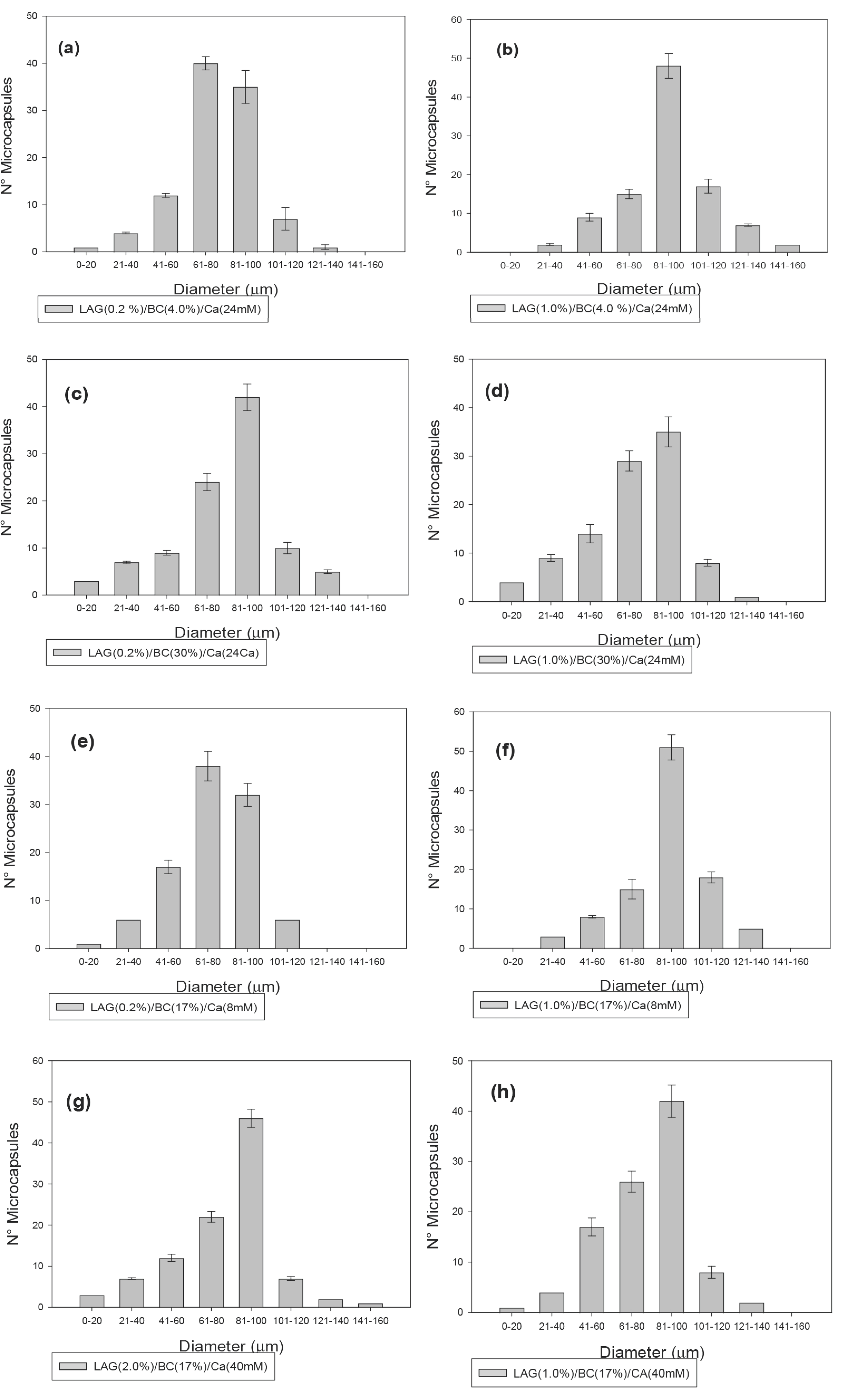
3.1. Microcapsule Size

Internal ionic gelation is a microencapsulation technique that involves forming an emulsion between two immiscible phases: a hydrophobic phase and a hydrophilic phase. Through agitation, numerous droplets are generated, which subsequently undergo gelation via mild acidification with gluconolactone. This process occurs due to the release of calcium ions (Ca2+) through an ion-exchange reaction with the calcium carbonate (CaCO3) salt used in the formulation [31].
Determining the microcapsule diameter is a critical factor in the food industry, as excessively large capsules (>1 mm) exhibit poor distribution within the food matrix, potentially altering sensory attributes. Conversely, smaller microcapsules may provide insufficient protection against adverse environmental conditions [32,33]. The microcapsule size was evaluated for all treatments, as presented in Figure 1, where a unimodal distribution pattern was observed across all samples. This behavior can be attributed to the gradual release of calcium ions from calcium carbonate, facilitated by gluconolactone hydrolysis. As calcium ions become available, they interact with the carboxyl groups of the gellan gum helix, initiating the gelation process [34,35]. This mechanism provides sufficient time for the microcapsules to develop their final size and morphology.
The microcapsule size distribution ranged from 15 to 120 µm, which is considered optimal for food applications, including both solid and liquid matrices—particularly in dairy products. A smaller diameter could compromise cell protection, whereas a larger diameter might lead to undesirable textural modifications in the final product [36]. Kim et al. [37] reported that microcapsules smaller than 80 µm are preferred in industrial applications, as they do not negatively impact the sensory properties of food [38]. Similarly, Burgain et al. [19] suggested that microcapsules below 120 µm are appropriate for liquid food applications, minimizing potential sensory alterations. Therefore, LAG, BC, and Ca can be effectively employed to produce microcapsules with an ideal size for food applications.

3.2. Optimization of the Probiotic Microencapsulation

In this study, response surface methodology (RSM) based on the Box–Behnken Design (BBD) was employed to minimize the number of experimental trials, resulting in a matrix of 15 experimental runs (Table 2). The objective was to optimize microencapsulation efficiency (%EE) and the viability of microencapsulated Lactobacillus plantarum after exposure to simulated gastric juice (SGJ) for 1 h. RSM is a crucial statistical tool that provides an efficient and cost-effective approach for analyzing the interactive effects of multiple variables on experimental outcomes [39].
Table 2 presents the BBD experimental design used to evaluate the encapsulation efficiency and viability of L. plantarum within a multicomponent system, where the independent variables included low-acyl gellan gum (LAG, X1), bacterial cellulose (BC, X2), and calcium concentration (Ca, X3). It is important to highlight that the initial counts of L. plantarum were set according to the minimum recommended levels for incorporation into food matrices. Several studies [40,41] have suggested that the intake of probiotic cells should be approximately 8 to 9 log10 CFU/g to confer beneficial health effects to consumers.
The experimental encapsulation efficiency values (%EE) ranged between 53.3% and 86.30%. These results align with those reported by Holkem et al. [42], who achieved an encapsulation efficiency of 89.71% for Bifidobacterium BB-12 microencapsulated via internal ionic gelation using alginate as the wall material. The lowest %EE (53.30%) was observed in microcapsules prepared with low LAG concentrations (0.2% w/v), low calcium levels (8 mM), and an intermediate BC concentration (17% w/v). Conversely, the highest %EE (86.30%) was obtained using intermediate concentrations of LAG (0.60% w/v), BC (17% w/v), and Ca (24 mM), suggesting that BC does not play a significant role in the microencapsulation efficiency of L. plantarum via internal ionic gelation.
Regarding the resistance of microencapsulated L. plantarum to simulated gastric juice, the highest viability percentages (90.7–92.40%) were achieved with intermediate concentrations of LAG (0.6% w/v), BC (17% w/v), and Ca (24 mM). In contrast, the lowest viability percentages (66.7–69.2%) were recorded when low concentrations of LAG (0.20% w/v) and BC (17% w/v) were used, coupled with either high (40 mM) or low (8 mM) calcium concentrations. These findings suggest that calcium saturation within the microencapsulation system significantly affects the viability of L. plantarum.
The encapsulation efficiency obtained in this study was higher than the values reported by Zou et al. [43], who microencapsulated Bifidobacterium bifidum F-35, achieving %EE values between 43% and 50% using alginate microcapsules produced through a similar microencapsulation technique. This difference could be attributed to the highly porous surface structure of alginate microcapsules, which makes them more susceptible to erosion by gastric acid and subsequent probiotic degradation [44].
The experimental results were subjected to a sequential response surface methodology (RSM) analysis, which included the following steps: (i) assessing the statistical significance of the developed model using analysis of variance (ANOVA); (ii) generating response surface plots to identify the optimal region and interactive effects of the variables; (iii) applying an optimization strategy to maximize the viability of Lactobacillus plantarum exposed to SGJ; and (iv) validating the predictive capability of the model by comparing its estimations with experimental data obtained under optimal conditions.
The second-order polynomial model (Equation (1)) was analyzed through multiple regression to achieve an optimal fit, and its statistical significance was evaluated using ANOVA. The ANOVA results for the response variables—microencapsulation efficiency (%EE) and viability—are presented in Table 3. A p-value lower than 0.05 indicates statistical significance [45], suggesting that at least one term in the regression equation is significantly associated with the response variable (%EE or viability).
Regarding %EE, the interaction terms X1·X2 and X2·X3 exhibited p-values greater than 0.05, indicating a lack of statistical significance. Consequently, these interactions were excluded from the model, leading to a reduced second-order polynomial equation (Equation (3)). Model fit was assessed using the coefficient of determination (R2), adjusted R2, and predicted R2. The obtained R2 value for %EE was 99.23%, indicating a strong correlation between the model and the experimental data [46]. This high R2 value suggests that the model effectively explains the variability in the response variable.
Moreover, the predicted R2 (97.49%) was reasonably aligned with the adjusted R2 (98.45%), as their difference was less than 0.2, further confirming the model’s reliability. The R2 value represents the proportion of data variability accounted for by the model. Adjusted R2 considers the number of factors included, making it valuable for assessing the impact of adding or removing terms, while predicted R2 provides insight into the model’s predictive accuracy [47].
Additionally, the lack-of-fit test yielded a p-value of 0.947 (>0.05), indicating that the model’s lack of fit was not statistically significant. This result confirms that the proposed regression model is suitable for predicting the %EE of Lactobacillus plantarum microencapsulated within a multicomponent system.
% E E = 84.000 + 8.000   X 1 + 1.337 X 2 + 3.838   X 3 11.737   X 1 2 4.312   X 2 2 10.112 X 3 2 + 3.200   X 1   ×   X 3 .
Regarding probiotic viability, the factors X1, X2, and X3 exhibited a significant influence (p < 0.05) on the survival of L. plantarum microencapsulated and exposed to SGJ. Similar to the results observed for %EE, the interaction terms X1·X2 and X2·X3 were not statistically significant, as their p-values exceeded 0.05. Consequently, these interactions were excluded from the polynomial equation to refine the predictive model (Equation (4)) and accurately describe the viability profile of L. plantarum under SGJ conditions. The optimized equation was subsequently used to generate response surface plots, illustrating the interactive effects of the remaining significant factors on probiotic survival.
V i a b i l i t y = 91.467 + 7.888   X 1 + 1.163 X 2 + 2.950 X 3 7.158 X 1 2 5.058   X 2 2 8.133 X 3 2 + 1.925   X 1   ×   X 3 .
The reduced polynomial equation developed to describe the viability response under SGJ conditions was evaluated using key statistical indices: R2, adjusted R2, and predicted R2. The obtained R2 value for viability was 99.64, with a predicted R2 of 98.07 and an adjusted R2 of 99.29. Additionally, the lack-of-fit test resulted in a p-value of 0.71, confirming the model’s suitability for predicting the tolerance of L. plantarum microencapsulated in a multicomponent system under SGJ conditions.
These findings suggest that the model can reliably explore the design space, effectively representing the relationship between the independent variables (LAG, BC, and Ca) and the response variables (encapsulation efficiency and probiotic viability) [48]. The strong agreement between experimental and predicted values further confirms the accuracy of the polynomial models, validating their applicability in predicting both encapsulation efficiency and probiotic survival in the proposed system.

3.3. Response Surface Plots

Based on the developed polynomial models (Equations (3) and (4)), three-dimensional (3D) response surface plots were generated to visualize the effects of the independent variables on the encapsulation efficiency (%EE) and viability of L. plantarum under SGJ conditions (Figure 2). These 3D plots provide a comprehensive statistical and visual representation, facilitating the identification of optimal experimental conditions [49]. This information is essential for optimizing both encapsulation efficiency and probiotic viability, ultimately enhancing probiotic protection.
Figure 2a illustrates the relationship between the statistically significant factors (LAG vs. Ca, p < 0.05) influencing the %EE of L. plantarum microcapsules. The plot reveals that increasing the LAG concentration up to approximately 0.60% leads to an increase in %EE, reaching around 84%. However, further increasing the LAG concentration beyond this point results in a decline in %EE to approximately 65% when LAG reaches 1%. This parabolic behavior can be attributed to the viscosity changes and gelation process of gellan gum [20]. Specifically, higher LAG concentrations increase viscosity, inducing adhesive forces that promote the formation of microcapsule droplets.
Additionally, insufficient calcium ion concentrations may fail to establish a densely crosslinked three-dimensional gel network through electrostatic interactions [50], leading to low encapsulation efficiency due to inadequate coverage of probiotic cells within the microcapsules. Conversely, at elevated calcium ion concentrations, the electrostatic interactions between gellan gum helices and the electrolyte balance of probiotic cells may be disrupted, reducing encapsulation efficiency [51]. Moreover, oversaturation of calcium-binding sites in gellan gum helices may cause osmotic stress, further decreasing %EE [52].
Figure 2b illustrates the viability of L. plantarum microcapsules exposed to SGJ for 1 h. A parabolic response pattern, similar to that observed for %EE, was identified. Specifically, low concentrations of calcium and gellan gum diminished the protective effect of the microcapsules against gastric juice. However, increasing LAG and calcium concentrations up to intermediate levels enhanced the tolerance of L. plantarum to acidic conditions.
LAG contributes to the formation of a stable three-dimensional barrier [22] that effectively restricts acid diffusion into the microcapsules. Consequently, this enhances the protection of encapsulated probiotic cells due to multiple electrostatic interactions between crosslinked polymers and strong hydrogen bonding among polysaccharide chains [53]. Additionally, ionic crosslinking occurs between the carboxyl groups of gellan gum helices and calcium ions, forming a dense and stable network that further improves microcapsule resistance [53]. This is supported by the significant interaction effect between LAG and calcium in the polynomial model. However, at elevated LAG concentrations, probiotic viability decreases, possibly due to the viscosity-related constraints previously mentioned, which may hinder the microencapsulation process.
The critical concentrations of LAG, BC, and Ca required to optimize the viability of microencapsulated Lactobacillus plantarum under SGJ conditions were determined using the ‘Response Optimiser’ function in Minitab® statistical software (version 17.00) (see Supplementary Material). According to the response surface model, the optimal LAG concentration (X1) was 0.6364% w/v, BC was 17.91% w/v, and the optimal Ca concentration (X3) was 25.13 mM, resulting in a predicted survival rate of 94.28%.
The viability of the optimized microcapsules was further assessed at different time intervals over a 1-h period. The enhanced survival of L. plantarum in SGJ could be attributed to the reduced penetration of gastric fluid into the microcapsule core, as well as the presence of negatively charged carboxylate groups, which enhanced the buffering effect against infiltrated acid. These findings suggest that this microencapsulation matrix could serve as an effective protective system for probiotics in harsh gastric conditions.

4. Conclusions

In this study, microcapsules composed of low-acyl gellan gum (LAG), bacterial cellulose (BC), and calcium ions (Ca), encapsulating Lactobacillus plantarum through internal ionic gelation, were identified as a promising alternative for delivering probiotics in food systems, particularly in solid food applications due to their appropriate microcapsule size. Both low-acyl gellan gum and calcium ions proved to be effective in enhancing encapsulation efficiency and improving the viability of L. plantarum under simulated gastric juice (SGJ) conditions.
Response surface methodology (RSM) based on the Box–Behnken design was successfully employed to optimize the concentrations of LAG and calcium. Under optimal conditions—0.63% w/v LAG, 17.91% w/v BC, and 25.12 mM Ca—the highest L. plantarum viability reached 94.28% after exposure to SGJ. These findings demonstrate that it is possible to develop multicomponent microcapsules that provide effective protection for probiotic bacteria against gastric fluids, representing a viable alternative for the food industry in designing probiotic-enriched food systems. The microcapsules developed in this study offer a promising approach for food applications, balancing enhanced microbial protection with compatibility within food matrices.

Supplementary Materials

The following supporting information can be downloaded at https://www.mdpi.com/article/10.3390/jcs9040189/s1: Figure S1: Viability optimization of microencapsulated Lactobacillus plantarum subjected to simulated gastric juice; Table S1: Information about the edible sunflower oil used to produce microcapsules.

Author Contributions

Conceptualization, R.G.-C. and R.O.-T.; data curation, R.G.-C.; formal analysis, R.G.-C.; investigation, R.G.-C.; methodology, R.G.-C., J.H.-F. and R.O.-T.; resources, J.H.-F. and R.O.-T.; software, R.G.-C. and J.H.-F.; validation, R.G.-C.; visualization, R.O.-T.; writing—original draft, R.G.-C.; writing—review and editing, J.H.-F. and R.O.-T. All authors have read and agreed to the published version of the manuscript.

Funding

This research received no external funding.

Data Availability Statement

The original contributions presented in the study are included in the article/Supplementary Materials, further inquiries can be directed to the corresponding authors.

Acknowledgments

The authors thank the Universidad de Cartagena for providing equipment and reagents to conduct this research.

Conflicts of Interest

The authors declare no conflicts of interest.

References

  1. Herdiana, Y. Functional Food in Relation to Gastroesophageal Reflux Disease (GERD). Nutrients 2023, 15, 3583. [Google Scholar] [CrossRef] [PubMed]
  2. Sgroi, F.; Sciortino, C.; Baviera-Puig, A.; Modica, F. Analyzing Consumer Trends in Functional Foods: A Cluster Analysis Approach. J. Agric. Food Res. 2024, 15, 101041. [Google Scholar] [CrossRef]
  3. Kheto, A.; Bist, Y.; Awana, A.; Kaur, S.; Kumar, Y.; Sehrawat, R. Utilization of Inulin as a Functional Ingredient in Food: Processing, Physicochemical Characteristics, Food Applications, and Future Research Directions. Food Chem. Adv. 2023, 3, 100443. [Google Scholar] [CrossRef]
  4. Martirosyan, D.M.; Singh, J. A New Definition of Functional Food by FFC: What Makes a New Definition Unique? Funct. Foods Health Dis. 2015, 5, 209. [Google Scholar] [CrossRef]
  5. Obayomi, O.V.; Olaniran, A.F.; Owa, S.O. Unveiling the Role of Functional Foods with Emphasis on Prebiotics and Probiotics in Human Health: A Review. J. Funct. Foods 2024, 119, 106337. [Google Scholar] [CrossRef]
  6. Sun, X.; Liu, H.; Duan, C.; Yan, G. Effects of Mixed Starters of Plant- and Wine-Derived L. plantarum on Hawthorn Juice Fermentation: Physicochemical Properties, Phenolic and Volatile Profiles. Food Biosci. 2023, 56, 103363. [Google Scholar] [CrossRef]
  7. FAO/OMS. Report of a Joint FAO/WHO Working Group on Drafting Guidelines for the Evaluation of Probiotics in Food; FAO/OMS: Rome, Italy, 2002. [Google Scholar]
  8. Le, B.; Yang, S.H. Efficacy of Lactobacillus plantarum in Prevention of Inflammatory Bowel Disease. Toxicol. Rep. 2018, 5, 314–317. [Google Scholar] [CrossRef]
  9. de Vries, M.C.; Vaughan, E.E.; Kleerebezem, M.; de Vos, W.M. Lactobacillus plantarum-Survival, Functional and Potential Probiotic Properties in the Human Intestinal Tract. Int. Dairy J. 2006, 16, 1018–1028. [Google Scholar] [CrossRef]
  10. Luxananil, P.; Promchai, R.; Wanasen, S.; Kamdee, S.; Thepkasikul, P.; Plengvidhya, V.; Visessanguan, W.; Valyasevi, R. Monitoring Lactobacillus plantarum BCC 9546 Starter Culture during Fermentation of Nham, a Traditional Thai Pork Sausage. Int. J. Food Microbiol. 2009, 129, 312–315. [Google Scholar] [CrossRef]
  11. Palomino, J.M.; del Arbol, J.T.; Benomar, N.; Abriouel, H.; Cañamero, M.M.; Gálvez, A.; Pulido, R.P. Application of Lactobacillus plantarum Lb9 as Starter Culture in Caper Berry Fermentation. LWT-Food Sci. Technol. 2014, 60, 788–794. [Google Scholar] [CrossRef]
  12. Ding, X.; Xu, Y.; Wang, Y.; Xie, L.; Liang, S.; Li, D.; Wang, Y.; Wang, J.; Zhan, X. Carboxymethyl Konjac Glucomannan-Chitosan Complex Nanogels Stabilized Double Emulsions Incorporated into Alginate Hydrogel Beads for the Encapsulation, Protection and Delivery of Probiotics. Carbohydr. Polym. 2022, 289, 119438. [Google Scholar] [CrossRef] [PubMed]
  13. Mojikon, F.D.; Kasimin, M.E.; Molujin, A.M.; Gansau, J.A.; Jawan, R. Probiotication of Nutritious Fruit and Vegetable Juices: An Alternative to Dairy-Based Probiotic Functional Products. Nutrients 2022, 14, 3457. [Google Scholar] [CrossRef] [PubMed]
  14. Yilmaz, M.T.; Taylan, O.; Karakas, C.Y.; Dertli, E. An Alternative Way to Encapsulate Probiotics within Electrospun Alginate Nanofibers as Monitored under Simulated Gastrointestinal Conditions and in Kefir. Carbohydr. Polym. 2020, 244, 116447. [Google Scholar] [CrossRef] [PubMed]
  15. Yao, M.; Xie, J.; Du, H.; McClements, D.J.; Xiao, H.; Li, L. Progress in Microencapsulation of Probiotics: A Review. Compr. Rev. Food Sci. Food Saf. 2020, 19, 857–874. [Google Scholar] [CrossRef]
  16. Çanga, E.M.; Dudak, F.C. Improved Digestive Stability of Probiotics Encapsulated within Poly (Vinyl Alcohol)/Cellulose Acetate Hybrid Fibers. Carbohydr. Polym. 2021, 264, 117990. [Google Scholar] [CrossRef]
  17. Ermis, E. A Review of Drying Methods for Improving the Quality of Probiotic Powders and Characterization. Dry. Technol. 2022, 40, 2199–2216. [Google Scholar] [CrossRef]
  18. Mohammadalinejhad, S.; Almonaitytė, A.; Jensen, I.-J.; Kurek, M.; Lerfall, J. Alginate Microbeads Incorporated with Anthocyanins from Purple Corn (Zea mays L.) Using Electrostatic Extrusion: Microencapsulation Optimization, Characterization, and Stability Studies. Int. J. Biol. Macromol. 2023, 246, 125684. [Google Scholar] [CrossRef]
  19. Burgain, J.; Gaiani, C.; Linder, M.; Scher, J. Encapsulation of Probiotic Living Cells: From Laboratory Scale to Industrial Applications. J. Food Eng. 2011, 104, 467–483. [Google Scholar] [CrossRef]
  20. González, R.; Ramos, G.; Cruz, A.; Salazar, A. Rheological Characterization and Activation Energy Values of Binary Mixtures of Gellan. Eur. Food Res. Technol. 2012, 234, 305–313. [Google Scholar] [CrossRef]
  21. Huang, H.; Yan, W.; Tan, S.; Zhao, Y.; Dong, H.; Liao, W.; Shi, P.; Yang, X.; He, Q. Frontier in Gellan Gum-Based Micro-Capsules Obtained by Emulsification: Core-Shell Structure, Interaction Mechanism, Intervention Strategies. Int. J. Biol. Macromol. 2024, 272, 132697. [Google Scholar] [CrossRef]
  22. Gomes, D.; Batista-Silva, J.; Sousa, A.; Passarinha, L. Progress and Opportunities in Gellan Gum-Based Materials: A Review of Preparation, Characterization and Emerging Applications. Carbohydr. Polym. 2023, 311, 120782. [Google Scholar] [CrossRef]
  23. Rajwade, J.M.; Paknikar, K.M.; Kumbhar, J.V. Applications of Bacterial Cellulose and Its Composites in Biomedicine. Appl. Microbiol. Biotechnol. 2015, 99, 2491–2511. [Google Scholar] [CrossRef] [PubMed]
  24. de Amorim, J.D.P.; de Souza, K.C.; Duarte, C.R.; da Silva Duarte, I.; de Assis Sales Ribeiro, F.; Silva, G.S.; Sarubbo, L.A. Plant and bacterial nanocellulose: Production, properties and applications in medicine, food, cosmetics, electronics, and engineering. A Review. Environ. Chem. Lett. 2020, 18, 851–869. [Google Scholar] [CrossRef]
  25. Rezaei, A.; Fathi, M.; Jafari, S.M. Nanoencapsulation of Hydrophobic and Low-Soluble Food Bioactive Compounds Within Different Nanocarriers. Food Hydrocoll. 2019, 88, 146–162. [Google Scholar] [CrossRef]
  26. Yuan, H.; Li, W.; Chen, C.; Yu, H.; Huang, J.; Tian, H. Novel Cinnamon Essential Oil-Bacterial Cellulose Microcapsules for Enhanced Preservation of Prefabricated Meat. Int. J. Biol. Macromol. 2024, 282, 136851. [Google Scholar] [CrossRef]
  27. Fasolo, D.; Pippi, B.; Meirelles, G.; Zorzi, G.; Fuentefria, A.M.; Poser, G.; Teixeira, H.F. Topical Delivery of Antifungal Brazilian Red Propolis Benzophenones-Rich Extract by Means of Cationic Lipid Nanoemulsions Optimized by Means of Box-Behnken Design. J. Drug Deliv. Sci. Technol. 2020, 56, 101573. [Google Scholar] [CrossRef]
  28. Zain, Z.M.; Abdulhameed, A.S.; Jawad, A.H.; Alothman, Z.A.; Yaseen, Z.M. A pH-Sensitive Surface of Chitosan/Sepiolite Clay/Algae Biocomposite for the Removal of Malachite Green and Remazol Brilliant Blue R Dyes: Optimization and Adsorption Mechanism Study. J. Polym. Environ. 2023, 31, 501–518. [Google Scholar] [CrossRef]
  29. González-Cuello, R.; Parada-Castro, A.L.; Ortega-Toro, R. Application of a Multi-Component Composite Edible Coating for the Preservation of Strawberry Fruit. J. Compos. Sci. 2024, 8, 515. [Google Scholar] [CrossRef]
  30. Cheow, W.; Yi, K.; Hadinoto, K. Controlled Release of Lactobacillus rhamnosus Biofilm Probiotics from Alginate-Locust Bean Gum Microcapsules. Carbohydr. Polym. 2014, 103, 587–595. [Google Scholar] [CrossRef]
  31. González, R.E.; Salazar, J.A.; Pérez, J.A. Obtaining Size-Controlled Microcapsules by Ionic Gelation with High and Low Acyl Gellans Containing Lactococcus lactis. Rev. Colomb. Biotecnol. 2013, 15, 70–80. [Google Scholar]
  32. Arepally, D.; Reddy, R.S.; Goswami, T.K.; Coorey, R. A Review on Probiotic Microencapsulation and Recent Advances of Their Application in Bakery Products. Food Bioproc. Technol. 2022, 15, 1677–1699. [Google Scholar] [CrossRef]
  33. Zarali, M.; Sadeghi, A.; Jafari, S.M.; Ebrahimi, M.; Mahoonak, A.S. Enhanced Viability and Improved In Situ Antibacterial Activity of the Probiotic LAB Microencapsulated Layer-by-Layer in Alginate Beads Coated with Nisin. Food Biosci. 2023, 53, 102593. [Google Scholar] [CrossRef]
  34. Larwood, V.; Howlin, B.; Webb, G. Solvation Effects on the Conformational Behavior of Gellan and Calcium Ion Binding to Gellan Double Helices. J. Mol. Model. 1996, 2, 175–182. [Google Scholar] [CrossRef]
  35. Tang, J.; Tung, M.; Zeng, Y. Gelling Properties of Gellan Solutions Containing Monovalent and Divalent Cations. J. Food Sci. 1997, 62, 688–712. [Google Scholar] [CrossRef]
  36. Lacroix, C.; Grattepanche, F.; Doleyres, Y.; Bergmaier, D. Immobilised Cell Technologies for the Dairy Industry. In Applications of Cell Immobilisation Biotechnology; Springer: Dordrecht, The Netherlands, 2005; pp. 295–319. [Google Scholar] [CrossRef]
  37. Kim, S.; Cho, S.; Kim, S.; Song, O.; Shin, I.; Cha, D.; Park, H. Effect of Microencapsulation on Viability and Other Characteristics in Lactobacillus acidophilus ATCC 43121. LWT-Food Sci. Technol. 2008, 41, 493–500. [Google Scholar] [CrossRef]
  38. Tyle, P. Effect of Size, Shape and Hardness of Particles in Suspension on Oral Texture and Palatability. Acta Psychol. 1993, 84, 111–118. [Google Scholar] [CrossRef]
  39. Fu, J.F.; Zhao, Y.Q.; Xue, X.D.; Li, W.C.; Babatunde, A.O. Multivariate-Parameter Optimization of Acid Blue-7 Wastewater Treatment by Ti/TiO2 Photoelectrocatalysis via Box-Behnken Design. Desalination 2009, 243, 42–51. [Google Scholar] [CrossRef]
  40. Aureli, P.; Capurso, L.; Castellazzi, A.; Clerici, M.; Giovannini, M.; Morelli, L.; Poli, A.; Pregliasco, F.; Salvini, F.; Zuccotti, G. Probiotics and Health: An Evidence-Based Review. Pharmacol. Res. 2011, 63, 366–376. [Google Scholar] [CrossRef]
  41. Salminen, S.; Kenifel, W.; Ouwehand, A. Probiotics, Applications in Dairy Products. In Encyclopedia of Dairy Sciences; Fuquay, J.W., Fox, P.F., McSweeney, P.L.H., Eds.; Academic Press: San Diego, CA, USA, 2011. [Google Scholar]
  42. Holkem, T.; Raddatz, G.; Nunes, L.; Cichoski, A.; Jacob, E.; Grosso, R.; Ragagnin, C. Development and Characterization of Alginate Microcapsules Containing Bifidobacterium BB-12 Produced by Emulsification/Internal Gelation Followed by Freeze Drying. LWT-Food Sci. Technol. 2016, 71, 302–308. [Google Scholar] [CrossRef]
  43. Zou, Q.; Zhao, J.; Liu, X.; Tian, F.; Zhang, H.; Zhang, H.; Zhang, H.; Chen, W. Microencapsulation of Bifidobacterium bifidum F-35 in Reinforced Alginate Microspheres Prepared by Emulsification/Internal Gelation. Int. J. Food Sci. Technol. 2011, 46, 1672–1678. [Google Scholar] [CrossRef]
  44. Wang, K.; Ni, J.; Li, H.; Tian, X.; Tan, M.; Su, W. Survivability of Probiotics Encapsulated in Kelp Nanocellulose/Alginate Microcapsules on Microfluidic Device. Food Res. Int. 2022, 160, 111723. [Google Scholar] [CrossRef] [PubMed]
  45. Kumar, A.; Prasad, B.; Mishra, I.M. Process Parametric Study for Ethene Carboxylic Acid Removal onto Powder Activated Carbon Using Box-Behnken Design. Chem. Eng. Technol. 2007, 30, 932–937. [Google Scholar] [CrossRef]
  46. Körbahti, B.K. Response Surface Optimization of Electrochemical Treatment of Textile Dye Wastewater. J. Hazard. Mater. 2007, 145, 277–286. [Google Scholar] [CrossRef]
  47. Montgomery, D.C. Introduction to Statistical Quality Control, 6th ed.; Wiley: New York, NY, USA, 2010. [Google Scholar]
  48. Ravilumar, K.; Ramalingam, S.; Krishnan, S.; Balu, K. Application of Response Surface Methodology to Optimize the Process Variables for Reactive Red and Acid Brown Dye Removal Using a Novel Adsorbent. Dye. Pigment. 2006, 70, 18–26. [Google Scholar] [CrossRef]
  49. Henseler, J.; Sarstedt, M. Goodness-of-Fit Indices for Partial Least Squares Path Modeling. Comput. Stat. 2013, 28, 565–580. [Google Scholar] [CrossRef]
  50. Li, J.; Wu, Y.; He, J.; Huang, Y. A New Insight to the Effect of Calcium Concentration on Gelation Process and Physical Properties of Alginate Films. J. Mater. Sci. 2016, 51, 5791–5801. [Google Scholar] [CrossRef]
  51. Thinkohkaew, K.; Jonjaroen, V.; Niamsiri, N.; Panya, A.; Suppavorasatit, I.; Potiyaraj, P. Microencapsulation of Probiotics in Chitosan-Coated Alginate/Gellan Gum: Optimization for Viability and Stability Enhancement. Food Hydrocoll. 2024, 151, 109788. [Google Scholar] [CrossRef]
  52. Lai, P.Y.; How, Y.H.; Pui, L.P. Microencapsulation of Bifidobacterium lactis Bi-07 with Galactooligosaccharides Using Co-Extrusion Technique. J. Microbiol. Biotechnol. Food Sci. 2022, 11, e2416. [Google Scholar] [CrossRef]
  53. Dong, K.; Xu, K.; Wei, N.; Fang, Y.; Qin, Z. Three-Dimensional Porous Sodium Alginate/Gellan Gum Environmentally Friendly Aerogel: Preparation, Characterization, Adsorption, and Kinetics Studies. Chem. Eng. Res. Des. 2022, 179, 227–236. [Google Scholar] [CrossRef]
Figure 1. Average diameter distribution of the microcapsules obtained with low-acyl gellan, bacterial cellulose, and calcium.
Jcs 09 00189 g001aJcs 09 00189 g001b
Figure 2. Three-dimensional response surfaces for the effects of the LAG and Ca concentrations on the encapsulation efficiency and viability of L. plantarum. %EE (a) and viability to SGF (b).
Jcs 09 00189 g002aJcs 09 00189 g002b
Table 1. Coded levels and experimental values of independent variables.
VariableFactor CodeRange and Levels of Factors
−11
LAG (w/v)X10.201
BC (w/v)X2430
Ca (mM)X3840
Table 2. Comparison between the experimental response and RSM predicted response.
RunX1: LAG (w/v)X2: BC
(w/v)
X3: Ca
(mM)
%EEViability (%)
Experimental DataRSM
Predicted
Experimental DataRSM
Predicted
10.2042457.90 ± 1.3457.8170.80 ± 1.4170.55
21.0042475.70 ± 0.9975.4185.20 ± 2.1785.62
30.20302461.80 ± 0.7462.0872.60 ± 0.9472.17
41.00302476.40 ± 0.2876.4888.40 ± 1.7488.65
50.2017853.30 ± 2.2553.5166.70 ± 1.7367.26
61.0017862.70 ± 1.7363.1179.30 ± 2.1179.18
70.20174055.20 ± 0.8754.7869.20 ± 1.0869.31
81.00174077.40 ± 2.0477.1889.50 ± 1.9488.93
90.604864.70 ± 1.4564.5774.80 ± 2.0474.48
100.6030867.40 ± 1.5566.9076.30 ± 1.6076.16
110.6044071.40 ± 1.7371.9079.60 ± 1.7179.73
120.60304074.80 ± 1.3974.9282.40 ± 1.9282.71
130.60172483.20 ± 2.3584.0090.70 ± 2.0791.46
140.60172482.50 ± 1.8884.0092.40 ± 2.3391.46
150.60172486.30 ± 2.0684.0091.30 ± 3.0491.46
Table 3. ANOVA and regression coefficient δ of the predicted reduced second-order polynomials model for viability and EE.
Factors%EEViability
Coefficient δp-ValueCoefficient δp-Value
Intercept84.000.0091.460.00
X1 (LAG)8.000.007.880.00
X2 (BC)1.330.031.160.00
X3 (Ca)3.830.002.950.00
Interaction
X1 X2−0.800.290.350.39
X1 X33.200.001.920.00
X2 X30.170.800.320.43
Quadratic
X 1 2 −11.730.00−7.150.00
X 2 2 −4.310.00−5.050.00
X 3 2 −10.110.00−8.130.00
p-value (Model) 0.00
p-value
(Lack of fit)
0.94 0.71
R299.23% 99.64%
Adjusted R298.45% 99.29%
Predicted R297.49% 98.07%
Disclaimer/Publisher’s Note: The statements, opinions and data contained in all publications are solely those of the individual author(s) and contributor(s) and not of MDPI and/or the editor(s). MDPI and/or the editor(s) disclaim responsibility for any injury to people or property resulting from any ideas, methods, instructions or products referred to in the content.

