Article

Structural Insights: In Situ Synthesis of Titanium Carbide by Magnesiothermic Method Using Carbon Nanotubes and Turbostratic Carbon as Carbon Sources

by
María Luisa Camacho-Ríos
1,2,*,
Guillermo Herrera-Pérez
3,
Luis Carlos Rodriguez-Pacheco
1,
Mariana Lizbeth Luján-Aguilar
1,
Antonio Ramírez-de la Cruz
4,
Nathaly Withney Aguilar-Cisneros
4,
Marco Antonio Ruiz Esparza-Rodriguez
1,
Daniel Lardizabal-Gutierrez
1,
Raúl Pérez-Bustamante
3 and
José Antonio Betancourt-Cantera
2
1
Centro de Investigación en Materiales Avanzados (CIMAV), Miguel de Cervantes 120, Chihuahua 31109, Chihuahua, Mexico
2
SECIHTI-Corporación Mexicana de Investigación en Materiales S.A. de C.V. (COMIMSA), Ciencia y Tecnología 790, Saltillo 400, Saltillo 25290, Coahuila, Mexico
3
SECIHTI-Centro de Investigación en Materiales Avanzados (CIMAV), Miguel de Cervantes 120, Chihuahua 31109, Chihuahua, Mexico
4
Faculty of Chemical Sciences, Autonomous University of Chihuahua, Circuit 1, New University Campus, Chihuahua 31125, Chihuahua, Mexico
*
Author to whom correspondence should be addressed.
J. Compos. Sci. 2025, 9(4), 171; https://doi.org/10.3390/jcs9040171
Submission received: 21 February 2025 / Revised: 28 March 2025 / Accepted: 29 March 2025 / Published: 31 March 2025
(This article belongs to the Section Carbon Composites)

Abstract

:
The current work presents the influence of the magnesiothermic synthesis method on titanium carbide (TiC). In this method, powdered titanium precursors and two carbon sources—turbostratic carbon and carbon nanotubes—were employed in proportions of 10 wt.% and 20 wt.%. The refinement of the X-ray diffraction (XRD) patterns using the Rietveld method for TiC suggests suggested coexistence of two phases, cubic with Fm-3m space group and hexagonal with P3121 space group. In particular, for the sample with 20 wt.% of carbon sources, the XRD refinement revealed that the cubic phase accounted for 94% of the composition, in contrast to a secondary hexagonal phase, Ti6C3.75, which comprised 6%. The influence of carbon on the morphology (particle size and shape) and crystallite size was monitored through bright-field transmission electron microscopy (BF-TEM) imaging and XRD. In samples containing 20 wt.% carbon, a homogeneous morphology in both size (around 11 microns) and shape was observed, along with a reduction in crystallite size (from 22.7 to 17.8 nm). Raman band analysis further revealed vibrational modes indicating that carbon induced disorder in the TiC structure. The magnesiothermic synthesis method developed in this work offers a low-cost approach of interest in the aerospace and automotive industries. Additionally, the study provides significant insights for particles used as additives or reinforcing agents to enhance the mechanical properties of metal matrix composites (MMCs).

1. Introduction

Titanium carbide (TiC) is characterized as one of the metal carbides with high hardness (28–35 GPa), a high melting point (3067–3340 °C), excellent abrasion resistance, superior chemical stability, and notable thermal conductivity (40 W/mK) and electrical conductivity (1.3 × 10⁶–1.47 × 10⁴ S/cm) [1]. Also, it is chemically inert, meaning that it does not react with most chemicals desirable for resistance applications in corrosion.
TiC is commonly employed as a reinforcement in other materials such as aluminum, titanium, and their alloys [2,3]. To produce metal matrix composites (MMCs) reinforced with TiC, it is important to consider various variables such as morphology, weight fraction, homogeneous distributions, and particle size of the reinforcement. The following studies allow us to discuss the benefits of TiC in composite preparation. Zemtsova et al. (2024) [4] employed the mold injection method to prepare their composites by dispersing TiC nanoparticles within an aluminum matrix. The study demonstrated that the use of reinforcement significantly improved mechanical properties (1.5 times higher than base Al) by increasing strength without compromising plasticity. Furthermore, it was confirmed that the material did not exhibit brittleness, even with high reinforcement content. Mikheev et al. (2023) [5] studied an aluminum matrix reinforced with a hybrid of TiC and an intermetallic compound Al3TiC. They employed casting and mechanical alloying methods. The study revealed that using a hybrid reinforcement enhanced wear properties compared to using a single type of reinforcement because a hybrid allows better load distribution during stress. Dwivedi et al. (2024) [6] synthesized and purified TiC particles through vacuum carbothermal reduction. They achieved a purity of 99.40% and particle sizes between 50 to 80 nm. They observed that incorporating 5% nano-TiC into aluminum A356 composites improved tensile strength by 19.41% and hardness by 50.81%.
Controlling variables is crucial to optimize composite performance. Another important aspect is the selection of the synthesis method. This depends on factors such as the desired purity of the material, specific required properties, and intended final applications. Each technique presents advantages and challenges that must be considered according to the production objectives.
In recent years, various synthesis methods have been explored for the production of TiC, including carbothermal reduction (with mineralizer), sol-gel, electrospinning, self-propagating high-temperature synthesis (SHS), mechanical milling, and chemical vapor deposition, gas-phase laser induced reactions, and high-energy mechano-chemical technique [7,8,9]. Because its production requires elevated temperatures—which entail high costs, long processing times, expensive precursors, and pretreatment steps—the industry has been seeking alternative approaches. One technique that has garnered significant interest is magnesiothermy [10,11], a pyrometallurgical process similar to the Goldschmidt method [12] exhibiting significant advantages.
The magnesiothermic reduction process is a traditional and effective self-propagating high-temperature synthesis (SHS) technique for producing ceramic powders [13]. The SHS method is highly efficient and energy-saving for the synthesis of non-stoichiometric titanium and titanium–niobium carbides and carbohydrides, which may be of commercial and industrial interest [14].
This synthesis is characterized by a negative Gibbs free energy [12], as it involves an exothermic reduction of various metal oxides using powdered magnesium. The reaction occurs at high temperatures, exceeding 2500 °C. During the process, the metal is displaced from oxygen (reduction), forming molten metal, while magnesium is oxidized to magnesium oxide (MgO), which is obtained as a whitish solid.
A method derived from magnesiothermy is the so-called dry ice method [15,16]. In this process, instead of using metals, carbon dioxide (CO2) is utilized. By burning magnesium within a cavity of dry ice (solid CO2) used as a carbon precursor, a reaction is initiated that results in the formation of magnesium oxide and carbon, as shown in Equation (1) [17]. The product obtained is known as turbostratic carbon. This form of carbon possesses unique characteristics, as it consists of only a few layers of graphene, similar to exfoliated carbon, which holds promising potential for applications in catalysis.
2Mg(s) + CO2(g) = 2MgO(s) + C(s)
The current work proposes a modification of the dry ice method by employing gaseous CO₂ within a steel reactor. In our approach, the generated heat is harnessed to synthesize titanium carbide (TiC), with the concomitant formation of reaction by-products—magnesium oxide (MgO) and turbostratic carbon. Another method for obtaining turbostratic carbon via magnesiothermy involves using CO2 gas and metallic magnesium (Mg) as precursors. The objective is to trap the maximum amount of gas. To achieve this, two pellets composed of compressed magnesium shavings are fabricated and placed inside a reactor, through which a flow of CO2 gas is circulated. An electric arc is then used to induce a spark capable of igniting the Mg. The CO2 decomposes into CO and O2, with the CO subsequently converting into turbostratic carbon, while, simultaneously, the Mg reacts with the O2 to form MgO. Upon completion of the reaction, the white layer covering the pellets—comprising MgO and residual Mg—is removed; thereafter, a process of leaching and filtration yields the turbostratic carbon.
The motivation behind this work was to synthesize in situ TiC composites via a novel magnesiothermic route. In the preparation, titanium powders possessing a hexagonal crystalline structure were employed along with two carbon sources: turbostratic carbon and carbon nanotubes (CNTs). This study proposes a variant of the magnesiothermic method that utilizes gaseous CO2. By maintaining a continuous gas flow, a controlled reducing atmosphere was established to prevent the exothermic reaction from becoming difficult to manage. The TiC composites were structurally characterized by X-ray diffraction (XRD), while the indexing of the selected area electron diffraction (SAED) patterns corroborated the material phase identified in the XRD analysis. The microstructural characteristics (average size, apparent shape, and dispersion) were examined using scanning electron microscopy (SEM) and bright-field transmission electron microscopy (BF-TEM). Chemical analysis was carried out through elemental mapping via energy-dispersive X-ray spectroscopy (EDX), and the structural study was further complemented by Raman vibrational mode analysis for TiC.

2. Materials and Methods

2.1. Materials

Combinations were prepared using titanium powders from Alfa Aesar (Ti, 99.5%) and two different carbon sources (CS)—turbostratic carbon and carbon nanotubes (CNTs)synthesized according to the literature [18,19,20]—as well as hydrochloric acid. Table 1 presents the nomenclature for the combinations of titanium and the carbon sources employed at different weight percentages (20 and 10 wt.%) of the constituent elements [21,22]. Samples weighing one gram were prepared, with a total of five grams produced per sample.

2.2. Experimental Procedure

Pellets of the two precursors were synthesized using multi-walled carbon nanotubes and turbostratic carbon. These were then mixed with metallic titanium in an agate mortar. Using a 12 mm die, the mixture was pressed in an MSI-brand press under a load of 500 kg/cm2. Figure 1a,b illustrates how the resulting pellets are positioned between two metallic magnesium wafers, which were fabricated using a die with a 2.5 cm diameter and 1 cm thickness by applying minimal manual pressure (20 to 40 psi). Once the sandwich—comprising a wafer–pellet–wafer assembly—was formed, it was introduced into the reactor with a continuous flow of carbon dioxide (Praxair, 99.99% purity) at a rate of 500 cm3/min.
Magnesium was ignited using a graphite electrode with alternating current, employing a commercial arc welding unit (Anirona Arc-225 Inverter Welder, Mexico). As soon as the reaction commenced, the reactor was sealed semi-hermetically with a steel lid, allowing the generated gases to vent while restricting the ingress of air. This process lasted approximately 5 min, from ignition until the complete consumption of the metallic magnesium and its subsequent cooling.
As a result of the reduction reaction, the metallic wafers transformed into turbostratic carbon coated with a whitish layer of magnesium oxide. This oxide predominantly migrated to the exterior of the carbon. From the center, the pellet—within which TiC had already formed—was extracted; it was surrounded by an external layer of turbostratic carbon that was slightly denser.
Figure 2 schematically illustrates the leaching and washing process for obtaining TiC powders. Using a spatula, the pellets were collected, the whitish layer was removed, and they were ground for 25 min to homogenize the powder size, thereby facilitating improved processing. Subsequently, the powders were washed with hydrochloric acid (J. T. Baker, 37% purity) diluted to 20% with distilled water. They were then heated at 80 °C for 2 h with the acid to remove any residual MgO. Finally, the mixture was filtered and dried at 100 °C for 1 h, resulting in TiC with minimal impurities.

2.3. Characterization

X-ray diffraction was used to determine the phases present in all synthesized materials and estimate the average crystallite size of the compounds. Diffraction patterns were collected using a Panalytical X’Pert PRO diffractometer in the Bragg–Brentano configuration with a Cu Kα radiation source (λ = 1.54108 Å). The conditions for acquiring the diffractograms were as follows: an acceleration voltage of 40 kV, a beam current of 30 mA, a 2θ range of 30–90°, a step size of 0.017°, and a step time of 18 min.
Phase indexing was performed using the International Centre for Diffraction Data Powder Diffraction File (ICDD PDF) 2010 database with the X’Pert HighScore Plus 5.1a software [23]. XRD patterns were refined through the Rietveld method [24] using the FullProf Suite software, version 2018 [25]. A pseudo-Voigt function was applied to model the diffraction patterns and determine lattice parameters. The refinement strategy followed McCusker’s recommendations [26]. The Thompson–Cox–Hastings pseudo-Voigt function [27], combined with spherical harmonics and anisotropy parameters, allowed for the determination of the average crystallite size. This refinement strategy has been previously reported by Uribe et al. [28].
The two-dimensional and three-dimensional visualization of the apparent anisotropic crystallite shape was carried out using the GFourier program, version 04.06 [29]. Raman spectroscopy was employed to identify the carbon allotrope types and analyze their structural features within the graphitic lattice. The Horiba LabRam HR Vis-633 spectrometer with a He-Ne laser source operating at 632.58 nm was used for measurements. The spectral scanning range (cm⁻1) was set to 200–900 cm−1. Lorentzian functions were applied for Raman band fitting using Fityk software, version 1.3.1 [30].
To evaluate the morphology and chemical composition of the samples, a Hitachi scanning electron microscope was used, model SU3500 (Hitachi High-Technologies Corporation, Tokyo, Japan) operating at a voltage of 10 kV. The morphological features of the synthesized particles, such as their average size, shape, and distribution, were examined through bright-field micrographs in scanning mode obtained using a transmission electron microscope (TEM) model JEOL JEM2200FS (Japan Electron Optics Laboratory Co., Ltd., Tokyo, Japan). Sample preparation for TEM involved dispersing the nanoparticles in isopropanol and subjecting the mixture to sonication for 1 h. A drop of the suspension was deposited onto a Lacey carbon film on a 300 Cu mesh grid. Selected Area Electron Diffraction (SAED) patterns were analyzed to verify the phases present in the materials. Phase indexing was performed using CrysTbox software, version 1.10 [31] through its graphical user GUI Rings interface, employing the crystallographic information file (CIF) generated during the refinement of the X-ray diffraction patterns with the FullProf Suite’s graphical editor (PCR interface).

3. Results and Discussion

3.1. Scanning Electron Microscopy (SEM) Analysis

3.1.1. Initial Materials

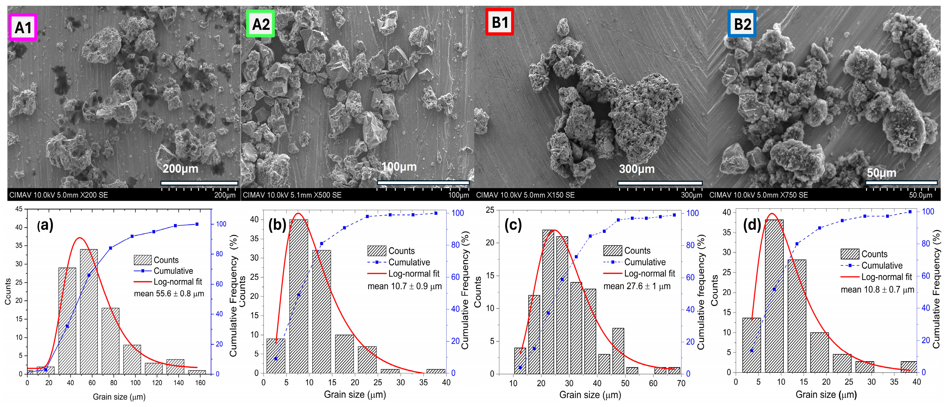
To monitor the morphological characteristics of the grains (average size, shape, and distribution), SEM micrographs were collected. In this analysis, a count of 200 grains was conducted to determine the average grain size. For each case, a graph of the count was included using the ImageJ software, version 1.50i [32], along with a histogram and its corresponding fit based on the logarithmic-normal function in combination with the iterative Levenberg-Marquardt method [33,34]. The graph visualization was performed using the Origin software, version 2018 [35].
Figure 3a shows a micrograph of the initial materials, where the grains were clustered in flake-like shapes with well-defined faces. The distribution of these grains was homogeneous. Figure 3b presents the result of the grain count along with the histogram fit. The Ti grains had an average size of 10.9 ± 0.3 µm.

3.1.2. Sources of Carbon

The following section presents the microstructural analysis from the SEM micrographs (panels a and c) in Figure 4, conducted for the two carbon sources: turbostratic carbon and CNTs. Figure 4a shows the clean turbostratic carbon particles after leaching. The micrograph displays a morphology of disordered plate clusters with a non-uniform distribution. In Figure 4b, the average grain size of 9.25 ± 0.49 µm is shown.
Figure 4c presents the micrograph containing CNTs obtained through the spray pyrolysis method. In this micrograph, the CNTs exhibited a fiber morphology resembling spaghetti. Figure 4d shows an average grain diameter of 62.52 ± 2.9 nm. Carbon-based materials generally exhibit excellent chemical stability and a large surface area, as seen in graphene and carbon nanotubes. This characteristic is crucial for their application in supercapacitors, where surface area is directly related to their energy storage capacity [36].

3.1.3. Morphology of the Composites

The morphology characteristics, such as the size, shape, and distribution of TiC powder composites obtained through the magnesiothermic method, were analyzed by SEM. This reaction belongs to the category of metallothermic reactions, involving the use of metallic magnesium, which possesses high reactivity. This characteristic enables it to break strong bonds, such as Si-O in SiO2 and C-O in graphene oxide [37,38]. Additionally, a spontaneous reaction occurs between Mg and gaseous CO2, allowing the formation of particles with various nanostructures and morphologies using CO2 as a precursor [39].
To synthesize the four samples, two carbon sources were used, as previously mentioned. Figure 5 presents SEM images forming a comparative panel illustrating the effect of the two carbon sources on grain formation, distribution, and size during the synthesis of TiC.
In the upper section of the panel, samples A1 and B1, containing 10 wt.% of the carbon sources (turbostratic carbon and CNTs), are displayed. The A1 sample showed a microstructure dominated by TiC particles with sharp-edged flat plate shapes, exhibiting a wide grain size distribution. Sample A1 exhibited a mean grain size of 56 microns compared to B1, which showed multiple plate formations with a mean grain size of 30 microns stacked in clusters. More than 100 grains were measured for each sample; the histogram analysis and the corresponding cumulative section size distribution plot are shown in panels (a–d) of Figure 5. Samples A2 and B2 contained 20 wt.% of the carbon source. Sample A2 exhibited clusters of small particles with a narrow grain size distribution and a mean grain size of 11 microns. In sample B2, more compact clusters were observed, formed by small, layered particles with a mean grain size of 11 microns, indicative of nucleation and rapid growth during the exothermic reaction. Our SEM microstructural results suggest that the increase in the wt.% of carbon source promoted a decrease in the grain size distribution width. According to Zhingang Fang et al. [40], the A2 and B2 samples showed a decrease in grain size distribution width, which was associated with a decrease in the rate of Ostwald ripening.

3.2. X-Ray Diffraction (XRD) Analysis

Figure 6 shows the diffraction patterns of powders obtained through the magnesiothermic method. All samples exhibited a Khamrabaevite-type structure for titanium carbide (TiC). The XRD pattern was indexed using the Inorganic Crystal Structure Database ICSD 01-089-3828 crystallographic card, associated with a cubic phase and space group Fm-3m (225).
This figure also includes the diffraction pattern of TiC to enable a comparison between the diffraction peaks of the samples. The characteristic XRD peaks (111), (200), (220), (311), and (222) are presented. Samples labeled A1 and B1, containing 10 wt.% of turbostratic carbon and CNTs, respectively, exhibited a higher intensity at the (200) peak compared to the (111) peak.
In samples A2 and B2, which contained 20 wt.% of the carbon sources, the intensities of both peaks were very similar. This behavior is attributed to the C/Ti ratio. According to Taehyung et al. [41], the structure of the (111) plane consists solely of Ti atoms, while the (222) plane is formed by a carbon layer between two (111) planes. Carbon acts as a barrier to interference during diffraction.
When carbon vacancies increased, this barrier weakened, enhancing the intensity of the (111) peak. The (200) plane, which lacked an intermediate layer, was formed by both Ti and C atoms. Therefore, carbon non-stoichiometry reduced diffraction to a certain extent, resulting in a gradual decrease in the diffraction intensity of the (200) peak.
The average crystallite size was determined using the spherical harmonics method implemented in the FullProf Suite software. Samples A1 and B1 showed average sizes of 24.4 and 24.5 nm, respectively. For samples A2 and B2, the sizes were 22.7 and 17.8 nm. It has been observed that the size of the carbon source used influences the size of the synthesized TiC. Gao et al. [21] synthesized TiC particles and found that the particle size decreased gradually. In their study, they used carbon black, CNTs, and a mixture of 50 wt.% pure carbon black and 50 wt.% CNTs as carbon sources. The particle sizes of TiC were 150 nm for carbon black, 96 nm for the mixture of both sources, and 67 nm when CNTs were used.
CNTs have specific properties, such as a reduced diameter from 10 nm to 20 nm, a length ranging from 20 nm to 100 µm, and a large surface area. No residual MgO or Mg was identified in the samples, which is evidence that these were removed after the washing process.

Rietveld Refinement Method

To determine the lattice parameters in samples A2 and B2, the XRD patterns were refined using the Rietveld method, implemented in the FullProf suite software. Figure 7a shows the A2 sample, and Figure 7b shows the B2 sample. Both figures present a comparison between the experimental XRD pattern (black line with symbol, Yobs) and the calculated diffraction pattern (solid red line, Ycalc).
In the figure panel, the residual line (solid black line, Yobs-Ycalc) and the Bragg positions (vertical lines) are also displayed. The reference pattern for the cubic structure with Fm-3m space group No. 225 having Z = 4 similar to NaCl (ICSD 01-089-3828) matches the identification of the characteristic peaks (111), (200), (220), (311), and (222). The presence of a second hexagonal phase with P3121 space group No. 152 (ICSD 01-079-0971) was associated with the peaks (006), (102), (104), (014), (108), (110), (018), (022), and (024).
Table 2 summarizes the refined parameters for samples A2 and B2. The XRD patterns of the samples showed the presence of two main phases: the cubic TiC phase and a second hexagonal phase, Ti6C3.75, both of which were considered in the refinement. The TiC phase comprised 93.85% and 91.17%, respectively, while the second hexagonal Ti6C3.75 phase was present at 6.15% and 8.83%.
In this study, it has been observed that the magnetothermic reaction is a vigorous combustion reaction that favors the formation of TiC, which can be found in a wide range of stoichiometries, as described by V. N. Lipatnikov et al. [42]. TiCy, with a basal B1 (NaCl)-type structure, belongs to a group of compounds that include cubic and hexagonal carbides, nitrides, and MXy and M2Xy oxides (M is a transition metal from group IV or V, and X can be C, O, or N). Depending on the composition, synthesis, and heat treatment conditions, TiCy can exist in a disordered state with thermodynamic equilibrium at temperatures higher than 1100 K, while it can exist in an ordered state at temperatures below 1000 K [43]. Experimental studies of TiCy carbides with compositions in the range of 0.5 ≤ y < 0.7 have revealed the existence of phases such as cubic Ti2C (Fd3m) [44,45], the trigonal phase (space group R-3 m or P3121), the orthorhombic Ti3C2 phase (space group C2221) [46,47], and the hexagonal Ti6C5 phase [48,49]. Tashmetov et al. [50] found that the carbide with a composition of TiC0.62 could result in a trigonal phase (space group P3121) of Ti2C with a superstructure (Ti6C3−x). They also reported that Ti2C underwent a phase transformation as follows: disordered phase (Fm-3m) at 1070 K ⇔ cubic phase (Fd3m) at 1050 K ⇔ trigonal phase (space group R-3 m or P3121). Later, de Novion et al. [51] concluded that the cubic Ti2C phase with a composition of y ≥ 0.58 was a metastable phase, with a transition temperature (Ttr) approximately 10 K lower than the trigonal phase.
The presence of a second phase, Ti6C3.75, has been reported by Chenglong Ma et al. [52]. These authors prepared TiC/Ti-Ni composites using the laser selective melting (LSM) process and reported the in situ formation of the Ti6C3.75 phase. In their study, they observed dendrites of this phase with two variants in their formation mechanism: (1) growth along the edges of partially melted TiC particles and (2) dissolution/precipitation of fine, fully melted TiC particles. When the marginal part of the TiC melted, the diffusion of carbon atoms in the melt increased. During solidification, TiC particles acted as a carbon source, enabling the epitaxial growth of Ti6C3.75 dendrites. Their XRD patterns showed the characteristic peaks of TiC (002) and Ti6C3.75 (014).
Xinyu Shen et al. [53] synthesized a coating layer on a Ti alloy by mixing Ti, C, B, and Ni powders. These authors used a combined method of self-propagating high-temperature synthesis (SHS) and hot-pressing sintering (HPS) at a temperature of 1373 K. The authors reported that the Ti-C reaction during the HPS process favored the formation of the Ti6C3.75 phase before the TiC phase. In the Vickers hardness tests performed on the coating surface, they obtained values ranging from 1300 to 1800 HV, demonstrating that the high hardness of the coating was dominated by the formation of Ti6C3.75 and TiB phases.

3.3. Bright-Field Transmission Electron Microscopy (BF-TEM) Micrographs

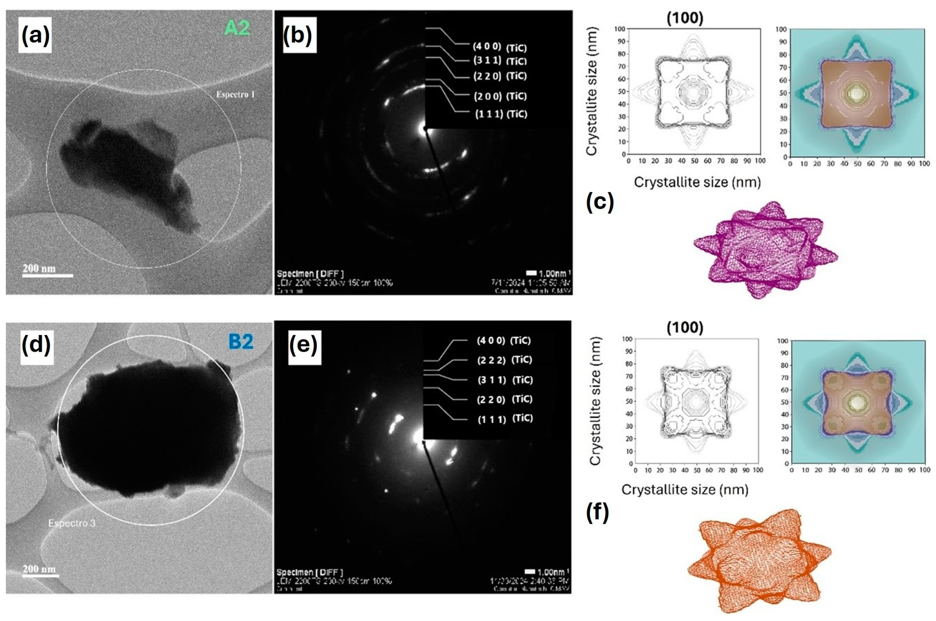
The microstructural characteristics of TiC, such as particle shape and size, were monitored in the bright-field transmission electron microscopy (BF-TEM) in scanning mode micrographs [54,55]. Selected area electron diffraction (SAED) allowed for the monitoring of the structure through the indexing of the Debye rings.
Figure 8a is a BF-TEM image corresponding to sample A2, which contained 20 wt.% of turbostratic carbon as the carbon source. The particles displayed a morphology of irregular faceted polyhedra. These particles exhibited aggregation, forming clusters with irregular shapes. This was due to the inherent morphology of the turbostratic carbon, which consisted of layers stacked one over the other in a disordered manner. Figure 8b shows the indexing of the Debye rings using the CrysTBoX program for sample A2, where the planes (111), (200), (220), (311), and (400) were identified. These results coincide with the analysis obtained from the XRD patterns. This indexing suggests that sample A2 predominantly contained the TiC cubic phase.
The determination of the average crystal shape was carried out using the FullProf suite program with the spherical harmonic method combined with the anisotropic criterion for the crystal. The apparent crystal shape visualization was performed using the GFourier program. Figure 8c contains two visualizations of the (100) plane, indicating the nanometric size of the crystal. Additionally, a three-dimensional representation of the crystal is included. The crystallite shape in the model had a cubic structure at its center, with layers growing on the faces that stack in the shape of cones with sharp tips. A similar analysis was performed for sample B2, which contained 20% CNTs. Figure 8d shows a BF-TEM micrograph of a particle agglomerate for sample B2. The agglomerate exhibited a quasi-cubic particle shape. Figure 8e presents the SAED with the indexing of the rings, identifying the cubic TiC phase. Figure 8f contains a simulation of the morphology, where it was observed to have a spherical center, and cones with more rounded tips also formed on its faces. This morphology is attributed to the carbon nanotubes; the TiC particles had a stable and defined shape, and the carbon nanotubes acted as templates or molds that help stabilize the particles [56].
The growth and formation mechanisms play a crucial role in determining the final morphology. The Wulff theorem is a tool used to predict the morphology of nanoparticles. It describes how the equilibrium morphology is determined by the minimization of surface energy, considering the surface energy of each face. For face-centered cubic crystals, the most representative indices are (111) and (100). Generally, the Wulff theorem can guide us in predicting the equilibrium morphology of a crystal [57,58]. It is known that compact planes are characteristic of high densities and large interplanar spaces, presenting low surface energy. Planes with low surface energy are more stable and eventually become the exposed crystalline planes. As particles grow, they merge into a larger one (Ostwald ripening) or form separate crystals with boundaries and multiple interfaces [59,60].

3.4. Raman Spectroscopy

Figure 9a–d shows the Raman spectra used to assess the effect of the magnesiothermic synthesis and the influence of the carbon sources on the formation of TiC in the active Raman modes. The high-purity titanium (Ti) starting powder, like most metals, did not produce a Raman spectrum, indicating that Ti does not exhibit active vibrational modes [61]. According to Amer et al. [62], stoichiometric TiC has no Raman active vibrational modes. However, Klein et al. [63] suggested that Raman scattering in TiC is due to disorder induced by carbon.
Comparing the region from 200 cm−1 to 800 cm−1 depicted in Figure 9 with previous reports, Raman spectra showed two small signals centered at 250 and 330 cm−1. In addition, two wide signals centered were observed around 410 and 610 cm−1, in agreement with Klein et al. [63], which suggests the disorder induced Raman signals. To identify the position of the TiCx Raman bands, Lorentzian fitting was applied, which is associated with the phonon lifetimes. The Fityk program was used to fit these bands, and the maximum intensity of each of these modes is marked with dotted vertical lines on the Raman spectra to monitor their evolution. Table 3 contains the results of the analysis with the vibration modes and the position of the Raman bands. It was observed that samples A2 and B2 showed more defined signals corresponding to the C-Ti bonds, indicating a higher proportion of this compound compared to samples A1 and B1.
Kaipoldayev et al. [64] suggested that the acoustic longitudinal and transverse modes (LA and TA) are determined by the Ti cations in the range from 150 to 350 cm−1. Meanwhile, the signals in the range from 400 to 650 cm−1 are optical longitudinal and transverse modes (LO and TO), attributed to lighter anions of C [64,65]. The spectra obtained in this study are also compared with previously reported results, as presented in Table 4, showing good consistency with earlier findings in the literature.

4. Conclusions

In this work, the low-cost synthesis of TiC using the magnesiotermic method was successfully achieved with two carbon sources: turbostratic carbon and carbon nanotubes. SEM micrographs for the samples with turbostratic carbon revealed agglomerated grains due to disordered stacking in layers. For the nanotubes, the agglomeration of the grains was due to an isotropic conformation with faceted surfaces. XRD patterns were indexed considering a cubic phase with space group Fm-3m, predominantly accompanied by a hexagonal phase with space group P3121. The refinement of the XRD patterns using the Rietveld method considered the presence of both cubic and hexagonal phases. The average crystallite size was determined by considering the anisotropic shape of the crystallite using the spherical harmonics method in the FullProf Suite program. In particular, a reduction in crystallite size (from 22.7 to 17.8 nm) was observed in samples with 20 wt.% of carbon sources. The identification of the cubic TiC phase in the A2 and B2 samples was carried out using the SAED technique. This result matches the phase quantifications obtained through the Rietveld method, which were 93.85% and 91.17%, indicating that Ti-C reaction is more inclined to generate the TiC phase in this synthesis method rather than the Ti₆C₃.₇₅ phase. The morphological characteristics of the particles were monitored through bright-field TEM micrographs and were influenced by the carbon source used. To complement this work, vibrational modes were identified in the Raman spectra for disorder TiC induced by carbon. In addition, the current work opens the viability to use these TiC particles as additives or reinforcing agents to enhance the mechanical properties of metal matrix composites (MMCs).

Author Contributions

Conceptualization, M.L.C.-R. and G.H.-P.; methodology, D.L.-G.; software, L.C.R.-P. and M.L.L.-A.; validation, D.L.-G. and R.P.-B.; formal analysis, M.L.C.-R.; investigation, N.W.A.-C. and A.R.-d.l.C.; resources, G.H.-P.; data curation, M.A.R.E.-R. and J.A.B.-C.; writing—original draft preparation, M.L.C.-R.; writing—review and editing, G.H.-P. and M.L.C.-R.; visualization, D.L.-G.; supervision, D.L.-G. All authors have read and agreed to the published version of the manuscript.

Funding

This research received no external funding.

Data Availability Statement

Data are available upon reasonable request.

Acknowledgments

M. L. Camacho-Rios acknowledges the postdoctoral project Scholarship No. 2866844 from the Secretary of Science, Humanities, Technology, and Innovation (SECIHTI). The authors appreciate the support from COMIMSA, CIATEQ, CIMAV, and Nanotech for providing the necessary infrastructure. Special thanks for data acquisition support to Andrés González (XRD), Karla Campos (SEM), and Pedro Pizá (Raman).

Conflicts of Interest

Author María Luisa Camacho-Ríos and José Antonio Betancourt-Cantera were employed by the company SECIHTI-Corporación Mexicana de Investigación en Materiales S.A. de C.V. (COMIMSA). The remaining authors declare that the research was conducted in the absence of any commercial or financial relationships that could be construed as a potential conflict of interest.

References

  1. Mhadhbi, M. Titanium Carbide: Synthesis, Properties and Applications. Brill. Eng. 2020, 2, 1–11. [Google Scholar] [CrossRef]
  2. Rao, V.R.; Ramanaiah, N.; Sarcar, M.M.M. Tribological properties of Aluminium Metal Matrix Composites (AA7075 Reinforced with Titanium Carbide (TiC) Particles). Int. J. Adv. Sci. Technol. 2016, 88, 13–26. [Google Scholar]
  3. He, B.; Ma, D.; Ma, F.; Xu, K. Microstructures and wear properties of TiC coating produced by laser cladding on Ti-6Al-4V with TiC and carbon nanotube mixed powders. Ferroelectrics 2019, 547, 217–225. [Google Scholar]
  4. Zemtsova, P.; Morozov, P.; Sidorova, Y.V.; Morozov, N.F.; Semenov, B.N.; Smirnov, V.M. The effect of carbide nanostructures of the reinforcing phase on the mechanical properties of an aluminum composite obtained by injection molding. Procedia Struct. Integr. 2024, 65, 317–323. [Google Scholar]
  5. Kovačević, S.; Mikheev, R.; Kalashnikov, I.; Kamyshev, K.V. Aluminium matrix hybrid composite materials reinforced with carbides and intermetallics. E3S Web Conf. 2023, 376, 01011. [Google Scholar]
  6. Dwivedi, S.P.; Chaudhary, V.; Sharma, S.; Pattanaik, A.; Kumar, P.V.; Bisht, Y.S.; Singh, V.; Abbas, M.; Kozak, D.; Lozanovic, J.; et al. Synthesis and purification of nano-sized titanium carbide particles through vacuum carbothermal reduction for enhanced mechanical, microstructural morphology, and tribological properties in friction stir processed A356-based aluminum composites. J. Mater. Res. Technol. 2024, 33, 5874–5887. [Google Scholar]
  7. Cochepin, B.; Gauthier, V.; Vrel, D.; Dubois, S. Crystal growth of TiC grains during SHS reactions. J. Cryst. Growth 2007, 304, 481–486. [Google Scholar]
  8. Zhang, L.; Hu, J.; Voevodin, A.A.; Fong, H. Synthesis of continuous TiC nanofibers and/or nanoribbons through electrospinning followed by carbothermal reduction. Nanoscale 2010, 2, 1670–1673. [Google Scholar] [CrossRef]
  9. Camacho-Ríos, M.L.; Herrera-Pérez, G.; Esparza-Rodríguez, M.A.R.; Pérez-Bustamante, R.; García-Herrera, J.E.; Betancourt-Cantera, J.A.; Lardizábal-Gutiérrez, D. Optimization of in situ formation of a titanium carbide nanohybrid via mechanical alloying using stearic acid and carbon nanotubes as carbon sources. J. Compos. Sci. 2023, 7, 502. [Google Scholar] [CrossRef]
  10. Yang, L.; Song, R.; Wan, D.; Ji, S.; Liu, J.; Hu, W.; Zhong, C. Magnesiothermic reduction SiO coated with vertical carbon layer as high-performance anode for lithium-ion batteries. J. Energy Storage 2024, 99, 113440. [Google Scholar]
  11. Ma, X.; Fei, W.; Liu, J.; Zhang, X.; Ji, J.; Zhou, X. Energetic characteristics of highly reactive Si nanoparticles prepared by magnesiothermic reduction of mesoporous SiO2. Chem. Eng. J. 2024, 481, 148542. [Google Scholar] [CrossRef]
  12. Smith, W.R. HSC Chemistry for Windows, 2.0. J. Chem. Inf. Comput. Sci. 1996, 36, 151–152. [Google Scholar]
  13. Davoodi, D.; Hassanzadeh-Tabrizi, S.A.; Emami, A.H.; Salahshour, S. A low temperature mechanochemical synthesis of nanostructured ZrC powder by a magnesiothermic reaction. Ceram. Int. 2015, 41, 8397–8401. [Google Scholar] [CrossRef]
  14. Hovhannisyan, A.A.; Dolukhanyan, S.K.; Ter-Galstyan, O.P.; Mnatsakanyan, N.L.; Asatryan, K.V.; Mnatsakanyan, S.E.; Mardanyan, S.S.; Muradyan, G.N. Synthesis of non-stoichiometric carbides and carbohydrides of Ti and Ti-Nb using self-propagating high-temperature synthesis technique. Materialia 2023, 30, 101820. [Google Scholar] [CrossRef]
  15. Cuadros-Lugo, E.; Piñon-Espitia, M.; Martinez-Rodriguez, H.A.; Lardizabal-Gutierrez, D.; Estrada-Guel, I.; Herrera-Ramirez, J.M.; Carreno-Gallardo, C. Turbostratic Carbon/Graphene Prepared via the Dry Ice in Flames Method and Its Purification Using Different Routes: A Comparative Study. Materials 2022, 15, 2501. [Google Scholar] [CrossRef]
  16. Zhang, J.; Tian, T.; Chen, Y.; Niu, Y.; Tang, J.; Qin, L.C. Synthesis of Graphene from Dry Ice in Flames and Its Application in Supercapacitors. Chem. Phys. Lett. 2014, 591, 78–81. [Google Scholar] [CrossRef]
  17. Jones, R.F. A Demonstration of Burning Magnesium and Dry Ice. J. Chem. Educ. 1978, 55, 450–451. [Google Scholar] [CrossRef]
  18. Aguilar-Elguézabal, A.; Antúnez, W.; Alonso, G.; Delgado, F.P.; Espinosa, F.; Miki-Yoshida, M. Study of carbon nanotubes synthesis by spray pyrolysis and model of growth. Diam. Relat. Mater. 2006, 15, 1329–1335. [Google Scholar] [CrossRef]
  19. Valenzuela-Muñiz, A.M.; Alonso-Nuñez, G.; Miki-Yoshida, M.; Botte, G.G.; Verde-Gómez, Y. High electroactivity performance in Pt/MWCNT and PtNi/MWCNT electrocatalysts. Int. J. Hydrogen Energy 2013, 38, 12640–12647. [Google Scholar] [CrossRef]
  20. Rosado, G.; Verde, Y.; Valenzuela-Muñiz, A.M.; Barbosa, R.; Yoshida, M.M.; Escobar, B. Catalytic activity of Pt-Ni nanoparticles supported on multi-walled carbon nanotubes for the oxygen reduction reaction. Int. J. Hydrogen Energy 2016, 41, 23260–23271. [Google Scholar] [CrossRef]
  21. Gao, Y.Y.; Qiu, F.; Liu, T.S.; Chu, J.G.; Zhao, Q.L.; Jiang, Q.C. Effects of Carbon Source on TiC Particles’ Distribution, Tensile, and Abrasive Wear Properties of In Situ TiC/Al-Cu Nanocomposites Prepared in the Al-Ti-C System. Nanomaterials 2018, 8, 610. [Google Scholar] [CrossRef] [PubMed]
  22. Jia, H.; Zhang, Z.; Qi, Z.; Liu, G.; Bian, X. Formation of nanocrystalline TiC from titanium and different carbon sources by mechanical alloying. J. Alloys Compd. 2009, 472, 97–103. [Google Scholar]
  23. Degen, T.; Sadki, M.; Bron, E.; König, U.; Nénert, G. The HighScore suite. Powder Diffraction 2014, 29 (Suppl. 2), S13–S18. [Google Scholar]
  24. Rietveld, H.M. A profile refinement method for nuclear and magnetic structures. J. Appl. Crystallogr. 1969, 2, 65–71. [Google Scholar]
  25. Rodriguez-Carbajal, J. Recent advances in magnetic structure determination by neutron powder diffraction. Phys. B Condens. Matter 1993, 192, 55–69. [Google Scholar]
  26. McCusker, L.B.; Von Dreele, R.B.; Cox, D.E.; Louër, D.; Scardi, P. Rietveld refinement guidelines. J. Appl. Crystallogr. 1999, 32, 36–50. [Google Scholar]
  27. Thompson, P.; Cox, D.E.; Hastings, J.B. Rietveld refinement of Debye–Scherrer synchrotron X-ray data from Al2O3. J. Appl. Crystallogr. 1987, 20, 79–83. [Google Scholar] [CrossRef]
  28. Uribe-Chavira, J.S.; Herrera-Pérez, G.; Santillán-Rodríguez, C.R.; Sáenz-Hernández, R.J.; Matutes-Aquino, J.A.; Grijalva-Castillo, M.C. X-ray diffraction analysis by Rietveld refinement of FeAl alloys doped with terbium and its correlation with magnetostriction. J. Rare Earths 2023, 41, 1217–1224. [Google Scholar]
  29. Gonzales-Platas, J.; Rodriguez-Carvajal, J. GFourier: A Windows/Linux Program to Calculate and Display Fourier Maps (version 04.06); Program Available Within the FullProf Suite. Available online: https://scholar.google.com/scholar_lookup?title=GFourier+Program&author=Gonzales-Platas,+J.&author=Rodriguez-Carvajal,+J.&publication_year=2007&journal=Incl.+Full-Prof+Suite+Packag.&volume=26&pages=12%E2%80%9319#d=gs_cit&t=1743399622461&u=%2Fscholar%3Fq%3Dinfo%3A8SlBr80Vy5MJ%3Ascholar.google.com%2F%26output%3Dcite%26scirp%3D0%26hl%3Den (accessed on 25 March 2025).
  30. Wojdyr, M. Fityk: A general-purpose peak fitting program. J. Appl. Crystallogr. 2010, 43, 1126–1128. [Google Scholar]
  31. Klinger, M. More features, more tools, more CrysTBox. J. Appl. Crystallogr. 2017, 50, 1226–1234. [Google Scholar]
  32. Schneider, C.A.; Rasband, W.S.; Eliceiri, K.W. NIH Image to ImageJ: 25 years of image analysis. Nat. Methods 2012, 9, 671–675. [Google Scholar]
  33. Levenberg, K. A Method for the Solution of Certain Non-Linear Problems in Least Squares. Q. Appl. Math. 1944, 2, 164–168. [Google Scholar]
  34. Marquardt, D.W. An Algorithm for Least-Squares Estimation of Nonlinear Parameters. SIAM J. Appl. Math. 1963, 11, 431–441. [Google Scholar] [CrossRef]
  35. Origin(Pro), version 2018; OriginLab Corporation: Northampton, MA, USA, 2018.
  36. Kumar, Y.A.; Koyyada, G.; Ramachandran, T.; Kim, J.H.; Sajid, S.; Moniruzzaman, M.; Alzahmi, S.; Obaidat, I.M. Carbon Materials as a Conductive Skeleton for Supercapacitor Electrode Applications: A Review. Nanomaterials 2023, 13, 1049. [Google Scholar] [CrossRef] [PubMed]
  37. Luo, W.; Wang, B.; Wang, X.; Stickle, W.F.; Ji, X. Production of graphene by reduction using a magnesiothermic reaction. Chem. Commun. 2013, 49, 10676–10678. [Google Scholar]
  38. Bao, Z.; Weatherspoon, M.R.; Shian, S.; Cai, Y.; Graham, P.D.; Allan, S.M.; Ahmad, G.; Dickerson, M.B.; Church, B.C.; Kang, Z.; et al. Chemical reduction of three-dimensional silica micro-assemblies into microporous silicon replicas. Nature 2007, 446, 172–175. [Google Scholar]
  39. Chakrabarti, A.; Lu, J.; Skrabutenas, J.C.; Xu, T.; Xiao, Z.; Maguire, J.A.; Hosmane, N.S. Conversion of carbon dioxide to few-layer graphene. J. Mater. Chem. 2011, 21, 9491–9493. [Google Scholar]
  40. Fang, Z.; Patterson, B.R.; Turner, M.E., Jr. Modeling Particle Size Distributions by the Weibull Distribution Function. Mater. Charact. 1993, 31, 177–182. [Google Scholar]
  41. Kim, T.; Kang, S. On the quantitative analysis of secondary carbide and carbon in (Ti1−xMx)C solid solutions via XRD measurements. Int. J. Refract. Met. Hard Mater. 2008, 26, 444–448. [Google Scholar] [CrossRef]
  42. Lipatnikov, V.N.; Gusev, A.I. Effect of ordering on the structure and specific heat of nonstoichiometric titanium carbide. JETP Lett. 1999, 69, 669–675. [Google Scholar]
  43. Gusev, A.I. Phase equilibria, phases and compounds in the Ti–C system. Russ. Chem. Rev. 2002, 71, 439–463. [Google Scholar]
  44. Horak, P.; Bakardjieva, S.; Vacik, J.; Rui, X.; Klie, R. Preparation of Ti2C MXene phase by ion beam sputtering and ion irradiation. Nucl. Instrum. Methods Phys. Res. Sect. B Beam Interact. Mater. At. 2020, 469, 49–51. [Google Scholar]
  45. Aydinyan, S. Synthesis of Ti2AlC MAX phase and Ti2C MXene by activated combustion. Ceram. Int. 2024, 50, 12263–12269. [Google Scholar]
  46. Mondal, K.; Ghosh, P. Exfoliation of Ti2C and Ti3C2 Mxenes from bulk trigonal phases of titanium carbide: A theoretical prediction. Solid State Commun. 2019, 299, 113657. [Google Scholar]
  47. He, R.; Wan, Y.; Zhao, P.; Guo, P.; Jiang, Z.; Zheng, J. First-principles investigation of native point defects in two-dimensional Ti3C2. Comput. Theor. Chem. 2019, 1150, 26–39. [Google Scholar]
  48. Jiang, C.; Jiang, W. Pressure–composition phase diagram of Ti–C from first principles. Phys. Status Solidi (B) 2013, 251, 533–536. [Google Scholar]
  49. Korzhavyi, P.A.; Pourovskii, L.V.; Hugosson, H.W.; Ruban, A.V.; Johansson, B. Ab initio study of phase equilibria in TiC(x). Phys. Rev. Lett. 2002, 88, 015505. [Google Scholar]
  50. Tashmetov, M.Y.; Em, V.T.; Lee, C.H.; Shim, H.S.; Choi, Y.N.; Lee, J.S. Neutron diffraction study of the ordered structures of nonstoichiometric titanium carbide. Physica B 2002, 311, 318–325. [Google Scholar]
  51. de Novion, C.H.; Beuneu, B.; Priem, T.; Lorenzelli, N.; Finel, A. Defect Structures and Order-Disorder Transfromations in Transition Metal Carbides and Nitrides. In The Physics and Chemistry of Carbides, Nitrides and Borides; Freer, R., Ed.; NATO ASI Series; Springer: Dordrecht, The Netherlands, 1990; Volume 185, pp. 423–440. [Google Scholar]
  52. Ma, C.; Gu, D.; Dai, D.; Yu, G.; Xia, M.; Chen, H. Thermodynamic behaviour and formation mechanism of novel titanium carbide dendritic crystals within a molten pool of selective laser melting TiC/Ti–Ni composites. CrystEngComm 2017, 19, 1089–1099. [Google Scholar]
  53. Shen, X.; Su, T.; Fan, Q.; Xu, S.; Yang, L.; Gong, H.; Yan, Q.; Cai, C. Preparation of millimeter-scale hard layer on the surface of titanium alloy via self-propagating high temperature synthesis combined with hot pressing sintering process. J. Mater. Res. Technol. 2022, 21, 4012–4022. [Google Scholar]
  54. Hassanbeygi, V.; Shafyei, A. Investigation on Microstructure and Grain Refining Performance of a New Type of Al-3Ti-1C Master Alloy. Open J. Met. 2014, 4, 49–55. [Google Scholar] [CrossRef]
  55. Twardowska, A.; Rajchel, B.; Jaworska, L. Low temperature Ti-Si-C thin film deposition by ion beam assisted methods. IOP Conf. Ser. Mater. Sci. Eng. 2010, 15, 012040. [Google Scholar]
  56. Saba, F.; Sajjadi, S.A.; Haddad-Sabzevar, M.; Zhang, F. Formation mechanism of nano titanium carbide on multi-walled carbon nanotube and influence of the nanocarbides on the load-bearing contribution of the nanotubes inner-walls in aluminum-matrix composites. Carbon 2017, 115, 720–729. [Google Scholar] [CrossRef]
  57. Wulff, G. On the question of speed of growth and dissolution of crystal surfaces. Z Kryst. Miner. 1901, 34, 449–530. [Google Scholar]
  58. Zucker, R.V.; Chatain, D.; Dahmen, U.; Hagège, S.; Carter, W.C. New software tools for the calculation and display of isolated and attached interfacial-energy minimizing particle shapes. J. Mater. Sci. 2012, 47, 8290–8302. [Google Scholar]
  59. Vengrenovitch, R.D. On the Ostwald ripening theory. Acta Metall. 1982, 30, 1079–1086. [Google Scholar] [CrossRef]
  60. Voorhees, P.W. The Theory of Ostwald Ripening. Theory Ostwald Ripening 1985, 38, 231–252. [Google Scholar]
  61. Lohse, B.H.; Calka, A.; Wexler, D. Raman spectroscopy as a tool to study TiC formation during controlled ball milling. J. Appl. Phys. 2005, 97, 114912. [Google Scholar]
  62. Amer, M.; Barsoum, M.W.; El-Raghy, T.; Weiss, I.; Leclair, S.; Liptak, D. The Raman spectrum of Ti3SiC2. J. Appl. Phys. 1998, 84, 5817. [Google Scholar] [CrossRef]
  63. Klein, M.V.; Holy, J.A.; Williams, W.S. Raman scattering induced by carbon vacancies in TiCx. Phys. Rev. B 1978, 17, 1546. [Google Scholar] [CrossRef]
  64. Kaipoldayev, O.E.; Baigarinova, G.A.; Nemkayeva, R.R.; Guseinov, N.R.; Mukhametkarimov, Y.S.; Tauasarov, K.; Prikhodko, O.Y. Influence of Substrate Temperature on the Formation of Titanium Carbide Film. J. Nano-Electron. Phys. 2019, 11, 04003. [Google Scholar] [CrossRef] [PubMed]
  65. Dreiling, I.; Stiens, D.; Chassé, T. Raman spectroscopy investigations of TiBxCyNz coatings deposited by low pressure chemical vapor deposition. Surf. Coat. Technol. 2010, 205, 1339–1344. [Google Scholar] [CrossRef]
  66. Pellegrino, S.; Trocellier, P.; Thomé, L.; Miro, S.; Costantini, J.M.; Jouanny, E. Raman investigation of ion irradiated TiC and ZrC. Nucl. Instrum. Methods Phys. 2019, 454, 61–67. [Google Scholar] [CrossRef]
  67. Munir, K.S.; Qian, M.; Li, Y.; Oldfield, D.T.; Kingshott, P.; Zhu, D.M.; Wen, C. Quantitative Analyses of MWCNT-Ti Powder Mixtures using Raman Spectroscopy: The Influence of Milling Parameters on Nanostructural Evolution. Adv. Eng. Mater. 2015, 17, 1660–1669. [Google Scholar] [CrossRef]
Figure 1. (a) Oxidation–reduction reaction among Mg, Ti, CNTs, and CO2; (b) formation of TiC.
Jcs 09 00171 g001
Figure 2. Leaching and washing process for the production of TiC powders.
Jcs 09 00171 g002
Figure 3. SEM micrographs of starting materials for: (a) titanium particle morphology, (b) the average particle size obtained by the analysis of histogram using log-normal fitting.
Jcs 09 00171 g003
Figure 4. SEM micrographs of starting materials for: (a) turbostratic carbon grain morphology. (b) The average grain size obtained by the analysis of histogram using log-normal fitting, (c) CNTs, and (d) the average outlet diameter size obtained by the analysis of histogram using log-normal fitting.
Jcs 09 00171 g004
Figure 5. SEM micrographs of morphological behavior of powders prepared by magnesiotermic TiC. (ad) The mean grain size obtained by the analysis of the histogram using log-normal fitting, grain size distribution width, and cumulative analysis.
Jcs 09 00171 g005
Figure 6. Indexation of X-ray diffraction patterns for samples obtained through the magnesiothermic method. The TiC samples exhibited 94% of the cubic phase.
Jcs 09 00171 g006
Figure 7. (a,b) Comparison between two refined diffraction patterns using the FullProf Suite software. The experimental diffraction pattern is labeled as (Yobs) and the refined pattern as (Ycal). The horizontal line represents the residual (Yobs-Ycal), and the vertical lines indicate the Bragg positions.
Jcs 09 00171 g007
Figure 8. Representative BF-TEM images of samples A2 and B2: (a,d) In these micrographs, two regions of interest are highlighted for both samples. (b,e) Determination of the TiC phase by SAED using CrysTbox software GUI Rings interface. (c,f) Simulation of the apparent crystallite shape in the 100 projection using the FullProf suite and GFourier programs.
Jcs 09 00171 g008
Figure 9. Raman spectra of Ti and C samples: (a,b) samples prepared with 20 wt.% carbon nanotubes, (c,d) samples with 20 wt.% turbostratic carbon. Ti and C modes vibrations are denoted by the dotted line. The acoustic longitudinal (LA) and transverse (TA) modes are represented with orange and yellow colors, while optical longitudinal (LO) and transverse (TO) modes can be observed with green and purple, respectively in each sample.
Jcs 09 00171 g009
Table 1. The nomenclature used to distinguish the samples containing titanium and the different carbon sources employed in the mechanical alloying technique to obtain in situ TiC.
Carbon SourcesSamplesTi
(wt.%)
CS
(wt.%)
Ti (g)FC (g)
CTurbostraticA190100.90.10
A280200.80.2
CNTsB190100.90.10
B280200.80.2
Table 2. Comparison of lattice parameters, volume, phase percentages, residual parameters (Rp and Rwp), and goodness of fit (χ2) for the TiC compound.
SamplesLattice Parameters
(Å)
Volume
3)
Phase
%
Rp
(%)
Rwp
(%)
χ2
TiC bcc
Reference
01-089-3828
a = b = c = 4.317880.50100---
A2a = b = c = 4.310480.0893.855.807.534.92
B2a = b = c = 4.295779.2791.178.089.494.96
TiC hcp
Reference
01-079-0971
a = b = 3.06
c = 14.91
120.91100---
A2a = b = 3.0587
c = 14.97
121.336.155.807.534.92
B2a = b = 3.0579
c = 14.994
121.438.838.089.494.96
Table 3. Raman band signals for samples with different wt.%.
SamplesTA
(cm−1)
LA
(cm−1)
TO
(cm−1)
LO
(cm−1)
B2276328410600
B1243323412574
A2255361413607
A1270329407611
Table 4. Raman band signals reported previously and the respective references used to compare our results.
StudiesTA
(cm−1)
LA
(cm−1)
TO
(cm−1)
LO
(cm−1)
References
1265340372596[62]
2288386580670[66]
3253333461664[64]
4260-420605[61]
5--435540[63]
6260-418605[67]
Disclaimer/Publisher’s Note: The statements, opinions and data contained in all publications are solely those of the individual author(s) and contributor(s) and not of MDPI and/or the editor(s). MDPI and/or the editor(s) disclaim responsibility for any injury to people or property resulting from any ideas, methods, instructions or products referred to in the content.