
Rapid and Highly Selective Dopamine Sensing with CuInSe2-Modified Nanocomposite


Abstract

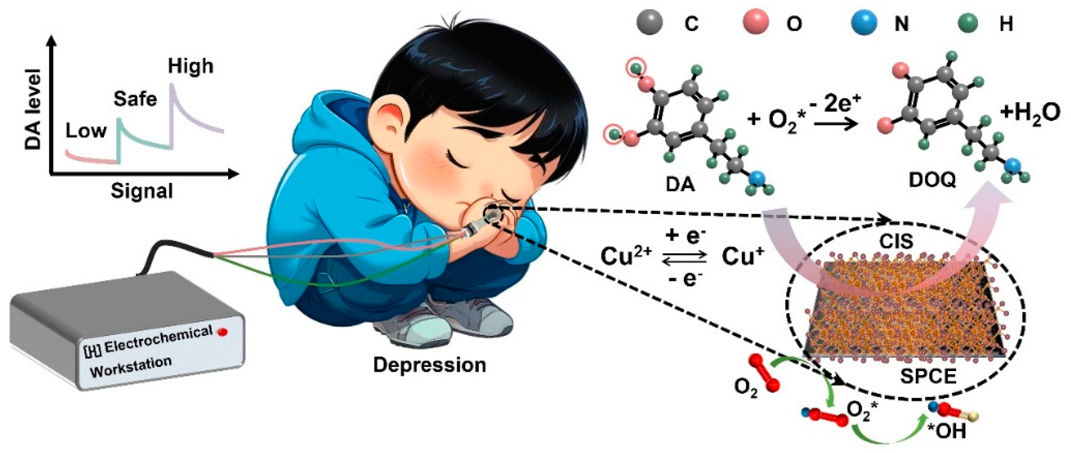
1. Introduction
2. Experimental Section
2.1. Reagents
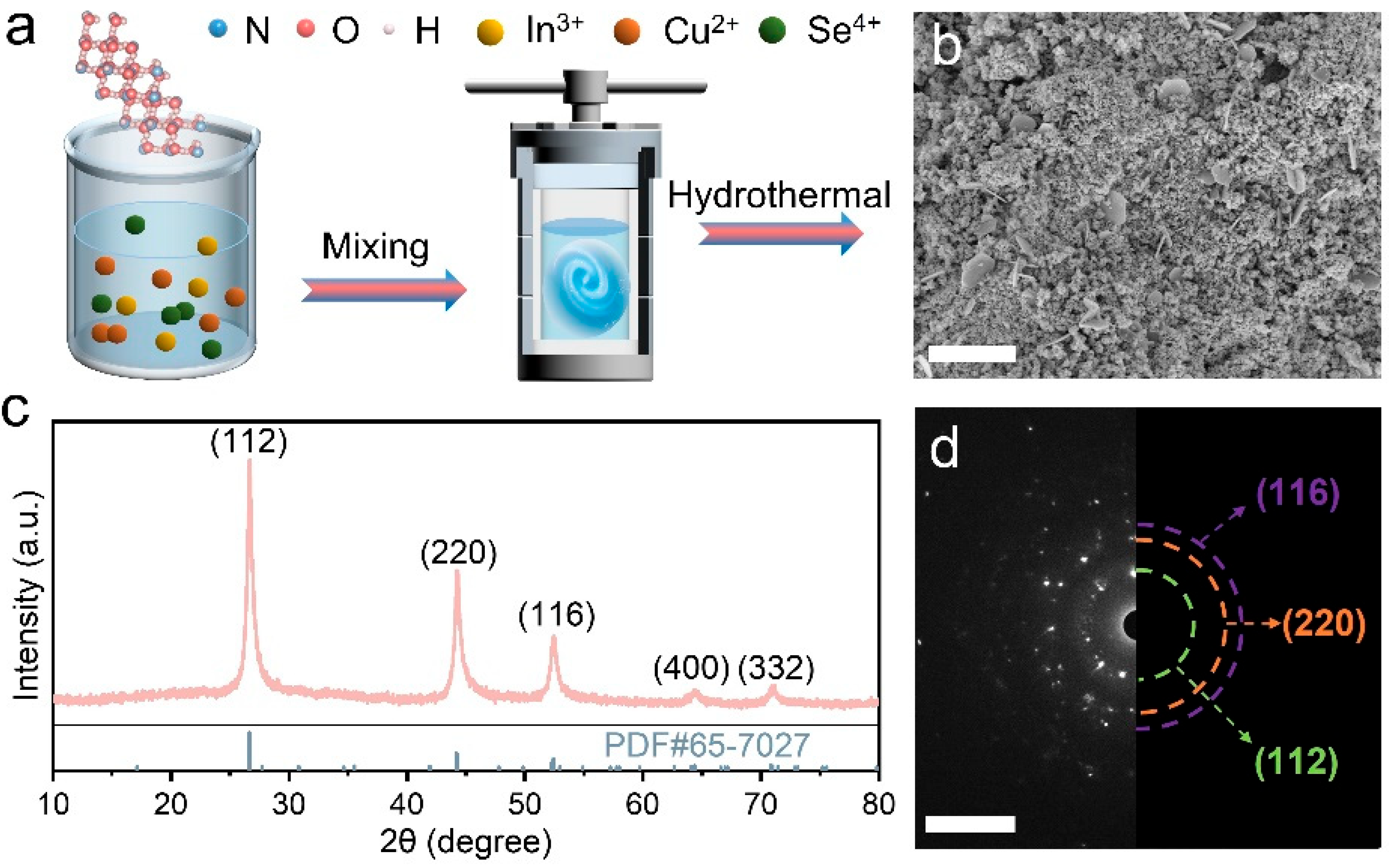
2.2. Synthesis of CuInSe2 Catalyst
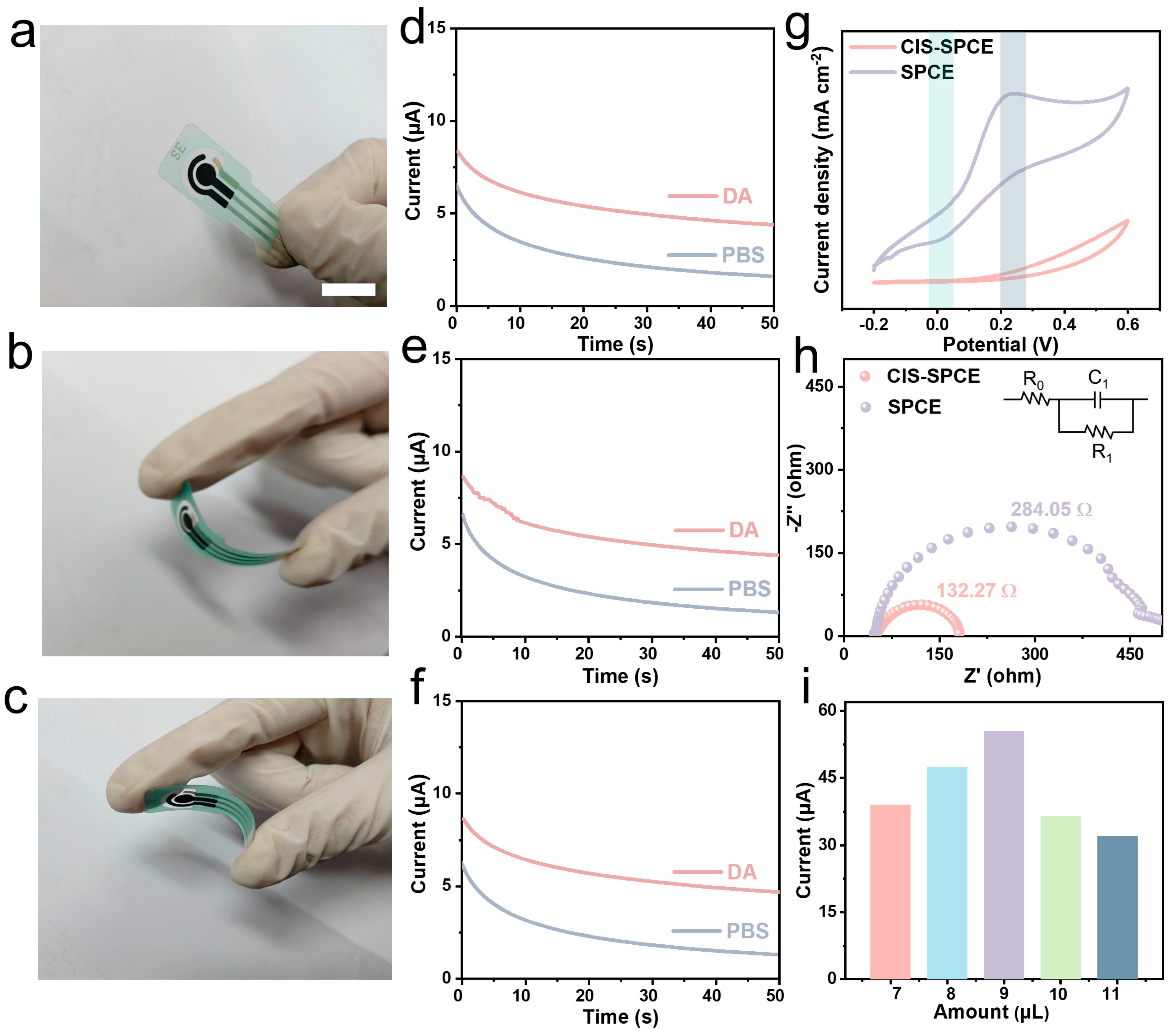
2.3. Fabrication of the CIS-Modified SPCE Sensor
2.4. Apparatus and Characterizations
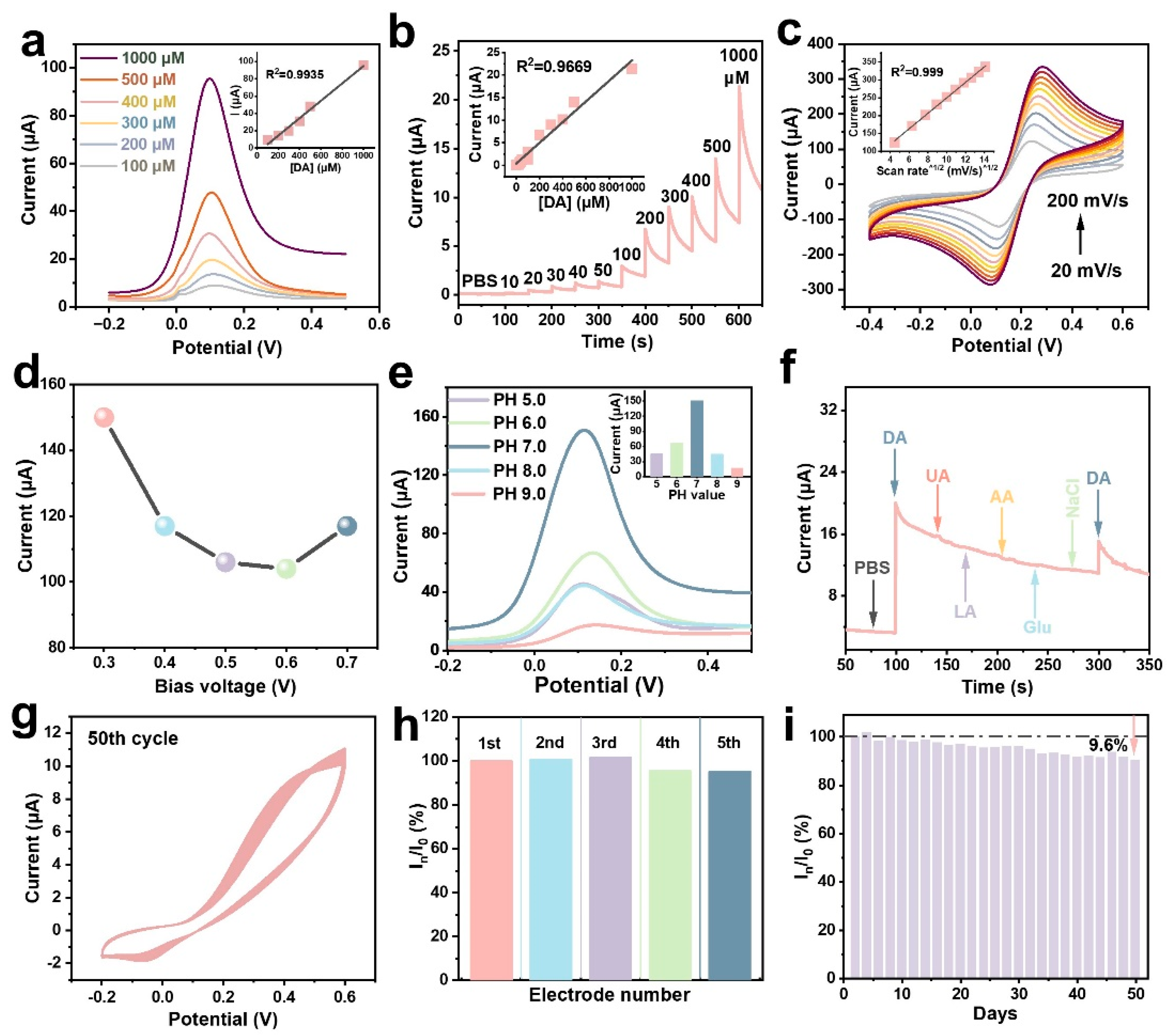
3. Results and Discussion
4. Conclusions
Supplementary Materials
Author Contributions
Funding
Data Availability Statement
Conflicts of Interest
References
- Lee, K.; Claar, L.D.; Hachisuka, A.; Bakhurin, K.I.; Nguyen, J.; Trott, J.M.; Gill, J.L.; Masmanidis, S.C. Temporally restricted dopaminergic control of reward-conditioned movements. Nat. Neurosci. 2020, 23, 209–216. [Google Scholar] [CrossRef] [PubMed]
- Liu, Z.; Le, Q.; Lv, Y.; Chen, X.; Cui, J.; Zhou, Y.; Cheng, D.; Ma, C.; Su, X.; Xiao, L.; et al. A distinct D1-MSN subpopulation down-regulates dopamine to promote negative emotional state. Cell Res. 2021, 32, 139–156. [Google Scholar] [CrossRef]
- Huang, Q.; Lin, X.; Tong, L.; Tong, Q.-X. Graphene Quantum Dots/Multiwalled Carbon Nanotubes Composite-Based Electrochemical Sensor for Detecting Dopamine Release from Living Cells. ACS Sustain. Chem. Eng. 2020, 8, 1644–1650. [Google Scholar] [CrossRef]
- Chowdhury, S.; Nugraha, A.S.; O’May, R.; Wang, X.; Cheng, P.; Xin, R.; Osman, S.M.; Hossain, M.S.; Yamauchi, Y.; Masud, M.K.; et al. Bimetallic metal-organic framework-derived porous one-dimensional carbon materials for electrochemical sensing of dopamine. Chem. Eng. J. 2024, 492, 152124. [Google Scholar] [CrossRef]
- Niu, B.; Liu, M.; Li, X.; Guo, H.; Chen, Z. Vein-Like Ni-BTC@Ni3S4 with Sulfur Vacancy and Ni3+ Fabricated In Situ Etching Vulcanization Strategy for an Electrochemical Sensor of Dopamine. ACS Appl. Mater. Interfaces 2023, 15, 13319–13331. [Google Scholar] [CrossRef]
- Lu, Z.; Li, Y.; Liu, T.; Wang, G.; Sun, M.; Jiang, Y.; He, H.; Wang, Y.; Zou, P.; Wang, X.; et al. A dual-template imprinted polymer electrochemical sensor based on AuNPs and nitrogen-doped graphene oxide quantum dots coated on NiS2/biomass carbon for simultaneous determination of dopamine and chlorpromazine. Chem. Eng. J. 2020, 389, 124417. [Google Scholar] [CrossRef]
- Zou, J.; Guan, J.-F.; Zhao, G.-Q.; Jiang, X.-Y.; Liu, Y.-P.; Yu, J.-G.; Li, W.-J. Construction of a highly sensitive signal electrochemical sensor based on self-assembled cobalt oxide-hydroxylated single-walled carbon nanotubes composite for detection of dopamine in bovine serum samples. J. Environ. Chem. Eng. 2021, 9, 105831. [Google Scholar] [CrossRef]
- Shukla, R.P.; Aroosh, M.; Matzafi, A.; Ben-Yoav, H. Partially Functional Electrode Modifications for Rapid Detection of Dopamine in Urine. Adv. Funct. Mater. 2021, 31, 2004146. [Google Scholar] [CrossRef]
- Dhiman, P.; Kumar, A.; Shekh, M.; Sharma, G.; Rana, G.; Vo, D.-V.N.; AlMasoud, N.; Naushad, M.; Alothman, Z.A. Robust magnetic ZnO-Fe2O3 Z-scheme hetereojunctions with in-built metal-redox for high performance photo-degradation of sulfamethoxazole and electrochemical dopamine detection. Environ. Res. 2021, 197, 111074. [Google Scholar] [CrossRef]
- Li, Z.; Liang, L.; Lin, W.; Huang, Y.; Huang, T.; Wang, W.; Ma, J.; Li, J.; Sun, L.-P.; Guan, B.-O. Optofluidic laser sensor for the detection of dopamine. Sens. Actuators B Chem. 2023, 390, 133941. [Google Scholar] [CrossRef]
- Zhou, L.; Yang, R.; Li, X.; Dong, N.; Zhu, B.; Wang, J.; Lin, X.; Su, B. COF-Coated Microelectrode for Space-Confined Electrochemical Sensing of Dopamine in Parkinson’s Disease Model Mouse Brain. J. Am. Chem. Soc. 2023, 145, 23727–23738. [Google Scholar] [CrossRef]
- Li, Y.-Y.; Kang, P.; Wang, S.-Q.; Liu, Z.-G.; Li, Y.-X.; Guo, Z. Ag nanoparticles anchored onto porous CuO nanobelts for the ultrasensitive electrochemical detection of dopamine in human serum. Sens. Actuators B Chem. 2021, 327, 128878. [Google Scholar] [CrossRef]
- Murugan, N.; Jerome, R.; Preethika, M.; Sundaramurthy, A.; Sundramoorthy, A.K. 2D-titanium carbide (MXene) based selective electrochemical sensor for simultaneous detection of ascorbic acid, dopamine and uric acid. J. Mater. Sci. Technol. 2021, 72, 122–131. [Google Scholar] [CrossRef]
- Xing, Y.; Lv, C.; Fu, Y.; Luo, L.; Liu, J.; Xie, X.; Chen, F. Sensitive sensing platform based on Co, Mo doped electrospun nanofibers for simultaneous electrochemical detection of dopamine and uric acid. Talanta 2024, 271, 125674. [Google Scholar] [CrossRef] [PubMed]
- Shi, S.; Zhou, C.; Wei, Y.; Chen, A.; Tang, N.; He, Q.; Deng, P. Ultrasensitive and selective electrochemical sensor for nanomolar dopamine determination based on MnS/Co3S4 hybrids embedded on electrochemically reduced graphene oxide. Microchem. J. 2023, 194, 109310. [Google Scholar] [CrossRef]
- Priyadarshini, P.; Senapati, S.; Bisoyi, S.; Samal, S.; Naik, R. Zn doping induced optimization of optical and dielectric characteristics of CuInSe2 nanosheets for optoelectronic device applications. J. Alloys Compd. 2023, 945, 169222. [Google Scholar] [CrossRef]
- Nithiananth, S.; Silambarasan, K.; Logu, T.; Harish, S.; Ramesh, R.; Muthamizhchelvan, C.; Shimomura, M.; Archana, J.; Navaneethan, M. Transition divalent metal substitution in chalcopyrite CuInSe2 (In = Co, Ni, and Mn) counter electrode for dye-sensitized solar cell applications. Mater. Lett. 2022, 308, 130887. [Google Scholar] [CrossRef]
- Reddy, Y.B.K.; Raja, V.S. Optical, structural and electrical properties of co-evaporated CuIn2Se3.5 thin films. Mater. Lett. 2004, 58, 1839–1843. [Google Scholar] [CrossRef]
- Fuhr, A.; Yun, H.J.; Crooker, S.A.; Klimov, V.I. Spectroscopic and Magneto-Optical Signatures of Cu1+ and Cu2+ Defects in Copper Indium Sulfide Quantum Dots. ACS Nano 2020, 14, 2212–2223. [Google Scholar] [CrossRef]
- Singh, H.; Bernabe, J.; Chern, J.; Nath, M. Copper selenide as multifunctional non-enzymatic glucose and dopamine sensor. J. Mater. Res. 2021, 36, 1413–1424. [Google Scholar] [CrossRef]
- Dashtian, K.; Hajati, S.; Ghaedi, M. Ti-Based Solid-State Imprinted-Cu2O/CuInSe2 Heterojunction Photoelectrochemical platform for Highly Selective Dopamine Monitoring. Sens. Actuators B Chem. 2021, 326, 128824. [Google Scholar] [CrossRef]
- Arya Nair, J.S.; Saisree, S.; Aswathi, R.; Sandhya, K.Y. Ultra-selective and real-time detection of dopamine using molybdenum disulphide decorated graphene-based electrochemical biosensor. Sens. Actuators B Chem. 2022, 354, 131254. [Google Scholar] [CrossRef]
- Xu, L.; Chen, M.; Hou, P.; Hou, X.; Wang, J.; Qi, Q.; Zhu, Y.; Yang, X.; Liu, X.; Li, X.; et al. Synthesis of CdSe Nanowires and CuInSe2 Nanosheets for Hydrogen Evolution. ACS Appl. Nano Mater. 2022, 5, 1935–1943. [Google Scholar] [CrossRef]
- Liu, F.; Zong, J.; Liang, Y.; Zhang, M.; Song, K.; Mi, L.; Feng, J.; Xiong, S.; Xi, B. Ordered Vacancies as Sodium Ion Micropumps in Cu-Deficient Copper Indium Diselenide to Enhance Sodium Storage. Adv. Mater. 2024, 36, 2403131. [Google Scholar] [CrossRef]
- Ren, S.; Feng, R.; Guo, T.; Cao, L.; Lv, R.; Liu, X.; Wang, Q.; Zheng, Z. Electroanalytical sensor based on CuInSe2/carbon sphere towards the non-invasive determination of dopamine. Electroanalysis 2023, 35, e202300203. [Google Scholar] [CrossRef]
- Sun, Z.; Sun, S.; Jiang, X.; Ai, Y.; Xu, W.; Xie, L.; Sun, H.B.; Liang, Q. Oligo-layer graphene stabilized fully exposed Fe-sites for ultra-sensitivity electrochemical detection of dopamine. Biosens. Bioelectron. 2022, 211, 114367. [Google Scholar] [CrossRef]
- Sheng, P.; Li, W.; Tong, X.; Wang, X.; Cai, Q. Development of a high performance hollow CuInSe2 nanospheres-based photoelectrochemical cell for hydrogen evolution. J. Mater. Chem. A 2014, 2, 18974–18987. [Google Scholar] [CrossRef]
- Liu, Y.; Wang, P.; Xie, L.; Xia, Y.; Zhan, S.; Hu, W.; Li, Y. Electronic Metal-Support Interactions Boost *OOH Intermediate Generation in Cu/In2Se3 for Electrochemical H2O2 Production. Angew. Chem. Int. Ed. 2024, 63, e202319470. [Google Scholar] [CrossRef]
- Dutková, E.; Bujňáková, Z.L.; Sphotyuk, O.; Jakubíková, J.; Cholujová, D.; Šišková, V.; Daneu, N.; Baláž, M.; Kováč, J.; Kováč, J.; et al. SDS-Stabilized CuInSe2/ZnS Multinanocomposites Prepared by Mechanochemical Synthesis for Advanced Biomedical Application. Nanomaterials 2020, 11, 69. [Google Scholar] [CrossRef]
- Umapathi, S.; Masud, J.; Coleman, H.; Nath, M. Electrochemical sensor based on CuSe for determination of dopamine. Microchim. Acta 2020, 187, 440. [Google Scholar] [CrossRef]
- Umapathi, S.; Singh, H.; Masud, J.; Nath, M. Nanostructured copper selenide as an ultrasensitive and selective non-enzymatic glucose sensor. Mater. Adv. 2021, 2, 927–932. [Google Scholar] [CrossRef]
- Saxena, A.; Liyanage, W.; Masud, J.; Kapila, S.; Nath, M. Selective electroreduction of CO2 to carbon-rich products with a simple binary copper selenide electrocatalyst. J. Mater. Chem. A 2021, 9, 7150–7161. [Google Scholar] [CrossRef]
- Guan, Y.; Li, S.; Su, Z.; Li, Y.; Wang, X.; Tian, M.; Guo, C.; Dong, T.; Chai, F. Fabrication of Cu-BTC@PW12/GO for multivariate sensing dopamine and acetaminophen as electrochemical sensor. J. Environ. Chem. Eng. 2024, 12, 114078. [Google Scholar] [CrossRef]
- Zhao, W.; Zhou, H.; Lu, K.; Li, Y.; Shi, W.; Ma, X. Electronic/ionic engineering inspired formation of carbon-encapsulated Cu3PSe4/Cu2Se heterostructured hollow nanosphere for trace level neurochemical monitoring. Appl. Surf. Sci. 2023, 613, 156142. [Google Scholar] [CrossRef]
- Liu, B.; Ouyang, X.; Ding, Y.; Luo, L.; Xu, D.; Ning, Y. Electrochemical preparation of nickel and copper oxides-decorated graphene composite for simultaneous determination of dopamine, acetaminophen and tryptophan. Talanta 2016, 146, 114–121. [Google Scholar] [CrossRef] [PubMed]
- Shin, J.-W.; Kim, K.-J.; Yoon, J.; Jo, J.; El-Said, W.A.; Choi, J.-W. Silver Nanoparticle Modified Electrode Covered by Graphene Oxide for the Enhanced Electrochemical Detection of Dopamine. Sensors 2017, 17, 2771. [Google Scholar] [CrossRef]
- Oh, J.-W.; Heo, J.; Kim, T.H. An electrochemically modulated single-walled carbon nanotube network for the development of a transparent flexible sensor for dopamine. Sens. Actuators B Chem. 2018, 267, 438–447. [Google Scholar] [CrossRef]
- Tan, W.; Zhu, Z.; Yang, J.; Li, H.; Li, S.; Wu, D.; Qin, Y.; Kong, Y. Synthesis of ZnO and CuO co-decorated porous carbon spheres with simultaneous accessibility to small biomelucules. Synth. Met. 2019, 258, 116193. [Google Scholar] [CrossRef]
- Zhao, Y.; Zhou, J.; Jia, Z.; Huo, D.; Liu, Q.; Zhong, D.; Hu, Y.; Yang, M.; Bian, M.; Hou, C. In-situ growth of gold nanoparticles on a 3D-network consisting of a MoS2/rGO nanocomposite for simultaneous voltammetric determination of ascorbic acid, dopamine and uric acid. Microchim. Acta 2019, 186, 92. [Google Scholar] [CrossRef]
- Manjula, N.; Vinothkumar, V.; Chen, S.-M.; Sangili, A. Simultaneous and sensitive detection of dopamine and uric acid based on cobalt oxide-decorated graphene oxide composite. J. Mater. Sci. Mater. Electron. 2020, 31, 12595–12607. [Google Scholar] [CrossRef]
- Mohamed, N.B.; El-Kady, M.F.; Kaner, R.B. Macroporous Graphene Frameworks for Sensing and Supercapacitor Applications. Adv. Funct. Mater. 2022, 32, 2203101. [Google Scholar] [CrossRef]
- Ahmed, J.; Faisal, M.; Alsareii, S.A.; Jalalah, M.; Harraz, F.A. A novel gold-decorated porous silicon-poly(3-hexylthiophene) ternary nanocomposite as a highly sensitive and selective non-enzymatic dopamine electrochemical sensor. J. Alloys Compd. 2023, 931, 167403. [Google Scholar] [CrossRef]
- Kunpatee, K.; Traipop, S.; Chailapakul, O.; Chuanuwatanakul, S. Simultaneous determination of ascorbic acid, dopamine, and uric acid using graphene quantum dots/ionic liquid modified screen-printed carbon electrode. Sens. Actuators B Chem. 2020, 314, 128059. [Google Scholar] [CrossRef]
- Kaya, H.K.; Cinar, S.; Altundal, G.; Bayramlı, Y.; Unaleroglu, C.; Kuralay, F. A novel design thia-bilane structure-based molecular imprinted electrochemical sensor for sensitive and selective dopamine determination. Sens. Actuators B Chem. 2021, 346, 130425. [Google Scholar] [CrossRef]
- Wu, R.; Yu, S.; Chen, S.; Dang, Y.; Wen, S.-H.; Tang, J.; Zhou, Y.; Zhu, J.-J. A carbon dots-enhanced laccase-based electrochemical sensor for highly sensitive detection of dopamine in human serum. Anal. Chim. Acta 2022, 1229, 340365. [Google Scholar] [CrossRef]
- Xu, Z.; Song, J.; Liu, B.; Lv, S.; Gao, F.; Luo, X.; Wang, P. A conducting polymer PEDOT:PSS hydrogel based wearable sensor for accurate uric acid detection in human sweat. Sens. Actuators B Chem. 2021, 348, 130674. [Google Scholar] [CrossRef]
- Al Kiey, S.A.; Khalil, A.M.; Kamel, S. Insight into TEMPO-oxidized cellulose-based composites as electrochemical sensors for dopamine assessment. Int. J. Biol. Macromol. 2023, 239, 124302. [Google Scholar] [CrossRef]


| Electrode Material | Linear Range (µM) | LOD (nM) | Sensitivity (μA·µM−1) | References |
|---|---|---|---|---|
| Ni-BTC@Ni3S4/CPE | 0.05–750 | 16 | 0.56 | [5] |
| Co3O4/SWCNTs a-OH/GCE | 1–100 | 80 | 1.427 | [7] |
| Ag/CuO/ITO | 0.04–10 | 7 | 14 | [12] |
| NiO–CuO/Graphene /GCE | 0.5–20 | 167 | 1.1238 | [35] |
| Ag/GO/ITO | 0.1–100 | 200 | / | [36] |
| Eox b-SWCNT/PET | 1.5–30 | 510 | 0.13 | [37] |
| ZnO–CuO/PCS c/GCE | 13–400 | 1.03 | 0.82 | [38] |
| MoS2/rGO/AuNPs/GCE | 0.3–198.3 | 110 | / | [39] |
| Co3O4/GO/SPCE | 0.2–1221 | 90 | 0.034 | [40] |
| FGH d/GCE | 0.5–200 | 110 | 2.294 | [41] |
| Au@PSi-P3HT e/GCE | 1–460 | 630 | 0.0205 | [42] |
| GQDs/IL f-SPCE | 0.2–15 | 60 | / | [43] |
| pS-BIL g MIP PeGE h | 0.5–250 | 130 | / | [44] |
| Lc i/CDs/GCE | 0.25–9.82, 9.82–76.81 | 80 | 3.586 | [45] |
| CIS-SPCE | 0.04–1000 | 96 | 2.511 | This work |
Disclaimer/Publisher’s Note: The statements, opinions and data contained in all publications are solely those of the individual author(s) and contributor(s) and not of MDPI and/or the editor(s). MDPI and/or the editor(s) disclaim responsibility for any injury to people or property resulting from any ideas, methods, instructions or products referred to in the content. |
© 2025 by the authors. Licensee MDPI, Basel, Switzerland. This article is an open access article distributed under the terms and conditions of the Creative Commons Attribution (CC BY) license (https://creativecommons.org/licenses/by/4.0/).
Share and Cite
Li, J.; Xie, G.; Dai, L.; Yang, M.; Su, Y. Rapid and Highly Selective Dopamine Sensing with CuInSe2-Modified Nanocomposite. J. Compos. Sci. 2025, 9, 123. https://doi.org/10.3390/jcs9030123
Li J, Xie G, Dai L, Yang M, Su Y. Rapid and Highly Selective Dopamine Sensing with CuInSe2-Modified Nanocomposite. Journal of Composites Science. 2025; 9(3):123. https://doi.org/10.3390/jcs9030123
Chicago/Turabian StyleLi, Jing, Guangzhong Xie, Luwei Dai, Min Yang, and Yuanjie Su. 2025. "Rapid and Highly Selective Dopamine Sensing with CuInSe2-Modified Nanocomposite" Journal of Composites Science 9, no. 3: 123. https://doi.org/10.3390/jcs9030123
APA StyleLi, J., Xie, G., Dai, L., Yang, M., & Su, Y. (2025). Rapid and Highly Selective Dopamine Sensing with CuInSe2-Modified Nanocomposite. Journal of Composites Science, 9(3), 123. https://doi.org/10.3390/jcs9030123