Article

Synthesis of Vectorized Nanoparticles Based on a Copolymer of N-Vinyl-2-Pyrrolidone with Allyl Glycidyl Ether and a Carbohydrate Vector

by
Dmitry Z. Vinnitskiy
1,
Anna L. Luss
2,
Vadim B. Krylov
1,
Nadezhda E. Ustyuzhanina
1,
Anastasiya V. Goryachaya
2,
Anna M. Nechaeva
2,
Mikhail I. Shtilman
2,
Nikolay E. Nifantiev
1,* and
Yaroslav O. Mezhuev
2,*
1
N.D. Zelinsky Institute of Organic Chemistry, Russian Academy of Sciences, Leninsky Prospekt, 47, 119991 Moscow, Russia
2
Mendeleev University of Chemical Technology of Russia, Miusskaya Square, 9, 125047 Moscow, Russia
*
Authors to whom correspondence should be addressed.
J. Compos. Sci. 2022, 6(9), 247; https://doi.org/10.3390/jcs6090247
Submission received: 30 July 2022 / Revised: 18 August 2022 / Accepted: 22 August 2022 / Published: 25 August 2022
(This article belongs to the Special Issue Nanocomposites for Biomedical and Environmental Applications)

Abstract

:
A method was developed for the conjugation of aminopropyl spacer-bearing carbohydrates with epoxy groups on the crown of nanoparticles consisting of a copolymer of N-vinyl-2-pyrrolidone and allyl glycidyl ether in basic buffer, opening prospects for the design of vectorized nanocomposite drug forms. A conjugate of the above copolymer and trisaccharide A, a synthetic blood group antigen, was synthesized. Meglumine was used to bind any unreacted epoxide groups of the allyl glycidyl fragment. One- and two-dimensional NMR spectroscopy showed quantitative opening of the epoxide ring as a result of carbohydrate immobilization. By integrating the characteristic signals in the 1H NMR spectrum, we determined the molar ratio of the immobilized vector and meglumine, as well as the composition and number-average molecular weight of the carrier copolymer. The results obtained point to the interesting possibilities in the further study of the polymer–carbohydrate ligand system as a platform for the development of several drug carriers and theranostics based on them.

1. Introduction

Targeted diagnostics and drug delivery is a promising approach toward increasing efficacy and reducing side effects in the treatment of the most socially significant diseases, which primarily include diseases of the cardiovascular system and cancer [1,2,3,4]. This concept is based upon the design of nanoscale composite systems consisting of a vector—a fragment responsible for detection and delivery to the source of the disease (tumor, thrombus, center of the inflammatory process, etc.), a carrier—a structural element that exposes vector fragments and deposits medicinal compounds, and the medicinal compounds responsible for the therapeutic effect themselves [2,5]. With this approach, several advantages are achieved: reduction of the therapeutic load and, hence, the reduction of side effects, as well as prolongation of the drug action due to the gradual release of active molecules [6]. In addition, these structures make it possible to avoid the body’s protective systems, for example, overcoming the blood–brain barrier, where it would be impregnable for the drug itself [7,8].
Theranostics based on nanoparticles of iron oxide, silicon, gold, carbon nanotubes, and quantum dots are being actively studied [9]. Various polymers have also become extremely widespread as carriers, having several significant advantages such as relative simplicity and low cost of production, biochemical inertness, etc. [10,11,12]. Amphiphilic copolymers seem to be promising representatives of this class due to their ability to self-assemble into nanoparticles. They use polyethylene oxide, poly(N-vinyl caprolactam), poly(N-vinyl-2-pyrrolidone) as a hydrophilic base, whereas the hydrophobic components are polyglycolide, polylactide, and poly(ε-caprolactone) [13,14,15,16,17,18,19].
When it comes to vectors, a promising approach is the use of synthetic oligosaccharides. These compounds, in contrast to natural oligosaccharides, have a determined structure, are easier to purify, and at the same time, have a high affinity for biological targets, providing the necessary targeted delivery [20,21,22,23]. The high diversity of carbohydrate structures, meanwhile, allows for a great variety of intended targets [24,25,26,27]. Thus, based on the carbohydrate-carrier system, supplemented depending on the target with a known or promising therapeutic agent, a number of theranostics can be developed.
This paper describes a first approach to the development of such a system based on an amphiphilic copolymer of N-vinyl-2-pyrrolidone and allyl glycidyl ether (NVPAGE) as a carrier and a synthetic oligosaccharide (trisaccharide A) containing an amino propyl spacer as a vector prototype. An effective protocol for conjugation was proposed, and the obtained vectored nanoparticles, prospective nanocontainers for the subsequent loading of drugs, were characterized by NMR spectroscopy, which confirmed a significant number of conjugated ligands.

2. Materials and Methods

N-vinyl-2-pyrrolidone and allyl glycidyl ether were purchased from Sigma-Aldrich and further purified by distillation, while azobisisobutyronitrile (AIBN) and n-octadecylmercaptan were used without further purification to synthesize the amphiphilic NVPAGE copolymer. The copolymerization was carried out in a 1,4-dioxane medium from Himmed. As a model vector, the synthetic trisaccharide 3-aminopropyl 2-acetamido-2-deoxy-α-d-galactopyranosyl-(l→3)-[(α-L-fucopyranosyl)-(l→2)]-β-D-galactopyranoside related to a blood group antigen (trisaccharide A) (obtained earlier [28,29] for biological studies [30,31]) was applied. Conjugation of the amphiphilic NVPAGE copolymer with a carbohydrate ligand was performed in distilled deionized water purified using a Merck Millipore Simplicity purification system. Reagents (meglumine and sodium carbonate) from Sigma-Aldrich were used without further purification.
Particle diameter distribution and surface tension isotherm were obtained by dynamic laser light scattering (NANO-flex II (Colloid Metrix, Meerbusch, Germany)) and hanging drop (KRÜSS DSA30 (KRÜSS, Hamburg, Germany)) after ultrasonic treatment of solutions (SONOPULS HD4400 (Bandelin, Germany)). NMR spectra were obtained on a Bruker Avance 600 spectrometer at 303 K after a single lyophilization of the samples from D2O and their subsequent dissolution in 99.96% D2O. Signals were assigned using homo- and heteronuclear two-dimensional COSY and HSQC correlation spectra. See all NMR spectra in Supplementary Materials. Statistical analysis was carried out according to the results of three experiments [32,33,34]; for the relative error of all points in the series, the maximum calculated error was taken.

2.1. Radical Copolymerization of N-vinyl-2-pyrrolidone and Allyl Glycidyl Ether

The NVPAGE amphiphilic copolymer was synthesized according to a procedure described in [35] at a temperature of 343 K, a molar ratio of N-vinyl-2-pyrrolidone and allyl glycidyl ether of 5.7, and concentrations of AIBN and n-octadecylmercaptan of 0.015 M and 0.03 M in a solution of 1,4-dioxane. The product was isolated by precipitation in diethyl ether, washed by 3 portions of 30 mL of diethyl ether on a filter and then dried at 323 K.

2.2. Conjugation of Trisaccharide A with NVPAGE Copolymer

To a solution of 20 mg NVPAGE in 380 μL of a buffer solution (0.1 M Na2CO3 aq.), 50 μL of a buffer solution containing 1.2 mg of trisaccharide A with a free amino group in the spacer fragment was added. The reaction mixture was well-stirred and left at room temperature. After 24 h, 70 μL of a buffer solution containing 5 mg of meglumine was added to the reaction mixture. The reaction mixture was kept for another 24 h at room temperature, after which it was transferred to a Vivaspin 20 centrifuge concentrator with a cut-off mass of 10 kDa. The tube was centrifuged at 5000 rpm for 15 min, after which 1 mL of deionized water was added to the concentrate and centrifuged again at 5000 rpm for 15 min. The procedure was repeated 2 more times. The final concentrate was dissolved in 1 mL of deionized water, transferred to a flask, and lyophilized. An amount of 20 mg of product was isolated. The NMR spectra of the synthesized conjugate are described in Table 1.

3. Results

The original amphiphilic copolymer NVPAGE was obtained by radical copolymerization of N-vinyl-2-pyrrolidone and allyl glycidyl ether. In the 1H NMR spectrum of the amphiphilic NVPAGE copolymer, all characteristic signals of protons included in the composition of N-vinyl-2-pyrrolidone residues, allyl glycidyl ether, and terminal n-octadecylthio groups are observed (Figure 1).
The number-average degree of polymerization of the NVPAGE amphiphilic copolymer, determined from the integral intensities ratio of the signals of the C17H35 fragment and protons in position II (N-vinyl-2-pyrrolidone residues) and J (allyl glycidyl ether residues), is about 7 × 10³. The mole fraction of epoxy-containing allyl glycidyl ether residues in the polymer chain is about 12 mol%.
An increase in the concentration of the NVPAGE amphiphilic copolymer leads to a decrease in the surface tension at the water/air interface to 60 mJ × m−2 (Figure 2).
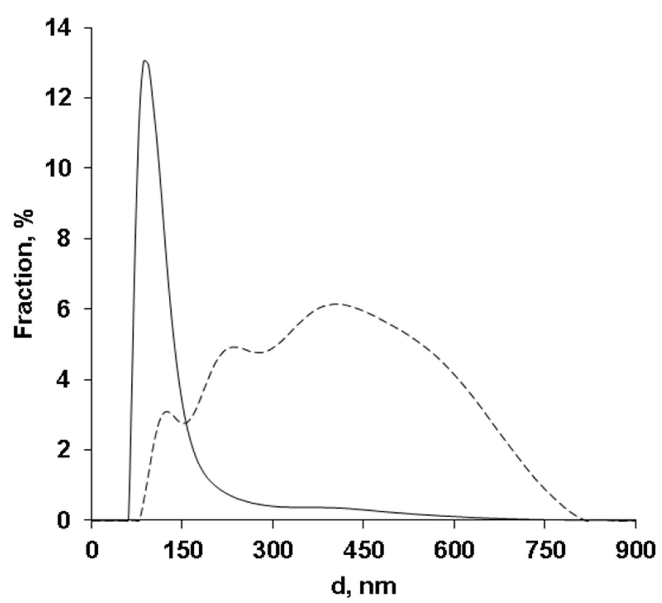
According to dynamic laser light scattering data (Figure 3), the number-average and intensity-averaged diameters for aggregates formed by NVPAGE chains are 106 nm and 287.4 nm at a PDI of 0.277.
Thus, the chains of the amphiphilic NVPAGE copolymer exhibit surface activity and can form nanoparticles as a result of self-organization in aqueous media. As has been shown earlier, amphiphilic NVPAGE copolymers can serve as carriers for anticancer drugs such as doxorubicin and paclitaxel and allow for the joint loading of these substances into nanoparticles [35]. At the same time, in the absence of molecules capable of specifically interacting with cell receptors, only passive targeting is possible, which indicates the need to find ways of immobilizing vector molecules that would ensure targeted delivery of anticancer drugs. The model carbohydrate frame we used in this work was a synthetic blood group antigen, trisaccharide A (Scheme 1).
At the first stage of our work, we studied the possibility of conjugation between the original compounds. On the surface of particles formed by NVPAGE chains, there are epoxy groups, which interact with trisaccharide A’s spacer amino group. Upon performing a literature search, we chose the following procedure [36,37]: copolymer nanoparticles and a vector containing an aminopropyl spacer are mixed in 0.1 M carbonate buffer; the reaction takes place within a day. Then, unreacted epoxy groups are bound by adding glucamine (meglumine, MeGlu), the reaction mixture is kept for another day, after which all low-molecular-weight compounds (unreacted vector and meglumine) are removed from the product by centrifugation using a centrifugal concentrator with a cut-off of 10 kDa (Scheme 1). If necessary, replacing MeGlu with doxorubicin allows covalent immobilization of this anticancer drug, as was previously established [35].
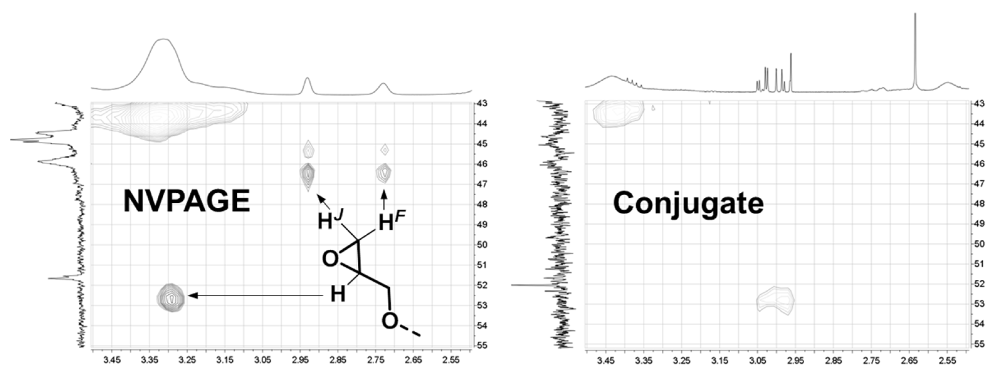
The obtained product was investigated by means of one-dimensional and two-dimensional NMR spectroscopy to confirm the conjugate’s structure and determine the content of the oligosaccharide vector and meglumine. First, the obtained spectra were compared with those of the original polymer. Figure 4 shows the heteronuclear HSQC correlations of the parent NVPAGE polymer corresponding to the terminal epoxy moiety. In the resulting conjugate, signals of this type are completely absent, which confirms the complete opening of the oxirane rings. The absence of a reactive three-membered ring is extremely important, because this group can act as an alkylator in biological systems and is a factor in drug toxicity. Thus, full opening of the epoxy ring led to formation of chains built by residues of N-vinyl-2-pyrrolidone and aminoglycerol derivatives, which, as is known, are biocompatible and do not induce toxic effects [38,39].
We also analyzed the signals of the carbohydrate residues in the vectored conjugate. Figure 5 shows fragments of the 1H NMR spectra of free meglumine and the resulting conjugate. The signals of the meglumine protons are clearly resolved against the background of broadened signals of the polymer backbone, which confirms the free uninhibited rotation of the carbohydrate residue.
Analysis of the chemical shifts of the 1H and 13C signals (Table 2) in the NMR spectra of free meglumine and its residue in the vectored conjugate showed a significant difference only in atoms next to the nitrogen atom through which attachment to the polymer carrier was carried out.
The presence of the oligosaccharide vector component in the conjugate is easily observed using 1H NMR spectra. First, the anomeric region is characteristic (Figure 6), where the signals of all three anomeric protons of the monosaccharide residues (~4.52, 5.16, 5.28 ppm) are clearly visible, present in the spectra of both trisaccharide A and the conjugate, while there are no signals in the spectrum of the initial NVPAGE polymer in this region. Characteristic signals of carbohydrates in the region of 4.4–4.2 ppm and the methyl group signal of the fucose residue at 1.20 ppm are also noticeable, while others are overlapped by signals from polymeric fragments and meglumine. Integration of characteristic signals in the 1H NMR spectrum made it possible to estimate the molar ratio of trisaccharide A and meglumine as 1:7. In the future, using an already-established protocol, the amount of the extremely valuable carbohydrate vector can be increased if necessary. The protocol for nanoparticle vectorization developed here ensures the quantitative involvement of available epoxy groups of the copolymer and is superior in efficiency to the previously described methods for immobilizing amine-containing compounds on the NVPAGE copolymer [35,40].

4. Conclusions

Thus, we have developed a protocol for the conjugation of carbohydrates with NVPAGE by opening the epoxy groups of allyl glycidyl residues in a basic buffer. Its effectiveness has been demonstrated using a synthetic trisaccharide A containing an aminopropyl spacer which acts as a nucleophile in the reaction with epoxy groups. The presence of the vector carbohydrate in the structure of the resulting conjugate was reliably confirmed by NMR spectroscopy based on the appearance of characteristic signals. In combination with the known possibility of immobilizing doxorubicin and paclitaxel with chain aggregates of the amphiphilic copolymer NVPAGE, the vectorization of these nanoparticles opens up new prospects for the targeted delivery of anticancer drugs.

Supplementary Materials

The following supporting information can be downloaded at: https://www.mdpi.com/article/10.3390/jcs6090247/s1. Copies of NMR spectra of all compounds and microphotographs of NVPAGE particles.

Author Contributions

Conceptualization, N.E.U., N.E.N., D.Z.V., and Y.O.M.; methodology, V.B.K.; investigation, D.Z.V., V.B.K., A.V.G., A.L.L., and A.M.N.; resources, N.E.U.; data curation, N.E.U.; writing—original draft preparation, D.Z.V., V.B.K., M.I.S., and A.L.L.; writing—review and editing, N.E.N., Y.O.M., and M.I.S.; supervision, N.E.N., Y.O.M., and M.I.S.; project administration, N.E.N.; funding acquisition, N.E.N. All authors have read and agreed to the published version of the manuscript.

Funding

This research was funded by the Ministry of Science and Higher Education of Russia (Grant Agreement № 075-15-2020-792).

Institutional Review Board Statement

Not applicable.

Data Availability Statement

Data are available from the corresponding author.

Acknowledgments

The authors thank A.I. Tokatly for reading this manuscript and its critical discussion.

Conflicts of Interest

The authors declare no conflict of interest. The funders had no role in the design of the study; in the collection, analyses, or interpretation of data; in the writing of the manuscript; or in the decision to publish the results.

References

  1. Chen, H.; Zhang, W.; Zhu, G.; Xie, J.; Chen, X. Rethinking cancer nanotheranostics. Nat. Rev. Mater. 2017, 2, 17024. [Google Scholar] [CrossRef] [PubMed]
  2. Hapuarachchige, S.; Artemov, D. Theranostic Pretargeting Drug Delivery and Imaging Platforms in Cancer Precision Medicine. Front. Oncol. 2020, 10, 1131. [Google Scholar] [CrossRef] [PubMed]
  3. MacRitchie, N.; Di Francesco, V.; Ferreira, M.; Guzik, T.J.; Decuzzi, P.; Maffia, P. Nanoparticle theranostics in cardiovascular inflammation. Semin. Immunol. 2021, 56, 101536. [Google Scholar] [CrossRef] [PubMed]
  4. Manners, N.; Priya, V.; Mehata, A.K.; Rawat, M.; Mohan, S.; Makeen, H.A.; Albratty, M.; Albarrati, A.; Meraya, A.M.; Muthu, M.S. Theranostic Nanomedicines for the Treatment of Cardiovascular and Related Diseases: Current Strategies and Future Perspectives. Pharmaceuticals 2022, 15, 441. [Google Scholar] [CrossRef]
  5. Shrivastava, S.; Jain, S.; Kumar, D.; Soni, S.; Sharma, M. A Review on Theranostics: An Approach to Targeted Diagnosis and Therapy. Asian J. Pharm. Res. Dev. 2019, 7, 63–69. [Google Scholar] [CrossRef]
  6. Chiari-Andréo, B.G.; Abucafy, M.P.; Manaia, E.B.; da Silva, B.L.; Rissi, N.C.; Oshiro-Junior, J.A.; Chiavacci, L.A. Drug delivery using theranostics: An Overview of its use, advantages and safety assessment. Curr. Nanosci. 2020, 16, 3–14. [Google Scholar] [CrossRef]
  7. Tang, W.; Fan, W.; Lau, J.; Deng, L.; Shen, Z.; Chen, X. Emerging blood-brain-barrier-crossing nanotechnology for brain cancer theranostics. Chem. Soc. Rev. 2019, 48, 2967–3014. [Google Scholar] [CrossRef]
  8. Samanta, S.; Le Joncour, V.; Wegrzyniak, O.; Rangasami, V.K.; Ali-Löytty, H.; Hong, T.; Selvaraju, R.K.; Aberg, O.; Hilborn, J.; Laakkonen, P.; et al. Heparin-Derived Theranostic Nanoprobes Overcome the Blood–Brain Barrier and Target Glioma in Murine Model. Adv. Ther. 2022, 5, 2200001. [Google Scholar] [CrossRef]
  9. Xie, J.; Lee, S.; Chen, X. Nanoparticle-based Theranostic Agents. Adv. Drug Deliv. Rev. 2010, 62, 1064–1079. [Google Scholar] [CrossRef]
  10. Ventola, C.L. The nanomedicine revolution: Part 1—Emerging concepts. Pharm. Ther. 2012, 37, 512–525. [Google Scholar]
  11. Sevastre, A.-S.; Horescu, C.; Carina Baloi, S.; Cioc, C.E.; Vatu, B.I.; Tuta, C.; Artene, S.A.; Danciulescu, M.M.; Tudorache, S.; Dricu, A. Benefits of Nanomedicine for Therapeutic Intervention in Malignant Diseases. Coatings 2019, 9, 628. [Google Scholar] [CrossRef] [Green Version]
  12. Soares, S.; Sousa, J.; Pais, A.; Vitorino, C. Nanomedicine: Principles, Properties, and Regulatory Issues. Front. Chem. 2018, 6, 360. [Google Scholar] [CrossRef] [PubMed]
  13. Martin, C.; Aibani, N.; Callan, J.F.; Callan, B. Recent advances in amphiphilic polymers for simultaneous delivery of hydrophobic and hydrophilic drugs. Ther. Deliv. 2016, 7, 15–31. [Google Scholar] [CrossRef]
  14. Nutan, B.; Singh Chandel, A.K.; Jewrajka, S.K. Synthesis and Multi-Responsive Self-Assembly of Cationic Poly(caprolactone)–Poly(ethylene glycol) Multiblock Copolymers. Chem. Eur. J. 2017, 23, 8166–8170. [Google Scholar] [CrossRef]
  15. Atanase, L.I.; Desbrieres, J.; Riess, G. Micellization of synthetic and polysaccharides-based graft copolymers in aqueous media. Prog. Polym. Sci. 2017, 73, 32–60. [Google Scholar] [CrossRef]
  16. Winninger, J.; Iurea, D.M.; Atanase, L.I.; Salhi, S.; Delaite, C.; Riess, G. Micellization of novel biocompatible thermo-sensitive graft copolymers based on poly(ε-caprolactone), poly(N-vinylcaprolactam) and poly(N-vinylpyrrolidone). Eur. Polym. J. 2019, 119, 74–82. [Google Scholar] [CrossRef]
  17. Atanase, L.I.; Winninger, J.; Delaite, C.; Riess, G. Reversible addition–fragmentation chain transfer synthesis and micellar characteristics of biocompatible amphiphilic poly(vinyl acetate)-graft-poly(N-vinyl-2-pyrrolidone) copolymers. Eur. Polym. J. 2014, 53, 109–117. [Google Scholar] [CrossRef]
  18. Daraba, O.M.; Cadinoiu, A.N.; Rata, D.M.; Atanase, L.I.; Vochita, G. Antitumoral Drug-Loaded Biocompatible Polymeric Nanoparticles Obtained by Non-Aqueous Emulsion Polymerization. Polymers 2020, 12, 1018. [Google Scholar] [CrossRef]
  19. Essa, D.; Kondiah, P.P.D.; Choonara, Y.E.; Pillay, V. The Design of Poly(lactide-co-glycolide) Nanocarriers for Medical Applications. Front. Bioeng. Biotechnol. 2020, 8, 48. [Google Scholar] [CrossRef]
  20. Bloise, N.; Okkeh, M.; Restivo, E.; Della Pina, C.; Visai, L. Targeting the “Sweet Side” of Tumor with Glycan-Binding Molecules Conjugated-Nanoparticles: Implications in Cancer Therapy and Diagnosis. Nanomaterials 2021, 11, 289. [Google Scholar] [CrossRef]
  21. Sampaolesi, S.; Nicotra, F.; Russo, L. Glycans in nanomedicine, impact and perspectives. Future Med. Chem. 2019, 11, 43–60. [Google Scholar] [CrossRef] [PubMed]
  22. Delbianco, M.; Bharate, P.; Varela-Aramburu, S.; Seeberger, P.H. Carbohydrates in Supramolecular Chemistry. Chem. Rev. 2016, 116, 1693–1752. [Google Scholar] [CrossRef] [PubMed]
  23. Hossain, F.; Andreana, P.R. Developments in Carbohydrate-Based Cancer Therapeutics. Pharmaceuticals 2019, 12, 84. [Google Scholar] [CrossRef] [PubMed]
  24. Shchegravina, E.S.; Sachkova, A.A.; Usova, S.D.; Nyuchev, A.V.; Gracheva, Y.A.; Fedorov, A.Y. Carbohydrate Systems in Targeted Drug Delivery: Expectation and Reality. Russ. J. Bioorg. Chem. 2021, 47, 71–98. [Google Scholar] [CrossRef]
  25. Stenzel, M.H. Glycopolymers for Drug Delivery: Opportunities and Challenges. Macromolecules 2022, 55, 4867–4890. [Google Scholar] [CrossRef]
  26. Zhang, C.-W.; Zhang, J.-G.; Yang, X.; Du, W.-L.; Yu, Z.-L.; Lv, Z.-Y.; Mou, X.-Z. Carbohydrates based stimulus responsive nanocarriers for cancer-targeted chemotherapy: A review of current practices. Expert Opin. Drug Deliv. 2022, 19, 623–640. [Google Scholar] [CrossRef]
  27. Chollet, L.; Saboural, P.; Chauvierre, C.; Villemin, J.-N.; Letourneur, D.; Chaubet, F. Fucoidans in Nanomedicine. Mar. Drugs 2016, 14, 145. [Google Scholar] [CrossRef]
  28. Kazakova, E.D.; Yashunsky, D.V.; Nifantiev, N.E. The Synthesis of Blood Group Antigenic A Trisaccharide and Its Biotinylated Derivative. Molecules 2021, 26, 5887. [Google Scholar] [CrossRef]
  29. Fomitskaya, P.A.; Argunov, D.A.; Tsvetkov, Y.E.; Lalov, A.V.; Ustyuzhanina, H.E.; Nifantiev, N.E. Further investigation of the 2-azido-phenylselenylation of glycals. Eur. J. Org. Chem. 2021, 2021, 5897–5904. [Google Scholar] [CrossRef]
  30. Iurisci, I.; Cumashi, A.; Sherman, A.A.; Tsvetkov, Y.E.; Tinari, N.; Piccolo, E.; D’Egidio, M.; Adamo, V.; Natoli, C.; Rabinovich, G.A.; et al. Synthetic inhibitors of galectin-1 and -3 selectively modulate homotypic cell aggregation and tumor cell apoptosis. Anticancer Res. 2009, 29, 403–410. [Google Scholar]
  31. Casset, F.; Peters, T.; Etzler, M.; Korchagina, E.; Nifant’ev, N.; Perez, S.; Imberty, A. Conformational analysis of blood group A trisaccharide in solution and in the binding site of Dolichos biflorus lectin using transient ans transferred NOE and ROE experiments. Eur. J. Biochem. 1996, 239, 710–719. [Google Scholar] [CrossRef]
  32. Qi, X.; Tong, X.; You, S.; Mao, R.; Cai, E.; Pan, W.; Zhang, C.; Hu, R.; Shen, J. Mild Hyperthermia-Assisted ROS Scavenging Hydrogels Achieve Diabetic Wound Healing. ACS Macro Lett. 2022, 11, 861–867. [Google Scholar] [CrossRef] [PubMed]
  33. You, S.; Xiang, Y.; Qi, X.; Mao, R.; Cai, E.; Lan, Y.; Lu, H.; Shen, J.; Deng, H. Harnessing a biopolymer hydrogel reinforced by copper/tannic acid nanosheets for treating bacteria-infected diabetic wounds. Mater. Today Adv. 2022, 15, 100271. [Google Scholar] [CrossRef]
  34. You, S.; Huang, Y.; Mao, R.; Xiang, Y.; Cai, E.; Chen, Y.; Shen, J.; Dong, W.; Qi, X. Together is better: Poly(tannic acid) nanorods functionalized polysaccharide hydrogels for diabetic wound healing. Ind. Crops Prod. 2022, 186, 115273. [Google Scholar] [CrossRef]
  35. Nechaeva, A.; Artyukhov, A.; Luss, A.; Shtilman, M.; Gritskova, I.; Shulgin, A.; Motyakin, M.; Levina, I.; Krivoborodov, E.; Toropygin, I.; et al. Synthesis of Amphiphilic Copolymers of N-Vinyl-2-pyrrolidone and Allyl Glycidyl Ether for Co-Delivery of Doxorubicin and Paclitaxel. Polymers 2022, 14, 1727. [Google Scholar] [CrossRef]
  36. Hermanson, G.T. Bioconjugate Techniques, 2nd ed.; Academic Press: Cambridge, MA, USA, 2008; pp. 215–228. [Google Scholar]
  37. Wong, S.S.; Jameson, D.M. Chemistry of Protein and Nucleic Acid Cross-Linking and Conjugation, 2nd ed.; CRC Press: Boca Raton, FL, USA, 2012; pp. 82–92. [Google Scholar]
  38. Tsatsakis, A.; Stratidakis, A.; Goryachaya, A.; Tzatzarakis, M.; Stivaktakis, P.; Docea, A.; Berdiaki, A.; Nikitovic, D.; Velonia, K.; Shtilman, M.; et al. In vitro blood compatibility and in vitro cytotoxicity of amphiphilic poly-N-vinylpyrrolidone nanoparticles. Food Chem. Toxicol. 2019, 127, 42–52. [Google Scholar] [CrossRef] [PubMed]
  39. Yingyongnarongkul, B.; Radchatawedchakoon, W.; Krajarng, A.; Watanapokasin, R.; Suksamrarn, A. High transfection efficiency and low toxicity cationic lipids with aminoglycerol–diamine conjugate. Bioorg. Med. Chem. 2009, 17, 176–188. [Google Scholar] [CrossRef]
  40. Mezhuev, Y.O.; Varankin, A.V.; Luss, A.L.; Dyatlov, V.A.; Tsatsakis, A.M.; Shtilman, M.I.; Korshak, Y.V. Immobilization of dopamine on the copolymer of N-vinyl-2-pyrrolidone and allyl glycidyl ether and synthesis of new hydrogels. Polym. Int. 2020, 69, 1275–1282. [Google Scholar] [CrossRef]
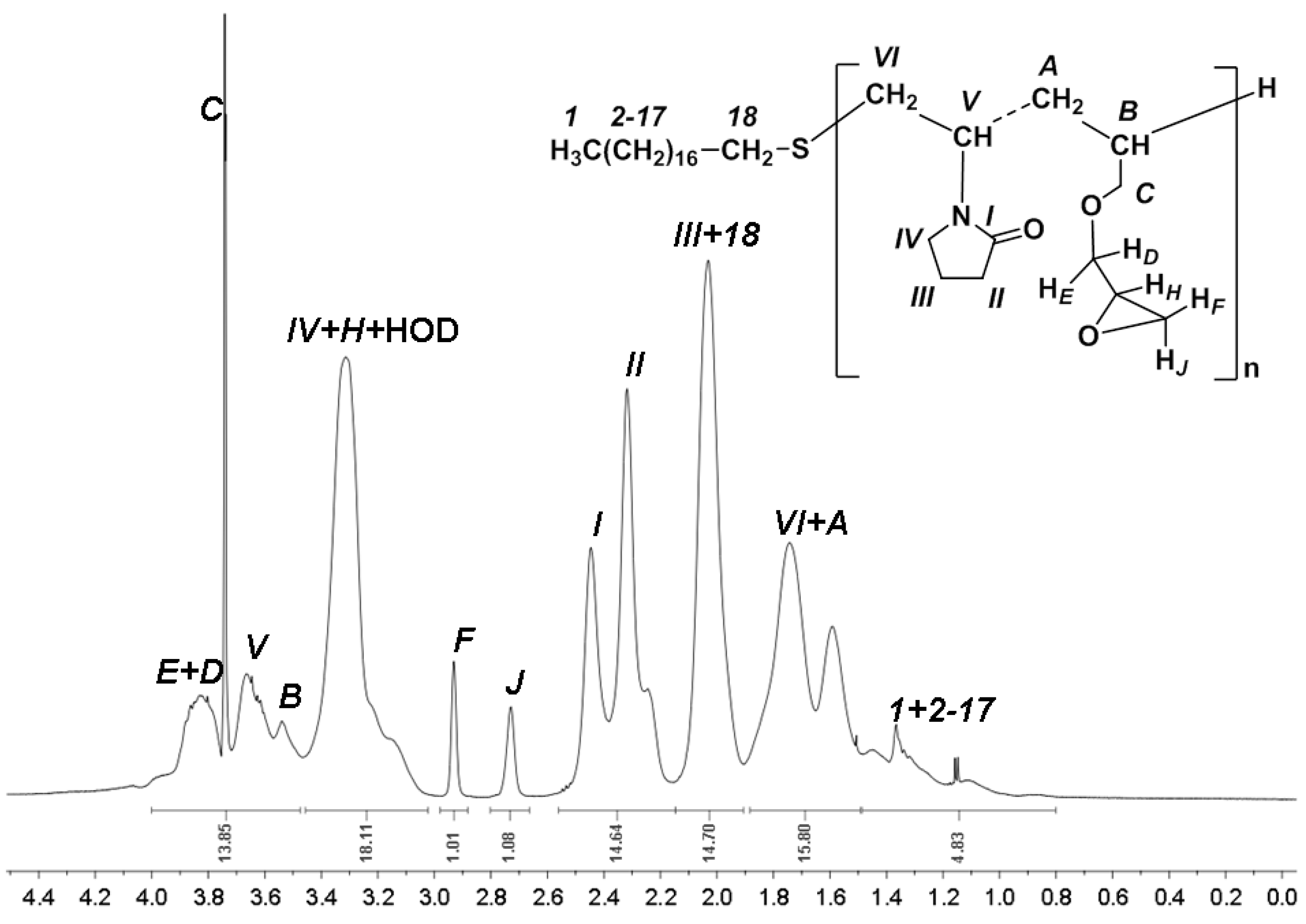
Figure 1. 1H NMR spectrum of the NVPAGE.
Jcs 06 00247 g001
Figure 2. Surface tension isotherm at the water/air interface of the NVPAGE amphiphilic copolymer at a temperature of 298 K.
Jcs 06 00247 g002
Figure 3. Numerical distribution (continuous) and intensity distribution (dotted line) of particle diameters in an aqueous solution of NVPAGE amphiphilic copolymer at a temperature of 298 K.
Jcs 06 00247 g003
Scheme 1. Conjugation of Trisaccharide A with the crown of NVPAGE nanoparticles.
Jcs 06 00247 sch001
Figure 4. Fragments of the HSQC spectra of the original NVPAGE polymer and the vectorized conjugate based on it.
Jcs 06 00247 g004
Figure 5. Fragments of 1H NMR spectra of meglumine and polymer-based conjugate.
Jcs 06 00247 g005
Figure 6. Fragments of 1H NMR spectra (anomeric region) of free trisaccharide A and polymer-based conjugate.
Jcs 06 00247 g006
Table 1. 1H- and 13C-NMR assignment for conjugate (Selected peaks, 600 MHz, D2O).
1-Gal1-GalNAc1-FucMe MeGlu1a
MeGlu
1b MeGlu2
MeGlu
3
MeGlu
4
MeGlu
5
MeGlu
6a MeGlu6b MeGlu
1H4.52 (t)5.16 (d)5.28 (d)2.53 (s)2.88 (m)2.92 (m)3.98 (m)3.76 (m)3.64 (m)3.76 (m)3.64 (m)3.80 (dd)
13C101.5991.3898.7534.8152.7970.5271.9271.9071.9263.74
Table 2. Chemical shifts of the 1H and 13C atoms in the NMR spectra of free meglumine and the meglumine residue in the polymer-based conjugate.
Me1a1b23456a6b
Meglumine
1H2.342.622.683.883.753.633.753.643.81
13C35.8353.5872.2372.2072.1072.2064.09
Conjugate
1H2.532.882.923.983.763.643.763.643.80
13C34.8152.7970.5271.9271.9071.9263.74
Publisher’s Note: MDPI stays neutral with regard to jurisdictional claims in published maps and institutional affiliations.