Article

Comparison of Coronal Discoloration Induced by White MTA and CEM Cement

1
Department of Endodontics, Dental Caries Prevention Research Center, Qazvin University of Medical Sciences, Qazvin 3419915315, Iran
2
Department of Endodontics, Dental School, Shahid Beheshti University of Medical Sciences, Tehran 19857-17443, Iran
3
Department Research Laboratory, Howard University College of Dentistry, Washington, DC 20059, USA
4
Dental Research Laboratory, Howard University College of Dentistry, Washington, DC 20059, USA
5
Department of Oral and Maxillofacial Sciences, Sapienza University of Rome, 00161 Rome, Italy
6
Department of Oral and Maxillofacial Radiology, Dental Caries Prevention Research Center, Qazvin University of Medical Sciences, Qazvin 3419915315, Iran
*
Authors to whom correspondence should be addressed.
J. Compos. Sci. 2022, 6(12), 371; https://doi.org/10.3390/jcs6120371
Submission received: 23 September 2022 / Revised: 14 November 2022 / Accepted: 30 November 2022 / Published: 5 December 2022
(This article belongs to the Special Issue Feature Papers in Journal of Composites Science in 2022)

Abstract

:
Coronal discoloration of endodontically treated teeth is a challenge in clinical dentistry. This study aimed to compare coronal discoloration induced by White Mineral Trioxide Aggregate and Calcium-enriched mixture cement. Fifty single-rooted, unrestored premolar teeth extracted for orthodontic reasons were selected. After access cavity preparation, all the root canals were instrumented with MTWO rotary files up to #40.6%. The specimens were randomly assigned to two experimental groups, White Mineral Trioxide Aggregate and Calcium-enriched mixture cement groups (n = 20), and two control groups (n = 5). In the White Mineral Trioxide Aggregate and Calcium-enriched mixture cement groups, the material was condensed via the access cavity 3 mm below the cementoenamel junction to a thickness of 3 mm. Tooth color was assessed using computer analysis of digital images. Tooth color measurements were recorded at eight time intervals: before material placement (but after tooth preparation), at 24 h, 48 h, one week, two weeks, four weeks, eight weeks, and sixteen weeks after material placement. Data were analyzed using t-test, ANOVA, repeated measure ANOVA, and Tukey HSD tests. The significance level was set at 5% for all the tests. Cervical discoloration of teeth in both experimental groups significantly increased over time (p < 0.05). However, samples in the White Mineral Trioxide Aggregate group showed more discoloration in cervical regions than Calcium-enriched mixture cement specimens after two, four, eight, and sixteen weeks (p < 0.05). Applying both White Mineral Trioxide Aggregate and Calcium-enriched mixture cement induced coronal discoloration; however, White Mineral Trioxide Aggregate samples exhibited greater cervical discoloration than Calcium-enriched mixture cement specimens after two, four, eight, and sixteen weeks.

1. Introduction

The unfavorable aesthetic appearance of endodontically treated teeth is challenging in clinical dentistry. Coronal discoloration is even one of the disadvantages of a new biologically based dental procedure called regenerative endodontic treatment [1].
Mineral trioxide aggregate (MTA) is used for several endodontic procedures, such as vital pulp therapy, apexification, and regenerative endodontic treatment [2]. However, it was reported that MTA induces coronal discoloration [3,4] and should be used with caution when it is used to fill pulp chambers as it may yield aesthetically displeasing outcomes [5]. Initially, MTA was a gray powder, but due to the coronal tooth discoloration associated with it [6,7], white MTA (wMTA) was developed and has been commercially available since 2002 [8]. Although wMTA was thought to be more suitable for use as a pulp capping material in teeth in the esthetic zone [9,10], it was shown that applying both gray and white MTA formulations induces a significant degree of tooth discoloration [5].
Calcium-enriched mixture (CEM) cement was introduced to dentistry in 2006 as an endodontic filling material [11]. It is biocompatible and has many favorable physical and biological properties [12,13,14]. The clinical applications of CEM cement are similar to MTA [14]. It can be successfully used for vital pulp therapy [15], managing internal root resorption [16], and repairing furcation perforations [17]. CEM cement was also used for the pulpotomy of permanent molar teeth with established irreversible pulpitis [18]. However, some studies have reported contradictory results with the use of MTA and CEM cement [19,20,21,22]. Therefore, this study aimed to evaluate and compare coronal discoloration induced by wMTA and CEM cement using the computer analysis of digital images.

2. Materials and Methods

The protocol of this study was approved by the Ethics Committees of Qazvin University of Medical, Qazvin, Iran (QUMS.REC.1394.388). Considering the effect size of 0.35 for repeated measured ANOVA statistical test, alpha error of 0.05 and power of the study equal to 0.80, using G*Power software, the total sample size was determined to be 40, and 20 samples in each experimental group were included in the study [19,20].
Fifty single-rooted, unrestored premolar teeth extracted for orthodontic reasons were selected. Teeth with enamel hypoplasia, fluorosis, or any other type of discoloration were excluded. To disinfect the samples, they were immersed in a 2% thymol solution for 24 h. Then, specimens were stored in normal saline before the experiment. The samples were examined radiographically and teeth with cracks, calcified root canals, carious lesions, or any restoration were excluded. Extrinsic stains were removed using a rubber cup and pumice.
The access cavity was prepared using an 008-diamond fissure bur in a high-speed handpiece. A #15 K-file (Dentsply Maillefer, Tulsa, Ok, USA) was used to determine the working length 1 mm shorter than the anatomic apical foramen. The working length was then confirmed radiographically.
Root canal preparation was carried out using the Mtwo rotary instrument (VDW, Germany) up to #40.6%. The root canals were then irrigated using 10 mL of 2.5% sodium hypochlorite to remove any remaining pulpal tissue. Next, 3.0 mL of 17% EDTA (Ariadent, Tehran, Iran) was introduced and allowed to remain in the root canals for 3 min. Then, a final flush with 1.0 mL of 2.5% sodium hypochlorite was performed, followed by 5.0 mL of normal saline. The specimens were randomly assigned to two experimental groups (n = 20) and two control groups: negative (n = 5) and positive (n = 5). MTA is a mixture of refined Portland cement and bismuth oxide and was reported to contain trace amounts of SiO2, CaO, MgO, K2SO4, and Na2SO4 [23]. The concentrations of carborundum (Al2O3), periclase (MgO), and FeO were lowered in white MTA compared to gray MTA, but these metal oxides are still present in white preparations [24,25] [Table 1].
In the MTA specimen group, wMTA (ProRoot MTA; Dentsply Tulsa Dental, TN) was mixed according to the manufacturer’s instructions. A sterile cotton pellet impregnated with normal saline solution (#4; Richmond Dental, Charlotte, NC) was placed 3 mm under the CEJ via the access cavity. Then, the mixture was condensed on the cotton pellet to a thickness of 3 mm with an endodontic plugger (#2/3 and #2/4; Maillefer, Dentsply, Switzerland). After MTA placement, its thickness was confirmed radiographically. Next, a sterile cotton pellet impregnated with normal saline solution (#4; Richmond Dental, Charlotte, NC) was loosely placed on the MTA. Finally, the tooth was restored with a temporary material (Coltozol, Coltene, Switzerland).
In the CEM cement group, the samples were prepared in the same manner as the MTA specimen group. However, CEM cement (BioniqueDent, Tehran, Iran) was placed instead of MTA. CEM cement contains CaO, SO3, P2O5, and SiO2 [25,26] [Table 1].
In the negative control groups, no filling material was used and just the moistened sterile cotton pellets were placed in the access cavity. In the positive control group, instead of MTA or CEM cement, high-copper dental amalgam (Dispersalloy, Dentsply Caulk, Milford, DE, USA) was used as a filling material. The teeth were stored individually in marked tubes containing distilled water in an incubator at 37 °C.
To examine tooth discoloration over time, color measurements were recorded at eight time intervals: before material placement (but after tooth preparation), at 24 h, 48 h, one week, two weeks, four weeks, eight weeks, and sixteen weeks after material placement. To prepare the specimens for color measurements, they were each attached to a fixed bed of sticky wax on a gray background. Standardized digital images were taken of the teeth with a digital camera (Olympus C-5000Zoom, 5.0 MP, 12X Optical, Tokyo, Japan) in a darkened room under two 60 W lights. To provide a reference for shade control during analysis, a shade reference card was included in each photograph. Then, images were imported into Adobe Photoshop CS6. The buccal surface of the teeth was divided into three horizontal sections (occlusal, middle, and cervical) and a circular area of 74 pixels in diameter in the center of the cervical section was used to perform color measurements.
L*a*b* values, according to Cal et al. [27], were determined in the cervical area where ‘L’ was the lightness, and ‘a’ and ‘b’ showed the chroma, in which +a, −a, +b, −b indicated red, green, yellow, and blue, respectively. The color difference (ΔE) was calculated using the following formula [28]: ΔE = [(ΔL)2 + (Δa)2 +(Δb)2]1/2.
The Kolmogorov–Smirnov test was used to determine the normality of the dispersal distribution of parameters. Afterward, the data were analyzed by t-test, ANOVA, repeated measures ANOVA, and Tukey HSD tests. The significance level was set at 5% for all the tests.

3. Results

Figure 1 shows a sample of each experimental group at 16 weeks. The findings related to the discoloration (ΔE) of specimens in the control and experimental groups are summarized in Table 2.
Based on the results of the repeated measures ANOVA, cervical discoloration for the MTA, CEM cement, and positive control groups significantly increased over time (p < 0.001). However, the MTA specimens displayed more discoloration than the CEM cement specimens at 2-, 4-, 8-, and 16-week intervals after material placement (p < 0.01). In addition, the results of the ANOVA and Tukey tests showed that the positive control group showed more cervical discoloration than the experimental groups at 4, 8 and 16 weeks, and the differences were significant (p < 0.05). On the other hand, the negative control group’s discoloration was not significantly different over time. Based on the results of the independent t-test at 24 and 48 h and one week after material application, discoloration in the MTA group was not significantly different from the CEM cement group (p = 0.132, p = 0.658, and p = 0.914).

4. Discussion

The findings of the present ex vivo study demonstrated that both wMTA and CEM cement discolored extracted teeth, and the amount of discoloration increased over time. Although previous publications concerning discoloration associated with wMTA reported similar results [2,3,5]. Some previous studies were consistent with the present study, indicating that CEM cement caused less coronal discoloration than MTA [29,30]. On the other hand, Araghi et al., Arman et al., and Madani et al. did not report any significant difference between discolorations caused by CEM cement and MTA [20,21,22]. It should be pointed out that the sample sizes were small in the above studies. In a study by Farhang et al., the discoloration caused by CEM cement at the 1-month interval was less than that caused by MTA; however, there were no significant differences between these two materials over time [31]. In the present study, over time, CEM had significantly less discoloration. In the study of Farhang R. et al., the entire pulp chamber was filled with the investigated material, which is different from this study [31]. On the other hand, according to the results of Rohani et al., who compared the discoloration of CEM and MTA by colorimeter, no coronal color change was reported for CEM [19].
It is important to note that MTA and CEM cement are chemically different [32]. Phosphorous is one of the major elements of CEM cement; however, it is only slightly detectable in MTA [32]. Our findings indicated that discoloration resulting from wMTA was significantly more noticeable than that of the CEM cement. The mechanism by which CEM cement and wMTA impact and alter coronal tooth color is currently unclear. It was suggested that the oxidation and incorporation of the remaining iron content within the dental material powder into the calcium aluminoferrite phase of the set material may cause tooth discoloration [3]. Asgary et al. [32] reported that wMTA contains more iron oxide than CEM cement. They also showed that the percentage of other metal oxides, such as titanium oxide, aluminum oxide, and magnesium oxide in CEM cement, is less than that of wMTA.
On the other hand, bismuth oxide, which was added to MTA for radiopacity, might cause coronal discoloration. CEM cement does not have bismuth oxide as one of its ingredients. New calcium silicate cement such as Biodentine, does not have bismuth oxide and causes less coronal discoloration. In the present study, two groups were included to confirm the study method. In the negative control group, only a sterile cotton pellet impregnated with normal saline solution was placed and no cement was applied. In the positive control group, amalgam was used, which causes dentin discoloration as a restorative material. Therefore, some studies have used amalgam in the positive control group [33,34,35].
Several techniques, such as visual subjective comparisons, spectrophotometric analysis, colorimetric analysis, and computer analysis of digital images, are currently used to assess tooth color. For instance, external light conditions, the observer’s experience, age, the human eye’s fatigue, and color blindness can all impact visual color determination, leading to inconsistent results [36,37]. Additionally, instruments such as spectrophotometers and colorimeters were used in several studies for the color measurement of a wide range of materials and substrates [38,39]. These instruments are successfully used to measure tooth discoloration; however, they have some disadvantages. For example, spectrophotometric equipment is complex and expensive [38]. In addition, previous studies have shown similar accuracy for computer analysis and spectrometry methods. On the other hand, colorimeters are designed to measure flat surfaces’ color, while teeth are usually not flat and may have surface anomalies [40]. Another approach for measuring and examining tooth color is a computer analysis of digital images. This technique was successfully used in several studies [3,41,42] and does not need complex equipment or outstanding bias. Furthermore, this method is sensitive, objective, and reproducible despite its limitations [39].
One of the principal applications of the studied cements that can cause coronal discoloration is in vital pulp therapy and regenerative treatments. The present study was designed accordingly. However, since the cement is placed in the vicinity of blood under clinical conditions during treatment, the results of the present study cannot be extended to clinical conditions, which is a limitation of the present study. Madani et al. [22] studied coronal discoloration caused by MTA Angelus, CEM, and Biodentine in the vicinity of blood, reporting that Biodentine caused less coronal discoloration than MTA Angelus, with no significant difference from CEM cement. In addition, all the studied cements resulted in more discoloration in the vicinity of blood compared to the vicinity of normal saline solution.

5. Conclusions

Within the limitations of the present ex vivo study, it can be concluded that applying both wMTA and CEM cement to teeth induced coronal discoloration. However, wMTA-treated specimens exhibited greater tooth color change than CEM cement-treated teeth at 2-, 4-, 8-, and 16-week intervals.

Author Contributions

Conceptualization, M.A. and R.R.; methodology, A.Z.; software, S.A.; validation, S.A., A.Z. and M.J.E.; formal analysis, A.D.; investigation, A.Z. and A.M.G.; resources, A.M.G.; data curation, R.R.; writing—original draft preparation, M.A., N.M.M. and M.T.; writing—review and editing, M.J.E. and L.T.; visualization, M.T.; supervision, L.T.; project administration, M.T.; All authors have read and agreed to the published version of the manuscript.

Funding

This research received no external funding.

Data Availability Statement

Not applicable.

Acknowledgments

The authors would like to thank from the Vice Chancellor for Research at Qazvin University of Medical Sciences and the Office of the Associate Dean for Research at Qazvin Dental School.

Conflicts of Interest

The authors declare no conflict of interest.

References

  1. Nosrat, A.; Homayounfar, N.; Oloomi, K. Drawbacks and Unfavorable Outcomes of Regenerative Endodontic Treatments of Necrotic Immature Teeth: A Literature Review and Report of a Case. J. Endod. 2012, 38, 1428–1434. [Google Scholar] [CrossRef] [PubMed]
  2. Parirokh, M.; Torabinejad, M. Mineral trioxide aggregate: A comprehensive literature review—Part III: Clinical applications, drawbacks, and mechanism of action. J. Endod. 2010, 36, 400–413. [Google Scholar] [CrossRef] [PubMed]
  3. Felman, D.; Parashos, P. Coronal Tooth Discoloration and White Mineral Trioxide Aggregate. J. Endod. 2013, 39, 484–487. [Google Scholar] [CrossRef] [PubMed]
  4. Krastl, G.; Allgayer, N.; Lenherr, P.; Filippi, A.; Taneja, P.; Weiger, R. Tooth discoloration induced by endodontic materials: A literature review. Dent. Traumatol. 2013, 29, 2–7. [Google Scholar] [CrossRef]
  5. Ioannidis, K.; Mistakidis, I.; Beltes, P.; Karagiannis, V. Spectrophotometric analysis of coronal discolouration induced by grey and white MTA. Int. Endod. J. 2013, 46, 137–144. [Google Scholar] [CrossRef]
  6. Karabucak, B.; Li, D.; Lim, J.; Iqbal, M. Vital pulp therapy with mineral trioxide aggregate. Dent. Traumatol. 2005, 21, 240–243. [Google Scholar] [CrossRef]
  7. Bogen, G.; Kim, J.S.; Bakland, L.K. Direct pulp capping with mineral trioxide aggregate: An observational study. J. Am. Dent. Assoc. 2008, 139, 305–315. [Google Scholar] [CrossRef]
  8. Asgary, S.; Parirokh, M.; Eghbal, M.J.; Brink, F. A Comparative Study of White Mineral Trioxide Aggregate and White Portland Cements Using X-ray Microanalysis. Aust. Endod. J. 2004, 30, 89–92. [Google Scholar] [CrossRef]
  9. Bogen, G.; Kuttler, S. Mineral Trioxide Aggregate Obturation: A Review and Case Series. J. Endod. 2009, 35, 777–790. [Google Scholar] [CrossRef]
  10. Bakland, L.K. Revisiting Traumatic Pulpal Exposure: Materials, Management Principles, and Techniques. Dent. Clin. N. Am. 2009, 53, 661–673. [Google Scholar] [CrossRef]
  11. Asgary, S.; Eghbal, M.J.; Parirokh, M.; Torabzadeh, H. Sealing Ability of Three Commercial Mineral Trioxide Aggregates and an Experimental Root-End Filling Material. Iran. Endod. J. 2006, 1, 101–105. [Google Scholar] [PubMed]
  12. Zanza, A.; D’Angelo, M.; Reda, R.; Gambarini, G.; Testarelli, L.; Di Nardo, D. An Update on Nickel-Titanium Rotary Instruments in Endodontics: Mechanical Characteristics, Testing and Future Perspective—An Overview. Bioengineering 2021, 8, 218. [Google Scholar] [CrossRef] [PubMed]
  13. Bhandi, S.; Mashyakhy, M.; Abumelha, A.; Alkahtany, M.; Jamal, M.; Chohan, H.; Raj, A.; Testarelli, L.; Reda, R.; Patil, S. Complete Obturation—Cold Lateral Condensation vs. Thermoplastic Techniques: A Systematic Review of Micro-CT Studies. Materials 2021, 14, 4013. [Google Scholar] [CrossRef] [PubMed]
  14. Utneja, S.; Nawal, R.R.; Talwar, S.; Verma, M. Current perspectives of bio-ceramic technology in endodontics: Calcium enriched mixture cement—review of its composition, properties and applications. Restor. Dent. Endod. 2015, 40, 1–13. [Google Scholar] [CrossRef]
  15. Asgary, S.; Eghbal, M.J.; Parirokh, M.; Ghanavati, F.; Rahimi, H. A comparative study of histologic response to different pulp capping materials and a novel endodontic cement. Oral Surg. Oral Med. Oral Pathol. Oral Radiol. Endodontol. 2008, 106, 609–614. [Google Scholar] [CrossRef]
  16. Asgary, S.; Ahmadyar, M. One-visit endodontic retreatment of combined external/internal root resorption using a calcium-enriched mixture. Gen. Dent. 2012, 60, e244–e248. [Google Scholar]
  17. Samiei, M.; Eghbal, M.J.; Parirokh, M.; Abbas, F.M.; Asgary, S. Repair of furcal perforation using a new endodontic cement. Clin. Oral Investig. 2010, 14, 653–658. [Google Scholar] [CrossRef]
  18. Asgary, S.; Ehsani, S. Permanent molar pulpotomy with a new endodontic cement: A case series. J. Conserv. Dent. 2009, 12, 31–36. [Google Scholar] [CrossRef] [Green Version]
  19. Rouhani, A.; Akbari, M.; Farhadi-Faz, A. Comparison of Tooth Discoloration Induced by Calcium-Enriched Mixture and Mineral Trioxide Aggregate. Iran. Endod. J. 2016, 11, 175–178. [Google Scholar] [CrossRef]
  20. Godiny, M.; Araghi, S.; Khavid, A.; Saeidipour, M. In vitro evaluation of coronal discoloration following the application of calcium-enriched mixture cement, Biodentine, and mineral trioxide aggregate in endodontically treated teeth. Dent. Res. J. 2019, 16, 53. [Google Scholar] [CrossRef]
  21. Arman, M.; Khalilak, Z.; Rajabi, M.; Esnaashari, E.; Saati, K. In Vitro Spectrophotometry of Tooth Discoloration Induced by Tooth-Colored Mineral Trioxide Aggregate and Calcium-Enriched Mixture Cement. Iran. Endod. J. 2015, 10, 226–230. [Google Scholar] [CrossRef] [PubMed]
  22. Madani, Z.; Alvandifar, S.; Bizhani, A. Evaluation of tooth discoloration after treatment with mineral trioxide aggregate, calcium-enriched mixture, and Biodentine® in the presence and absence of blood. Dent. Res. J. 2019, 16, 377–383. [Google Scholar]
  23. Roberts, H.W.; Toth, J.M.; Berzins, D.W.; Charlton, D.G. Mineral trioxide aggregate material use in endodontic treatment: A review of the literature. Dent. Mater. 2008, 24, 149–164. [Google Scholar] [CrossRef] [PubMed] [Green Version]
  24. Asgary, S.; Parirokh, M.; Eghbal, M.J.; Brink, F. Chemical Differences Between White and Gray Mineral Trioxide Aggregate. J. Endod. 2005, 31, 101–103. [Google Scholar] [CrossRef]
  25. Ertas, H.; Kucukyilmaz, E.; Ok, E.; Uysal, B. Push-out bond strength of different mineral trioxide aggregates. Eur. J. Dent. 2014, 8, 348–352. [Google Scholar] [CrossRef]
  26. Asgary, S.; Ahmadyar, M. Vital pulp therapy using calcium-enriched mixture: An evidence-based review. J. Conserv. Dent. 2013, 16, 92–98. [Google Scholar] [CrossRef]
  27. Cal, E.; Sonugelen, M.; Güneri, P.; Kesercioglu, A.; Köse, T. Application of a digital technique in evaluating the reliability of shade guides. J. Oral Rehabil. 2004, 31, 483–491. [Google Scholar] [CrossRef]
  28. O’Brien, W. Dental Materials and Their Selection, 2nd ed.; Quintessence: Chicago, IL, USA, 1997; pp. 28–30. [Google Scholar]
  29. Eghbal, M.J.; Torabzadeh, H.; Bagheban, A.A.; Shamszadeh, S.; Marvasti, L.A.; Asgary, S. Color stability of mineral trioxide aggregate and calcium enriched mixture cement. J. Investig. Clin. Dent. 2016, 4, 341–346. [Google Scholar] [CrossRef]
  30. Esmaeili, B.; Alaghehmand, H.; Kordafshari, T.; Daryakenari, G.; Ehsani, M.; Bijani, A. Coronal Discoloration Induced by Calcium-Enriched Mixture, Mineral Trioxide Aggregate and Calcium Hydroxide: A Spectrophotometric Analysis. Iran. Endod. J. 2016, 11, 23–28. [Google Scholar] [CrossRef]
  31. Farhang, R.; Hekmatfar, S.; Samadi, V.; Meraji, A.; Jafari, K. Comparison of Tooth Discoloration Induced by Calcium-enriched Mixture, Mineral Trioxide Aggregate, and Endocem. World J. Dent. 2020, 11, 392–395. [Google Scholar] [CrossRef]
  32. Asgary, S.; Eghbal, M.J.; Parirokh, M.; Ghoddusi, J.; Kheirieh, S.; Brink, F. Comparison of mineral trioxide aggregate’s composition with Portland cements and a new endodontic cement. J. Endod. 2009, 35, 243–250. [Google Scholar] [CrossRef] [PubMed]
  33. Scholtanus, J.D.; Özcan, M.; Huysmans, M.-C.D. Penetration of amalgam constituents into dentine. J. Dent. 2009, 37, 366–373. [Google Scholar] [CrossRef]
  34. Scholtanus, J.D.; Özcan, M. Clinical longevity of extensive direct composite restorations in amalgam replacement: Up to 3.5 years follow-up. J. Dent. 2014, 42, 1404–1410. [Google Scholar] [CrossRef] [PubMed]
  35. Enan, E.T.; Yousef, E.A. Discoloration of MTA Filled Teeth With and Without Dentine Bonding Agent. Ann. Int. Med. Dent. Res. 2018, 4, 34–37. [Google Scholar] [CrossRef]
  36. Watts, A.; Addy, M. Tooth discolouration and staining: A review of the literature. Br. Dent. J. 2001, 190, 309–316. [Google Scholar] [CrossRef]
  37. Hill, A.R. How we see colour. In Colour Physics for Industry; McDonald, R., Ed.; H. Charlesworth & Co Ltd.: Huddersfield, UK, 1987; pp. 211–281. [Google Scholar]
  38. Joiner, A. Tooth colour: A review of the literature. J. Dent. 2004, 32, 3–12. [Google Scholar] [CrossRef]
  39. Ahrari, F.; Akbari, M.; Mohammadpour, S.; Forghani, M. The efficacy of laser-assisted in-office bleaching and home bleaching on sound and demineralized enamel. Laser Ther. 2015, 24, 257–264. [Google Scholar] [CrossRef] [Green Version]
  40. Bolt, R.A.; Ten Bosch, J.J.; Coops, J.C. Influence of window size in small-window colour measurements, particularly of teeth. Phys. Med. Biol. 1994, 39, 1133–1142. [Google Scholar] [CrossRef]
  41. Lath, D.L.; Guan, Y.H.; Lilley, T.H. Comparison of colorimetry and image analysis for quantifying tooth whiteness—A preliminary study. In Dental Morphology 2001; Brook, A., Ed.; Academic Press: Sheffield, UK, 2001; pp. 229–237. [Google Scholar]
  42. Gerlach, R.W.; Barker, M.L.; Sagel, P.A. Objective and subjective whitening response of two self-directed bleaching systems. Am. J. Dent. 2002, 15, 7A–12A. [Google Scholar]
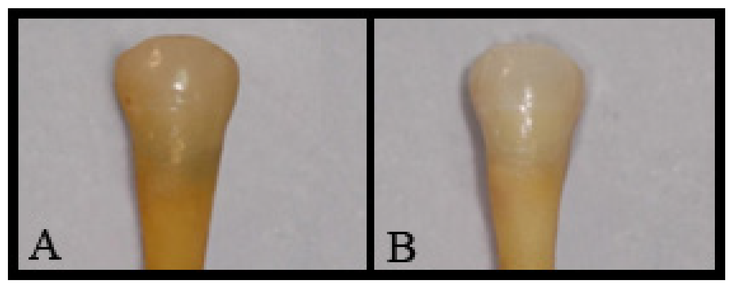
Figure 1. A sample of each experimental group at 16 weeks. (A) MTA; (B) CEM cement.
Jcs 06 00371 g001
Table 1. Composition of wMTA and CEM cement.
MaterialswMTA (%)CEM Cement (%)
CaO44.1651.75
SiO221.256.32
Bi2O316.13ND
Al2O31.870.93
Cl0.390.21
Na2O0.030.34
FeO0.39ND
P2O50.278.49
SO30.559.53
MgO1.360.24
TiO20.09ND
H2O and CO13.5122.19
Table 2. Discoloration values expressed as mean ± standard deviation for experimental and control groups over the time intervals.
TimeGroupsMean ± SD
Cervical discoloration
at 24 h
MTA0.24 ± 0.47
CEM cement0.28 ± 0.92
Positive Control0.28 ± 1.42
Negative Control0.24 ± 1.73
Cervical discoloration
at 48 h
MTA0.86 ± 0.47
CEM cement0.67 ± 0.90
Positive Control0.54 ± 1.52
Negative Control0.23 ± 0.75
Cervical discoloration
at 1 week
MTA1.74 ± 1.48
CEM cement0.97 ± 2.03
Positive Control1.41 ± 1.50
Negative Control0.33 ± 0.72
Cervical discoloration
at 2 weeks
MTA2.57 ± 1.47
CEM cement1.42 ± 1.79
Positive Control4.86 ± 1.39
Negative Control0.31 ± 0.69
Cervical discoloration
at 4 weeks
MTA3.31 ± 1.45
CEM cement2.20 ± 1.95
Positive Control6.59 ± 1.63
Negative Control0.52 ± 0.91
Cervical discoloration
at 8 weeks
MTA4.50 ± 1.43
CEM cement3.94 ± 2.04
Positive Control6.18 ± 1.52
Negative Control0.47 ± 0.82
Cervical discoloration
at 16 weeks
MTA5.50 ± 1.43
CEM cement3.94 ± 2.04
Positive Control6.18 ± 1.52
Negative Control0.47 ± 0.82
Publisher’s Note: MDPI stays neutral with regard to jurisdictional claims in published maps and institutional affiliations.