Article

Multifunctional Polysaccharide Hydrogel Ameliorates Cardiac Function After Myocardial Infarction via Antioxidant, Immunomodulatory, and Pro-Angiogenic Activities


1
State Key Laboratory of Advanced Medical Materials and Devices, Engineering Research Center of Pulmonary and Critical Care Medicine Technology and Device (Ministry of Education), Tianjin Key Laboratory of Biomaterial Research, Institute of Biomedical Engineering, Tianjin Institutes of Health Science, Chinese Academy of Medical Science & Peking Union Medical College, Tianjin 300192, China
2
Institute of Basic Medical Sciences, Chinese Academy of Medical Sciences & Peking Union Medical College, Beijing 100005, China
3
Cancer Institute, University College London, London WCIE 6DD, UK
*
Author to whom correspondence should be addressed.
These authors contributed equally to this work.
J. Compos. Sci. 2026, 10(6), 287; https://doi.org/10.3390/jcs10060287
Submission received: 18 April 2026 / Revised: 15 May 2026 / Accepted: 18 May 2026 / Published: 25 May 2026
(This article belongs to the Special Issue Functional Composites: Fabrication, Properties and Applications)

Abstract

Myocardial infarction (MI) triggers excessive oxidative stress, a detrimental immune response, and insufficient angiogenesis, which collectively impede effective cardiac repair. This study developed a multifunctional composite polysaccharide hydrogel, termed KgXdgel, based on konjac glucomannan (KGM) and xanthan gum (XG) functionalized with gallic acid (GA) and dopamine (DA), respectively, to integrate reactive oxygen species (ROS) scavenging, macrophage polarization, and pro-angiogenic activities. In vitro assays demonstrated that the KgXdgel hydrogel exhibited excellent cytocompatibility, effectively scavenged ROS, promoted the polarization of macrophages towards the reparative M2 phenotype, and enhanced the migration and tube formation of human umbilical vein endothelial cells. In a rat MI model, treatment with KgXdgel significantly improved cardiac function (e.g., left ventricular ejection fraction, LVEF; left ventricular fractional shortening, LVFS), attenuated left ventricular dilation (LVIDs), and favorably modulated the post-infarction microenvironment. This was evidenced by the upregulation of the M2 marker CD163 and the angiogenic factor VEGF, alongside the downregulation of pro-inflammatory cytokines (e.g., IL-1β, TNF-α) and the M1 marker iNOS. These findings conclusively demonstrate that the KgXdgel hydrogel synergistically promotes cardiac repair post-MI through its integrated antioxidant, immunomodulatory, and pro-angiogenic functions, presenting a promising multi-targeted therapeutic strategy.

1. Introduction

MI remains one of the leading causes of global morbidity and mortality from cardiovascular diseases, posing a significant public health challenge [1,2]. The pathological essence of MI is the acute interruption of coronary blood flow, leading to massive coagulative necrosis of cardiomyocytes due to sustained ischemia and hypoxia [3,4]. Although current reperfusion therapies can rapidly restore epicardial coronary blood flow, they often fail to salvage microvascular perfusion and may even induce “reperfusion injury,” where oxidative stress is a key pathological mediator [5]. The infarct zone, due to disturbed energy metabolism and mitochondrial dysfunction, produces excessive ROS, which not only directly damage cellular structures but also act as signaling molecules to perpetuate the inflammatory response and suppress endogenous repair mechanisms [6,7]. Concurrently, the innate immune system, particularly macrophages, plays a dual role: early pro-inflammatory M1 macrophages dominate the clearance of necrotic debris, while the repair phase requires a shift towards anti-inflammatory, pro-reparative M2 macrophages to suppress excessive inflammation and promote angiogenesis and tissue remodeling [8,9]. However, the persistent oxidative stress microenvironment severely impedes M2 polarization, creating a vicious cycle of “oxidative stress-chronic inflammation” that ultimately leads to repair failure and pathological fibrosis [10,11]. Furthermore, inadequate angiogenesis limits the delivery of oxygen and nutrients, thereby weakening the repair potential [12,13]. Thus, oxidative stress, immune imbalance, and insufficient angiogenesis are intricately intertwined, forming a complex pathological network that often renders single-target intervention strategies ineffective due to constraints from other pathways [14,15]. Therefore, developing combined therapeutic strategies capable of simultaneously targeting these multiple aspects is crucial for breaking this vicious cycle and creating a favorable microenvironment for cardiac repair.
In recent years, hydrogel-based cardiac tissue engineering has brought new hope for MI treatment [16,17,18]. Recent high-impact studies have further demonstrated that multifunctional injectable hydrogels enable synergistic regulation of oxidative stress, immune microenvironment, and angiogenesis, representing a promising direction for cardiac repair [19]. Owing to their three-dimensional network structure, high water content, and physical properties resembling the natural extracellular matrix (ECM), hydrogels serve as excellent implantable carriers or scaffolds. They can not only provide mechanical support and inhibit adverse ventricular remodeling but also act as platforms for the localized and sustained delivery of bioactive molecules, enabling precise microenvironmental regulation [20,21]. However, most conventional hydrogels are functionally singular, making it difficult to address the complex and dynamic pathological changes following MI [22,23]. Consequently, developing “smart” hydrogels that integrate multiple bioactive functions and synergistically target various repair processes has become a frontier and important direction in current research [24,25].
Based on this background, this study proposes an innovative combined therapeutic strategy: constructing a multifunctional composite hydrogel aimed at synchronously achieving efficient ROS scavenging, guiding macrophage polarization towards the M2 phenotype, and promoting angiogenesis, thereby cooperatively facilitating myocardial repair through multiple pathways. Regarding the material basis, natural polysaccharides have garnered significant attention due to their good biocompatibility, biodegradability, and ease of functionalization. As reported in latest top-tier publications, natural polysaccharide-based hydrogels exhibit superior biocompatibility and tunable bioactivity, making them particularly attractive for cardiac tissue engineering [26,27,28]. This study selected KGM and XG as the hydrogel backbone (KGM-XG, named KXgel). KGM possesses good biocompatibility and biodegradability, and the abundant hydroxyl groups on its molecular chain facilitate functionalization [29,30]. XG exhibits unique shear-thinning behavior and stable physicochemical properties; when blended with KGM, it can form a synergistic three-dimensional network structure, enhancing the mechanical performance and stability of the hydrogel [31,32]. To further endow the hydrogel with targeted bioactivities, we functionalized the polysaccharide backbones with GA and DA, respectively. GA is a natural polyphenolic compound proven to possess remarkable antioxidant activity due to the presence of multiple phenolic hydroxyl groups in its molecule, effectively scavenging various ROS and exhibiting anti-inflammatory properties, which help alleviate oxidative stress during myocardial ischemia–reperfusion injury [33]. Recent advanced studies further confirm that GA functionalization effectively enhances the ROS-scavenging and immunomodulatory performance of hydrogels in myocardial repair systems [34]. DA is renowned for its excellent tissue adhesiveness and recognized ROS-scavenging capacity; its catechol groups can not only efficiently quench free radicals but may also regulate immune responses by participating in cell signal transduction [35,36]. We hypothesized that combining these two functionalized polymers (KGM-GA and XG-DA) would yield a composite hydrogel (named KgXdgel) that integrates the advantageous properties of each component, creating a favorable regenerative microenvironment capable of simultaneously clearing ROS, regulating the immune microenvironment, and supporting angiogenesis.
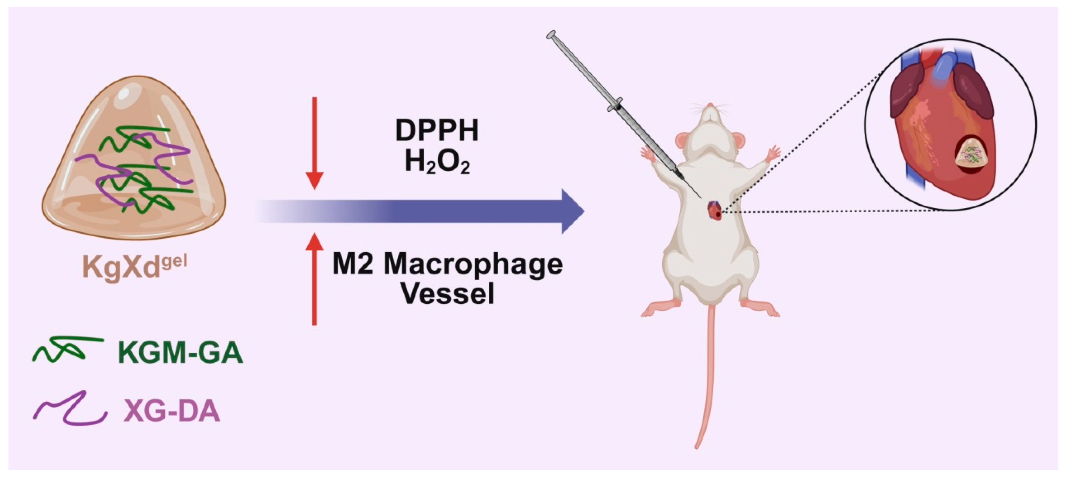
This study systematically evaluated the ROS scavenging efficiency, macrophage polarization regulation, and pro-angiogenic effects of the KgXdgel hydrogel in vitro. As schematically illustrated in Scheme 1, combined with validation in a rat MI model for its comprehensive therapeutic efficacy, the research aims to verify its effectiveness in promoting cardiac repair through the synergistic mechanism of “ROS Scavenging—Immune Regulation—Angiogenesis Promotion”, providing a new paradigm for developing next-generation intelligent cardiac repair materials.

2. Materials and Methods

2.1. Materials

KGM and XG were purchased fromSolarbio Science & Technology Co., Ltd. (Beijing, China). GA, DA, N-Hydroxysuccinimide (NHS), 4-Dimethylaminopyridine (DMAP), and 1-Ethyl-3-(3-dimethylaminopropyl) carbodiimide hydrochloride (EDC·HCl) were obtained from Heowns Biochemical Technology Co., Ltd. (Tianjin, China). 1,1-Diphenyl-2-picrylhydrazyl (DPPH) was acquired from TCI (Shanghai, China). ROS detection kits and Live/Dead Cell Staining Kits were sourced from Beyotime Biotechnology (Shanghai, China). Hematoxylin and Eosin (H&E) Staining Kit was procured from Solarbio Science & Technology Co., Ltd. (Beijing, China). Rat ELISA Kits for VEGF, TNF-α, IL-6, IL-4, IL-1β, IL-10, IFN-γ, Arg-1, and TGF-β were purchased from Jianglai Biotechnology Co., Ltd. (Shanghai, China). Antibodies against VEGF, iNOS, CX43, and CD163 were bought from Proteintech Group, Inc. (Wuhan, China).

2.2. Cells and Animals

The mouse L929 cell line, RAW 264.7 macrophage cell line, Rat cardiomyocyte cell line H9C2, and Human Umbilical Vein Endothelial Cells (HUVECs) were obtained from iCell Bioscience Inc. (Shanghai, China). Sprague Dawley (SD) rats (male, six weeks old) were purchased from Vital River Laboratory Animal Technology Co., Ltd. (Beijing, China). All animal experiments were approved by the Animal Care and Ethics Committee of the Institute of Radiation Medicine, Chinese Academy of Medical Sciences (Approval No. SYXK (Jin) 2019-0002).

2.3. Preparation and Characterization of KgXdgel Hydrogel

KGM-GA was synthesized via an esterification reaction according to a previous method. Briefly, KGM (1 g) was dissolved in 200 mL distilled water under magnetic stirring at 50 °C until completely dissolved. Separately, a mixture of GA (0.21 g), EDC·HCl (0.355 g, 1.85 mM), and DMAP (0.226 g, 1.85 mM) was dissolved in 50 mL distilled water and stirred for 0.5 h for condensation reaction activation. The two solutions were then mixed thoroughly and stirred at 50 °C for 72 h. After cooling to room temperature, the reaction mixture was precipitated in anhydrous ethanol, repeatedly dissolved, and precipitated three times. The final product (KGM-GA) was obtained after drying under vacuum at 60 °C.
XG-DA was synthesized via an amidation reaction based on a previous protocol. XG (0.933 g) was dissolved in 150 mL distilled water under magnetic stirring at room temperature until a uniform liquid state was achieved. Then, EDC·HCl (0.1.920 g, 1 mM) and NHS (0.115 g, 0.999 mM) were added to the solution and stirred in the dark for 1 h to activate XG. Subsequently, DA (0.190 g) was added to the solution, and the reaction proceeded under stirring in the dark at room temperature for 48 h. The reaction mixture was then precipitated in anhydrous ethanol, repeatedly dissolved, and precipitated three times. The final product (XG-DA) was obtained after drying under vacuum at 60 °C.
The KgXdgel hydrogel was formed by physically blending KGM-GA and XG-DA polymers at a 1:1 concentration ratio in pure water under mechanical stirring for 24 h. The chemical structures of KGM-GA, XG-DPA and KgXdgel were confirmed via Fourier transform infrared (FTIR) spectroscopy (Thermo Fisher Scientific, Waltham, MA, USA) and UV-Vis spectra. The morphology and structure of the hydrogel were observed using a Scanning Electron Microscope (SEM, TESCAN MIRA LMS, Brno, Czech Republic). The rheological properties were assessed using an AR 2000ex rheometer (TA Instruments, New Castle, DE, USA). The hydrogel sample was placed between parallel plates with a diameter of 10 mm and a gap of 1 mm. The storage modulus (G′) and loss modulus (G″) were measured within a frequency range of 1–100 rad/s and a strain range of 1–100% at 25 °C. The self-healing capability was evaluated by monitoring the changes in G′ and G″ under continuous step strain scans alternating between high and low strain.

2.4. Biocompatibility Evaluation of KgXdgel

For cytotoxicity assessment, the rat cardiomyocyte cell line H9C2 was used. Cells (5000 per well) were seeded in a 96-well plate using high-glucose DMEM supplemented with 10% fetal bovine serum (FBS) and incubated for 24 h. Subsequently, the medium was replaced with 100 μL of fresh medium containing the lyophilized KXgel or KgXdgel powder dissolved at designated concentrations, and cells were incubated for another 24 h. The hydrogel solution was homogeneously mixed into the culture medium. Cell viability was evaluated using a Cell Counting Kit-8 (CCK-8, Beyotime, C0037). Additionally, cells were stained using the Calcein/PI Live/Dead Viability/Cytotoxicity Assay Kit (Beyotime, C2015S) and imaged with a fluorescence microscope.

2.5. HUVEC Migration Assay (Transwell)

HUVECs (1 × 10⁴ cells) were seeded in the upper chamber of a 24-well Transwell plate. The lower chamber contained 400 μL of cell culture medium homogeneously mixed with 100 μL of KXgel or KgXdgel solution prepared from lyophilized powder. After 24 h of incubation, cells that migrated to the lower chamber were stained with 0.5% crystal violet solution for 30 min and counted.

2.6. HUVEC Tube Formation Assay

Matrigel (50 μL, BD, 356234) was added to a pre-cooled 96-well plate and incubated at 37 °C for 30 min for polymerization. Subsequently, 40 μL of cell culture medium containing 5 × 10³ HUVECs was homogeneously mixed with 10 μL of hydrogel solution and added onto the polymerized Matrigel. After further incubation for 6 h, tube formation was observed, and the tube formation density was quantified using ImageJ (version 1.50i) software.

2.7. ROS Scavenging Ability Evaluation

The free radical scavenging capacity of KXgel and KgXdgel was first evaluated using the DPPH assay. A DPPH ethanol solution (200 μg/mL) was prepared. KXgel and KgXdgel samples were prepared in deionized water, and a series of concentrations of the hydrogel solutions were added to equal volumes of the DPPH solution and incubated at 37 °C for 1 h. The absorbance of the reaction solution at 517 nm was recorded using a microplate reader and used to calculate the free radical scavenging capacity.
To investigate the intracellular ROS scavenging ability of KgXdgel, L929 cells (1 × 10⁵ cells/well) were seeded in a 24-well plate and incubated for 24 h. Except for the negative control group, all other groups were treated with 200 μM H2O2 for 2 h. After removing the medium, the cells were treated with 500 μL of fresh medium (positive control) or fresh medium homogeneously containing dissolved KXgel or KgXdgel. Following 2 h of incubation, cells were stained with 10 μM 2′,7′-Dichlorodihydrofluorescein diacetate (DCFH-DA) at 37 °C for 30 min and observed under an inverted fluorescence microscope.

2.8. Immunomodulation of RAW 264.7 Macrophages by KgXdgel

For macrophage morphology analysis, RAW 264.7 cells (1 × 10⁵ cells/well) were seeded in a 24-well plate and incubated at 37 °C for 24 h. Cells were then treated with 500 μL of culture medium homogeneously mixed with dissolved KXgel or KgXdgel for 48 h. Afterward, cells were washed three times with PBS to remove excess material, and macrophage morphology was captured using a microscope. Cell elongation was statistically analyzed.

2.9. Rat MI Model

Male SD rats (180–200 g, six weeks old) were acclimatized for 3–4 days before MI model induction. Rats were randomly divided into four groups (n = 3 per group): Sham, MI, KXgel, and KgXdgel. After fasting for 12 h, rats were anesthetized with inhaled isoflurane. Tracheal intubation was performed using a 16G catheter needle, and mechanical ventilation was maintained using a small animal ventilator (tidal volume: 3 mL/100 g, frequency: 90 breaths/min). The rat was fixed on a surgical board, the left anterior chest hair was shaved and disinfected, and a ~2 cm longitudinal incision was made along the left side of the sternum. The skin and fascia were sequentially cut, and the pectoralis major and serratus anterior muscles were bluntly separated using hemostatic forceps. A thoracotomy was performed at the fourth intercostal space to expose the heart. The pericardium was carefully torn open with forceps to fully expose the heart structure. The left anterior descending coronary artery was ligated with a 6-0 nylon suture at a point 2–3 mm below the midpoint between the arterial conus and the left auricle. After 30 min of myocardial ischemia, 100 μL of saline or hydrogel was injected into the infarct area using a 1 mL sterile syringe. The Sham group underwent the same thoracotomy procedure without ligation or injection. Finally, the chest was carefully closed, the skin was sutured, and the tracheal tube was removed after the rat resumed spontaneous breathing. Penicillin (200,000 U/rat) was administered intraperitoneally to prevent infection.

2.10. Echocardiography

Rats were anesthetized with inhaled isoflurane. On day 7 post-MI, echocardiography was performed using an ultrasound imaging system. Parameters including left ventricular ejection fraction (LVEF), left ventricular fractional shortening (LVFS), and left ventricular internal diameter at end-systole (LVIDs) were collected and quantitatively analyzed.

2.11. Western Blot Analysis

On day 28, heart tissues were harvested. Approximately 0.1 g of tissue sample was weighed, and 1 mL of RIPA lysis buffer (supplemented with 10 μL PMSF per 1 mL) was added. The tissue was homogenized using a tissue homogenizer, lysed at 4 °C for 30 min, and then centrifuged at 12,000 rpm, 4 °C for 10 min. The supernatant was collected for BCA protein quantification. Approximately 80 μg of total protein was mixed with 5× loading buffer, denatured in boiling water for 10 min, briefly centrifuged, and loaded onto an SDS-PAGE gel. Electrophoresis was performed at 140 V until the loading dye just migrated out of the separating gel. The gel was then equilibrated in 1× transfer buffer for 20 min. PVDF membrane and filter paper were cut to size, and the PVDF membrane was activated with methanol. The transfer stack was assembled in the order: cathode–filter paper–gel–PVDF membrane–filter paper–anode. Transfer was carried out at 200 mA constant current for 1 h. The membrane was blocked with 5% skim milk in TBST. Primary antibodies (VEGF, iNOS, CX43, CD163; 1:1000 dilution) were incubated with the membrane overnight at 4 °C on a shaker. After washing three times with TBST (10 min each), the membrane was incubated with HRP-conjugated secondary antibody (1:3000 dilution) at room temperature for 1 h. Following another three washes with TBST, the membrane was incubated with ECL working solution according to the manufacturer’s instructions for 2 min at room temperature. Excess solution was removed, and the membrane was imaged using a chemiluminescence imaging system. Band intensities were analyzed using Gelpro32 grayscale analysis software.

2.12. ELISA Analysis

On day 28, rats were anesthetized with isoflurane, and blood was collected from the abdominal aorta. The blood samples were centrifuged at 3000 rpm for 10 min at 4 °C to obtain the supernatant (plasma) The levels of VEGF, TNF-α, IL-6, IL-4, IL-1β, IL-10, IFN-γ, Arg-1, and TGF-β in the rat plasma were measured using the corresponding Rat ELISA Kits according to the manufacturers’ instructions. Briefly, 100 μL of sample or standard was added to each well, covered with a sealing tape, and incubated at 37 °C for 2 h. After discarding the liquid, 100 μL of biotinylated detection antibody (1×) was added to each well, covered with new sealing tape, and incubated at 37 °C for 1 h. After washing five times, 90 μL of TMB substrate was added to each well and incubated at 37 °C for 15–30 min. Finally, 50 μL of stop solution was added to each well, and the absorbance at 450 nm was measured within 5 min using a microplate reader.

2.13. Statistical Analysis

All experimental data are presented as the mean ± standard deviation (SD). Differences between groups were assessed using one-way or two-way analysis of variance (ANOVA) in GraphPad Prism 8. A p-value < 0.05 was considered statistically significant (ns: not significant, * p < 0.05, ** p < 0.01, *** p < 0.001). The sample size was 3 unless otherwise stated.

3. Results

3.1. Characterization and Structural Analysis of KgXdgel Hydrogel

The synthesis of KGM-GA and XG-DA polymers involved EDC/DMAP-mediated esterification and EDC/NHS-mediated amidation reactions. The structural configuration of KGM-GA was initially substantiated through the utilisation of Fourier Transform Infrared (FTIR) spectroscopy. As illustrated in Supplementary Figure S1a, the FTIR spectra of KGM and KGM-GA are displayed. The carboxyl group (-COOH) of GA reacts with the hydroxyl group (-OH) of KGM, forming an ester bond that shifts the C=O stretching vibration peak from 1726 cm−1 to 1723 cm−1. The UV-vis spectra of GA, KGM, and KGM-GA are presented in Supplementary Figure S1b. Absorbance peaks were not observed for the KGM solution within the detection range. In contrast, the KGM-GA solution exhibited a distinct peak of absorption at 262 nm, which also appeared in the UV-vis spectrum of GA. These results thus confirm the successful synthesis of KGM-GA.
In addition, the successful synthesis of XG-DA was analysed further using FTIR and UV-Vis spectroscopy. This analysis revealed that the amino group (-NH2) of DA reacted with the carboxyl group (-COOH) of XG to form an amide bond. As demonstrated in Supplementary Figure S2a, the FTIR spectrum of XG-DA displays an absorption peak at 1532 cm−1, which is indicative of the in-plane vibration of the C=C bond within the benzene ring. The UV-visible spectra of DA, XG, and XG-DA are displayed in Supplementary Figure S2b. Absorbance peaks were not detected within the specified detection range for the XG solution. In contrast, the XG-DA solution exhibited a distinct peak of absorption at 278 nm, which was also observed in the UV-visible spectrum of DA. These results thus confirm the successful synthesis of XG-DA.
The KgXdgel hydrogel was subsequently formed by physically blending KGM-GA and XG-DA at a 1:1 concentration ratio. The successful synthesis of KgXdgel was subjected to further analysis via FTIR and UV-Vis spectroscopy. As illustrated in Supplementary Figure S3a, the FTIR spectra of KGM and XG exhibited peaks at 3418 cm−1 and 3422 cm−1, respectively, corresponding to the -OH functional groups. In comparison with KGM and XG, the stretching vibration peaks of -OH in KXgel and KgXdgel exhibited a gradual shift towards lower wavelengths. This observation suggests the presence of robust hydrogen bonding interactions between the molecular chains of KGM and XG. An additional peak at 3066 cm−1 was observed in the FTIR spectrum of KgXdgel, indicating the formation of new hydrogen bonds either within or between molecules. Furthermore, the O-H bending vibration peak in KgXdgel shifted to a lower wavelength, suggesting redistribution of water molecules during gel formation. In comparison with KXgel hydrogel, KgXdgel hydrogel demonstrates an absorption peak at 1532 cm−1, which is consistent with the in-plane C=C vibration of the benzene ring. As demonstrated in Supplementary Figure S3b, the UV-Vis spectrum also detects an absorption peak at 278 nm for the catechol group. These results preliminarily support the successful conjugation of GA and DA onto the polysaccharide chains, characterized by FTIR and UV–vis spectra. Further structural verification (e.g., 1H NMR) will be performed in our future extended research.
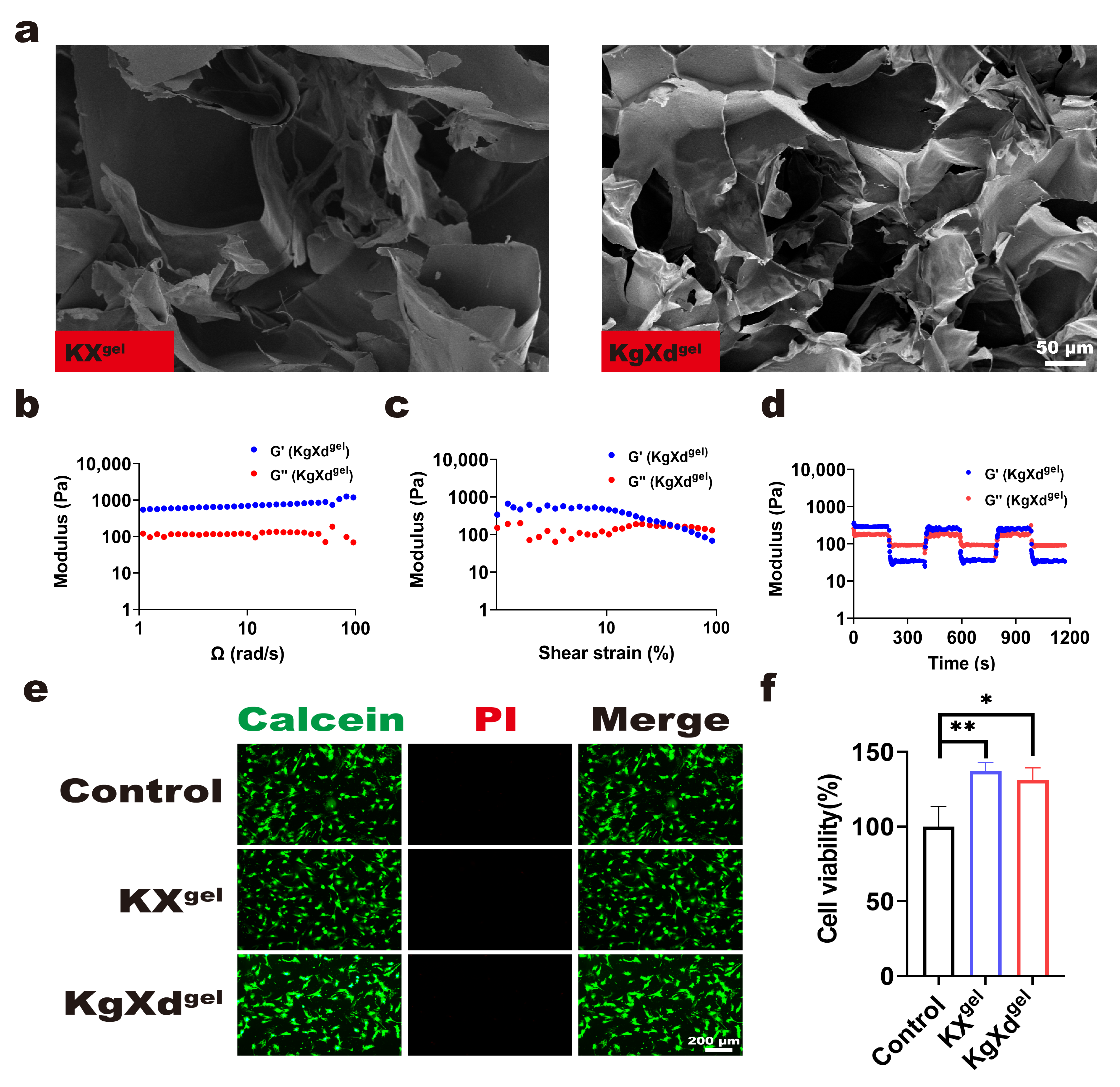
The microstructure, a critical determinant of biomaterial bioactivity and function, was examined using SEM. As shown in Figure 1a, the KgXdgel hydrogel exhibited an interconnected porous network structure with pore sizes ranging from tens to hundreds of micrometers. This structure facilitates the transport of nutrients and metabolic waste and provides an ideal physical space for cell migration and infiltration.
Post-MI degradation of the ECM weakens myocardial mechanical support, making the heart susceptible to overload stress, thereby exacerbating heart failure. Therefore, hydrogels are expected to provide appropriate mechanical stability against hemodynamic pressure to establish a durable cell-regulatory niche. Rheological tests evaluated the mechanical properties of KgXdgel. As shown in Figure 1b–d, within the angular frequency sweep range of 1–100 rad/s, the storage modulus (G′) was consistently greater than the loss modulus (G″), confirming the elastic, solid-like character of the hydrogel. This mechanical stability helps resist the cyclical stress of the beating heart, providing necessary mechanical support for the infarcted area and offering potential physical space for cell migration and tissue ingrowth. Simultaneously, the KgXdgel hydrogel exhibited non-Newtonian shear-thinning behavior; as shear strain increased, the G′ value decreased more than the G″ value. This property allows it to be smoothly injected through a syringe and recover its structure after cessation of shear, demonstrating excellent injectability. Furthermore, the self-healing ability was assessed by alternating high and low step-strain tests. When a large strain (e.g., 50%) was applied, KgXdgel behaved like a solution, but when the strain was reduced (e.g., to 20%), it rapidly recovered to a solid state. This self-healing property ensures the hydrogel can rapidly integrate into a cohesive mass after injection into the beating heart, prolonging its retention time at the infarction site. These results confirm that the KgXdgel hydrogel, with its mechanical stability, shear-thinning behavior, and self-healing capability, is suitable for cardiac injection.

3.2. Cytocompatibility of KgXdgel Hydrogel

Good biocompatibility is a prerequisite for the clinical application of biomaterials. The cytocompatibility of the KgXdgel hydrogel with the rat cardiomyocyte cell line (H9C2) was evaluated using Live/Dead staining and the CCK-8 assay. As shown in Figure 1e, after 24 h of co-culture with the KgXdgel hydrogel, a large number of viable H9C2 cells (green fluorescence) were detected, with almost no dead cells (red fluorescence), comparable to the control group. Consistent with this, cell viability remained at a high level when co-cultured with KgXdgel (Figure 1f). These results indicate that the KgXdgel hydrogel possesses good cytocompatibility, laying the foundation for its subsequent biological applications.

3.3. KgXdgel Hydrogel Promotes Endothelial Cell Migration and Tube Formation

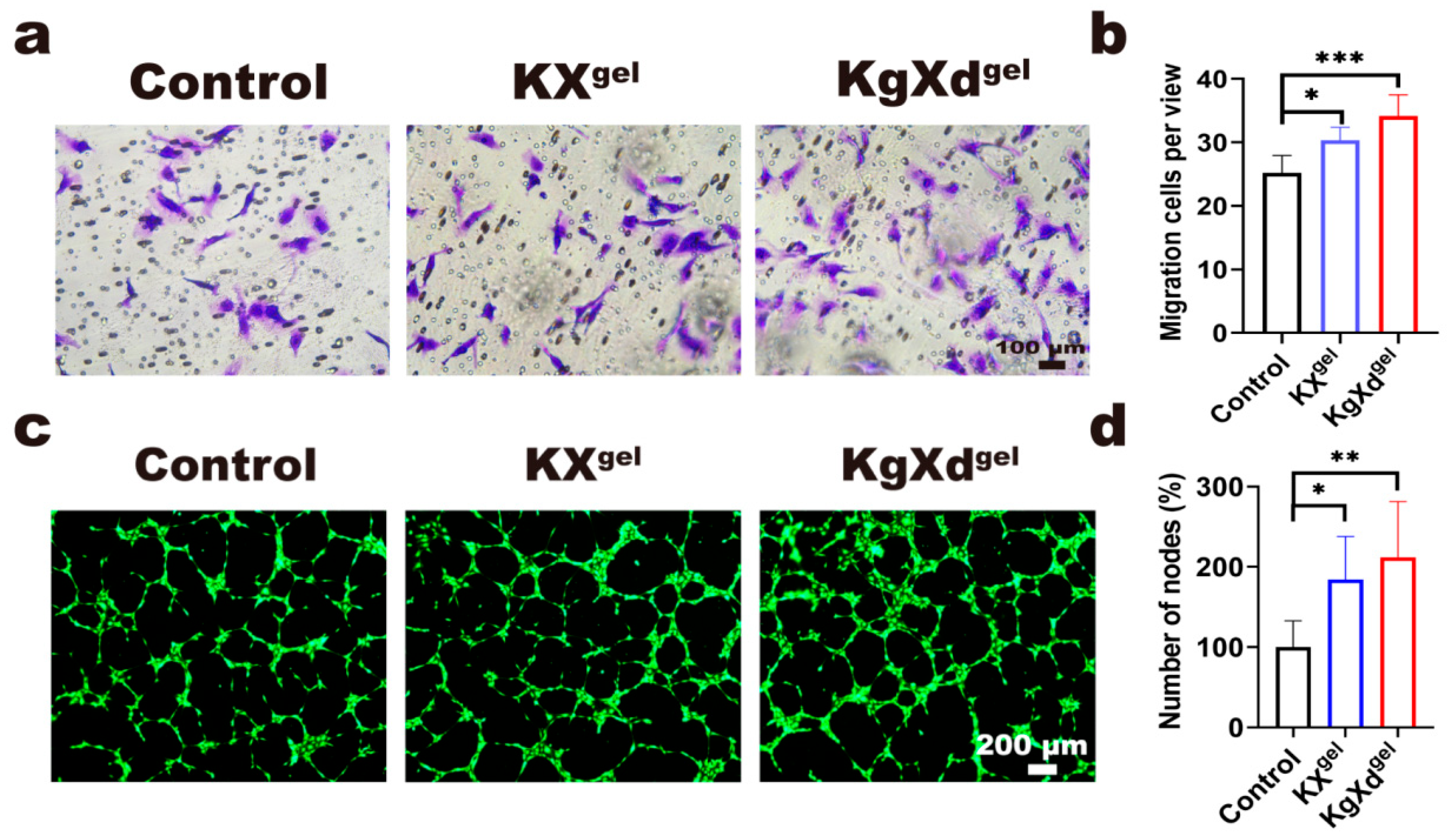
The reconstruction of a functional vascular network is crucial for the repair of infarcted myocardium. The porous microstructure of the KgXdgel hydrogel is expected to provide a favorable environment for cell homing and proliferation. The effect of KgXdgel on the migration ability of HUVECs was assessed using a transwell assay. As shown in Figure 2a,b, after 24 h of co-culture with KgXdgel, the number of HUVECs migrating to the lower chamber was significantly higher than in the control group. Mechanistically, the interconnected porous structure of KgXdgel provides a suitable physical microenvironment that supports cell adhesion, spreading, and directional migration, thereby facilitating endothelial cell infiltration and the early stages of endothelialization. This suggests that the porous microenvironment created by KgXdgel, along with its inherent bioactivity, provides favorable conditions for promoting cell migration, indicating potential for inducing cell infiltration and facilitating the endothelialization process during cardiac repair.
Furthermore, the pro-angiogenic capability of KgXdgel was evaluated in vitro using a tube formation assay. As shown in Figure 2c,d, after 6 h of culture, cells co-cultured with KgXdgel exhibited denser tube networks compared to the control group. The improved tube formation can be attributed to the structural support and bioactive cues provided by the hydrogel, which promote endothelial cell–matrix interaction, cytoskeleton organization, and the formation of intact vascular networks. These results collectively indicate that the KgXdgel hydrogel not only promotes endothelial cell migration but also significantly enhances their tube-forming ability, demonstrating strong pro-angiogenic potential and providing compelling in vitro evidence for its ability to restore blood supply during in vivo repair.

3.4. In Vitro ROS Scavenging Activity of KgXdgel Hydrogel

Oxidative stress is a central element in myocardial reperfusion injury and chronic inflammation. The intrinsic free radical scavenging ability of KgXdgel was first evaluated. The DPPH free radical scavenging assay (Supplementary Figure S4a,b) showed that KgXdgel exhibited concentration-dependent free radical scavenging activity, primarily attributed to the abundant phenolic hydroxyl groups in GA and DA molecules.
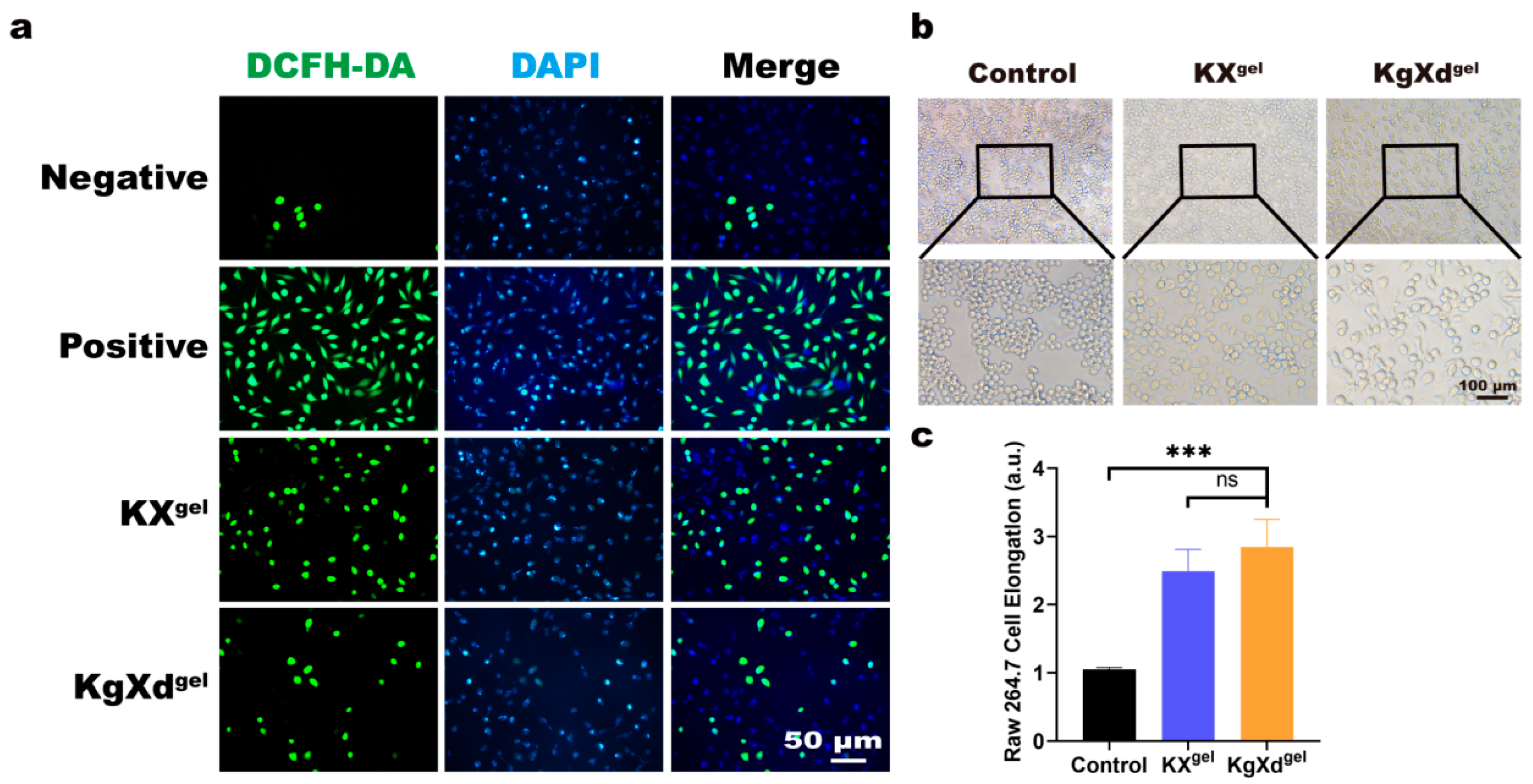
To verify its antioxidant effect at the cellular level, an oxidative stress model was established by stimulating L929 cells with H2O2, and intracellular ROS levels were detected using the DCFH-DA probe. As shown in Figure 3a, H2O2 stimulation led to a sharp increase in intracellular green fluorescence intensity. In contrast, treatment with KgXdgel significantly attenuated the fluorescence intensity, with a superior effect compared to the non-functionalized KXgel hydrogel. This indicates that KgXdgel could establish a local antioxidant microenvironment around cells, thereby scavenging excess ROS and protecting cells from oxidative damage.

3.5. M2 Macrophage Polarization by KgXdgel Hydrogel

Macrophages play a pivotal regulatory role in the pathophysiology of post-MI cardiac remodeling, possessing high plasticity that allows them to polarize into different phenotypes influenced by the microenvironment. Precise regulation of macrophage polarization is key to reversing the immune microenvironment in the infarct area. The effect of KgXdgel on the morphology of RAW264.7 macrophages was first observed. As shown in Figure 3b,c, untreated macrophages mostly exhibited a round morphology. After co-culture with KgXdgel, a large number of cells extended pseudopodia and transformed into the characteristic elongated spindle shape typical of pro-reparative M2 macrophages. This preliminary morphological evidence suggests that KgXdgel can guide macrophages towards the M2 phenotype. This regulatory effect may be related to its excellent ROS scavenging capacity, as ROS are key signaling molecules maintaining the pro-inflammatory M1 phenotype.

3.6. KgXdgel Modulates Cardiac Inflammation and Tissue Regeneration in a Rat MI Model

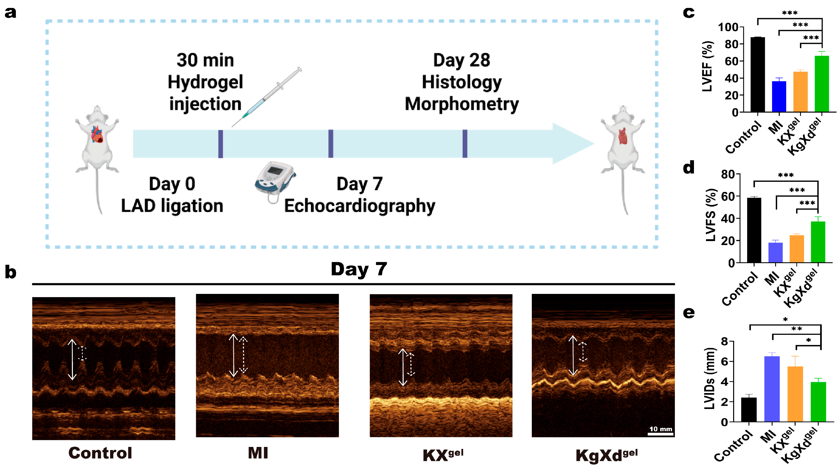
Based on the favorable in vitro performance of KgXdgel in anti-ROS, pro-angiogenesis, and M2 macrophage polarization, its reparative effects were next evaluated in a rat MI model. Figure 4a shows a schematic diagram of the experimental procedure, where the left anterior descending (LAD) coronary artery ligation model was used in this study. As observed in the echocardiograms (Figure 4b), rats in the MI group showed significantly weakened motion of the left ventricular anterior wall and thickened interventricular septum, whereas the KgXdgel hydrogel treatment group markedly improved these conditions.
Quantitative analysis of cardiac function parameters was performed. As shown in Figure 4c–e, compared to the Sham group, the MI group exhibited a sharp decrease in LVEF and LVFS, along with a significant increase in LVIDs, indicating successful establishment of the heart failure model. Although KXgel hydrogel treatment somewhat alleviated the functional deterioration, the KgXdgel hydrogel treatment showed the best effect, more effectively maintaining LVEF and LVFS and suppressing the expansion of LVIDs. These data demonstrate that the KgXdgel hydrogel not only significantly improves cardiac contractile function post-MI but also effectively inhibits adverse left ventricular dilation and remodeling. The improved cardiac function and inhibited ventricular dilation also indicated alleviated myocardial fibrosis and adverse remodeling.
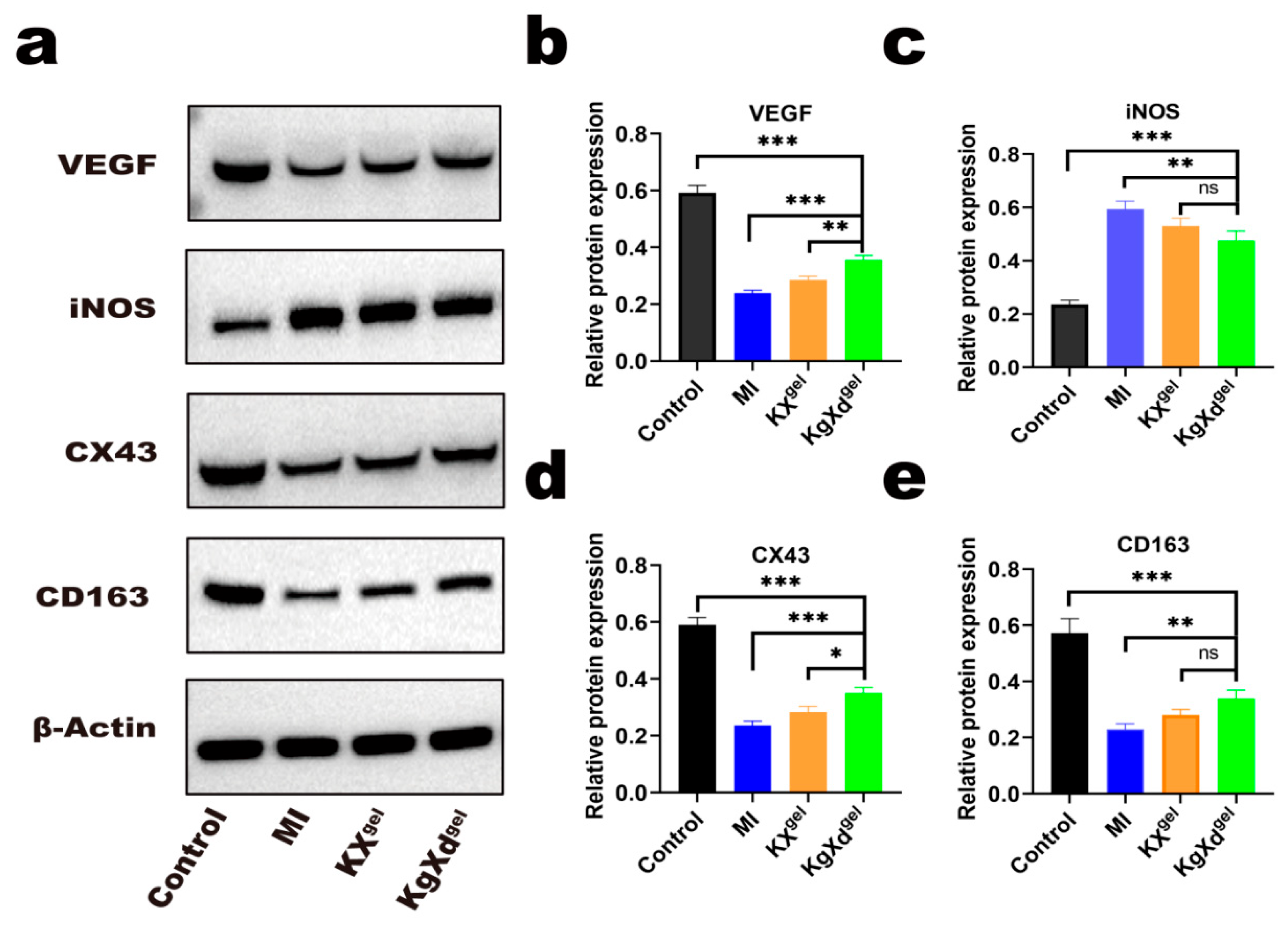
To delve deeper into the mechanism of action, the expression of key proteins in the infarcted myocardial tissue was analyzed by Western Blot. As shown in Figure 5a–e, KgXdgel treatment significantly upregulated the expression of Vascular Endothelial Growth Factor (VEGF) and Connexin 43 (CX43), consistent with its pro-angiogenic role and potential to improve electrical signal conduction by strengthening gap junction–mediated intercellular coupling in the myocardium [37,38]. Concurrently, KgXdgel downregulated the expression of the M1 macrophage marker iNOS and upregulated the M2 marker CD163. These results indicated that KgXdgel treatment could modulate macrophage phenotypic status in vivo, showing a trend toward a reduction in pro-inflammatory M1 phenotype and an increase in anti-inflammatory reparative M2 phenotype.
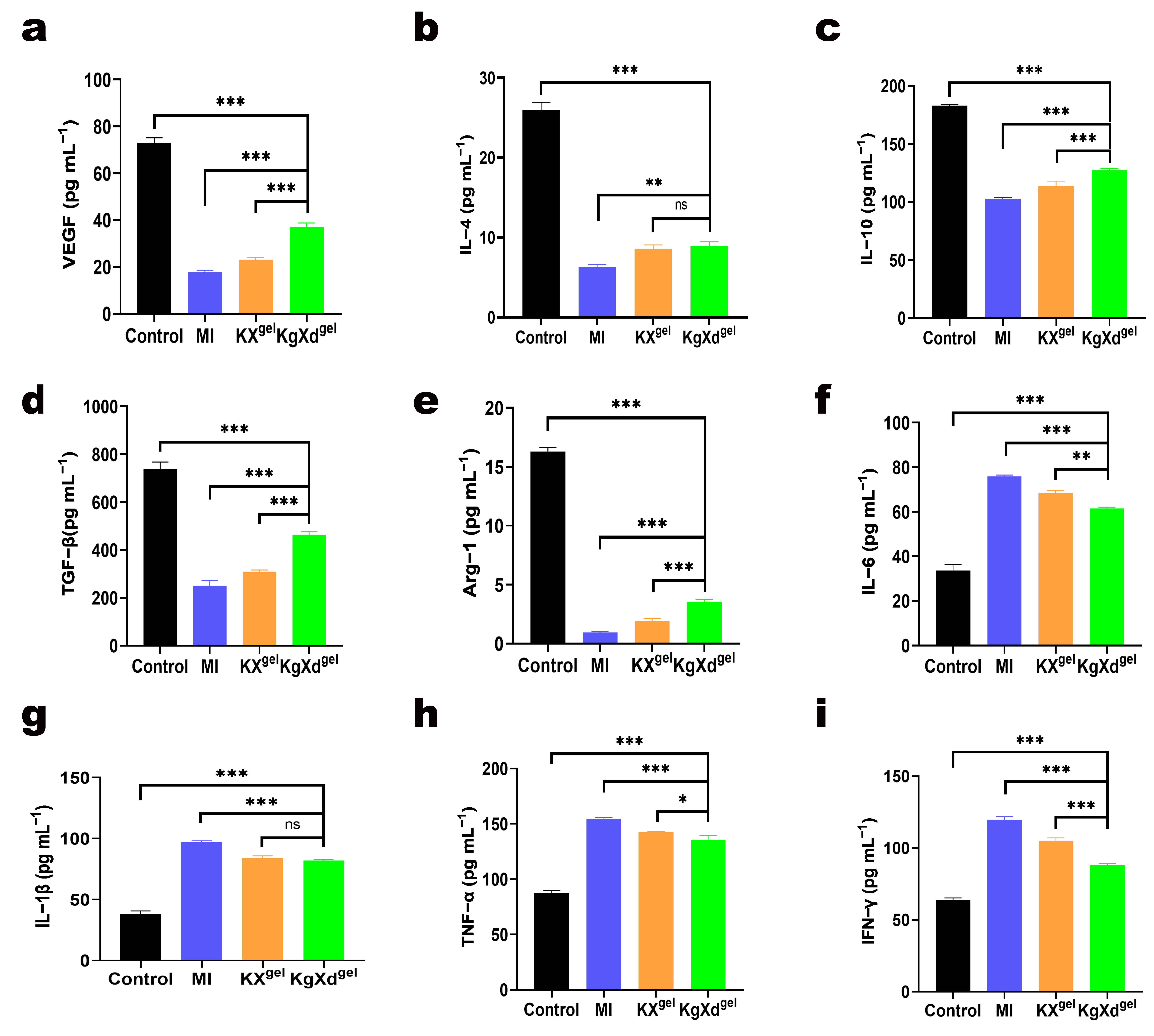
ELISA analysis of cytokines in the infarct area further confirmed this mechanism. As shown in Figure 6a–i, in the KgXdgel treatment group, the levels of pro-inflammatory cytokines (e.g., IL-1β, IFN-γ, TNF-α, IL-6) were significantly reduced, while the levels of factors associated with repair, angiogenesis, and M2 polarization (e.g., VEGF, TGF-β, IL-4, IL-10, Arg-1) were significantly elevated. This paints a clear picture: through its antioxidant properties, the KgXdgel hydrogel breaks the “oxidative stress-chronic inflammation” vicious cycle, shifting the infarct microenvironment from a pro-inflammatory state to a pro-reparative state. The polarization of macrophages towards the M2 phenotype, in turn, promotes the secretion of repair factors like VEGF, accelerating angiogenesis and tissue repair, ultimately collectively contributing to the improvement in cardiac function.

4. Discussion

This study successfully developed a multifunctional KgXdgel hydrogel that, by integrating the antioxidant activities of GA and DA with the structural advantages of the KGM and XG backbone, achieved synergistic regulation of the complex pathological microenvironment post-MI. The results indicate that KgXdgel not only provides a favorable microenvironment for cells through its porous structure and suitable mechanical properties but also effectively scavenges ROS, which is a critical step in breaking the “oxidative stress-chronic inflammation” vicious cycle. Scavenging ROS helped guide macrophage polarization from the pro-inflammatory M1 phenotype towards the reparative M2 phenotype, a finding supported by both in vitro morphological observations and in vivo protein and cytokine-level analyses. The increase in M2 macrophages further promoted the secretion of angiogenic factors like VEGF, thereby enhancing angiogenesis, improving blood supply and nutrient delivery to the infarct zone. Ultimately, these synergistic effects collectively contributed to the improvement in cardiac function and the inhibition of adverse ventricular remodeling.
Cardiac function was evaluated at day 7 post-MI, representing the critical early repair phase. The significantly improved LVEF, LVFS, and attenuated ventricular dilation at day 7 demonstrated the rapid and effective cardioprotective effect of KgXdgel. Meanwhile, molecular analyses at day 28 (including VEGF, CD163, iNOS, and cytokine profiles) further confirmed its sustained beneficial effects on long-term cardiac remodeling. This study validates the feasibility of promoting cardiac repair through a multi-targeted strategy, and the KgXdgel hydrogel demonstrates great potential as a next-generation cardiac repair material. A more comprehensive time-course investigation will be systematically conducted in future studies.

5. Limitations and Future Perspectives

Although the present study demonstrates the therapeutic potential of KgXdgel for myocardial infarction repair, some aspects can be further explored in future investigations. The current study evaluated cardiac function at the early stage and molecular indicators at the late stage, and a continuous time-course evaluation would help better reveal the dynamic repair process. In addition, the pro-angiogenic effects were mainly evaluated through molecular indicators, and more intuitive histological evidence could be combined in follow-up research. Furthermore, the therapeutic efficacy was validated in a rat model, and studies in more clinically relevant large animals would help promote its translational application. Future work will focus on optimizing the hydrogel system, exploring more comprehensive evaluation indicators, and carrying out translational studies in large animals to further improve its therapeutic effect and clinical potential.

6. Conclusions

In this study, a functionalized polysaccharide composite hydrogel, KgXdgel, was designed and fabricated. This hydrogel exhibited good biocompatibility, injectability, and self-healing capacity. Both in vitro and in vivo experiments confirmed that KgXdgel effectively scavenged ROS, induced macrophage polarization towards the M2 phenotype, and promoted angiogenesis. In a rat MI model, KgXdgel significantly improved cardiac function and inhibited adverse ventricular remodeling by modulating the inflammatory microenvironment and promoting tissue repair. These findings suggest that the KgXdgel hydrogel is a highly promising biomaterial for the treatment of MI, offering new insights and experimental evidence for developing combined therapeutic strategies targeting the complex process of cardiac repair.

Supplementary Materials

The following supporting information can be downloaded at: https://www.mdpi.com/article/10.3390/jcs10060287/s1.Figure S1: The FTIR spectra (a) and UV absorption spectra (b) of KGM−GA; Figure S2: The FTIR spectra (a) and UV absorption spectra (b) of XG−DA; Figure S3: The FTIR spectra (a) and UV absorption spectra (b) of KgXdgel; Figure S4: The free radical scavenging capacity of (a) KXgel and (b) KgXdgel.

Author Contributions

Conceptualization, E.-C.Z. and C.-N.Z.; Methodology, E.-C.Z. and X.-Y.L.; Investigation, E.-C.Z., X.-Y.L., Z.C. and J.-Y.Y.; Formal analysis, E.-C.Z., X.-Y.L., Z.C. and J.-Y.Y.; Software, E.-C.Z.; Writing—original draft, E.-C.Z. and X.-Y.L.; Data curation, X.-Y.L., Z.C., Q.-H.Y. and J.-Y.Y.; Validation, Z.C. and J.-Y.Y.; Visualization, Q.-H.Y.; Writing—review and editing, Q.-H.Y. and C.-N.Z.; Supervision, C.-N.Z.; Resources, C.-N.Z.; Funding acquisition, C.-N.Z.; Project administration, C.-N.Z. All authors have read and agreed to the published version of the manuscript.

Funding

This research was supported by Tianjin Municipal Science and Technology Commission Grant, No. 24ZXRKSY00010; and CAMS Innovation Fund for Medical Sciences (CIFMS), No. 2023-I2M-2-008.

Data Availability Statement

Data are contained within the article.

Conflicts of Interest

The authors declare no conflicts of interest.

References

  1. Tsao, C.W.; Aday, A.W.; Almarzooq, Z.I.; Alonso, A.; Beaton, A.Z.; Bittencourt, M.S.; Boehme, A.K.; Buxton, A.E.; Carson, A.P.; Commodore-Mensah, Y.; et al. Heart disease and stroke statistics—2022 update: A report from the American heart association. Circulation 2022, 145, e153–e639. [Google Scholar] [CrossRef] [PubMed]
  2. Vaduganathan, M.; Mensah, G.A.; Turco, J.V.; Fuster, V.; Roth, G.A. The global burden of cardiovascular diseases and risk: A compass for future health. J. Am. Coll. Cardiol. 2022, 80, 2361–2371. [Google Scholar] [CrossRef]
  3. Reed, G.W.; Rossi, J.E.; Cannon, C.P. Acute myocardial infarction. Lancet 2017, 389, 197–210. [Google Scholar] [CrossRef]
  4. Davidson, S.M.; Ferdinandy, P.; Andreadou, I.; Bøtker, H.E.; Heusch, G.; Ibáñez, B.; Ovize, M.; Schulz, R.; Yellon, D.M.; Hausenloy, D.J.; et al. Multitarget strategies to reduce myocardial ischemia/reperfusion injury: JACC review topic of the week. J. Am. Coll. Cardiol. 2019, 73, 89–99. [Google Scholar] [CrossRef]
  5. Heusch, G. Myocardial ischaemia-reperfusion injury and cardioprotection in perspective. Nat. Rev. Cardiol. 2020, 17, 773–789. [Google Scholar] [CrossRef]
  6. Münzel, T.; Camici, G.G.; Maack, C.; Bonetti, N.R.; Fuster, V.; Kovacic, J.C. Impact of oxidative stress on the heart and vasculature: Part 2 of a 3-part series. J. Am. Coll. Cardiol. 2017, 70, 212–229. [Google Scholar] [CrossRef]
  7. Hu, D.; Li, R.; Li, Y.; Wang, M.; Wang, L.; Wang, S.; Cheng, H.; Zhang, Q.; Fu, C.; Qian, Z.; et al. Inflammation-targeted nanomedicines alleviate oxidative stress and reprogram macrophages polarization for myocardial infarction treatment. Adv. Sci. 2024, 11, e2308910. [Google Scholar] [CrossRef] [PubMed]
  8. Frangogiannis, N.G. The inflammatory response in myocardial injury, repair, and remodeling. Nat. Rev. Cardiol. 2014, 11, 255–265. [Google Scholar] [CrossRef] [PubMed]
  9. Prabhu, S.D.; Frangogiannis, N.G. The biological basis for cardiac repair after myocardial infarction: From inflammation to fibrosis. Circ. Res. 2016, 119, 91–112. [Google Scholar] [CrossRef]
  10. Somasuntharam, I.; Yehl, K.; Carroll, S.L.; Maxwell, J.T.; Martinez, M.D.; Che, P.-L.; Brown, M.E.; Salaita, K.; Davis, M.E. Knockdown of TNF-α by DNAzyme gold nanoparticles as an anti-inflammatory therapy for myocardial infarction. Biomaterials 2016, 83, 12–22. [Google Scholar] [CrossRef]
  11. Frumento, D.; Țălu, Ș. Immunomodulatory potential and biocompatibility of chitosan–hydroxyapatite biocomposites for tissue engineering. J. Compos. Sci. 2025, 9, 305. [Google Scholar] [CrossRef]
  12. Carmeliet, P.; Jain, R.K. Molecular mechanisms and clinical applications of angiogenesis. Nature 2011, 473, 298–307. [Google Scholar] [CrossRef]
  13. Rafii, S.; Butler, J.M.; Ding, B.-S. Angiocrine functions of organ-specific endothelial cells. Nature 2016, 529, 316–325. [Google Scholar] [CrossRef]
  14. Dai, F.; Zhang, J.; Chen, F.; Chen, X.; Lee, C.J.; Liang, H.; Zhao, L.; Tan, H. A multi-responsive hydrogel combined with mild heat stimulation promotes diabetic wound healing by regulating inflammatory and enhancing angiogenesis. Adv. Sci. 2024, 11, e2408783. [Google Scholar] [CrossRef] [PubMed]
  15. Hu, C.; Liu, W.; Long, L.; Wang, Z.; Zhang, W.; He, S.; Lu, L.; Fan, H.; Yang, L.; Wang, Y. Regeneration of infarcted hearts by myocardial infarction-responsive injectable hydrogels with combined anti-apoptosis, anti-inflammatory and pro-angiogenesis properties. Biomaterials 2022, 290, 121849. [Google Scholar] [CrossRef]
  16. Xu, Q.; Xiao, Z.; Yang, Q.; Yu, T.; Deng, X.; Chen, N.; Huang, Y.; Wang, L.; Guo, J.; Wang, J. Hydrogel-based cardiac repair and regeneration function in the treatment of myocardial infarction. Mater. Today Bio 2024, 25, 100978. [Google Scholar] [CrossRef] [PubMed]
  17. Zheng, Z.; Lei, C.; Liu, H.; Jiang, M.; Zhou, Z.; Zhao, Y.; Yu, C.-Y.; Wei, H. A ROS-responsive liposomal composite hydrogel integrating improved mitochondrial function and pro-angiogenesis for efficient treatment of myocardial infarction. Adv. Healthc. Mater. 2022, 11, e2200990. [Google Scholar] [CrossRef]
  18. Wu, T.; Liu, W. Functional hydrogels for the treatment of myocardial infarction. NPG Asia Mater. 2022, 14, 9. [Google Scholar] [CrossRef]
  19. Yang, J.; Wang, J.; Zeng, Z.; Chen, Z.; Wang, D.; Wu, Y. Injectable sustained-release danshensusodium-loaded nanoparticle hydrogel targets macrophages to improve myocardial microenvironment for myocardial infarction treatment. Bioact. Mater. 2025, 54, 159–178. [Google Scholar]
  20. Li, Y.; Chen, X.; Jin, R.; Chen, L.; Dang, M.; Cao, H.; Dong, Y.; Cai, B.; Bai, G.; Gooding, J.J.; et al. Injectable hydrogel with MSNs/microRNA-21-5p delivery enables both immunomodification and enhanced angiogenesis for myocardial infarction therapy in pigs. Sci. Adv. 2021, 7, eabd6740. [Google Scholar] [CrossRef]
  21. Zheng, Z.; Sun, J.; Wang, J.; He, S.; Liu, Z.; Xie, J.; Yu, C.-Y.; Wei, H. Enhancing myocardial infarction treatment through bionic hydrogel-mediated spatial combination therapy via mtDNA-STING crosstalk modulation. J. Control. Release 2024, 371, 570–587. [Google Scholar] [CrossRef]
  22. Liu, Z.; Zheng, Z.; Xie, J.; Wei, H.; Yu, C.-Y. Hydrogel-based cardiac patches for myocardial infarction therapy: Recent advances and challenges. Mater. Today Bio 2024, 29, 101331. [Google Scholar] [CrossRef] [PubMed]
  23. Rumon, M.M.H.; Akib, A.A.; Sultana, F.; Moniruzzaman, M.; Niloy, M.S.; Shakil, M.S.; Roy, C.K. Self-healing hydrogels: Development, biomedical applications, and challenges. Polymers 2022, 14, 4539. [Google Scholar] [CrossRef]
  24. Luo, Q.; Gao, Z.; Bai, L.; Ye, H.; Ye, H.; Wang, Y.; Gao, Y.; Chen, T.; Chen, H.; Liu, Y.; et al. Bioactive peptide-based composite hydrogel for myocardial infarction treatment: ROS scavenging and angiogenesis regulation. Acta Biomater. 2025, 197, 167–183. [Google Scholar] [CrossRef]
  25. Zhong, Y.; Yang, Y.; Xu, Y.; Qian, B.; Huang, S.; Long, Q.; Qi, Z.; He, X.; Zhang, Y.; Li, L.; et al. Design of a Zn-based nanozyme injectable multifunctional hydrogel with ROS scavenging activity for myocardial infarction therapy. Acta Biomater. 2024, 177, 62–76. [Google Scholar] [CrossRef] [PubMed]
  26. Qin, J.; Wu, Z.; Ma, S.; Wu, Y.; Chen, Y.; Qian, C.; Lin, H.; Tan, G.; Liu, Q.; Zhao, J.; et al. Hierarchical structures of protein-fiber-infused aminoglycan hydrogels to promote myocardial repair. ACS Nano 2025, 19, 32254–32269. [Google Scholar] [CrossRef]
  27. Liu, Z.; Xuan, L.; Hou, Y.; Xie, T.; Li, J.; Cai, J.; Zhang, S.; Miao, Y.; Hou, N.; He, G.; et al. Microgel-based hierarchical porous hydrogel patch with adhesion and resilience for myocardial infarction repair. Adv. Sci. 2026, 13, e18646. [Google Scholar] [CrossRef]
  28. Rakshit, P.; Giri, T.K.; Mukherjee, K. Progresses and perspectives on natural polysaccharide based hydrogels for repair of infarcted myocardium. Int. J. Biol. Macromol. 2024, 269, 132213. [Google Scholar] [CrossRef]
  29. Yang, D.; Yuan, Y.; Wang, L.; Wang, X.; Mu, R.; Pang, J.; Xiao, J.; Zheng, Y. A review on konjac glucomannan gels: Microstructure and application. Int. J. Mol. Sci. 2017, 18, 2250. [Google Scholar] [CrossRef] [PubMed]
  30. Pan, X.; Zong, Q.; Liu, C.; Wu, H.; Fu, B.; Wang, Y.; Sun, W.; Zhai, Y. Konjac glucomannan exerts regulatory effects on macrophages and its applications in biomedical engineering. Carbohydr. Polym. 2024, 345, 122571. [Google Scholar] [CrossRef]
  31. Hua, D.; Gao, S.; Zhang, M.; Ma, W.; Huang, C. A novel xanthan gum-based conductive hydrogel with excellent mechanical, biocompatible, and self-healing performances. Carbohydr. Polym. 2020, 247, 116743. [Google Scholar] [CrossRef] [PubMed]
  32. Alves, A.; Miguel, S.P.; Araujo, A.R.T.S.; Valle, M.J.D.J.; Navarro, A.S.; Correia, I.J.; Ribeiro, M.P.; Coutinho, P. Xanthan gum-konjac glucomannan blend hydrogel for wound healing. Polymers 2020, 12, 99. [Google Scholar] [CrossRef]
  33. Krupska, T.; Wei, Q.; Zheng, J.; Yang, W.; Holovan, A.; Borysenko, M.; Turov, V. Design of composite systems based on hydrophilic silica and organic acids: Gallic, glycyrrhizic and its salts. J. Compos. Sci. 2025, 9, 247. [Google Scholar] [CrossRef]
  34. Das, A.; Nikhil, A.; Shiekh, P.A.; Yadav, B.; Jagavelu, K.; Kumar, A. Ameliorating impaired cardiac function in myocardial infarction using exosome-loaded gallic-acid-containing polyurethane scaffolds. Bioact. Mater. 2024, 33, 324–340. [Google Scholar] [CrossRef]
  35. Liu, Y.; Ai, K.; Lu, L. Polydopamine and its derivative materials: Synthesis and promising applications in energy, environmental, and biomedical fields. Chem. Rev. 2014, 114, 5057–5115. [Google Scholar] [CrossRef]
  36. Lee, H.; Lee, B.P.; Messersmith, P.B. A reversible wet/dry adhesive inspired by mussels and geckos. Nature 2007, 448, 338–341. [Google Scholar] [CrossRef] [PubMed]
  37. Greener, I.D.; Sasano, T.; Wan, X.; Igarashi, T.; Strom, M.; Rosenbaum, D.S.; Donahue, J.K. Connexin43 gene transfer reduces ventricular tachycardia susceptibility after myocardial infarction. J. Am. Coll. Cardiol. 2012, 60, 1103–1110. [Google Scholar] [CrossRef]
  38. Pimentel, R.C.; Yamada, K.A.; Kléber, A.G.; Saffitz, J.E. Autocrine regulation of myocyte Cx43 expression by VEGF. Circ. Res. 2002, 90, 671–677. [Google Scholar] [CrossRef] [PubMed]
Scheme 1. Schematic representation of the synthesis of KgXdgel hydrogel and its application in cardiac repair following myocardial infarction. The arrows indicate the material preparation process and the key biological functions, including ROS scavenging, macrophage regulation, and pro-angiogenic activities, which improve the infarct microenvironment and accelerate cardiac repair.
Jcs 10 00287 sch001
Figure 1. Preparation and characterization of KgXdgel hydrogel. (a) Representative SEM images of KXgel and KgXdgel hydrogels. (b,c) Rheological properties of KgXdgel hydrogel as a function of angular frequency and shear strain, respectively. (d) Self-healing performance of KgXdgel hydrogel. (e) Live/Dead staining analysis and (f) CCK-8 analysis of H9C2 cells co-cultured with the hydrogels. (* indicates significant difference between groups, * p < 0.05, ** p < 0.01).
Jcs 10 00287 g001
Figure 2. KgXdgel hydrogel promotes endothelial cell migration and tube formation. (a,b) Representative images and quantification of HUVECs migration determined by transwell assay after 24 h. Migrated cells were stained with crystal violet (n = 6). (c,d) Representative images and quantification of HUVECs tube formation. Cells were stained with calcein-AM for visualization. (* indicates significant difference between groups, * p < 0.05, ** p < 0.01, *** p < 0.001).
Jcs 10 00287 g002
Figure 3. In vitro ROS scavenging and macrophage polarization regulation by KgXdgel hydrogel. (a) Fluorescence images of ROS detection in L929 cells using DCFH-DA probe. (b) Morphology of Raw264.7 macrophages observed under an optical microscope after 48 h treatment under different conditions. (c) Statistical data of elongation changes in Raw 264.7 macrophages. (* indicates significant difference between groups, *** p < 0.001, ns: no significance).
Jcs 10 00287 g003
Figure 4. KgXdgel hydrogel modulates cardiac repair in a rat myocardial infarction model. (a) Schematic diagram of the treatment in the rat MI model. (b) Representative echocardiographic images reflecting cardiac function. White solid line indicates LVIDd; white dashed line indicates LVIDs. Quantification of (c) LVEF, (d) LVFS, and (e) LVIDs by echocardiography. (* indicates significant difference between groups, * p < 0.05, ** p < 0.01, *** p < 0.001).
Jcs 10 00287 g004
Figure 5. KgXdgel hydrogel modulates cardiac repair in a rat myocardial infarction model. (a) Western Blot analysis of VEGF, iNOS, CX43, and CD163 protein expression. (be) Quantitative expression levels of VEGF, iNOS, CX43, and CD163. (* indicates significant difference between groups, * p < 0.05, ** p < 0.01, *** p < 0.001, ns: no significance).
Jcs 10 00287 g005
Figure 6. KgXdgel hydrogel modulates cardiac repair in a rat myocardial infarction model. Levels of (a) VEGF, (b) IL−4, (c) IL−10, (d) TGF−β, (e) Arg−1, (f) IL−6, (g) IL−1β, (h) TNF−α, and (i) IFN−γ in the infarct area measured by ELISA. (* indicates significant difference between groups, * p < 0.05, ** p < 0.01, *** p < 0.001, ns: no significance).
Jcs 10 00287 g006
Disclaimer/Publisher’s Note: The statements, opinions and data contained in all publications are solely those of the individual author(s) and contributor(s) and not of MDPI and/or the editor(s). MDPI and/or the editor(s) disclaim responsibility for any injury to people or property resulting from any ideas, methods, instructions or products referred to in the content.