Article

Efficacy of Hypertonic Sodium Chloride in the Management of Oral Bacterial Infections and Inflammation in Companion Animals


by
Suttiwan Wunnoo
1,
Chanawee Jakkawanpitak
2,3,
Nattanan Methaspornpong
4,
Saowakon Indoung
4,
Teeraporn Kongtawee
4,
Chonlada Namdokmai
4,
Natthanit Hemtanon
4,
Supayang Piyawan Voravuthikunchai
1 and
Krittee Dejyong
4,*
1
Center of Antimicrobial Biomaterial Innovation-Southeast Asia, Faculty of Science, Prince of Songkla University, Songkhla 90110, Thailand
2
Center of Excellence for Biochemistry, Faculty of Science, Prince of Songkla University, Songkhla 90110, Thailand
3
Division of Health and Applied Sciences, Faculty of Science, Prince of Songkla University, Songkhla 90110, Thailand
4
Faculty of Veterinary Science, Prince of Songkla University, Songkhla 90110, Thailand
*
Author to whom correspondence should be addressed.
Sci 2026, 8(7), 168; https://doi.org/10.3390/sci8070168
Submission received: 30 May 2026 / Revised: 3 July 2026 / Accepted: 9 July 2026 / Published: 13 July 2026
(This article belongs to the Section Clinical Medicine and Healthcare)

Abstract

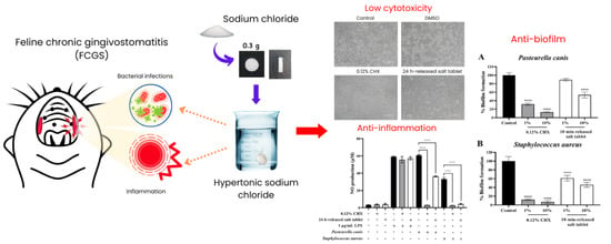
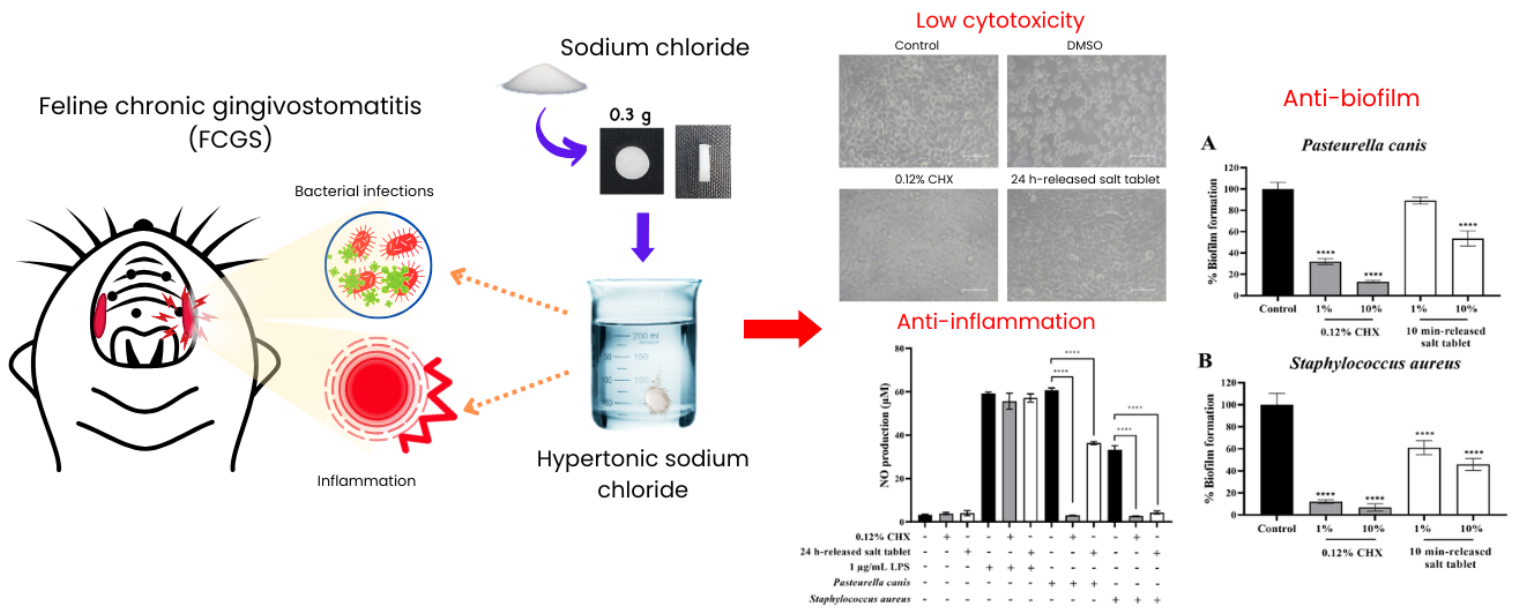
Oral infections are common health problems in companion animals, often associated with bacterial colonization, biofilm formation, and inflammation. This study investigates the efficacy of hypertonic salt tablets as an antibacterial approach and their effect on bacteria-associated inflammatory stimulation against important oral pathogens, Staphylococcus aureus and Pasteurella canis. Agar well diffusion showed that 0.3 g salt tablets exhibited antibacterial activity, with inhibition zones of 13.33 ± 0.89 mm for S. aureus and 28.75 ± 1.18 mm for P. canis, compared with 23.10 ± 0.87 mm and 31.43 ± 0.81 mm for 0.12% chlorhexidine, respectively. In vitro cytotoxicity assessment demonstrated that 1% v/v and 10% v/v released salt solutions maintained high cell viability (>80%), while morphology assays confirmed non-cytotoxicity of 1% v/v 24 h released salt solution. The 1% v/v and 10% v/v 10 min-released salt solution reduced relative biofilm formation in S. aureus to approximately 61% and 45%, respectively, and in P. canis to approximately 89% and 52%, respectively. Salt-treated bacterial suspensions induced lower NO production in RAW264.7 macrophages, decreasing NO levels from 33.17 ± 2.01 to 4.34 ± 0.65 µM for S. aureus and from 60.73 ± 0.99 to 36.33 ± 0.69 µM for P. canis, suggesting attenuation of bacteria-associated inflammatory stimulation. In a preliminary mouse oral mucosal wound model, local application of NaCl crystals for 30 min reduced total bacterial counts from 8.9 × 10² to 2.0 × 10² CFU/mL, corresponding to a 77.5% reduction. These findings suggest that hypertonic sodium chloride tablets may provide a simple localized approach for short-term reduction in oral bacterial burden and bacteria-associated inflammatory stimulation; however, further validation is required in clinically relevant oral disease models and companion animals.

Graphical Abstract

1. Introduction

Oral infections are among the most prevalent health problems in companion animals, particularly dogs and cats, and they can significantly affect overall well-being. Oral lesions may arise from various causes, including immune-mediated diseases, viral and bacterial infections, metabolic disorders, and adverse drug reactions [1]. The oral cavity of companion animals harbors a highly diverse microbiota, with studies estimating hundreds of bacterial species, comparable in scale to humans but differing in composition [2,3]. Furthermore, the microbial composition differs markedly between healthy and diseased animals, with dysbiosis and increased antibiotic resistance frequently documented in affected individuals [4]. Most bacteria detected in free-roaming cats with oral disease consisted of Streptococcus, Staphylococcus, Neisseria, and Pasteurella species [5]. Razali et al. [6] demonstrated that the pathogenic bacteria present in the oral cavity of cats consisted of some Gram-negative (Proteus, Pasteurella, Escherichia, Moraxella, Klebsiella, Acinetobacter, Enterobacter, Pseudomonas, Aeromonas, and Neisseria) and four Gram-positive (Staphylococcus, Streptococcus, Corynebacterium, and Bacillus). Moreover, 8 of Pasteurella isolates were found to be multidrug-resistant. Recent studies have examined domestic cats’ subgingival microbial communities, focusing on periodontal health and disease. These findings indicated that diseased sites had higher bacterial diversity in their microbiomes compared with healthy sites [7,8]. The formation of bacterial biofilms leading to plaque deposits activates the immune system and causes gingivitis, initially affecting individual teeth and gradually spreading to surrounding tissues [9]. Collectively, these findings underscore the central role of bacterial colonization and biofilm formation in the pathogenesis of gingivitis and periodontal disease, which, if left untreated, can progress to chronic oral inflammation. Effective management of periodontitis is therefore essential in companion animals, as persistent oral inflammation drives disease progression and necessitates careful evaluation for appropriate treatment.
Current therapeutic approaches for companion animal oral diseases include dental cleaning, tooth extraction, and medical management using antimicrobials, anti-inflammatory agents, or immunomodulatory drugs. However, treatment responses may vary, and repeated or inappropriate use of antibiotics and antiseptics can contribute to antimicrobial resistance and local tissue irritation [10]. Therefore, simple localized approaches that reduce oral bacterial burden while maintaining host–cell compatibility remain of interest. Sodium chloride (NaCl) is physiologically relevant because sodium and chloride are normal mammalian electrolytes, and isotonic 0.9% NaCl has an osmolality close to that of serum [11]. At higher concentrations, hypertonic NaCl can affect bacterial growth through osmotic stress and disturbance of membrane homeostasis. Previous work showed that 3.5% and 5% NaCl inhibited E. coli biofilm formation and bacterial growth [12], while a NaCl-based mouth rinse has been reported to show postoperative anti-inflammatory effects comparable to 0.12% chlorhexidine after periodontal surgery [13]. However, the potential use of a standardized NaCl tablet format for localized oral application against representative companion animal oral pathogens remains insufficiently characterized.
In this study, S. aureus and P. canis were selected as representative oral pathogens relevant to companion animal oral infections. S. aureus was included as a Gram-positive opportunistic bacterium associated with oral colonization and inflammatory infection, whereas P. canis was selected as a Gram-negative bacterium commonly associated with the oral microbiota of dogs and cats and with potential zoonotic and antimicrobial-resistance relevance [5,6]. Although oral diseases in companion animals are polymicrobial, these two organisms were used as initial representative models to evaluate the antibacterial and antibiofilm potential of the NaCl tablet system. Although the antimicrobial activity of hypertonic NaCl is well established, limited information is available on its use as a standardized local tablet format for companion animal oral applications. In particular, its effects on representative oral pathogens, biofilm biomass formation, host–cell cytocompatibility, and bacteria-associated inflammatory stimulation have not been evaluated together in a single study. Therefore, this study aimed to evaluate 0.3 g hypertonic NaCl tablets against S. aureus and P. canis using antibacterial, antibiofilm, cytotoxicity, and RAW264.7 macrophage NO-production assays. A preliminary mouse oral mucosal wound model was also used to assess short-term bacterial burden reduction after local NaCl application.

2. Materials and Methods

2.1. Sodium Chloride Tablet Preparation

NaCl tablets were prepared using a single-punch tablet press (TDP series, Finetech Pharmaceutical Equipment, Ruian City, Wenzhou, Zhejiang, China). Briefly, the required amount of 99.5% pure NaCl was accurately weighed and milled to obtain a uniform particle size before compression. The NaCl powder was then loaded into the die and compressed to form white cylindrical tablets. The target tablet weight was 0.30 g, and the resulting tablets contained approximately 0.32 g of NaCl, with an approximate diameter of 11 mm and thickness of 4 mm. Tablet weight and dimensions were recorded as basic characterization parameters. However, tablet hardness and compression force were not recorded in the present preliminary formulation study because hardness testing was not performed and the tablet press did not provide a recorded compression-force output. The finished NaCl tablets were stored in a dry container to protect them from moisture.

2.2. Microorganisms and Culture Conditions

A reference strain, Staphylococcus aureus ATCC 25923, was used to represent Gram-positive bacteria. Pasteurella canis clinical isolate was obtained from the diagnostic center, the Faculty of Veterinary Science, Prince of Songkla University, Thailand. The microorganisms were subcultured overnight on Mueller-Hinton agar (MHA) and incubated at 37 °C for 18–24 h. The cultures were maintained in Mueller-Hinton broth (MHB) containing 20% glycerol and stored at −80 °C until use.

2.3. Cell Culture

Murine fibroblast (L929) and murine macrophage (RAW264.7) cell lines were cultured in complete Dulbecco’s modified Eagle’s medium (cDMEM, Sigma-Aldrich, St. Louis, MO, USA) containing 10% fetal bovine serum (FBS, Gibco, Thermo Fisher Scientific (Waltham, MA, USA)) and 1× Antibiotic-Antimycotic (Gibco). Cells were incubated in a humidified atmosphere containing 5% CO2 at 37 °C.

2.4. Antibacterial Susceptibility Testing

The antibacterial activity of salt tablets against S. aureus and P. canis was determined using agar well diffusion method according to Clinical and Laboratory Standards Institute guidelines [14]. The pathogens were maintained on MHA and incubated at 37 °C for 24 h. A single colony was picked and transferred into MHB and incubated at 37 °C for 18–24 h. The organism suspensions were adjusted to a 0.5 McFarland standard (1.5 × 10⁸ CFU/mL). Then, the suspensions were swabbed on the surface of MHA. A hole of 8 mm in diameter was punched with a sterile tip. Each well was filled with a 0.3 g salt tablet, 100 µL of 0.85% normal saline, and 100 µL of 0.12% chlorhexidine (CHX) as a positive control. After incubation, the diameter zone of inhibition (mm) was assessed by a quantitative measurement from the edge of the hole by vernier caliper.

2.5. In Vitro Salt Release

The release study of salt from 0.3 g salt tablets was carried out at 37 °C using 1 mL of distilled water as the release medium. Sample solutions (1 mL) were taken at time intervals of 1 min, 5 min, 10 min, 20 min, 30 min, 40 min, 50 min, 60 min, and 24 h. For each release time point, an independent 0.3 g NaCl tablet was immersed in 1 mL of distilled water, and the entire solution was collected at the designated time point. Then, the cytotoxicity of 1% v/v and 10% v/v of the released solutions at different times and 1% v/v and 10% v/v of 0.12% CHX was estimated in murine fibroblast (L929) and murine macrophage (RAW264.7) cell lines. Concentrations of 1% and 10% of the non-toxic salt solution were used in further experiments. This release experiment was designed as a simplified preliminary dissolution assessment and did not fully simulate the ionic, proteinaceous, and enzymatic environment of saliva.

2.6. Cytotoxicity

To determine the cytotoxic effects of salt tablets, L929 mouse fibroblast cells (NCTC clone 929; ATCC CCL-1; RRID) were obtained from ATCC and used based on the identity provided by the supplier without independent authentication in our laboratory. Experiments were performed using cells between passages 10 and 20. The released salt solutions collected at 1–60 min time points were used in this study. L929 cells were seeded into a 96-well plate at densities of 3 × 10⁴ cells/well in cDMEM. The next day, cells were treated with either the release solutions (1% v/v and 10% v/v) or 0.12% chlorhexidine (1% v/v and 10% v/v). 10% DMSO solution was included as a cytotoxic agent control. Cell viability was assessed using 3-(4,5-dimethylthiazol-2-yl)-2,5-diphenyltetrazolium bromide (MTT) assay as previously described [15]. Briefly, following treatment, 0.5 mg/mL at the final concentration of MTT solution in cDMEM was added to each well and incubated for 4 h at 37 °C in 5% CO2. Formazan crystals formed by metabolically active cells were solubilized with 200 µL DMSO, and absorbance was measured at 570 nm using a microplate reader (Tecan Spark, Männedorf, Switzerland). The percentage of cell viability was calculated from (OD value of sample)/(OD value of control) × 100.

2.7. Antibiofilm Activity of Salt Tablets

The salt solutions released for 10 min at concentrations of 1% v/v and 10% v/v were chosen to assess biofilm formation by pathogens using crystal violet assay. Crystal violet staining was used to determine the amount of biofilm formed (biomass). This method binds to negatively charged molecules, effectively staining both live and dead bacteria and the surrounding biofilm matrix. Briefly, the microorganism suspensions were prepared at 1 × 10⁸ CFU/mL in culture mediums. An aliquot (500 µL) of each bacterial suspension was transferred into a 24-well plate containing an equal amount of the released salt solutions and incubated at 37 °C for 24 h. After incubation, the effects of the agents on bacterial growth were evaluated by plate count assay. Subsequently, the wells were washed twice with phosphate-buffered saline (pH 7.4) to eliminate planktonic cells, air-dried, and stained with 1 mL of 0.1% crystal violet solution for 30 min. The wells were washed with water and dried, then the stained biofilms were dissolved in DMSO. Biofilm was dissolved in 1 mL of DMSO. The absorbance was measured at 595 nm with a microplate reader (Tecan Spark, Männedorf, Switzerland). The percentage of biofilm inhibition was defined as: (OD values of treated well)/(OD values of control well) × 100.

2.8. Morphological Identification of Cytotoxicity

Cell morphology analysis was employed to determine the cytotoxicity of a 1% v/v of the released salt tablet over 24 h. In brief, L929 cells at densities of 3 × 10⁴ cells/well in cDMEM were seeded into a 96-well plate and incubated at 37 °C for 24 h. After incubation, the adhered cells were treated with the released salt solutions and 1% v/v of 0.12% CHX. 10% DMSO solution was included as a control. Following 24 h of incubation, the cell morphology was observed under phase-contrast microscopy, and images were captured using a microscope at 20× magnification (CKX53, Olympus, Tokyo, Japan). Then, the cell survival rate was evaluated using the MTT assay.

2.9. Nitric Oxide Production Assay

RAW 264.7 mouse macrophage cells (ATCC TIB-71; RRID) were obtained from ATCC and used based on the identity provided by the supplier without independent authentication in our laboratory. Experiments were performed using cells between passages 10 and 20. The cells were seeded at a density of 7.5 × 10⁴ cells/well in a 96-well plate and incubated at 37 °C for 24 h. Next, the cells were treated with 1% v/v of salt solutions released for 24 h and 1% v/v of 0.12% CHX. 10% DMSO solution was included as a cytotoxic agent control. Cell viability was assessed using the MTT assay as described above.
To evaluate the anti-inflammatory effects of salt tablets against bacteria-induced inflammation, nitric oxide (NO) production in RAW264.7 macrophages was assessed as previously described, with slight modifications [16]. Briefly, bacterial suspensions (1.5 × 10⁸ CFU/mL) of S. aureus and P. canis in culture medium were treated with or without 10% (v/v) salt solutions released for 10 min and incubated at 37 °C for 24 h. A 10% (v/v) solution of 0.12% chlorhexidine (CHX) was included as a commercial control. RAW264.7 cells seeded in 96-well plates were then incubated with 1% (v/v) bacterial suspensions, 1% (v/v) of salt-treated bacterial suspensions, or 1% (v/v) of CHX-treated bacterial suspensions in cDMEM without 1× Antibiotic–Antimycotic for 24 h. Lipopolysaccharide (LPS, 1 µg/mL) was used as a positive control for inflammation induction. Following treatment, the culture supernatants were collected, and NO production was quantified using a Griess reagent kit according to the manufacturer’s instructions (Promega, Madison, WI, USA).

2.10. Preliminary Evaluation of NaCl Crystals in a Mouse Surgically Induced Buccal Wound Model

The effects of salt tablets were evaluated using mouse surgically induced buccal wound model. The study was approved by the Prince of Songkla University Ethical Committee, with protocol code 2022-VET07-070 and acceptable use policy reference AR097/2022 for the mouse trials. The approval was granted by the Institutional Animal Care and Use Committee, Prince of Songkla University, under Approval Code Ref. AR097/2022, Approval Date 8 November 2022. The approved project title was “A preliminary study using hypertonic sodium chloride on the oral ulcer.
Five 10–12-week-old male ICR mice were obtained from Nomura Siam International and acclimatized at Southern Laboratory Animal Facility, Faculty of Science, Prince of Songkla University, Thailand, for 10 days before the experiment.
At day 0, the mice were anesthetized with a combination of 5 mg/kg of Xylazine (X-LAZINE® 20 mg, L.B.S. Laboratory LTD., Bangkok, Thailand), 30 mg/kg of tiletamine/zolazepam (Zoletil®, Virbac Corporation, Carros, France), and 15 mg/kg of Tramadol HCl (TRAMAL®, Seqirus Pty Ltd., Aachen, Germany) administered intraperitoneally (IP) for pain control. Anesthesia and vital signs were continuously monitored using a Doppler ultrasonic heartbeat measuring device, along with toe and tail pinch reflex tests every 5 min. The mice were observed until they fully recovered from anesthesia.
During anesthesia, an average of 0.26 ± 0.19 g of tissue from the buccal serosa on both the left and right sides was dissected using micro scissors, leaving the traumatic surface exposed to normal oral bacteria for 2 days. On day 2 of the experiment, the mice were re-anesthetized according to the protocol and positioned in right lateral recumbency. Buccal wound surface bacteria were collected separately from the left and right sides using mini sterile cotton buds (Baby Moby®, Earthdezign Co., Ltd., Bangkok, Thailand) and transferred into test tubes containing 0.85% NaCl solution (NSS) for quantitative bacterial assessment. In the experimental group, 15 µg of 99.5% sterile NaCl crystals were applied to the right buccal wound and incubated for 30 min. In the control group, the left buccal wounds were left untreated. After the incubation period, both the left and right buccal wounds were swabbed with mini sterile cotton buds for quantitative bacterial assessment. This paired left–right buccal wound design was used as a preliminary approach to reduce inter-animal variability. However, because both wounds were located within the same oral cavity, possible transfer of NaCl through saliva between the treated and untreated sides cannot be completely excluded. After the incubation period, both the left and right buccal wounds were swabbed with mini sterile cotton buds for quantitative bacterial assessment.

2.11. Bacterial Quantitative Analysis

The samples collected from the buccal wounds of mice were stored in sterile tubes on ice to prevent saliva from drying. Quantitative analysis of the bacteria was conducted using the ten-fold dilution method [17]. Samples were placed into test tubes containing 9 mL of 0.85% NaCl and gently mixed. Subsequently, 1 mL from the first tube (10−1) was transferred using a sterile micropipette into another tube containing 9 mL of 0.85% NaCl, continuing the series of ten-fold dilutions until the last tube (10−7). Bacterial growth was assessed using the spread plate technique. Briefly, 100 µL of each dilution was transferred onto MHA, and the sample was spread over the surface of the agar using a sterile glass spreader. After 24 h of incubation, total viable bacteria were counted and recorded.

2.12. Statistical Analysis

All in vitro experiments were performed as three independent biological experiments, each with technical triplicates, unless otherwise stated. Data are expressed as the mean ± standard deviation. The normality of data from both in vitro and in vivo experiments was assessed using the Shapiro–Wilk test. Parametric data from the in vitro experiments were analyzed for statistical differences using the independent t-test, while non-parametric data from the in vivo experiments were analyzed using the Wilcoxon Matched-Pairs Signed-Rank test. For the in vivo experiment, the Wilcoxon matched-pairs signed-rank test was applied because bacterial counts were obtained from paired measurements within the same animal. Nevertheless, the biological interpretation of this paired design was considered preliminary because possible cross-contamination within the oral cavity could not be fully excluded.

3. Results and Discussion

3.1. Antibacterial Susceptibility Testing

The antibacterial susceptibility assay was employed to evaluate the activity of salt tablets against key bacterial pathogens associated with oral infections. Before testing, salt tablets of various weights (0.3 g, 0.35 g, and 0.45 g) were assessed for their diffusion ability. The results found that the 0.35 g and 0.45 g salt tablets did not completely dissolve on incubated MHA after 24 h. This may be due to the saturation of the salt tablets. In contrast, the 0.3 g salt tablets completely dissolved after incubating for approximately 8 h. Thus, 0.3 g salt tablets were chosen to assess antibacterial activity. The results demonstrated that hypertonic NaCl from 0.3 g salt tablets inhibited S. aureus and P. canis with zones measuring 13.33 ± 0.89 mm and 28.75 ± 1.18 mm, respectively (Table 1). Whereas 100 µL of 0.12% CHX resulted in inhibitory zones of 23.10 ± 0.87 mm for S. aureus and 31.43 ± 0.81 mm for P. canis. Notably, the inhibition zone produced by the 0.3 g NaCl tablet against S. aureus was smaller than that produced by 0.12% CHX, indicating that the NaCl tablet should not be interpreted as clinically equivalent or superior to CHX based on agar diffusion results alone. Rather, these findings should be considered preliminary antibacterial screening data for the solid tablet format. On the other hand, 100 µL of 0.85% normal saline did not exhibit any antibacterial activity against either S. aureus or P. canis. Hypertonic NaCl can affect bacterial cell integrity by creating osmotic stress, leading to water efflux, cellular dehydration, and disruption of membrane homeostasis. Previous studies revealed that hypertonic salt enhances the activity of antimicrobial agents by increasing membrane permeability and causing the destruction of bacterial cell membranes [18,19]. Although the antimicrobial effect of hypertonic NaCl is a well-established principle, the present study evaluated a standardized NaCl tablet format in the context of representative companion animal oral pathogens. The tablet format may provide a simple localized dosage form; however, this study did not directly compare NaCl tablets with other delivery approaches, such as salt crystals or hypertonic saline solution. Therefore, the current findings should not be interpreted as evidence that the tablet format is superior to other NaCl delivery methods. Further comparative formulation studies are required to determine the practical advantages, release behavior, mucosal retention, and safety profile of NaCl tablets under clinically relevant oral conditions.

3.2. Toxicity Effects of Salt Tablets

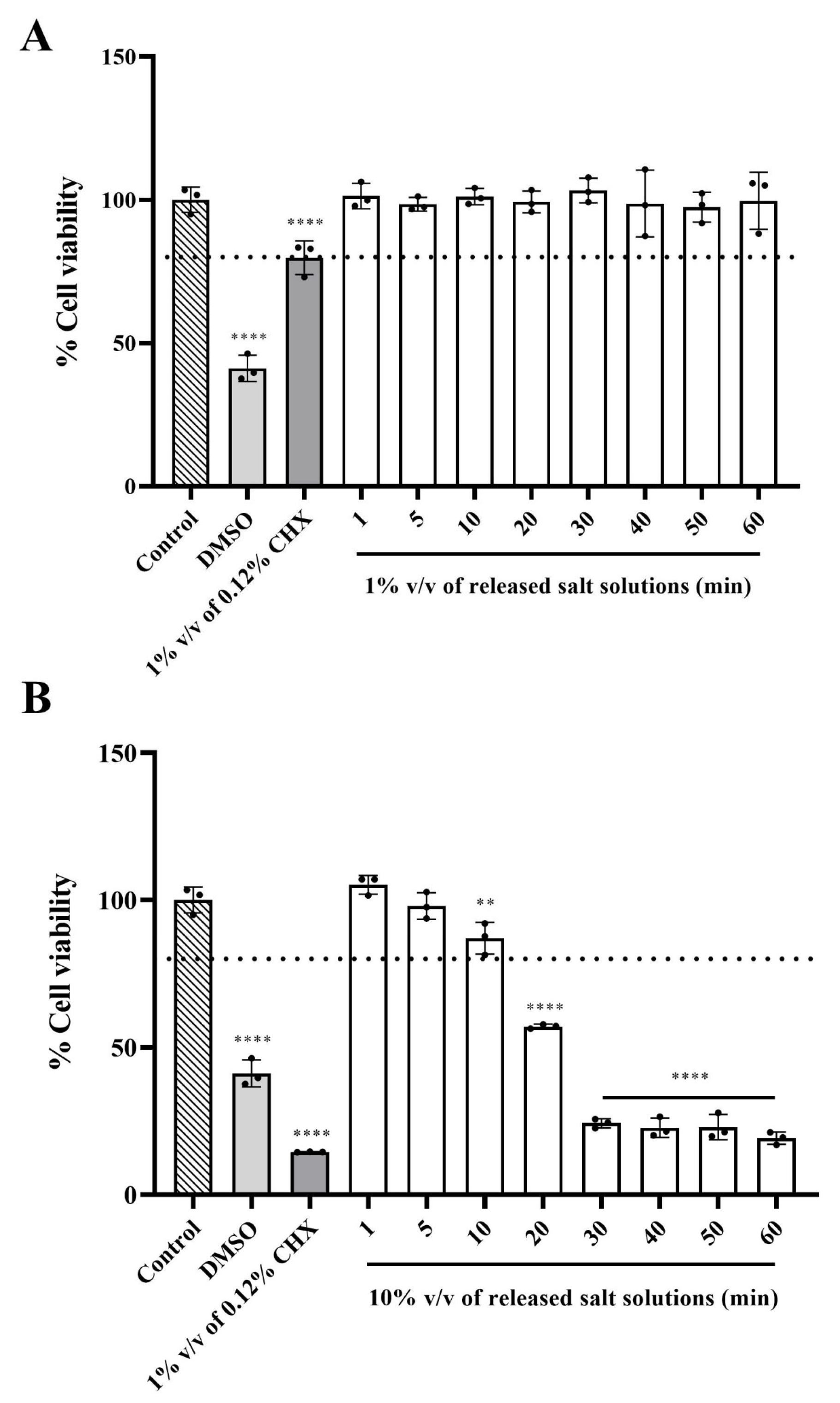
The cytotoxicity of salt tablets was evaluated against the L929 skin fibroblasts using MTT (3-(4,5-Dimethylthiazol-2-yl)-2,5-Diphenyltetrazolium Bromide) assay. According to ISO 10993-5, cell viability percentages more than 80% are considered non-cytotoxic, 80–60% as weak, 60–40% as moderate, and below 40% indicated as strong cytotoxicity [20]. Our findings revealed that a 1% v/v solution of the released salt tablet over 1 to 60 min did not induce cytotoxicity in L929 cells, with cell viability remaining above 80%. In contrast, a 1% v/v solution of 0.12% CHX significantly reduced cell viability to 79.72 ± 5.87%, indicating weak cytotoxic effects (p < 0.0001, Figure 1A). Furthermore, it was demonstrated that the cells could withstand up to 10% v/v of the released salt tablet for 1 to 10 min, with cell viability remaining above 80%. However, a 10% v/v solution of the released salt tablet after 20 min exhibited moderate cytotoxicity, resulting in a cell viability of 57.04 ± 0.74%. Whereas 10% v/v of 0.12% CHX and 10% v/v of the released salt solution for more than 30 min were considered strongly cytotoxic to L929 fibroblasts (cell viability < 40%, Figure 1B). The results suggested that the released salt solutions exhibited lower cytotoxicity than CHX in L929 fibroblasts under the tested conditions. However, this cytotoxicity assessment was limited to L929 fibroblasts as an initial screening model. Because oral epithelial cells were not evaluated, the current cytocompatibility results should be interpreted cautiously, and further studies using oral epithelial cell models are required to better assess the clinical relevance of NaCl tablets for oral mucosal application.
Salt-based approaches have several potential advantages for biomedical and oral-care applications, including availability, simple composition, and antimicrobial properties under hypertonic conditions. Previous studies reported that rinsing with 1.8% NaCl significantly enhanced type-I collagen expression and promoted wound healing in human gingival fibroblasts [21]. In addition, a 7% table salt solution was shown to accelerate wound healing in mice [22]. These findings support the potential usefulness of salt-based approaches in tissue repair and oral-care applications. However, because the present study evaluated only short-term cytotoxicity in L929 fibroblasts, further studies using oral epithelial cells and longer exposure periods are required before clinical safety or long-term application can be concluded.

3.3. Effects of Salt Tablets on Biofilm-Forming Microorganisms

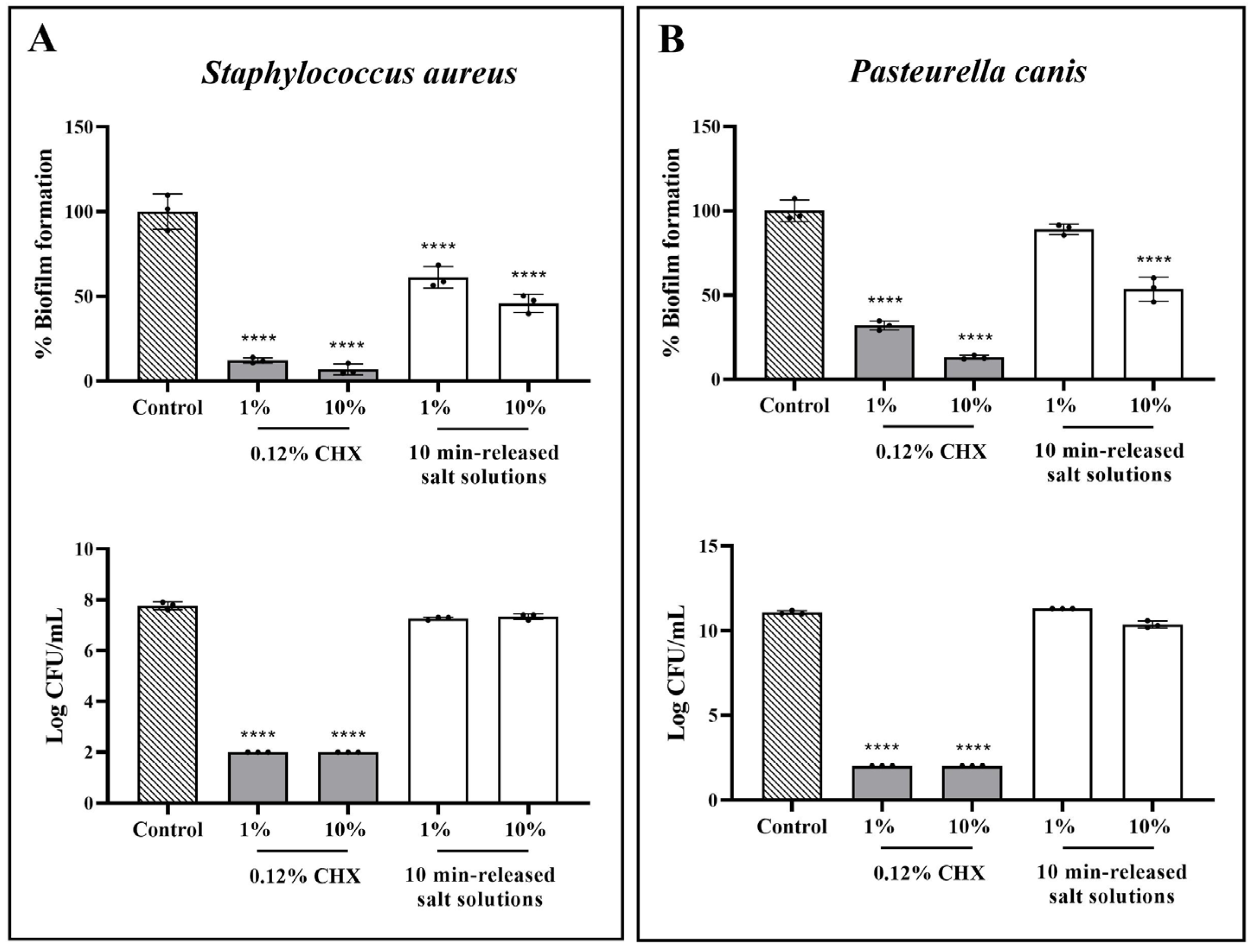
The formation of biofilms represents a major factor in the pathogenesis and chronicity of oral infections. A previous study revealed that natural extracts, such as propolis, can reduce oral pathogens and inhibit biofilm or plaque formation, aiding in the prevention of periodontal disease in animals [23]. Biofilms are structured communities of bacteria that adhere to surfaces and are embedded in a self-produced extracellular matrix composed of polysaccharides, proteins, lipids, and nucleic acids. The presence of extracellular polysaccharides (EPS) plays an essential role in biofilm accumulation. In this study, the efficiency of 1% and 10% v/v of 10 min-released salt solution to suppress biofilms in S. aureus and P. canis was examined using the crystal violet assay. Because crystal violet staining measures total biofilm biomass, the data are described as relative biofilm formation rather than viable biofilm killing. The results demonstrated that 1% and 10% v/v of the released salt solution could significantly decrease biofilm production in S. aureus compared with the control group (p < 0.0001). The released salt solution (10% v/v) significantly reduced biofilm formation in P. canis compared with the control group (p < 0.0001). While 1% v/v of the released salt solution slightly prevented P. canis biofilm formation. The percentage of biofilm formation in both S. aureus and P. canis treated with 1% and 10% v/v of 0.12% CHX was significantly reduced compared with the control group (p < 0.0001) (Figure 2A,B). The reduction in biofilm formation was interpreted together with the viable bacterial count data. In S. aureus, treatment with 1% and 10% v/v 10 min-released NaCl solutions significantly reduced relative biofilm formation, whereas viable bacterial counts remained comparable to the untreated control. A similar pattern was observed for P. canis, in which 10% v/v 10 min-released NaCl solution significantly reduced relative biofilm formation without significantly decreasing viable bacterial counts. These findings suggest that the released NaCl solution reduced biofilm biomass under the tested conditions without directly inhibiting bacterial growth. In contrast, 0.12% CHX reduced both biofilm formation and viable bacterial counts, indicating that the antibiofilm effect of CHX was largely associated with bacterial growth inhibition. A previous investigation discovered that the presence of proteins in EPS could affect bacterial auto-aggregation when microorganisms were exposed to hypertonic saline stress conditions [23,24]. This relates to our results, which indicated that 1% v/v of the released salt solution slightly reduced biofilm formation, suggesting that the appropriate concentration of hypertonic NaCl may be a key factor in inhibiting biofilms. Nevertheless, crystal violet staining does not distinguish viable from non-viable biofilm cells, and viable biofilm cell counts were not directly quantified. Microscopic analyses, such as confocal microscopy or SEM, would further strengthen the interpretation of biofilm structural changes in future studies.

3.4. Effects of Salt Tablets on Cell Morphology

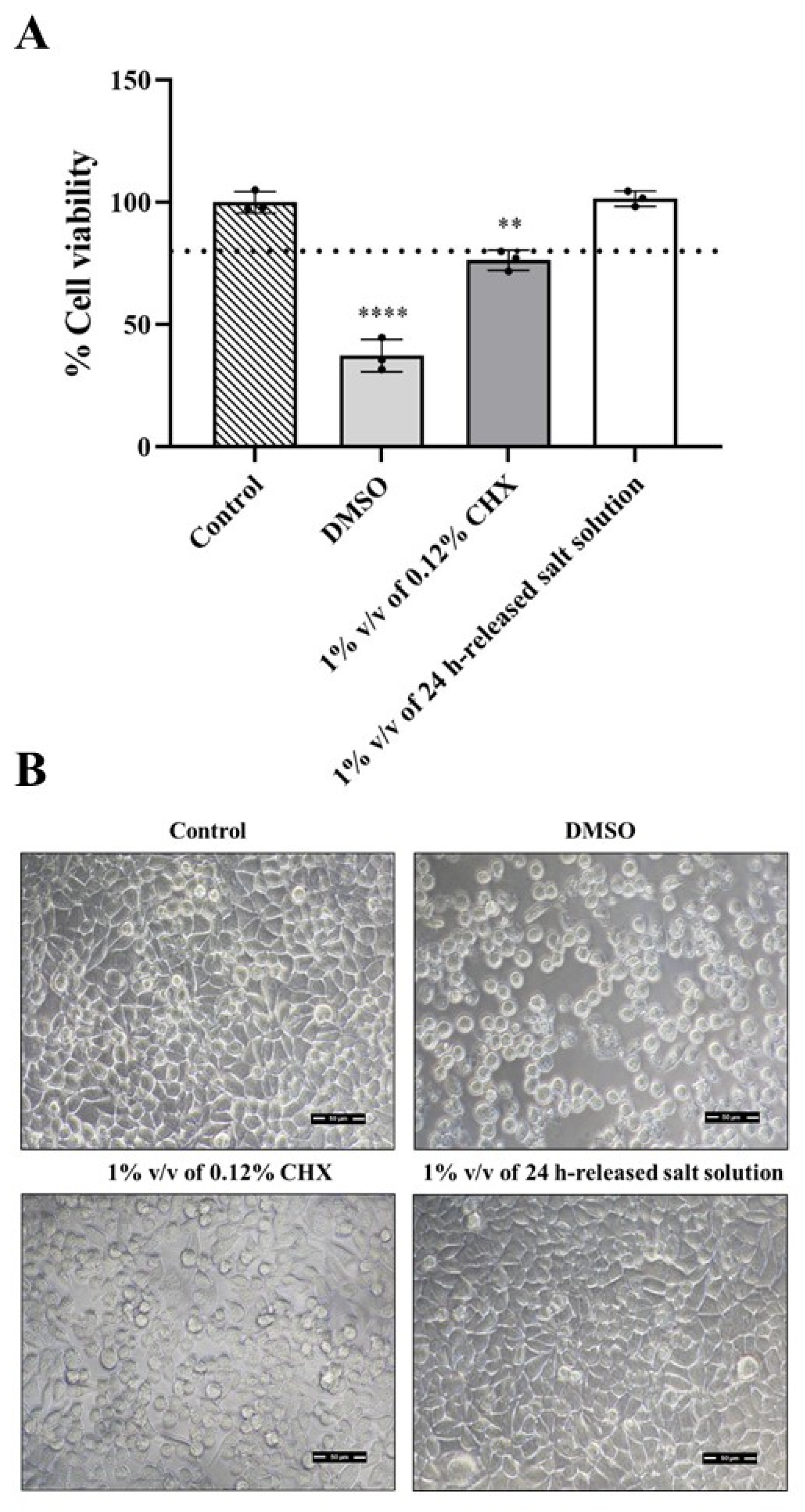
According to the data presented in Figure 1A, 1% v/v concentration of the released salt tablet from 1 to 60 min showed no cytotoxicity on L929 cells. Therefore, we further tested the cytotoxicity of the salt tablet using a 1% v/v salt solution that dissolved the tablet for 24 h. MTT results showed that treatment with a 1% v/v of the 24 h-released salt tablet did not significantly affect the cell viability of L929 fibroblasts compared with the control groups (101.52% ± 3.15 vs. 100.00% ± 4.41, p > 0.05) (Figure 3A). The morphology of the cells is shown in Figure 3B. After 24 h of treatment, the morphology of L929 cells treated with the 24 h-released salt tablet (1% v/v) was similar to that of the control group, with signs of cell–cell interaction. By contrast, changes in the morphology of L929 cells were observed after adding either DMSO (10% v/v) or 0.12% CHX (1% v/v). There was more rounding up of the cells and less cell–cell interaction compared to the control group. This phenomenon is consistent with the cell viability results, where both DMSO and chlorhexidine showed cytotoxic effects and significantly reduced the viability of L929 cells to 37.32% ± 6.62 (p < 0.01) and 76.33% ± 4.11 (p < 0.0001), respectively.
Chlorhexidine is known as an antiseptic and disinfectant. It is widely used in healthcare settings for skin disinfection. In dental care, chlorhexidine is often found in mouthwashes to reduce bacteria in the mouth, thereby preventing gingivitis and other periodontal diseases. However, several studies have been revealed that the use of chlorhexidine can be harmful to the cells and tissues. Our data showed that the morphology of L929 cells was obviously changed after exposure to CHX (Figure 3B). An explanation of this phenomenon is that chlorhexidine could induce cytotoxicity in L929 fibroblasts. Chlorhexidine was reported to trigger necrosis and apoptosis in L929 cells via ER stress. A marker of activation of the unfolded protein response (UPR) such as 78 kDa glucose-regulated protein 78 (Grp78) was significantly upregulated when the cells were exposed to 0.00025% CHX [25]. Moreover, the cytotoxicity of chlorhexidine was confirmed by the other cell types such as human keratinocyte (HaCaT) and human fibroblast (hFB) cell lines. 0.02% of CHX significantly reduced cell viability of both cell types and three minutes application of chlorhexidine could also inhibit cell migration capacity in HaCaT cells [26]. Thereby, an alternative agent with lower toxicity is highly desirable for use in clinical and dental applications.

3.5. Effects of Salt Tablets on Nitric Oxide Production

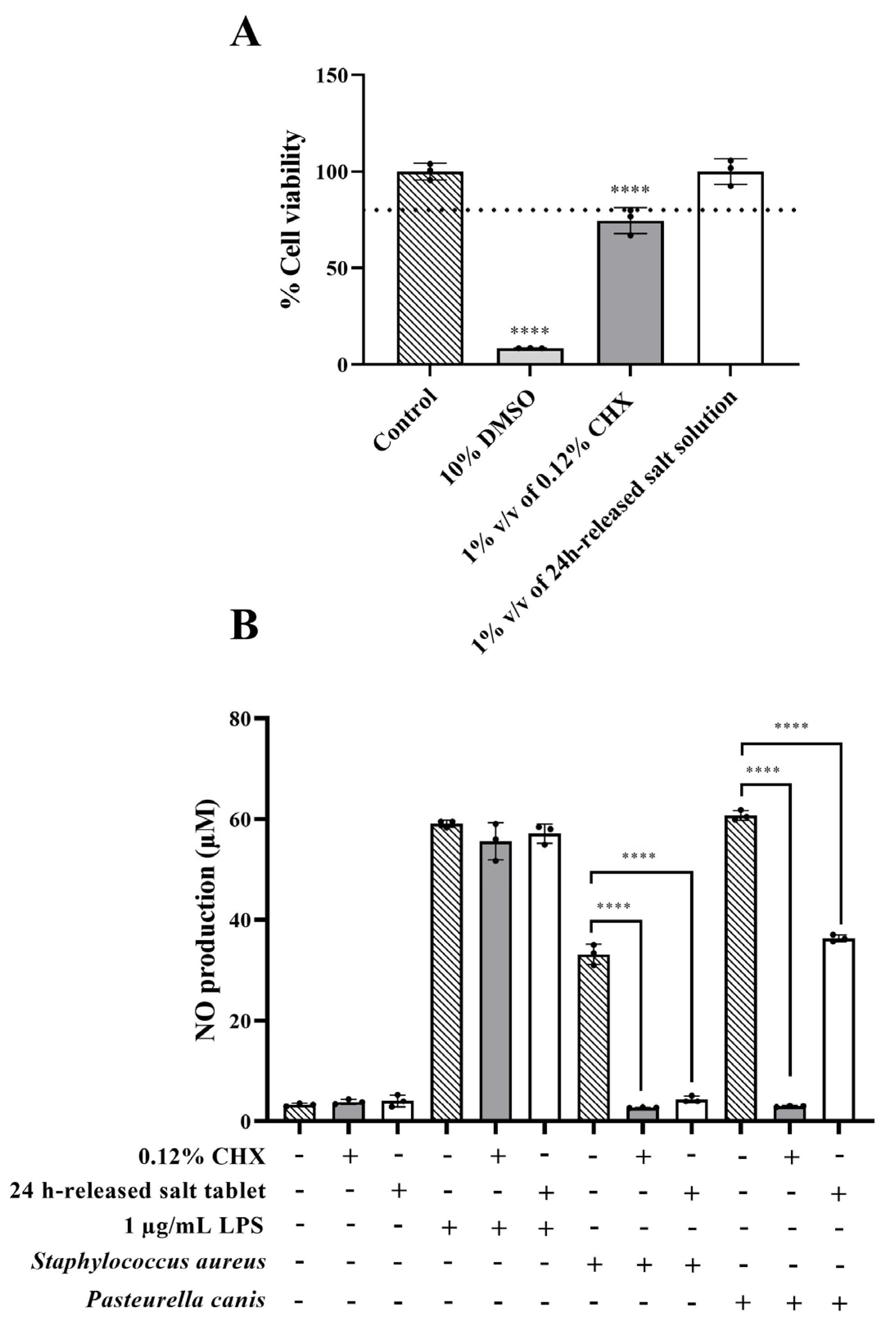
RAW264.7 cells are a murine macrophage cell line widely used in research to study inflammatory responses, including the production of nitric oxide (NO). The NO is a hallmark of the inflammatory marker in macrophages that plays an important role in mediating cytotoxic activities against pathogens. Therefore, to investigate the effects of salt tablets on RAW264.7 cells, their cytotoxic effects were initially evaluated. Similar to L929 fibroblasts, 1% v/v of 24 h-released salt tablet showed no cytotoxic effects on the RAW264.7 cells (cell viability > 80%), while 1% v/v of 0.12% CHX significantly reduced cell viability to 74.52 ± 6.77% (considered as weak-cytotoxic effects, p < 0.0001, Figure 4A). To investigate the effects of salt tablet on inflammation induced by S. aureus and P. canis, the RAW264.7 cells were cultured with either 1% v/v of 24 h-incubated bacterial solution, 1% v/v of 24 h-incubated bacterial/salt tablet solution, and 1% v/v of 24 h-incubated bacterial/chlorhexidine solution. 1 µg/mL of LPS was used as an inflammatory stimulus control. As shown in Figure 4B, the salt tablet and chlorhexidine solutions did not stimulate an increase in NO release, while LPS, S. aureus, and P. canis treatments remarkably triggered NO production in RAW264.7 cells compared with the control group (59.10 ± 0.69, 33.17 ± 2.01, and 60.73 ± 0.99 µM, respectively vs. 3.28 ± 0.32 µM, p < 0.0001). The results indicated that both S. aureus and P. canis can induce inflammation in RAW264.7 cells. The co-culture of LPS with either the salt tablet or chlorhexidine solutions showed no statistically significant difference in NO production from the cells compared with the LPS alone (57.11 ± 1.92, 55.60 ± 3.67, and 59.10 ± 0.69 µM. respectively, p > 0.05). However, the presence of either salt tablet or chlorhexidine solution significantly attenuated bacteria-induced NO production in RAW 264.7 cells compared with the treatment with either S. aureus alone (4.34 ± 0.65 µM and 2.71 ± 0.05 vs. 33.17 ± 2.01 µM, p < 0.0001) or P. canis (36.33 ± 0.69 µM and 2.98 ± 0.16 vs. 60.73 ± 0.99 µM, p < 0.0001). Together, the results suggest that the salt tablet is effective in protecting against inflammation induced by bacterial infections.
Although our data showed that the salt tablet solution has no effect on the inhibition of LPS-induced NO production in murine macrophages (Figure 4), several studies have shown the beneficial effects of salt on the suppression of inflammation caused by LPS. For example, hypertonic saline (NaCl 7.5%) was shown to reduce LPS-induced acute lung injury in rats by attenuating metalloproteinase 9 (MMP-9) activity in tissue as well as decreasing nitric oxide synthase (iNOS or NOS2) and NO production [27]. In addition, hypertonic saline solution (NaCl 7.5%) treatment significantly lowered levels of IL-6, NO, and inflammatory response in endotoxemic rats, compared to 0.9% normal saline [28]. A previous study revealed that IL-6 plays a crucial role in driving the expression of immune- and inflammation-related genes and pathways in the oral mucosal tissues of cats with FCGs [29]. Regarding the anti-inflammatory effects of salt tablets, our data revealed that the presence of hypertonic salt solution significantly attenuated S. aureus and P. canis -induced NO production in RAW264.7 cells (Figure 4). Bacteria and biofilms play significant roles in inflammation, particularly in chronic and persistent infections. Various studies have demonstrated that bacterial infection significantly induces inflammatory responses in numerous cell types. For example, elevated IL-6 and IL-8 responses were found in a keratinocyte cell line (HaCaT) upon exposure to S. aureus [30]. A high biofilm-producing strain of P. aeruginosa, as well as its conditioned media, also induced the expression of TNF-α, IL-6, PGE2, iNOS, and COX-2 in neutrophils and macrophages [31]. Combining our data in Figure 2, we demonstrated that the salt tablet effectively inhibited bacterial growth and biofilm formation. These findings suggest the potential of hypertonic NaCl solutions as a therapeutic option for managing bacterial-induced inflammation.

3.6. In Vivo Study

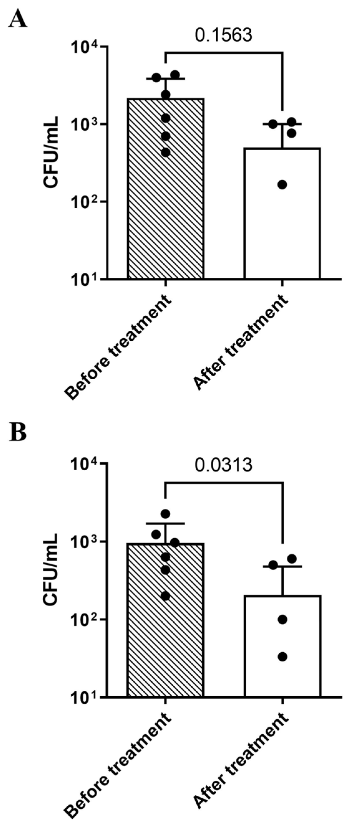
Oral mucosa wound healing is a crucial, sequential process that closes tissue ruptures to prevent microbial invasion and chronic inflammation. Due to constant exposure to trauma and infection, facilitating the healing of oral wounds poses a significant challenge. A previous study has shown that short-term rinsing with NaCl promotes gingival fibroblast migration, enhances extracellular matrix gene expression and cytoskeletal organization, providing scientific support for its traditional use as a mouth rinse to promote oral wound healing and improve oral health [13,21]. In our study, we simulated an oral wound treatment approach in a mouse model by applying salt crystals to the wound area for a short period to promote healing and reduce bacterial counts at the site. The average total bacterial count was assessed after 2 days to establish baseline data before treatment in both the control and experimental groups. The results demonstrated that the number of bacteria in the control group did not show a statistically significant difference before and after 30 min of untreated period (p = 0.1563) (Figure 5A). This finding suggests that the bacterial load remained relatively stable under untreated conditions. In contrast, treatment with NaCl crystals for 30 min significantly reduced the total bacterial count at the treatment site (p = 0.0313) (Figure 5B). However, this result should be interpreted as preliminary evidence of short-term bacterial burden reduction rather than definitive therapeutic efficacy. Because the untreated and treated wounds were located within the same oral cavity, possible NaCl transfer through saliva could not be completely excluded. In addition, because histological confirmation and inflammatory marker analysis were not performed, this model should not be interpreted as demonstrating resolution of faucitis or in vivo anti-inflammatory activity. The in vivo findings are limited to short-term reduction in total bacterial counts at the wound surface.
This study has several limitations. First, the in vivo experiment used only five mice and a short observation period; therefore, the animal findings should be interpreted as preliminary. Second, the paired left–right buccal wound design may be affected by possible NaCl transfer through saliva within the same oral cavity. Third, only two representative bacterial species were evaluated, and the results may not fully represent the polymicrobial nature of companion animal oral diseases. Fourth, oral epithelial cells, histological analysis, inflammatory markers, and long-term safety assessments were not included. Finally, clinical studies in dogs or cats with naturally occurring oral disease are required before clinical efficacy or long-term safety can be concluded.

4. Conclusions

Hypertonic salt tablets demonstrated antibacterial activity against S. aureus and P. canis under the tested in vitro conditions. The released salt solutions exhibited lower cytotoxicity than chlorhexidine (CHX) in L929 fibroblasts, and treatment with a 1% v/v 24 h-released salt solution maintained normal cell morphology, supporting its cytocompatibility in this preliminary cell model. The 1% and 10% v/v 10 min-released salt solutions reduced relative biofilm biomass in both bacterial species without significantly decreasing viable bacterial counts, suggesting that the released NaCl solution affected biofilm biomass formation rather than reducing biofilm indirectly through bacterial growth inhibition. In contrast, CHX reduced both biofilm formation and viable bacterial counts, indicating a growth-dependent antibiofilm effect. In RAW 264.7 macrophages, although salt itself did not exhibit anti-inflammatory activity against LPS stimulation, salt-treated bacterial suspensions significantly reduced NO production. This suggests that salt treatment may attenuate bacteria-associated inflammatory stimulation by reducing bacterial inflammatory burden rather than directly inhibiting intracellular inflammatory signaling in macrophages. In a preliminary mouse oral mucosal wound model, local application of NaCl crystals reduced total bacterial counts after a single 30 min treatment; however, because the in vivo experiment used a small sample size, short observation period, and paired left–right wound design, these results should be interpreted as preliminary evidence of short-term bacterial burden reduction rather than definitive therapeutic efficacy. Taken together, these findings suggest that hypertonic NaCl tablets may represent a simple localized approach for short-term reduction in oral bacterial burden and bacteria-associated inflammatory stimulation. Further studies are required to confirm actual NaCl release concentration and osmolality, long-term safety, disease-resolution outcomes, activity against broader oral pathogens, and clinical relevance in dogs or cats with naturally occurring oral disease.

Author Contributions

Conceptualization, S.W., C.J. and K.D.; methodology, S.W., C.J., N.M. and K.D.; validation, S.W. and S.I.; formal analysis, S.W., C.J., N.M., T.K., C.N. and N.H.; investigation, S.W., C.J., N.M., S.I., T.K., C.N. and N.H.; resources, S.P.V. and K.D.; writing—original draft preparation, S.W. and C.J.; writing—review and editing, S.W., S.P.V. and K.D.; visualization, K.D.; supervision, S.P.V. and K.D.; funding acquisition, K.D. All authors have read and agreed to the published version of the manuscript.

Funding

This research was supported by Faculty of Veterinary Science Research Fund.

Institutional Review Board Statement

All experimental procedures were approved by the Prince of Songkla University Ethical Committee, with protocol code 2022-VET07-070 and acceptable use policy reference AR097/2022 for the mouse trials.

Informed Consent Statement

Not applicable.

Data Availability Statement

The original contributions presented in this study are included in the article. Further inquiries can be directed to the corresponding author.

Conflicts of Interest

The authors declare no conflicts of interest.

References

  1. Dosenberry, C.; Arzi, B.; Palm, C.; Vapniarsky, N.; Soltero-Rivera, M. An Update on Oral Manifestations of Systemic Disorders in Dogs and Cats. Front. Vet. Sci. 2024, 11, 1511971. [Google Scholar] [CrossRef] [PubMed]
  2. Dewhirst, F.E.; Klein, E.A.; Thompson, E.C.; Blanton, J.M.; Chen, T.; Milella, L.; Buckley, C.M.F.; Davis, I.J.; Bennett, M.L.; Marshall-Jones, Z.V. The Canine Oral Microbiome. PLoS ONE 2012, 7, e36067. [Google Scholar] [CrossRef] [PubMed]
  3. Krishnan, K.; Chen, T.; Paster, B.J. A Practical Guide to the Oral Microbiome and Its Relation to Health and Disease. Oral Dis. 2017, 23, 276–286. [Google Scholar] [CrossRef] [PubMed]
  4. Lisjak, A.; Correa Lopes, B.; Pilla, R.; Nemec, A.; Suchodolski, J.S.; Tozon, N. A Comparison of the Oral Microbiota in Healthy Dogs and Dogs with Oral Tumors. Animals 2023, 13, 3594. [Google Scholar] [CrossRef] [PubMed]
  5. Whyte, A.; Gracia, A.; Bonastre, C.; Tejedor, M.T.; Whyte, J.; Monteagudo, L.V.; Simón, C. Oral Disease and Microbiota in Free-Roaming Cats. Top. Companion Anim. Med. 2017, 32, 91–95. [Google Scholar] [CrossRef] [PubMed]
  6. Razali, K.; Kaidi, R.; Abdelli, A.; Menoueri, M.N.; Ait-Oudhia, K. Oral Flora of Stray Dogs and Cats in Algeria: Pasteurella and Other Zoonotic Bacteria. Vet. World 2020, 13, 2806–2814. [Google Scholar] [CrossRef] [PubMed]
  7. Rodrigues, M.X.; Fiani, N.; Bicalho, R.C.; Peralta, S. Preliminary Functional Analysis of the Subgingival Microbiota of Cats with Periodontitis and Feline Chronic Gingivostomatitis. Sci. Rep. 2021, 11, 6896. [Google Scholar] [CrossRef] [PubMed]
  8. Rodrigues, M.X.; Bicalho, R.C.; Fiani, N.; Lima, S.F.; Peralta, S. The Subgingival Microbial Community of Feline Periodontitis and Gingivostomatitis: Characterization and Comparison between Diseased and Healthy Cats. Sci. Rep. 2019, 9, 12340. [Google Scholar] [CrossRef] [PubMed]
  9. Könönen, E.; Gursoy, M.; Gursoy, U.K. Periodontitis: A Multifaceted Disease of Tooth-Supporting Tissues. J. Clin. Med. 2019, 8, 1135. [Google Scholar] [CrossRef] [PubMed]
  10. Tsang, W.; Linde, A.; Krumbeck, J.A.; Wu, G.; Kim, Y.J.; Lushington, G.H.; Melgarejo, T. Occurrence of Antimicrobial Resistance Genes in the Oral Cavity of Cats with Chronic Gingivostomatitis. Animals 2021, 11, 3589. [Google Scholar] [CrossRef] [PubMed]
  11. Son, M.; Lee, Y.S.; Lee, M.J.; Park, Y.K.; Bae, H.R.; Lee, S.Y.; Shin, M.G.; Yang, S. Effects of Osmolality and Solutes on the Morphology of Red Blood Cells According to Three-Dimensional Refractive Index Tomography. PLoS ONE 2021, 16, e0262106. [Google Scholar] [CrossRef] [PubMed]
  12. Li, F.; Xiong, X.S.; Yang, Y.Y.; Wang, J.J.; Wang, M.M.; Tang, J.W.; Liu, Q.H.; Wang, L.; Gu, B. Effects of NaCl Concentrations on Growth Patterns, Phenotypes Associated With Virulence, and Energy Metabolism in Escherichia Coli BW25113. Front. Microbiol. 2021, 12, 05326. [Google Scholar] [CrossRef] [PubMed]
  13. Collins, J.R.; Veras, K.; Hernández, M.; Hou, W.; Hong, H.; Romanos, G.E. Anti-Inflammatory Effect of Salt Water and Chlorhexidine 0.12% Mouthrinse after Periodontal Surgery: A Randomized Prospective Clinical Study. Clin. Oral Investig. 2021, 25, 4349–4357. [Google Scholar] [CrossRef] [PubMed]
  14. CLSI M02; Performance Standards for Antimicrobial Disk Susceptibility Tests: Approved Standard—Eleventh Edition. Clinical and Laboratory Standards Institute: Wayne, PA, USA, 2012.
  15. Jakkawanpitak, C.; Wunnoo, S.; Sermwittayawong, D.; Runsaeng, P.; Amnuaikit, T.; Voravuthikunchai, S.P. Quercus infectoria Gall Extract Modulates Adipocyte Differentiation and Lipid Metabolism through Dual Regulation of Adipogenesis and Lipolysis in 3T3-L1 Adipocytes. Anim. Cells Syst. 2026, 30, 161–182. [Google Scholar] [CrossRef] [PubMed]
  16. Wunnoo, S.; Wae-Asae, P.; Kaewrak, S.; Ekchaweng, K.; Voravuthikunchai, S.P.; Jakkawanpitak, C. Internal Gelation-Based Alginate Hydrogel Films Incorporating Quercus infectoria Gall Extract for Multifunctional Wound Dressing Applications. Sci. Rep. 2026, 16, 12501. [Google Scholar] [CrossRef] [PubMed]
  17. Vongmanee, N.; Bunmak, A.; Visitsattapongse, S. An Automated Colony Counter for Serial-Dilution Culture Method. ACM Int. Conf. Proc. Ser. 2018, 42–45. [Google Scholar] [CrossRef]
  18. Yang, S.; Wang, M.; Gao, J.; Liu, J.; Jin, R.; Lin, R.; Weng, W.; Aweya, J.J. Sodium Chloride Augments the Antibacterial Activity of a Novel Penaeid Shrimp-Derived Peptide (GPCR10) against Halotolerant Staphylococcus aureus. LWT 2023, 184, 115096. [Google Scholar] [CrossRef]
  19. Lin, H.L.; Chiu, L.C.; Wan, G.H.; Huang, C.C.; Lee, Z.T.; Lin, Y.T.; Wu, S.R.; Chen, C.S. Hypertonic Saline Enhances the Efficacy of Aerosolized Gentamicin against Pseudomonas aeruginosa. Sci. Rep. 2020, 10, 4325. [Google Scholar] [CrossRef] [PubMed]
  20. Mohd Shafiee, M.A.; Muhamad Asri, M.A.; Syed Alwi, S.S. Review on the in Vitro Cytotoxicity Assessment in Accordance to the International Organization for Standardization (ISO). Malays. J. Med. Health Sci. 2021, 17, 261–269. [Google Scholar]
  21. Huynh, N.C.N.; Everts, V.; Leethanakul, C.; Pavasant, P.; Ampornaramveth, R.S. Rinsing with Saline Promotes Human Gingival Fibroblast Wound Healing in Vitro. PLoS ONE 2016, 11, e0159843. [Google Scholar] [CrossRef] [PubMed]
  22. Samidah, S.; Prihantono; Ahmad, M.; Jompa, J.; Rafiah, S.; Usman, A.N. The Effectiveness of 7% Table Salt Concentration Test to Increase Collagen in the Healing Process of Wound. Gac. Sanit. 2021, 35, S199–S201. [Google Scholar] [CrossRef] [PubMed]
  23. Figueiredo, C.G.F.; dos Santos, M.S.; Santos, A.S.; Silva, E.d.S.; Lima, B.; Junior, W.d.L.; de Araujo, Y.L.F.M.; Batista, M.V.d.A. In Vitro Evaluation of the Antibacterial Effect of Brazilian Red Propolis Ethanol Extract in the Prevention of Periodontal Disease in Dogs. Comp. Immunol. Microbiol. Infect. Dis. 2023, 92, 01924. [Google Scholar] [CrossRef] [PubMed]
  24. Li, D.; Liang, X.; Wu, C. Characteristics of Nitrogen Removal and Extracellular Polymeric Substances of a Novel Salt-Tolerant Denitrifying Bacterium, Pseudomonas sp. DN-23. Front. Microbiol. 2020, 11, 335. [Google Scholar] [CrossRef] [PubMed]
  25. Faria, G.; Cardoso, C.R.B.; Larson, R.E.; Silva, J.S.; Rossi, M.A. Chlorhexidine-Induced Apoptosis or Necrosis in L929 Fibroblasts: A Role for Endoplasmic Reticulum Stress. Toxicol. Appl. Pharmacol. 2009, 234, 256–265. [Google Scholar] [CrossRef] [PubMed]
  26. Ortega-Llamas, L.; Quiñones-Vico, M.I.; García-Valdivia, M.; Fernández-González, A.; Ubago-Rodríguez, A.; Sanabria-De la Torre, R.; Arias-Santiago, S. Cytotoxicity and Wound Closure Evaluation in Skin Cell Lines after Treatment with Common Antiseptics for Clinical Use. Cells 2022, 11, 1395. [Google Scholar] [CrossRef] [PubMed]
  27. Petroni, R.C.; Biselli, P.J.C.; de Lima, T.M.; Theobaldo, M.C.; Caldini, E.T.; Pimentel, R.N.; Barbeiro, H.V.; Kubo, S.A.; Velasco, I.T.; Soriano, F.G. Hypertonic Saline (NaCl 7.5%) Reduces LPS-Induced Acute Lung Injury in Rats. Inflammation 2015, 38, 2026–2035. [Google Scholar] [CrossRef] [PubMed]
  28. Theobaldo, M.C.; Barbeiro, H.V.; Barbeiro, D.F.; Petroni, R.; Soriano, F.G. Hypertonic Saline Solution Reduces the Inflammatory Response in Endotoxemic Rats. Clinics 2012, 67, 1463–1468. [Google Scholar] [CrossRef] [PubMed]
  29. Peralta, S.; Grenier, J.K.; Webb, S.M.; Miller, A.D.; Miranda, I.C.; Parker, J.S.L. Transcriptomic Signatures of Feline Chronic Gingivostomatitis Are Influenced by Upregulated IL6. Sci. Rep. 2023, 13, 13437. [Google Scholar] [CrossRef] [PubMed]
  30. Ngo, Q.V.; Faass, L.; Sähr, A.; Hildebrand, D.; Eigenbrod, T.; Heeg, K.; Nurjadi, D. Inflammatory Response Against Staphylococcus aureus via Intracellular Sensing of Nucleic Acids in Keratinocytes. Front. Immunol. 2022, 13, 828626. [Google Scholar] [CrossRef] [PubMed]
  31. Ciszek-Lenda, M.; Strus, M.; Walczewska, M.; Majka, G.; Machul-Żwirbla, A.; Mikołajczyk, D.; Górska, S.; Gamian, A.; Chain, B.; Marcinkiewicz, J. Pseudomonas aeruginosa Biofilm Is a Potent Inducer of Phagocyte Hyperinflammation. Inflamm. Res. 2019, 68, 397–413. [Google Scholar] [CrossRef] [PubMed]
Figure 1. Toxicity effects of 1% v/v (A) and 10% v/v (B) solutions of the released salt tablet over 1 to 60 min on L929 fibroblast cell line. The values indicate the means ± SD from three independent experiments performed in triplicate. **, **** Significant difference between treatment and control group (p < 0.01 and 0.0001, respectively).
Sci 08 00168 g001
Figure 2. Effects of 1% and 10% v/v 10 min-released NaCl solutions on relative biofilm formation and bacterial growth of Staphylococcus aureus (A) and Pasteurella canis (B). The upper graphs show relative biofilm formation determined by crystal violet staining, while the lower graphs show viable bacterial counts determined by plate count assay and expressed as Log CFU/mL. Data are presented as mean ± SD from three independent experiments performed in triplicate. **** indicates a significant difference compared with the untreated control (p < 0.0001).
Sci 08 00168 g002
Figure 3. Toxicity effects (A) and cell morphology (B) on L929 fibroblast cell line upon treatment with 1% v/v solution of a 24 h released salt tablet for 24 h at 37 °C in a humidified atmosphere containing 5% CO2. Scale bar = 50 µm. The values indicate the means ± SD from three independent experiments performed in triplicate. **, **** Significant difference between treatment and control group (p < 0.01 and 0.0001, respectively).
Sci 08 00168 g003
Figure 4. Anti-inflammatory activity of 1% v/v solution of a 24 h released salt tablet on RAW264.7 cells stimulated by lipopolysaccharides (LPS). The viability was investigated using MTT assay (A). The nitric oxide levels in the cell media were measured by Griess assay (B). The values indicate the means ± SD from three independent experiments performed in triplicate. **** Significant differences compared with the bacterial control group (p < 0.0001).
Sci 08 00168 g004
Figure 5. Comparison of total bacterial count (CFU/mL) in control group (A) and NaCl-treated group (B) before and after the application of 99.5% sterile NaCl crystals for 30 min. Data are presented as individual values with mean ± SD from five mice (n = 5). CFU measurements were performed using technical replicate plating for each sample.
Sci 08 00168 g005
Table 1. Antibacterial activity of 0.3 g salt tablets using agar well diffusion assay.
MicroorganismsMean of Inhibition Zone (mm)
Salt Tablet 0.3 g0.12% Chlorhexidine
Staphylococcus aureus13.33 ± 0.8923.10 ± 0.87
Pasteurella canis28.75 ± 1.1831.43 ± 0.81
Values are mean ± standard deviation for three independent experiments performed in triplicate.
Disclaimer/Publisher’s Note: The statements, opinions and data contained in all publications are solely those of the individual author(s) and contributor(s) and not of MDPI and/or the editor(s). MDPI and/or the editor(s) disclaim responsibility for any injury to people or property resulting from any ideas, methods, instructions or products referred to in the content.