


Abstract
Critical-sized bone defects remain a major reconstructive challenge, and the Masquelet technique has become an important option in traumatic, infectious, and post-resection settings. This review examines the role of acrylic bone cement in this technique, emphasizing its evolution from a temporary spacer to an active biomaterial that shapes the induced membrane and the local regenerative microenvironment. The article discusses the surgical and biological basis of the technique, the composition and handling characteristics of polymethyl methacrylate (PMMA) bone cement, the rationale for antibiotic-loaded spacers, and the influence of spacer-related variables such as formulation, surface properties, and geometry on membrane quality. It also addresses emerging strategies, including bioactive PMMA modifications, multifunctional cements, and degradable alternatives aimed at improving osteogenesis, angiogenesis, and infection control. Current evidence, derived mainly from in vitro studies and animal models, suggests that the spacer may play a role beyond space maintenance by participating in induced membrane formation and influencing biological signaling related to bone repair. In contrast, the clinical evidence primarily supports the reproducible use of PMMA spacers for dead-space management, infection control, and bone reconstruction. However, important gaps still remain in the translational validation of these biological properties and in the standardization of spacer formulations and antibiotic protocols.
1. Introduction
1.1. The Challenge of Critical Segmental Bone Defects
Reconstruction of large bone defects remains a major challenge for orthopedic surgeons, particularly in managing critical segmental defects in long bones. Their clinical relevance extends across a wide range of scenarios, including high-energy trauma, infected nonunion, osteomyelitis sequelae, and tumor resection, in which the objective is not only to restore bone continuity but also to recover function, control infection, and preserve the affected limb [1,2].
The concept of a critical bone defect has evolved from a purely dimensional definition to a more comprehensive view in which defect size interacts with the host’s biological status and the local conditions at the recipient site. This complexity helps explain why no universal solution exists and why versatile, biologically favorable reconstructive strategies are still required [1].
Traditionally, bone defects have been treated with autologous bone grafts, which remain the gold standard for bone regeneration. Autologous cancellous bone grafts, harvested either from the iliac crest or by reamer–irrigator–aspirator (RIA) techniques, mainly provide an osteoconductive scaffold with additional osteogenic and osteoinductive properties. However, this approach has important limitations: the amount of available bone is restricted, donor-site morbidity is not negligible, and the defect to be filled is usually smaller than 5 cm, as larger defects are associated with a high risk of failure due to physiological graft resorption [3].
Other reconstructive alternatives, such as distraction osteogenesis and vascularized fibular grafting, have also demonstrated utility in treating bone defects. Nevertheless, distraction osteogenesis is associated with prolonged reconstruction times, whereas vascularized fibular grafting requires advanced microsurgical expertise and is accompanied by considerable donor-site morbidity, which limits its applicability in certain clinical settings [3].
In osteomyelitis, the reconstructive challenge becomes even greater. This inflammatory bone disease, most commonly caused by bacterial infection and more frequently affecting the lower extremities, requires surgical management based on radical debridement of infected bone and soft tissue, removal of fixation hardware when indicated, dead-space management, adequate soft-tissue coverage, and restoration of sufficient vascular supply. In extensive bone defects, bone transport and the Masquelet-induced membrane technique are among the most relevant reconstructive options [2].
1.2. Why the Masquelet Technique Remains Relevant
In this context, the induced membrane technique remains relevant in contemporary reconstructive practice. The management of large diaphyseal bone defects, particularly those exceeding 5 cm, was substantially transformed by this surgical approach, first introduced by Alain-Charles Masquelet in 1986 and later established as the Masquelet induced membrane technique [4].
Its continued relevance is largely explained by its ability to address extensive bone defects through a two-stage procedure that integrates dead-space management, defect stabilization, local infection control, and the creation of a biologically favorable environment for subsequent bone graft incorporation. In the first stage, nonviable tissue is thoroughly debrided, and the defect is filled with a spacer; in the second stage, the spacer is removed, and the defect is reconstructed with bone graft while preserving the induced membrane formed around the spacer [3,4,5].
Unlike other reconstructive strategies that are more complex or require greater logistical resources, the Masquelet technique has demonstrated applicability in both post-traumatic defects and in infected and non-infected settings, which has contributed to its wider adoption and consolidation as a reconstructive option of broad clinical interest. Its potential advantages include the absence of a need for specialized equipment or high-cost infrastructure, relatively lower surgical complexity, the opportunity to optimize soft-tissue management between stages, reduced dependence on patient compliance, and a consolidation time that does not appear to be directly determined by defect size [3,5].
Another factor reinforcing the relevance of this technique is that bone defect size does not appear to correlate significantly with the healing time, which has been reported to be approximately 7 months in some clinical scenarios. This figure compares favorably with other reconstructive strategies, such as distraction osteogenesis, which may require approximately 10 months for defects smaller than 5 cm and nearly 20 months for those between 5 and 10 cm [3].
However, the persistence of the technique should not be interpreted solely as a consequence of its relative simplicity, but also as a reflection of a biological and surgical rationale that remains highly relevant. The induced membrane is no longer regarded merely as a passive peri-implant finding; rather, it is now recognized as a structure with angiogenic, osteogenic, and immunobiological potential, capable of protecting the graft, promoting revascularization, and contributing to the regenerative microenvironment of the defect [4].
1.3. The Historical Use of PMMA as a Spacer in the Masquelet Technique
Within this framework, polymethyl methacrylate (PMMA) has historically occupied a central role as a temporary spacer. Its use in orthopedic surgery long predates the Masquelet technique, particularly in arthroplasty, dead-space management, and the local treatment of osteoarticular infections through antibiotic-loaded cements. Its practical advantages, including broad availability, ease of intraoperative molding, relative structural stability, and the capacity for local antimicrobial delivery, help explain why it was adopted as the material of choice during the first stage of the technique [4,6].
A key feature of the induced membrane technique is the preparation of the graft bed by creating a biologically favorable membrane at the defect site. During the first stage, a PMMA spacer is molded to fill the bone defect. Implantation of this spacer initiates a physiological immune response known as the foreign body reaction, which leads to the formation of a fibrous encapsulating membrane, referred to as the induced membrane, surrounding the spacer. In most cases, a second surgical procedure is performed six to eight weeks later to remove the spacer while preserving the integrity of the membrane as much as possible [4].
In its classical interpretation, PMMA has been regarded primarily as a means of maintaining the defect cavity, preventing fibrous tissue invasion, obliterating dead space, and facilitating a more controlled second-stage reconstruction. In infected defects, moreover, antibiotic loading enables high local drug concentrations, further reinforcing its value as a useful tool for local infection control [2,6].
1.4. Reframing Bone Cement Not Merely as a Temporary Filler, but as an Active Biomaterial That Modulates the Regenerative Microenvironment
To regard acrylic bone cement exclusively as a space-maintaining device is to oversimplify a complex biological process in which the biomaterial actively interacts with the surrounding tissue environment [4]. Accumulating evidence, derived mainly from in vitro studies and animal models, suggests that the PMMA spacer does not behave merely as an inert element, but may instead induce local changes in gene expression and cellular composition within the bone defect [7].
During the first stage of the technique, spacer implantation triggers a foreign body reaction that leads to the formation of the induced membrane, a biologically active tissue with angiogenic and osteogenic functions [4]. This process has been characterized as an organized host response that not only encapsulates the material but also conditions the environment in which subsequent bone regeneration will occur [8].
At the molecular level, transcriptomic and experimental studies have shown enrichment of genes associated with osteoblastic activity and pathways related to bone repair in the presence of PMMA; however, much of this evidence remains preclinical, and its direct translation into clinical outcomes still requires further validation [7].
These findings reinforce the notion that the host–cement interface is not passive, but actively participates in the regulation of the local tissue microenvironment by promoting signaling networks associated with extracellular matrix formation and the activation of key regenerative pathways, including Wnt, transforming growth factor-beta (TGF-β), and Hedgehog signaling, all of which are implicated in bone repair. Consequently, the cement does more than preserve space; it directly influences the cellular and molecular processes that determine graft performance and regenerative success [4,7].
In addition, several studies have shown that spacer-related characteristics, including composition, surface features, and antibiotic loading, modulate the local inflammatory response and the quality of the induced membrane. These variables affect critical parameters such as vascularization, membrane thickness, and cellular content, which, in turn, influence the membrane’s capacity to support bone graft integration [4,9].
This multifunctional role becomes particularly relevant in septic settings, where PMMA serves not only a structural purpose, but also antimicrobial and biologically modulatory functions. In such contexts, the spacer simultaneously contributes to infection control and to the restoration of conditions favorable for regeneration, thereby integrating mechanical, biological, and pharmacological functions within a single system [4,9].
Overall, acrylic bone cement plays an active role in the Masquelet technique by modulating not only induced membrane formation, but also the cellular and molecular dynamics of the regenerative environment. Therefore, although PMMA should not be regarded merely as a temporary filler, the specific clinical relevance of its biological properties still depends on more robust translational and clinical evidence [4,7].
For these reasons, revisiting the role of acrylic bone cement in the Masquelet technique from a broader perspective than that historically adopted is both timely and necessary. Interpreting PMMA exclusively as a temporary spacer limits understanding of a reconstructive process in which the biomaterial not only preserves space, but also shapes the regenerative microenvironment, influences the biological quality of the induced membrane, affects graft interaction, and may ultimately contribute to the final clinical outcome. Accordingly, this review examines the surgical and biological foundations of the Masquelet technique, the historical and current roles of PMMA, the influence of spacer-related variables on induced membrane biology, and emerging strategies to improve reconstructive outcomes [4,8,9]. To support this analysis, a thematic literature search was conducted in major biomedical databases, as described in the following section.
2. Literature Search Strategy
A thematic literature search was performed in Scopus, Web of Science Core Collection, and PubMed to inform the conceptual framework of this review. The search strategy was organized around four major themes: (i) the clinical problem of critical segmental bone defects; (ii) the contemporary relevance of the Masquelet technique; (iii) the historical use of polymethyl methacrylate (PMMA) as a temporary spacer in orthopedic surgery and local infection control; and (iv) emerging evidence supporting acrylic bone cement as an active biomaterial capable of modulating induced membrane biology and the regenerative microenvironment.
The search included combinations of free-text terms such as “critical-sized bone defect”, “segmental bone defect”, “Masquelet”, “induced membrane technique”, “PMMA”, “bone cement”, “cement spacer”, “foreign body reaction”, “vascularization”, “osteogenesis”, “infection control”, and “bone union”. Original studies and review articles published in English between 2000 and 2026 were prioritized based on their relevance to the objectives of this review.
3. The Masquelet Technique: Surgical Foundations and Biological Basis
3.1. Clinical Indications for the Masquelet Technique
The induced membrane technique has become an established strategy for the reconstruction of critical-sized bone defects, defined as those in which spontaneous regeneration is biologically unfeasible [10]. Its relevance in trauma and reconstructive orthopedics lies in its ability to address complex clinical scenarios characterized by the coexistence of mechanical instability, biological insufficiency of the recipient bed, soft-tissue compromise, and infection, all of which limit the effectiveness of single-stage reconstruction. In this setting, the technique enables a staged reconstructive approach that supports limb preservation and functional recovery in patients with extensive segmental bone loss [3,11].
In acute trauma, post-traumatic bone defects represent the most frequent indication, particularly in the setting of high-energy open fractures, severe comminution, or failure of previous fixation procedures. The technique offers clear advantages by allowing for initial stabilization of the affected segment and management of dead space through spacer implantation before definitive reconstruction with bone grafting. This surgical sequence is applicable to both diaphyseal and metaphyseal defects, and its effectiveness does not depend exclusively on defect length, provided that the integrity and biological function of the induced membrane are preserved [3,10,12].
Bone infection, including chronic osteomyelitis and infected nonunion, represents another major clinical setting in which this technique offers significant advantages. In these cases, the procedure incorporates core surgical principles such as radical debridement, dead space obliteration, and implantation of a PMMA spacer, with or without antibiotic loading, thereby allowing for local infection control and preparing a biologically active environment for subsequent reconstruction [12].
In particular, infected nonunion is a well-established indication due to the complex interplay among mechanical instability, bone loss, local fibrosis, and persistent bacterial contamination. In this context, the Masquelet technique enables the progressive restoration of favorable biological conditions through early infection control, followed by reconstruction within a functional, induced membrane, thereby improving the potential for bone union in scenarios that are traditionally difficult to manage [3].
Finally, segmental bone defects following oncologic resection have emerged as an expanding field of application. In selected patients, the technique offers a biological alternative to complex reconstructions based on microsurgery or massive implants by creating a suitable biological chamber for autologous grafts or bone substitutes. Nevertheless, its use in this setting must be carefully considered based on the extent of resection, oncologic status, and overall patient condition, while leveraging its capacity to orchestrate coordinated bone regeneration in large defects [10,11].
Overall, the versatility of the Masquelet technique lies in its ability to integrate mechanical control, biological management, and infection resolution within a single reconstructive framework, which explains its consolidation as a key tool in modern orthopedic surgery.
3.2. Two-Stage Procedure
The Masquelet technique is based on a two-stage surgical procedure with clearly differentiated steps. In the first stage, thorough debridement of all necrotic, infected, or nonviable tissues is performed, followed by stabilization of the bone segment and placement of a polymethyl methacrylate (PMMA) spacer within the defect. At this stage, the spacer fulfills both mechanical and biological functions: it maintains the defect space, prevents fibrous tissue invasion, contributes to dead-space management, and, when loaded with antibiotics, supports local infection control [3,13].
Following spacer implantation, the host response to the biomaterial is initiated. This process has traditionally been described as a physiological foreign body reaction that progressively leads to the formation of an encapsulating membrane around the PMMA. However, the induced membrane is not merely an inert fibrous capsule; rather, it is a biologically active structure that develops during the weeks following the first surgery. Its formation constitutes the central biological basis of the technique, as it creates a favorable environment for graft incorporation during the second stage [8,13].
The second stage is typically performed several weeks later, once the induced membrane has matured. During this procedure, the membrane is carefully opened, the PMMA spacer is removed while preserving membrane integrity as much as possible, and the cavity is filled with bone graft, most commonly autologous, although in some cases other bone substitutes or biological extenders may be used. The membrane is then closed over the graft, creating a contained environment that promotes revascularization, limits graft dispersion, and contributes to bone consolidation [3,13]. A schematic representation of the induced membrane technique is shown in Figure 1.
Figure 1.
Schematic representation of the two-stage Masquelet technique. Stage 1 includes tissue debridement, stabilization, and PMMA spacer implantation, followed by the formation of an induced membrane over 6–8 weeks. Stage 2 involves membrane incision, spacer removal, autologous bone grafting, and membrane closure, ultimately leading to bone healing. Created in BioRender. Grande tovar, C. D. (2026) https://BioRender.com/ncbmyyu.
This sequential approach explains much of the technique’s clinical utility. The first stage enables local control of the defect environment and stabilization of the affected segment, whereas the second stage leverages the preformed membrane as a biologically privileged interface to optimize graft integration. Thus, the technique should not be viewed merely as a delayed reconstruction procedure, but rather as a strategy that deliberately uses surgical timing to transform a hostile defect bed into a more favorable microenvironment for bone regeneration [3,13].
3.3. Biology of the Induced Membrane
The biology of the induced membrane constitutes the central component of the Masquelet technique, as it represents an organized tissue response capable of transforming a critical bone defect into an environment with regenerative potential. This structure is characterized by a highly vascularized architecture, with particularly high capillary density on its inner surface, thereby ensuring a continuous supply of oxygen, nutrients, and bioactive mediators to the reconstruction site. This vascular pattern has been extensively documented in histological studies, which demonstrate sufficient perfusion to sustain graft viability during the second surgical stage [13,14].
At the molecular level, the induced membrane exhibits an active biological profile defined by the expression of angiogenic and osteoinductive factors, including vascular endothelial growth factor (VEGF), transforming growth factor beta 1 (TGF-β1), and bone morphogenetic protein 2 (BMP-2), thereby confirming its role as a functional tissue in bone regeneration [8]. However, most of these findings derive from histological studies, transcriptomic analyses, and experimental models, whereas their direct correlation with clinical outcomes remains incompletely established.
This biological behavior is further supported by transcriptomic analyses that show significant enrichment of genes associated with osteoblastic activity relative to nonunion tissues, suggesting activation of molecular pathways involved in early bone formation [7].
From a functional perspective, the induced membrane serves as a biologically active secretory structure that releases mediators involved in key processes, including angiogenesis, cellular differentiation, and tissue remodeling [14]. In this context, the expression of additional osteogenic factors, including BMP-7 and CXCL3, has also been reported, further supporting the establishment of a microenvironment conducive to bone regeneration [15]. This sustained secretory activity generates a biochemical milieu that promotes the activation and maintenance of local regenerative processes [8].
In addition, the induced membrane plays a relevant role in organizing the cellular environment of the defect by facilitating the presence and maintenance of cells with osteogenic potential within a favorable biological niche. This capability has been associated with regulating chemotactic signals involved in cell migration and differentiation, thereby contributing to the formation of functional bone tissue [14,15].
From a structural standpoint, the membrane also serves a biological containment function, acting as a barrier that delimits the defect and protects its contents from invasion by peripheral fibrous tissue. This role helps maintain local stability and supports an appropriate balance between bone formation and remodeling [8,13].
Taken together, the induced membrane represents a highly specialized tissue interface in which vascularization, secretory activity, and cellular organization converge to create a microenvironment conducive to bone regeneration [7]. Its role as an active biological component supports the Masquelet technique as a biologically driven reconstructive strategy [3].
4. The Importance of Bone Cement in the Masquelet Technique
4.1. Structural and Clinical Role of the PMMA Spacer
Polymethyl methacrylate (PMMA) spacers play a central structural and biological role in the Masquelet technique by initiating the host response that leads to the formation of an induced membrane [4]. This intervention effectively prevents the collapse of the surrounding soft tissues into the defect cavity during the waiting period. In addition, the device maintains the lesion’s alignment and geometric stability. The spacer also functions as a physical barrier that prevents undesirable fibrous tissue invasion at the defect site, thereby preserving the conditions required for subsequent grafting. Moreover, when previously loaded with antimicrobial agents, it enables local delivery of high antibiotic concentrations, which is particularly relevant in the treatment of bone infections [16,17,18].
PMMA remains the clinically validated reference biomaterial for membrane induction due to its ability to elicit the biological response required by the technique. In most cases, the spacer consists of bone cement loaded with heat-stable antibiotics such as gentamicin or vancomycin. Cement polymerization is a critical technical step that determines spacer formation at the surgical site. One of the major advantages of PMMA is its ease of intraoperative molding, which allows for precise adaptation to the dimensions of the segmental defect. This versatility has contributed to its widespread use in clinical practice, with scientific production led mainly by institutions in China and the United States. The effectiveness of PMMA as a membrane-inducing material has been documented in the reconstruction of critical bone defects in both the upper and lower extremities [16,17,18,19].
4.2. The Spacer as an Active Biological Modulator
From a tissue engineering perspective, predominantly experimental evidence suggests that the spacer may act as a modulator of the tissue repair microenvironment beyond its structural function. The physiological immune response triggered by the cement drives the formation of a membrane with both angiogenic and osteogenic properties [4]. This response corresponds to a controlled foreign body reaction that gives rise to an organized biological structure with specific functions in bone regeneration. As schematically illustrated in Figure 2, the host response to PMMA progresses through a temporally ordered sequence that includes rapid protein adsorption, acute and chronic inflammatory phases, macrophage activation and fusion into foreign-body giant cells, and fibroblast-mediated fibrous encapsulation, ultimately culminating in the formation of an induced membrane.
Figure 2.
Conceptual representation of the foreign body reaction after PMMA spacer implantation and its potential contribution to induced membrane formation in the Masquelet technique. Created in BioRender. Grande tovar, C. D. (2026) https://BioRender.com/p7crvnz.
Although several components shown in Figure 2, including inflammatory cell recruitment, macrophage activation, fibrous encapsulation, vascularization, and growth factor expression, are supported by histological, transcriptomic, and experimental evidence, the complete temporal integration of these events into a unified mechanistic pathway remains partly conceptual. It has not yet been fully validated clinically [4,7,8,14,15].
The induced membrane therefore acts as a vascularized bioreactor-like chamber that secretes key growth factors, including vascular endothelial growth factor (VEGF) and bone morphogenetic protein 2 (BMP-2), both of which are essential for graft survival and bone consolidation [14,16].
The material itself and its interaction with the surrounding tissue determine the thickness, vascularization, and histological composition of the resulting membrane [20]. This behavior is directly influenced by spacer-related properties, including composition and surface characteristics, which modulate cellular processes such as adhesion, proliferation, and differentiation [14]. In this regard, current research increasingly focuses on how spacer type and surface features influence the local inflammatory response and the cellular mechanisms involved in fibrogenesis [19].
Accordingly, cement indirectly affects graft integration by creating a protected, biologically active environment that supports definitive bone repair. This environment also acts as a physical and molecular barrier that prevents undesirable fibrous tissue invasion while retaining factors and cells that promote bone regeneration [4,14,16].
Beyond this structural role, recent evidence indicates that the spacer establishes an active biological microenvironment within the bone defect, characterized by the induction of stromal tissue rich in mesenchymal cells with osteotropic properties. In line with this observation, the induced membrane has been shown to contain and recruit mesenchymal stem cells, identified by specific markers and capable of osteogenic differentiation, thereby confirming its role as a key cellular reservoir for regeneration. In this context, the defect enclosed by the induced membrane appears to be regulated differently from that observed in conventional procedures, favoring conditions more compatible with bone metabolism than with fibrous healing. This phenomenon may be explained by the membrane’s ability to promote progenitor cell recruitment and migration via chemotactic axes such as SDF-1/CXCR4 [14,15].
At the molecular level, this environment is associated with overexpression of key mediators of bone regeneration, including BMP-2, BMP-7, and RANKL, suggesting coordinated activation of osteogenic and remodeling processes. Likewise, multiple studies have shown that the induced membrane expresses high levels of osteogenic and angiogenic factors, including TGF-β1, VEGF, fibroblast growth factor 2 (FGF-2), and Runx2, thereby establishing a highly bioactive microenvironment [14,15].
The biological activity of the PMMA spacer can also be understood as a temporally organized process involving mechanotransduction, membrane formation, and subsequent regenerative signaling. As summarized in Figure 3, implantation initiates early interface-mediated signaling events that evolve into induced membrane formation and, later, into pathways associated with remodeling and bone regeneration.
Figure 3.
Conceptual representation of the biological and mechanical interactions involved in the Masquelet technique and their potential influence on bone regeneration. Created in BioRender. Grande tovar, C. D. (2026) https://BioRender.com/vxs35oq.
Figure 3 integrates different biological and mechanical processes associated with bone regeneration in the Masquelet technique. Although several of these mechanisms have been described individually in experimental and translational studies, the functional relationships among the spacer, the induced membrane, the bone graft, and the mechanical environment should be interpreted as an integrative representation intended to summarize currently accepted biological hypotheses rather than as a fully demonstrated pathophysiological model [4,7,8,14,15].
This concept is supported by convergent evidence from experimental models, human-induced membrane samples, transcriptomic analyses, and studies using antibiotic-loaded PMMA formulations. Collectively, these studies show that PMMA spacers may influence membrane biology by modulating angiogenic and osteogenic signaling, inflammatory organization, membrane thickness, cellular recruitment, and regenerative potential. Representative studies illustrating these biological effects are summarized in Table 1.
Table 1.
Representative studies reporting biological effects of PMMA and antibiotic-loaded PMMA spacers in the Masquelet technique, arranged chronologically.
As shown in Table 1, the available evidence consistently indicates that PMMA spacers influence membrane biology by affecting angiogenic, osteogenic, inflammatory, and cell-recruitment pathways. However, the magnitude and direction of these effects may vary with spacer formulation and antibiotic loading.
Gene expression analyses further indicate that these factors are present at significantly higher levels than in models lacking an induced membrane, particularly during early phases of regeneration, suggesting that the spacer modulates the temporal dynamics of the local microenvironment. This dynamic behavior has been corroborated by studies showing temporal variations in factor secretion, with peaks of biological activity at specific stages of the repair process [14,15].
From a functional standpoint, this microenvironment is not only biochemically active but also biologically instructive, as the induced stromal tissue significantly enhances osteoblastic differentiation, as evidenced by increased alkaline phosphatase activity in coculture systems. Similarly, the induced membrane has been shown to enhance osteogenic differentiation of stem cells by activating pathways such as the Smad and MAPK pathways, further reinforcing its active role in regeneration [14,15].
These findings suggest that the spacer actively regulates the intradefect cellular niche by promoting the activity of host-derived osteoprogenitor cells. Therefore, the role of the spacer extends beyond that of a conventional space maintainer, acting instead as a modulator of the regenerative environment that integrates structural, cellular, and molecular signals.
Although this microenvironment is osteoinductive, it is not sufficient on its own to achieve complete healing of a critical defect in the absence of bone grafting, indicating that its primary role is to potentiate the regenerative response rather than replace it. This limitation further underscores the need to understand and optimize the factors governing the biological performance of both the membrane and the spacer in the technique [14,15].
Taken together, these findings support a broader interpretation of the PMMA spacer within the Masquelet technique. Rather than functioning solely as a mechanical space maintainer, the spacer should be regarded as a multifunctional component that integrates structural, biological, and pharmacological roles. Its capacity to preserve defect geometry, modulate host tissue responses, and, when antibiotic-loaded, contribute to local infection control provides the conceptual basis for redefining acrylic bone cement as an active participant in the reconstructive process rather than as a passive surgical adjunct. This shift in perspective is essential for understanding why spacer-related variables may ultimately influence not only membrane formation, but also graft performance and overall reconstructive success [4,14,16,17,18]. Nevertheless, much of the evidence supporting these biological effects continues to derive from experimental and translational studies rather than from direct clinical correlation studies.
A schematic overview of the proposed structural, biological, and pharmacological roles of PMMA spacers in the Masquelet technique is presented in Figure 4.
Figure 4.
Conceptual overview of the structural, biological, and pharmacological roles of PMMA spacers in the Masquelet technique. Created in BioRender. Grande tovar, C. D. (2026) https://BioRender.com/yrl81r4.
5. Acrylic Bone Cements: Composition, Properties, and Relevance in the Masquelet Technique
5.1. Composition, Setting Reaction, and Handling Characteristics of PMMA Bone Cement
Polymethyl methacrylate (PMMA) is the standard reference biomaterial used for membrane induction in the Masquelet technique. The system is typically supplied as a two-component formulation consisting of a polymer powder phase and a liquid monomer phase. The solid phase contains prepolymerized PMMA particles, a chemical initiator such as benzoyl peroxide (BPO), and radiopacifying agents, most commonly barium sulfate or zirconium dioxide. The liquid phase consists of methyl methacrylate (MMA) monomer and an activator or accelerator, typically N,N-dimethyl-p-toluidine. When both components are mixed, a free-radical polymerization reaction is initiated, transforming the material into a rigid solid [16,24].
During mixing, cement viscosity evolves through several phases, providing a working window that allows intraoperative shaping of the spacer. The surgeon must precisely adapt the spacer volume to the dimensions of the bone defect to ensure intimate contact with the surrounding soft tissues. PMMA polymerization is highly exothermic, resulting in heat release to the surrounding tissues. The biological implications of this thermal output include the possibility of localized thermal necrosis, although this effect appears to be mitigated by local irrigation and peripheral vascularization [16,20,25].
5.2. Mechanical Properties, Biological Interface, and Limitations in the Masquelet Setting
PMMA bone cement has considerable mechanical rigidity, which helps maintain the geometric alignment of the bone defect. However, it is an inherently brittle material with lower tensile and fatigue strength than cortical bone [16,20].
Although limited, the internal porosity of the cement plays a crucial role in the release kinetics of incorporated antibiotics. At the tissue–cement interface, the spacer induces a physiological immune response that culminates in the organization of a fibrous capsular structure. This interface is dynamic and influences both the vascularization and the biological potential of the resulting membrane [4,17,24].
The use of acrylic bone cements offers major advantages, including broad commercial availability and reproducible clinical performance. PMMA allows for excellent dead-space management and acts as an effective carrier for the local delivery of heat-stable antibiotics [16,17,18]. However, the material also presents important limitations, most notably its nondegradable nature, which necessitates an additional surgical procedure for removal [4,16]. There is also evidence of incomplete antibiotic release, since a substantial proportion of the drug may remain trapped within the polymer matrix [26]. In addition, the lack of intrinsic bioactivity and the potential cytotoxicity associated with residual monomer remain relevant concerns in ongoing efforts to optimize the technique [16,20].
6. Antibiotic-Loaded Acrylic Cements in the Masquelet Technique
6.1. Rationale for Local Antibiotic Delivery
Reconstruction of large bone defects is frequently complicated by local infection, thereby requiring robust management strategies. The use of antibiotic-loaded polymethyl methacrylate (PMMA) spacers enables infection control through direct local drug delivery at the defect site. This mode of administration ensures high local concentrations of the antimicrobial agent, exceeding those typically achievable with systemic therapy. By concentrating treatment at the site of infection, this approach also significantly reduces systemic toxicity and the adverse effects associated with prolonged pharmacological treatment [21,23].
6.2. Antibiotic Design of the Spacer: Interplay Among Formulation, Elution, and Biological Impact
Antibiotic selection is a critical determinant of spacer performance, with gentamicin, vancomycin, teicoplanin, and fusidic acid among the most extensively studied agents incorporated into bone cement. Vancomycin has become one of the most widely used antibiotics in the Masquelet technique because of its broad activity against Gram-positive pathogens and its stability during PMMA polymerization. Its clinical use spans a broad range of concentrations, typically between 1 and 10 g per cement unit, depending on infection severity and the clinical context. However, this variability reflects a lack of consensus on optimal dosing, thereby introducing substantial heterogeneity in both experimental and clinical outcomes [22,23].
To summarize the main antibiotic-related design variables of PMMA spacers in the Masquelet technique, their rationale, biological implications, and current limitations are presented in Table 2.
Table 2.
Antibiotic-related design variables in PMMA spacers and their biological implications in the Masquelet technique.
As shown in Table 2, antibiotic-loaded PMMA spacers provide clear advantages for local infection control, but their biological performance depends strongly on antibiotic type, dose, and spacer design. Current evidence supports their clinical relevance, although it remains insufficient to define universally optimal regimens.
From a formulation standpoint, incorporation of antibiotics alters the physicochemical properties of PMMA and may affect setting time, porosity, and mechanical strength. In particular, high antibiotic loads increase cement porosity, thereby enhancing drug release while compromising the spacer’s structural integrity. This trade-off between mechanical stability and release capacity has encouraged the development of strategies, such as porous or modified spacer architectures, that may optimize the elution of antibiotics like clindamycin without causing a critical loss of mechanical resistance. Nevertheless, these approaches still lack standardization and robust clinical validation [21,23].
In terms of release kinetics, antibiotic elution from PMMA is characterized by an initial burst, which enables high local drug concentrations in the early phase after implantation. This early peak is followed by a limited sustained-release phase, because a substantial fraction of the antibiotic remains trapped within the polymer matrix. Beyond physicochemical considerations, the influence of antibiotics on the biology of the Masquelet technique has become an increasingly important area of interest. A dose-dependent relationship has been demonstrated between antibiotic loading and bone regeneration: low vancomycin doses (1–4 g) do not impair bone union and may even be beneficial, whereas higher concentrations (6–10 g) negatively affect bone repair. This effect has been linked to the potential cytotoxicity of high local antibiotic concentrations toward mesenchymal and osteoprogenitor cells, which may compromise osteogenic differentiation and new bone formation [21,22].
In addition, antibiotic-loaded spacers have been reported to improve the biological quality of the induced membrane by increasing its thickness and regenerative potential compared with PMMA spacers without antibiotics. This finding suggests that the role of antibiotics is not exclusively antimicrobial, but may also involve modulation of the regenerative microenvironment. Likewise, local antibiotic release contributes to restoration of pro-osteogenic gene expression, including RANKL and OPG, in environments previously compromised by infection, thereby promoting conditions more favorable for bone regeneration. This observation indicates that bacterial load control directly affects the biology of tissue repair [21,23].
In the clinical setting, the relevance of these systems is particularly evident in the treatment of infected bone defects, where the Masquelet technique has achieved bone union rates of nearly 85% after the second surgical stage. Although these clinical studies support the effectiveness of antibiotic-loaded PMMA spacers for infection control and bone reconstruction, they do not yet fully establish which specific biological or molecular properties related to the spacer are directly responsible for the improved clinical outcomes. In such scenarios, eradication of infection through radical debridement remains an indispensable prerequisite for procedural success. The combination of the induced membrane technique and antibiotic-loaded PMMA spacers has also proven effective in preventing recurrence of infection in complex post-traumatic and post-infectious reconstructions. Accordingly, the current literature supports the use of these systems as a therapeutic standard, with reported cure rates of up to 92% in patients with infected critical-sized defects [27].
Taken together, these findings indicate that the antibiotic design of the spacer should be understood as a multifunctional system in which mechanical properties, release kinetics, and biological effects converge. Optimizing these parameters is a major challenge for maximizing clinical efficacy. It requires a careful balance between antimicrobial activity and the preservation of the regenerative potential of the local bone environment [22,23].
Despite the growing body of evidence suggesting that antibiotic loading and cement physicochemical properties influence membrane quality and regenerative performance, the available data remain highly heterogeneous. Differences in antibiotic type, concentration, spacer architecture, porosity, and experimental models make direct comparison difficult and currently preclude the definition of universally applicable optimal formulations. As a result, although local antibiotic delivery and spacer design clearly affect the biological behavior of the induced membrane, current evidence remains insufficient to establish standardized protocols that reliably balance antimicrobial efficacy, mechanical integrity, and the preservation of osteogenic potential across clinical scenarios [21,22,23,27].
7. Influence of Cement Characteristics on the Induced Membrane
The microarchitecture of the spacer plays a decisive role in the biological organization of the induced membrane. Porous PMMA space maintainers have been shown to outperform conventional smooth designs, as porosity promotes closer integration with the surrounding tissue. This rough and porous texture not only enhances cell adhesion but also allows for more effective restoration of pro-osteogenic gene expression, including receptor activator of nuclear factor kappa-B ligand (RANKL) and osteoprotegerin (OPG), compared with unmodified surfaces. Material topography, therefore, influences the membrane’s ability to function as a reservoir of biological factors essential for the reconstructive phase [21].
In addition to surface texture, spacer geometry is fundamental in defining the volume and shape of the resulting biological cavity. Recent studies have validated the use of hollow cylindrical spacers, which, when fixed intramedullarily, for example, with Kirschner wires, help maintain defect stability while promoting peripheral cortical bone formation. This filling strategy ensures that the membrane is organized concentrically around the bone axis, thereby facilitating the formation of a functional medullary cavity during the repair process. In large segmental defects, which average approximately 6.8 cm in clinical practice, precision in spacer geometry is critical for preventing soft-tissue collapse and ensuring a well-vascularized bed for graft incorporation [22,27].
Thermal and chemical cues also contribute significantly to membrane development. Although cement polymerization generates an exothermic reaction and releases residual monomers, these early thermal and chemical stimuli also signal the inflammatory response required for fibrogenesis. It has likewise been observed that adding certain antibiotics to the cement can modulate the early inflammatory response, improving the histological quality of the membrane without compromising its biological function. This chemical balance is crucial, as an excessively toxic environment, whether due to chemical exposure or extreme heat, could impair early vascularization [24,25].
Spacer retention time may also influence membrane biology, although the optimal interval remains incompletely defined. Experimental evidence has suggested early peaks in membrane bioactivity, including increased expression of osteogenic factors during the first weeks after implantation. In contrast, clinical practice commonly uses an interval of approximately 4–8 weeks before the second surgical stage, with variability depending on the clinical context [13]. Likewise, spacer composition may modulate membrane performance, as modified or bioactive PMMA-related strategies have been associated experimentally with enhanced osteogenic and angiogenic signaling compared with more conventional formulations [9,28,29].
Taken together, these findings indicate that the biological performance of the induced membrane is shaped by a combination of physicochemical and design-related spacer variables, as summarized in Figure 5. As illustrated, spacer-related variables such as surface roughness, porosity, geometry, antibiotic loading, polymerization-related factors, residence time, and PMMA composition may collectively influence membrane vascularization, cellular recruitment, osteogenic signaling, and inflammatory balance. In turn, these effects may contribute to differences in graft integration, bone union, and overall regenerative potential. However, many of these associations are supported predominantly by experimental and preclinical evidence, and their direct translation into consistent clinical outcomes remains to be fully established [9,13,21,22,23,24,28,29].
Figure 5.
Conceptual representation of how physicochemical and design variables of the PMMA spacer may influence induced membrane biology and downstream bone regenerative outcomes in the Masquelet technique. Created in BioRender. Grande tovar, C. D. (2026) https://BioRender.com/73m9iul.
8. Beyond Conventional PMMA: Emerging Cement Modifications and Alternative Biomaterials
Recent developments in biomaterials used in the Masquelet technique have driven a conceptual shift from polymethyl methacrylate (PMMA) toward bioactive systems that actively modulate bone regeneration and influence membrane biology. This evolution aligns with tissue engineering approaches that aim to optimize the molecular signature of the regenerative microenvironment through rational biomaterial design. In this context, emerging strategies seek not only to preserve space but also to enhance osteogenesis, angiogenesis, and local infection control while introducing resorbable materials that may eliminate the need for a second surgical procedure [9,28].
8.1. Bioactive and Topographical Modifications of PMMA
PMMA modifications aim to overcome its biological inertness by incorporating components with osteoconductive and biofunctional properties [28]. In this regard, spacer engineering is not limited to chemical composition, but also includes modification of surface topography to modulate the host tissue response. Increased surface roughness has been shown to induce thicker membranes with greater vascular density, which is associated with higher expression of angiogenic factors such as vascular endothelial growth factor (VEGF) and cytokines involved in inflammatory regulation [9].
The incorporation of calcium-based materials has been widely explored because of their role in bone regeneration. Among these, calcium sulfate has attracted particular attention, as it can induce membranes with structural characteristics comparable to those of PMMA membranes, but with greater osteogenic and angiogenic activity. In addition, these materials may promote processes such as endochondral ossification within the defect, suggesting superior bioactivity compared with conventional acrylic cement [29].
Complementarily, ceramic biomaterials and osteoconductive composites have been used as fillers or partial substitutes, promoting bone tissue formation and improving interaction with the induced membrane. Along the same line, bioactive glasses have demonstrated the ability to form hydroxyapatite-like layers after implantation, thereby facilitating chemical bonding to host bone and supporting tissue regeneration. Taken together, these strategies aim to transform PMMA into a functional platform that actively participates in bone regeneration [28,30].
8.2. Antibacterial Additives and Multifunctional Systems
One of the main limitations of PMMA is its restricted capacity for sustained release of therapeutic agents, which has stimulated the development of multifunctional systems with both antimicrobial and bioactive properties. Incorporation of antibiotics such as gentamicin or vancomycin is compatible with the formation of a biologically competent membrane and may even increase membrane thickness without compromising osteogenic potential [9,29].
In contrast, biodegradable materials such as calcium sulfate allow for more complete and sustained antibiotic release owing to their progressive degradation, thereby overcoming one of the main elution-related limitations of PMMA. This characteristic is particularly relevant in the treatment of bone infections, where local control of bacterial burden is critical for reconstructive success [28,29].
In addition, multifunctional systems incorporating biomolecules with osteogenic and angiogenic properties have been developed. For example, platelet-rich plasma (PRP)-based matrices release growth factors such as VEGF, platelet-derived growth factor (PDGF), and transforming growth factor beta 1 (TGF-β1), thereby simultaneously promoting angiogenesis and bone formation. Fibrin-based matrices further enable the controlled, sustained release of these factors, thereby prolonging their biological activity within the defect microenvironment. These strategies reflect a broader trend toward “smart” cements that act as local delivery systems with multiple therapeutic functions [28,31].
8.3. Injectable and Degradable Alternatives
The development of injectable and degradable biomaterials represents one of the most relevant advances beyond conventional PMMA. Among these materials, calcium sulfate stands out for its biocompatibility, osteoconductive properties, and complete resorption capacity, which enable the complete release of incorporated agents and allow for single-stage reconstruction [28,29].
Likewise, other systems such as calcium phosphate cements and composite scaffolds have demonstrated the ability to induce the formation of a functional membrane comparable to that generated by PMMA. In particular, composite materials such as platelet-rich plasma/fibrin gel/nanohydroxyapatite/polyamide 66 (PRP-FG-nHA/PA66) scaffolds have been shown to improve angiogenesis and reduce the need for bone grafting during the second stage, while maintaining comparable outcomes in defect repair [9,31].
On the other hand, biomaterials such as putty-type bioactive glass have shown membrane-inducing capacity. However, they still have structural strength limitations that prevent them from fully replacing PMMA in the technique. Overall, these alternatives aim to combine biodegradability, bioactivity, and ease of application, thereby moving closer to strategies aligned with regenerative medicine [28,30].
8.4. Can Non-Acrylic Spacers Replace PMMA?
Despite advances in biomaterials science, PMMA remains the clinical standard in the Masquelet technique because of its predictable ability to elicit a controlled foreign-body reaction culminating in membrane formation. This property, together with its capacity to provide immediate mechanical stability and prevent both defect collapse and fibrous tissue invasion, explains its continued role as the clinical benchmark [4]. Comparative studies have consistently confirmed that PMMA maintains reliable performance in both membrane induction and structural support of the regenerative environment [29].
Nevertheless, the intrinsic limitations of PMMA, including its nondegradable nature and the exothermic nature of polymerization, have spurred the development of alternative materials to optimize the regenerative microenvironment. In this context, non-acrylic biomaterials, such as calcium phosphates and calcium sulfates, have demonstrated the ability to induce membranes with biological characteristics comparable to, or even superior to, those of PMMA in terms of growth factor expression and osteogenic potential [4,29]. However, most of these observations derive from preclinical models and experimental studies, and their translation into consistent clinical benefits still requires further validation.
At the same time, the development of bioactive scaffolds and composite systems has enabled integration of osteogenic and angiogenic properties into the spacer itself, thereby promoting bone regeneration and potentially reducing the need for large volumes of autologous graft. These approaches are based on the premise that the biomaterial is not a passive element, but rather a direct modulator of the regenerative microenvironment [31,32].
Similarly, alternative materials such as bone wax have shown in experimental models the ability to induce thicker, more highly vascularized membranes, suggesting a potentially beneficial effect on bone regeneration. Collectively, these strategies reflect a transition toward bioactive and even degradable spacers conceived not merely as space maintainers, but as platforms for delivery of biological and pharmacological signals [4,33].
Despite these advances, most non-acrylic biomaterials remain at the preclinical validation stage, and evidence of their performance in complex clinical scenarios remains limited. One of the major challenges, in particular, is to match the mechanical predictability and biological consistency that PMMA offers in forming a functional and stable membrane [9,28].
Consequently, although non-acrylic alternatives represent a promising line of research aligned with the principles of regenerative medicine, they should currently be regarded as complementary or experimental strategies rather than definitive substitutes for PMMA. The transition toward intelligent, bioactive, and degradable spacers is a plausible future goal. However, it still requires robust clinical validation before acrylic bone cement can be displaced as the current reference standard [4,28].
To synthesize the comparative profile of conventional PMMA and emerging spacer alternatives, the main material-related characteristics relevant to the Masquelet technique are summarized in Table 3.
Table 3.
Comparison of conventional PMMA and emerging spacer alternatives in the Masquelet technique.
As shown in Table 3, PMMA remains the benchmark material because of its clinical predictability and ease of use. In contrast, emerging alternatives offer potential biological advantages but remain supported mainly by preclinical evidence. This comparison reinforces the view that current innovation is directed not toward immediate replacement of PMMA, but toward the development of more bioactive and clinically practical spacer systems.
A key message emerging from the current literature is that PMMA remains the clinical standard not because it is biologically ideal, but because it offers a level of predictability, availability, and mechanical reliability that alternative biomaterials have yet to match in translational terms. In contrast, emerging spacer systems, including calcium-based cements, bioactive glasses, multifunctional composites, and degradable scaffolds, offer promising biological advantages, particularly for osteogenesis, angiogenesis, and controlled therapeutic delivery. However, these potential benefits are still supported predominantly by preclinical or early-stage evidence, and robust clinical validation remains limited. Therefore, the most realistic interpretation at present is not that PMMA is being replaced, but that the field is progressively moving toward a new generation of bioactive spacers that may eventually complement or refine, rather than immediately displace, the current standard [4,9,28,29,30,31,32,33].
9. Current Gaps and Controversies
9.1. Lack of Standardization in Spacer Formulation
One of the major unresolved issues in the Masquelet technique is the absence of standardization in PMMA spacer formulation. In clinical practice, variables such as cement type, antibiotic incorporation, antibiotic concentration, and molding technique still depend largely on surgeon preference rather than on robust evidence. This variability is reflected in clinical studies, in which antibiotic use in the cement has been reported in up to 94% of cases, often with heterogeneous combinations and without a clear consensus regarding optimal regimens [27,34].
From a critical perspective, this lack of experimental control limits reproducibility and hinders meaningful comparison across studies. More importantly, it prevents the establishment of causal relationships between spacer formulation and biological or clinical outcomes, thereby posing a substantial barrier to the rational optimization of the biomaterial.
9.2. Unclear Relationship Between Cement Composition and Membrane Biology
Although the induced membrane is recognized as a source of key factors such as vascular endothelial growth factor (VEGF), transforming growth factor beta 1 (TGF-β1), and bone morphogenetic protein 2 (BMP-2), the direct relationship between cement composition and the membrane’s biological quality remains insufficiently characterized. In addition, much of the available evidence derives from experimental studies and animal models, whereas clinical studies remain limited and heterogeneous. Indeed, the mechanisms by which the membrane supports bone healing are still debated, with several non-mutually exclusive hypotheses involving barrier effects, secretion of trophic factors, and vascular support [24,34].
9.3. Limited Direct Comparison of Antibiotic Regimens
Despite the widespread use of antibiotic-loaded cements, there is a notable lack of direct comparative studies evaluating different antibiotic regimens in terms of both clinical efficacy and biological effects. The currently available evidence is based predominantly on heterogeneous retrospective studies involving variable combinations such as vancomycin, gentamicin, or tobramycin, without a solid comparative framework [27].
Moreover, although local antibiotic delivery contributes to infection control, it remains unclear whether different combinations or concentrations may adversely affect osteogenesis or induce membrane quality. This lack of comparative evidence limits the establishment of optimized protocols and underscores the urgent need for controlled studies.
9.4. Scarcity of Translational Studies Linking Material Design to Clinical Outcomes
Although interest in tissue engineering and bone cement modification has increased considerably, most of these developments remain at the experimental or preclinical stage. The literature shows a clear emphasis on mechanistic studies and animal models, but only limited integration of these findings into clinical investigations. This limitation makes it difficult to establish robust causal relationships between experimentally observed spacer properties and reproducible clinical outcomes in patients [19].
This translational gap is further exacerbated by the lack of studies that correlate specific material properties, such as porosity, drug-release behavior, or bioactivity, with clinical outcomes, such as bone union rate or infection recurrence. As a result, the development of novel biomaterials has not yet translated into consistent clinical improvements.
9.5. Need for Clinically Practical Bioactive Spacers
There is a growing consensus that the spacer should not be limited to a passive structural role, but should instead function as an active biomaterial capable of modulating bone regeneration. However, current proposals still face major challenges regarding complexity, cost, and clinical applicability [34].
From a critical standpoint, the challenge is not only to develop more bioactive materials but also to achieve an appropriate balance between biological functionality and clinical feasibility. The Masquelet technique has gained widespread acceptance precisely because of its simplicity and reproducibility; therefore, any innovation should preserve these advantages. In this context, the future of the field will depend on the ability to design spacers that incorporate controlled bioactivity without compromising surgical practicality.
Taken together, these gaps indicate that, despite the clinical success of the technique, its evolution toward a truly biomaterial-driven strategy based on rational design remains in its early stages.
10. Future Directions
Future perspectives for the induced membrane technique are increasingly focused on optimizing the host biological response through spacer manipulation and enrichment of the defect microenvironment. The variability observed in current clinical outcomes underscores the need to standardize surgical protocols and to move toward a truly “bioactive membrane” concept that does not rely exclusively on the patient’s baseline regenerative capacity [35].
10.1. Smart Multifunctional Cements
The development of smart cements seeks to move beyond the purely mechanical role of polymethyl methacrylate (PMMA), transforming it into an active delivery system for molecular signals. One promising direction is the design of spacers capable of the controlled release of osteoinductive and angiogenic factors, which could enhance endogenous secretion of VEGF and BMP-2 by the membrane. Likewise, the incorporation of specific antibiotics and the assessment of their effects on induced membrane cell viability represent critical areas of research for identifying formulations that control infection without compromising tissue regenerative potential. The direct integration of osteoconductive properties into the spacer material could also enable a smoother transition to the ossification phase [34,35].
10.2. Personalized Spacers and 3D Manufacturing
Spacer personalization through additive manufacturing technologies represents a key frontier for the treatment of bone defects with complex geometries. The use of synthetic scaffolds with controlled porosity and patient-specific geometries could optimize the interface between the membrane and the future graft, while improving mechanical stability and integration with internal fixation systems. This approach could enable the creation of in situ bioreactors tailored to the specific anatomy of the defect [34,35].
10.3. In Vitro and In Vivo Models Specifically Designed for Masquelet Biomaterials
A deeper understanding of the induced membrane technique requires the development of more precise experimental models that correlate material properties with the membrane’s molecular signature. Current animal models, ranging from rats to large mammals such as sheep and goats, have been instrumental in identifying mesenchymal stem cells within the membrane. Still, they also pose challenges for translating biological timing to the human setting. It is therefore essential to establish standardized in vitro and in vivo models to evaluate how changes in spacer composition affect extracellular matrix protein expression. These models should also focus on elucidating the cell-signaling mechanisms that mediate the interaction between the synthetic material and the host immune response [34,35].
10.4. Translational Roadmap
The translational pathway for clinical validation of new biomaterials in the induced membrane technique requires a rigorous progression from experimental formulations to large-scale preclinical studies. Clinical validation should focus on identifying the optimal combination of the induced membrane and grafting materials, thereby reducing exclusive reliance on autologous bone grafts by incorporating bioactive substitutes. Ultimately, the translational success of the technique will depend on the ability to simplify the procedure and improve its initial success rate, which remains lower than that of competing strategies such as distraction osteogenesis [34,35].
Author Contributions
Conceptualization, M.F.M.-V., C.D.G.-T., C.H.V.-L. and J.H.M.-H.; methodology, M.F.M.-V., C.D.G.-T., C.H.V.-L. and J.H.M.-H.; investigation, J.P.R.-O., M.F.M.-V., N.A.A.-P., C.D.G.-T., C.H.V.-L. and J.H.M.-H.; resources, J.P.R.-O., M.F.M.-V., N.A.A.-P., C.D.G.-T., C.H.V.-L. and J.H.M.-H.; writing—original draft preparation, J.P.R.-O., N.A.A.-P. and M.F.M.-V.; writing—review and editing, J.P.R.-O., M.F.M.-V., N.A.A.-P., C.D.G.-T., C.H.V.-L. and J.H.M.-H.; visualization, J.P.R.-O.; supervision, M.F.M.-V., C.D.G.-T., C.H.V.-L. and J.H.M.-H. All authors have read and agreed to the published version of the manuscript.
Funding
This research was funded by the Ministerio de Ciencia, Tecnología e Innovación (MinCiencias), Colombia, and ICETEX, under Project code 82175 and Contract No. 2022-0474.
Institutional Review Board Statement
Not applicable.
Informed Consent Statement
Not applicable.
Data Availability Statement
No new data were created or analyzed in this study. Data sharing is not applicable to this article.
Acknowledgments
The authors gratefully acknowledge MinCiencias, ICETEX, Universidad del Valle, Universidad del Atlántico, and ValleSalud-IPS for their contributions to the development of this project. During the preparation of this manuscript, the authors used BioRender.com to create the figures included in this paper. Graphical Abstract created in BioRender, Grande Tovar, C. D. (2026), https://BioRender.com/9p0hw1v. The authors reviewed and approved the final figures and take full responsibility for the content of this publication.
Conflicts of Interest
The authors declare no conflicts of interest.
Abbreviations
The following abbreviations are used in this manuscript:
| BMP-2 | Bone morphogenetic protein 2 |
| BMP-7 | Bone morphogenetic protein 7 |
| BPO | Benzoyl peroxide |
| CXCL3 | C-X-C motif chemokine ligand 3 |
| FGF-2 | Fibroblast growth factor 2 |
| MAPK | Mitogen-activated protein kinase |
| MMA | Methyl methacrylate |
| OPG | Osteoprotegerin |
| PA66 | Polyamide 66 |
| PDGF | Platelet-derived growth factor |
| PMMA | Polymethyl methacrylate |
| PRP | Platelet-rich plasma |
| PRP-FG-nHA/PA66 | Platelet-rich plasma/fibrin gel/nanohydroxyapatite/polyamide 66 |
| RANKL | Receptor activator of nuclear factor kappa-B ligand |
| RIA | Reamer–irrigator–aspirator |
| Runx2 | Runt-related transcription factor 2 |
| SDF-1 | Stromal cell-derived factor 1 |
| TGF-β1 | Transforming growth factor beta 1 |
| VEGF | Vascular endothelial growth factor |
References
- Tsang, S.T.J.; van Rensburg, A.J.; van Heerden, J.; Epstein, G.Z.; Venter, R.; Ferreira, N. The management of critical bone defects: Outcomes of a systematic approach. Eur. J. Orthop. Surg. Traumatol. 2024, 34, 3225. [Google Scholar] [CrossRef] [Scilit] [PubMed]
- Townsend, O.; Koç, T. Diagnosis and management of lower limb osteomyelitis. Surgery 2026, 44, 150–156. [Google Scholar] [CrossRef] [Scilit]
- Pederiva, D.; De Luca, L.; Faldini, C.; Vergano, L.B. Masquelet’s induced membrane technique in the upper limb: A systematic review of the current outcomes. J. Orthop. Traumatol. 2025, 26, 4. [Google Scholar] [CrossRef] [Scilit]
- Durand, M.; Mathieu, L.; Venant, J.; Masquelet, A.-C.; Collombet, J.-M. Engineering the bone reconstruction surgery: The case of the Masquelet-induced membrane technique. Eur. J. Trauma Emerg. Surg. 2025, 51, 138. [Google Scholar] [CrossRef] [Scilit] [PubMed]
- Bernstein, M.; Little, M.T.M.; Marecek, G. Current management of acute and posttraumatic critical bone defects. J. Orthop. Trauma 2024, 38, S1–S8. [Google Scholar] [CrossRef] [Scilit]
- Van Vugt, T.A.G.; Arts, J.J.; Geurts, J.A.P. Antibiotic-loaded polymethylmethacrylate beads and spacers in treatment of orthopedic infections and the role of biofilm formation. Front. Microbiol. 2019, 10, 1626. [Google Scholar] [CrossRef] [Scilit]
- Gohel, N.; Senos, R.; Goldstein, S.A.; Hankenson, K.D.; Hake, M.E.; Alford, A.I. Evaluation of global gene expression in regenerate tissues during Masquelet treatment. J. Orthop. Res. 2021, 38, 2120–2130. [Google Scholar] [CrossRef] [Scilit]
- Gruber, H.E.; Ode, G.; Hoelscher, G.; Ingram, J.; Bethea, S.; Bosse, M.J. Osteogenic, stem cell and molecular characterisation of the human induced membrane from extremity bone defects. Bone Jt. Res. 2016, 5, 106. [Google Scholar] [CrossRef] [Scilit]
- Liodakis, E.; Giannoudis, V.P.; Sehmisch, S.; Jha, A.; Giannoudis, P.V. Bone defect treatment: Does the type and properties of the spacer affect the induction of Masquelet membrane? Evidence today. Eur. J. Trauma Emerg. Surg. 2022, 48, 4403–4424. [Google Scholar] [CrossRef] [Scilit]
- Azi, M.L.; De Almeida Teixeira, A.A.; Cotias, R.B.; Joeris, A.; Kfuri, M. Induced-membrane technique in the management of posttraumatic bone defects. JBJS Essent. Surg. Tech. 2019, 9, e22. [Google Scholar] [CrossRef] [Scilit]
- Giannoudis, V.P.; Kanakaris, N.K.; Giannoudis, P.V. The Masquelet technique for severe open fractures: Current insights. J. Orthop. Surg. Res. 2026, 21, 1. [Google Scholar] [CrossRef] [Scilit]
- Kanakaris, N.K.; Rodham, P.; Giannoudis, V.P.; Giannoudis, P.V. Modern management of severe open fractures of the extremities: The role of the induced membrane technique. J. Bone Jt. Surg. 2025, 107, 504–517. [Google Scholar] [CrossRef] [Scilit]
- Pelissier, P.; Masquelet, A.C.; Bareille, R.; Mathoulin Pelissier, S.; Amedee, J. Induced membranes secrete growth factors including vascular and osteoinductive factors and could stimulate bone regeneration. J. Orthop. Res. 2004, 22, 73–79. [Google Scholar] [CrossRef] [Scilit]
- Wu, J.H.; Bao, Q.W.; Wang, S.K.; Zhou, P.Y.; Xu, S.G. Mechanisms of the Masquelet technique to promote bone defect repair and its influencing factors. Chin. J. Traumatol. 2025, 28, 157–163. [Google Scholar] [CrossRef] [Scilit] [PubMed]
- Suzuki, M.; Kamitakahara, M.; Kihara, W.; Xie, C.; Kato, H. Osteotropic properties of mesenchymal stromal cells generated in Masquelet’s induced membrane technique. Sci. Rep. 2025, 15, 18304. [Google Scholar] [CrossRef] [Scilit]
- Han, W.; Shen, J.; Wu, H.; Yu, S.; Fu, J.; Xie, Z. Induced membrane technique: Advances in the management of bone defects. Int. J. Surg. 2017, 42, 110–116. [Google Scholar] [CrossRef] [Scilit]
- Chadayammuri, V.; Hake, M.; Mauffrey, C. Innovative strategies for the management of long bone infection: A review of the Masquelet technique. Patient Saf. Surg. 2015, 9, 32. [Google Scholar] [CrossRef] [Scilit] [PubMed]
- Kołodziejczyk, K.; Ropielewski, A.; Garlewicz, R.; Złotorowicz, M.; Czubak, J. Clinical observations of the effectiveness of the Masquelet induced membrane technique in the treatment of critical long-bone defects of the lower and upper extremities. Medicina 2024, 60, 12. [Google Scholar] [CrossRef] [Scilit]
- Zhang, W.; Wu, X.; Ou, S.; Xu, C.; Qi, Y.; Yang, Y. Knowledge mapping of induced membrane technique: A scientometric study from 2004 to 2023. J. Orthop. Surg. Res. 2024, 19, 1. [Google Scholar] [CrossRef] [Scilit]
- Morelli, I.; Drago, L.; George, D.A.; Gallazzi, E.; Scarponi, S.; Romanò, C.L. Masquelet technique: Myth or reality? A systematic review and meta-analysis. Injury 2016, 47, S68–S76. [Google Scholar] [CrossRef] [Scilit] [PubMed]
- Shah, S.R.; Smith, B.T.; Tatara, A.M.; Molina, E.R.; Lee, E.J.; Piepergerdes, T.C.; Uhrig, B.A.; Guldberg, R.E.; Bennett, G.N.; Wenke, J.C.; et al. Effects of local antibiotic delivery from porous space maintainers on infection clearance and induction of an osteogenic membrane in an infected bone defect. Tissue Eng. Part A 2017, 23, 91–100. [Google Scholar] [CrossRef] [Scilit]
- Xie, J.; Wang, W.; Fan, X.; Li, H.; Wang, H.; Liao, R.; Hu, Y.; Zeng, M. Effects of PMMA spacer loaded with varying vancomycin concentrations on bone regeneration in the Masquelet technique. Sci. Rep. 2022, 12, 4255. [Google Scholar] [CrossRef] [Scilit]
- Ziroglu, N.; Koluman, A.; Kaleci, B.; Tanriverdi, B.; Tanriverdi, G.; Kural, A.; Bilgili, M.G. The antibiotics supplemented bone cement improved the Masquelet’s induced membrane in a rat femur critical size defect model. Injury 2023, 54, 329–338. [Google Scholar] [CrossRef] [Scilit] [PubMed]
- Ahmed, H.; Shakshak, M.; Trompeter, A. A review of the Masquelet technique in the treatment of lower limb critical-size bone defects. Ann. R. Coll. Surg. Engl. 2025, 107, 383–389. [Google Scholar] [CrossRef] [Scilit] [PubMed]
- Karger, C.; Kishi, T.; Schneider, L.; Fitoussi, F.; Masquelet, A.C. Treatment of posttraumatic bone defects by the induced membrane technique. Orthop. Traumatol. Surg. Res. 2012, 98, 97–102. [Google Scholar] [CrossRef] [Scilit]
- Hsu, C.A.; Hsu, C.A.; Chen, S.H.; Chan, S.Y.; Yu, Y.H. The induced membrane technique for the management of segmental tibial defect or nonunion: A systematic review and meta-analysis. BioMed Res. Int. 2020, 2020, 5893642. [Google Scholar] [CrossRef] [Scilit] [PubMed]
- Shen, J.; Wei, Z.; Wang, S.; Wang, X.; Lin, W.; Liu, L.; Wang, G. Treatment of infected bone defects with the induced membrane technique. Bone Jt. Res. 2023, 12, 546–558. [Google Scholar] [CrossRef] [Scilit]
- Saab, M.; Zobrist, C.; Blanchemain, N.; Martel, B.; Chai, F. Systematic literature review of in vivo rat femoral defect models using biomaterials to improve the induced membrane technique: A comprehensive analysis. EFORT Open Rev. 2024, 9, 138–145. [Google Scholar] [CrossRef] [Scilit]
- Ma, Y.F.; Jiang, N.; Zhang, X.; Qin, C.H.; Wang, L.; Hu, Y.J.; Lin, Q.R.; Yu, B.; Wang, B.W. Calcium sulfate induced versus PMMA-induced membrane in a critical-sized femoral defect in a rat model. Sci. Rep. 2018, 8, 637. [Google Scholar] [CrossRef] [Scilit]
- Silva Júnior, J.I.S.; Rahal, S.C.; Coris, J.G.F.; da Silva, B.M.; Brasileiro, F.C.S.; Nascimento, D.; Lacerda, Z.A.; da Silva, J.P.; Mamprim, M.J.; Souza, M.T. Use of F18 bioglass putty for induced membrane technique in segmental bone defect of the radius in rabbits. Acta Cir. Bras. 2024, 39, 392424. [Google Scholar] [CrossRef] [Scilit]
- Wang, X.; Huang, Y.; Liu, D.; Zeng, T.; Wang, J.; Al Hasan, M.J.; Liu, W.; Wang, D. The Masquelet induced membrane technique with PRP-FG-nHA/PA66 scaffold can heal a rat large femoral bone defect. BMC Musculoskelet. Disord. 2024, 25, 455. [Google Scholar] [CrossRef] [Scilit]
- Gaio, N.; Martino, A.; Toth, Z.; Watson, J.T.; Nicolaou, D.; McBride-Gagyi, S. Masquelet technique: The effect of altering implant material and topography on membrane matrix composition, mechanical and barrier properties in a rat defect model. J. Biomech. 2018, 72, 53–62. [Google Scholar] [CrossRef] [Scilit]
- Nakatani, T.; Mishima, H.; Totsuka, S.; Watanabe, R.; Arai, N.; Tomaru, Y.; Sugaya, H.; Nishino, T.; Yamazaki, M. Bone wax spacers increase induced membrane thickness and promote osteogenesis more than polymethylmethacrylate spacers in the Masquelet technique. Eur. J. Trauma Emerg. Surg. 2025, 51, 236. [Google Scholar] [CrossRef] [Scilit]
- Alford, A.I.; Nicolaou, D.; Hake, M.; McBride-Gagyi, S. Masquelet’s induced membrane technique: Review of current concepts and future directions. J. Orthop. Res. 2021, 39, 707–718. [Google Scholar] [CrossRef] [Scilit]
- Masquelet, A.C. The evolution of the induced membrane technique: Current status and future directions. Tech. Orthop. 2016, 31, 3–8. [Google Scholar] [CrossRef] [Scilit]
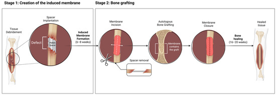
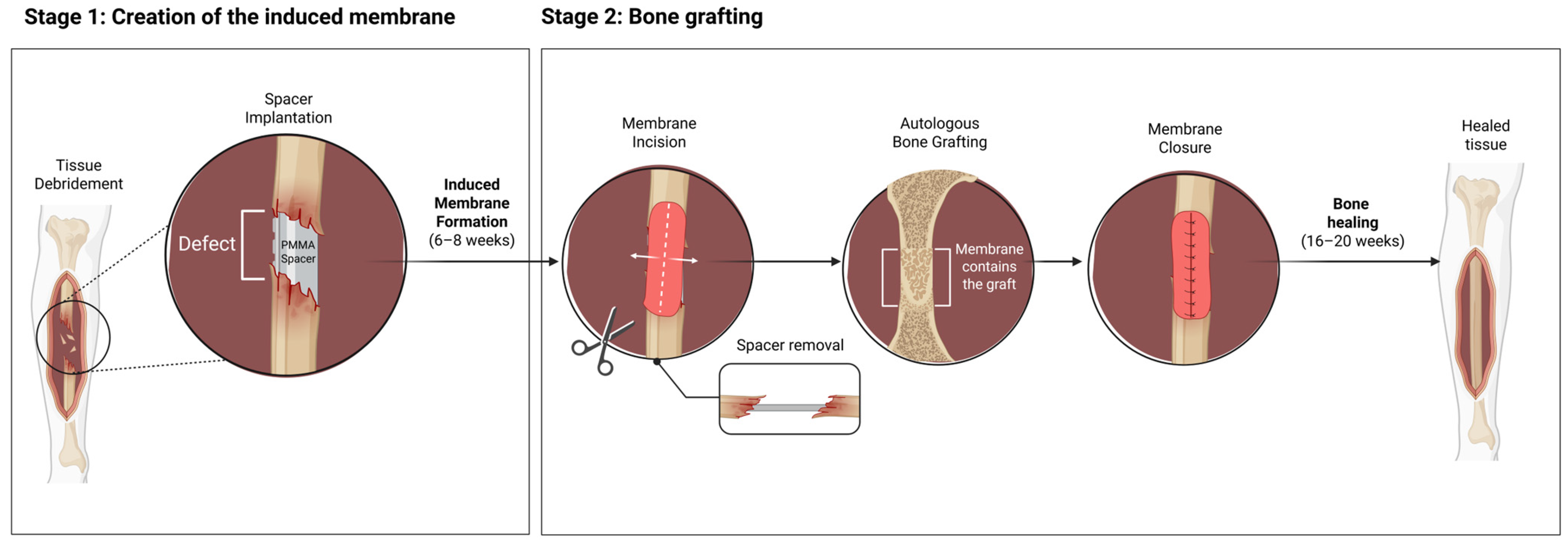
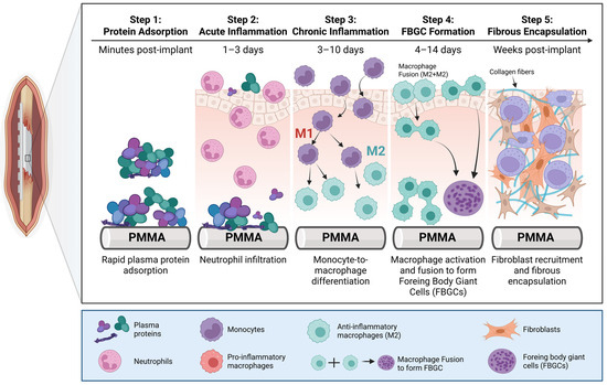
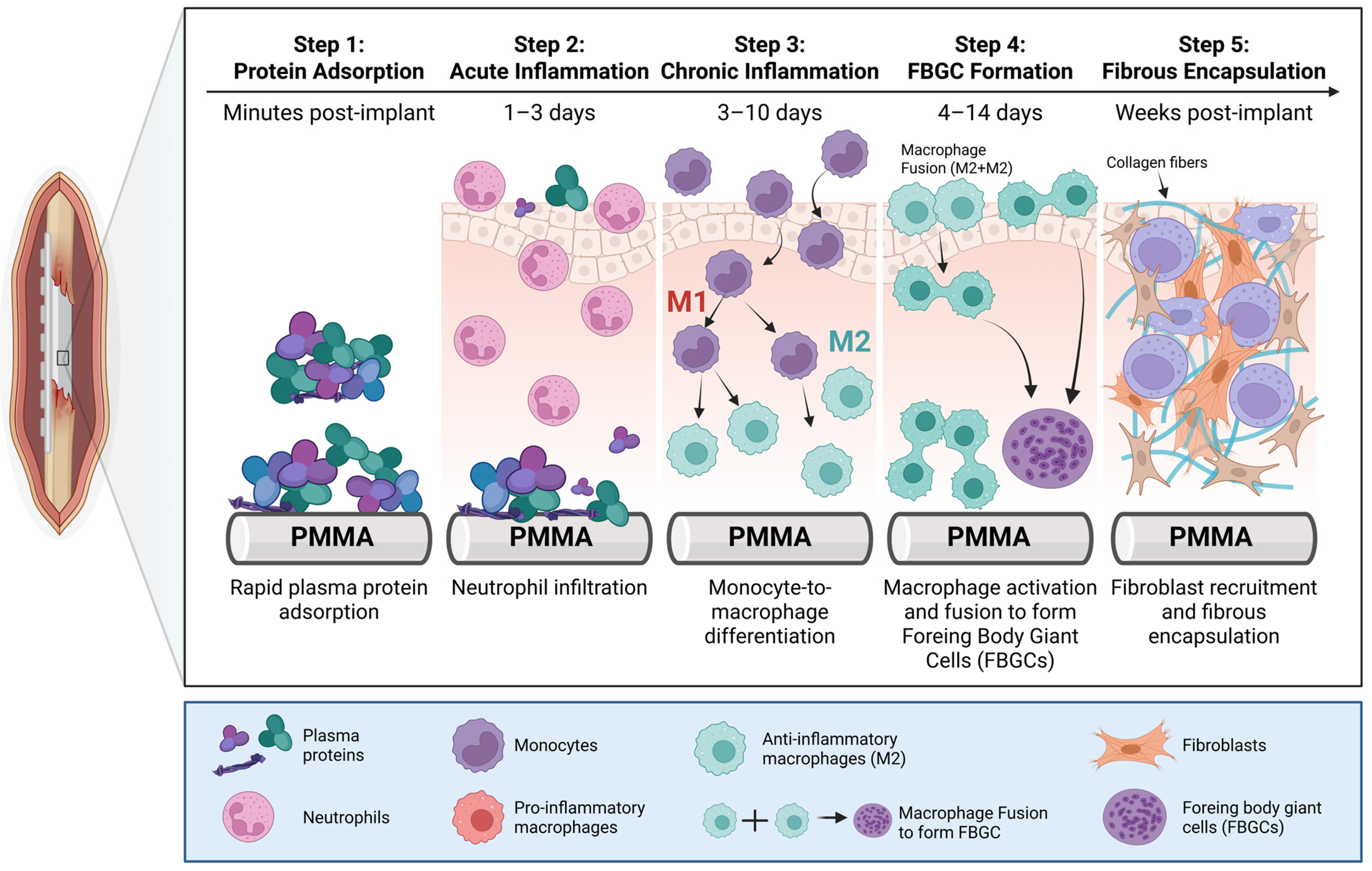
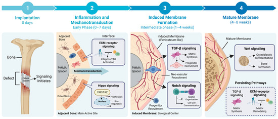
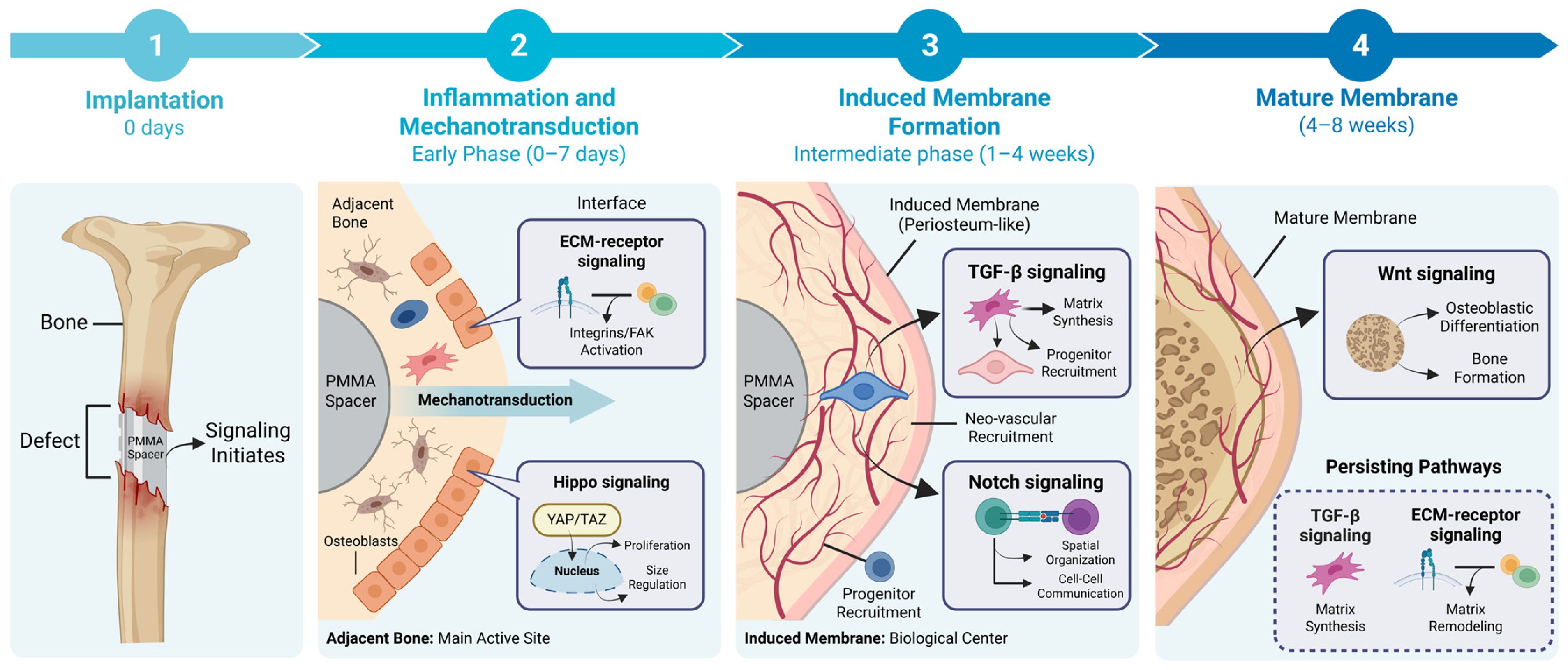
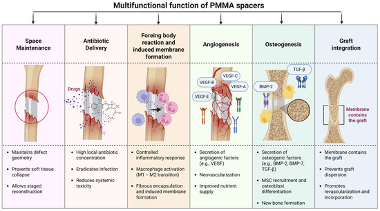
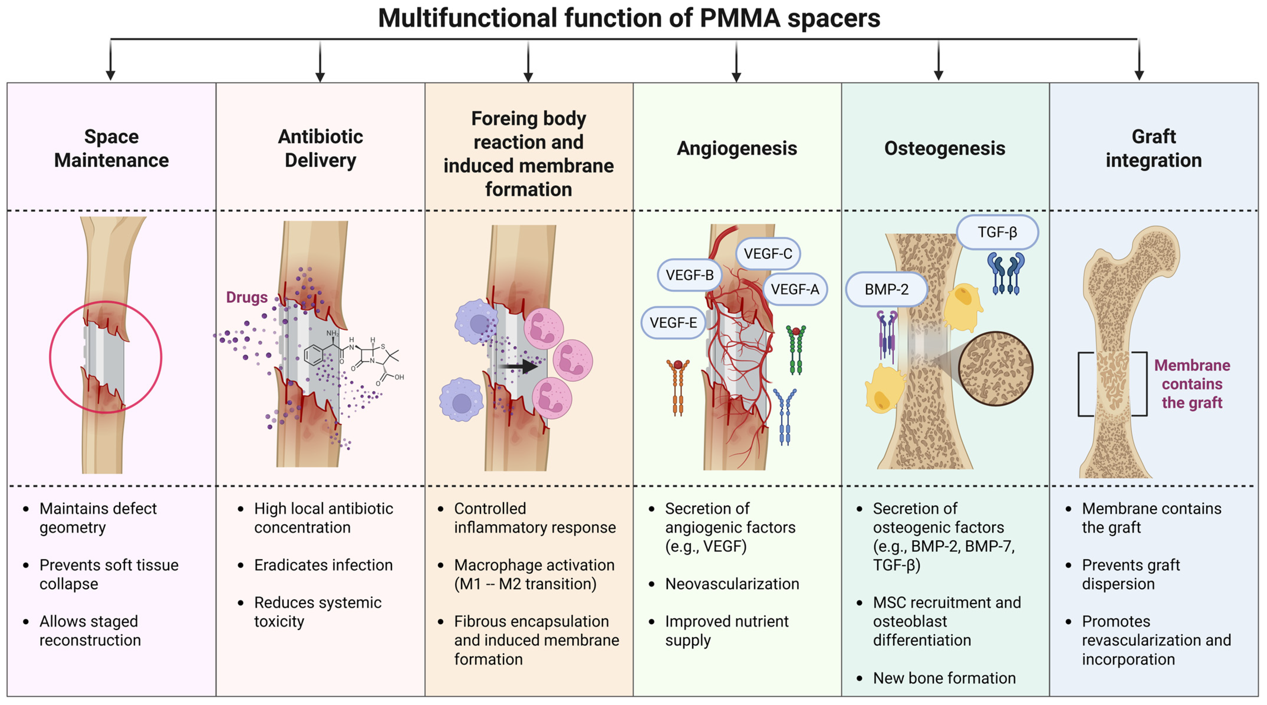
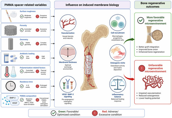
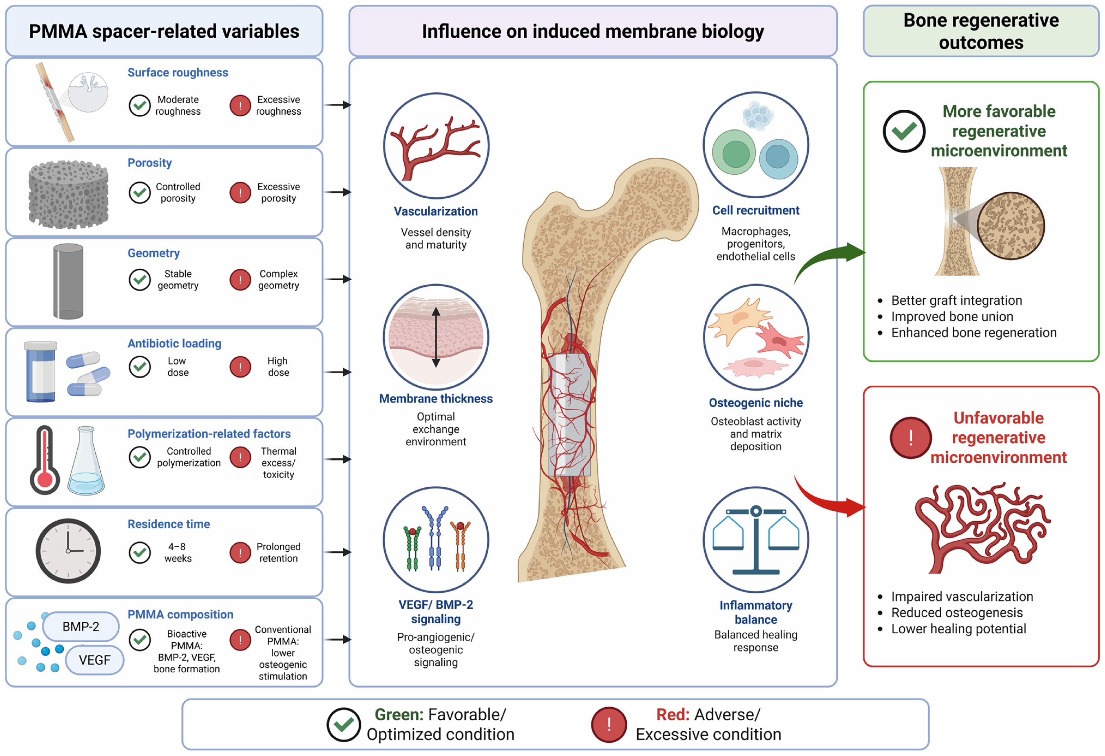

Disclaimer/Publisher’s Note: The statements, opinions and data contained in all publications are solely those of the individual author(s) and contributor(s) and not of MDPI and/or the editor(s). MDPI and/or the editor(s) disclaim responsibility for any injury to people or property resulting from any ideas, methods, instructions or products referred to in the content. |
© 2026 by the authors. Licensee MDPI, Basel, Switzerland. This article is an open access article distributed under the terms and conditions of the Creative Commons Attribution (CC BY) license.