Article

Evaluation of the Demineralization Potential of Medicinal Plant Infusions (Baccharis trimera, Moringa oleifera, and Hibiscus sabdariffa) on Bovine Teeth Using ICP OES


by
Eliane Silva de Pádua Melo
1,
Elaine Silva de Pádua Melo
1,2,* and
Valter Aragão do Nascimento
1
1
Group of Spectroscopy and Bioinformatics Applied to Biodiversity and Health, Postgraduation Program in Health and Development in the Midwest Region, Faculty of Medicine, Federal University of Mato Grosso do Sul, Campo Grande 79079-900, Mato Grosso do Sul, Brazil
2
Medicine Course, Campo Grande Unit, State University of Mato Grosso do Sul, Campo Grande 79115-898, Mato Grosso do Sul, Brazil
*
Author to whom correspondence should be addressed.
Sci 2026, 8(10), 278; https://doi.org/10.3390/sci8100278
Submission received: 9 April 2026 / Revised: 17 July 2026 / Accepted: 20 July 2026 / Published: 4 October 2026
(This article belongs to the Section Clinical Medicine and Healthcare)

Abstract

The consumption of medicinal plant infusions has increased worldwide due to their perceived health benefits; however, their effects on dental enamel integrity remain insufficiently understood. This study investigated dental enamel demineralization induced by infusions of Baccharis trimera, Hibiscus sabdariffa, and Moringa oleifera using an in vitro erosive challenge. Bovine teeth were incubated in the infusions for 0, 1, 5, and 60 min, with ultrapure water used as a control. pH was monitored throughout the experiment, and the concentrations of Ca, P, Mg, Na, K, Fe, Mn, and Zn released into the solutions were quantified by inductively coupled plasma optical emission spectrometry (ICP-OES). In addition, Ca/P and Ca/Mg molar ratios were calculated to assess hydroxyapatite stability. H. sabdariffa and M. oleifera exhibited consistently acidic pH values (3.0–3.8), with significant time-dependent increases in Ca and P release during the 60-min incubation period (p < 0.001), accompanied by marked deviations from the ideal Ca/P ratio (1.67), indicating unfavorable conditions for enamel remineralization. In contrast, B. trimera maintained pH values closer to the critical threshold for enamel dissolution (5.4–5.7), resulting in smaller mineral changes over time. Despite the high mineral content of the infusions, acidic pH prevailed over their remineralization potential. These findings demonstrate that medicinal plant infusions may contribute to enamel demineralization depending on their acidity and exposure time, highlighting the importance of evaluating their erosive potential.

1. Introduction

Oral health is recognized by the World Health Organization (WHO) as an essential component of overall health, and oral diseases affect approximately 3.5 billion people worldwide, representing a major public health concern [1]. Among these conditions, dental erosion has gained increasing attention due to its high prevalence and its association with modern dietary habits, particularly the frequent consumption of acidic foods and beverages [2,3,4,5]. Dental erosion is an irreversible process characterized by mineral loss from enamel, which may result in surface roughness, reduced microhardness, hypersensitivity, and aesthetic impairment [6].
The erosive potential of beverages is influenced by several factors, including pH, type and concentration of organic acids, buffering capacity, and exposure time [2,3,6]. Enamel demineralization occurs when the balance between mineral loss and remineralization is disrupted, a process strongly modulated by salivary flow and ionic composition [4]. When pH values fall below the critical threshold for hydroxyapatite dissolution (≈5.5), calcium and phosphate ions are progressively released from the enamel surface, compromising its structural integrity [7,8,9,10].
In recent years, the consumption of medicinal plant infusions has increased due to their perceived therapeutic benefits. However, several of these infusions exhibit acidic pH values and contain organic acids capable of promoting enamel demineralization [9,11]. Given their natural origin, these beverages are often perceived as harmless and are frequently consumed repeatedly throughout the day, which may favor progressive mineral loss from dental enamel. In this context, understanding not only the occurrence but also the systematic monitoring of enamel demineralization, particularly through time-dependent mineral release, is essential to assess their potential impact on oral health. For example, Hibiscus sabdariffa infusions have been reported to present acidogenic properties and significant erosive potential, with previous studies demonstrating mineral loss and mass reduction in dental tissues following exposure [11,12]. Conversely, Moringa oleifera has been investigated for potential remineralizing effects, including improvements in enamel microhardness under specific experimental conditions [13,14]. Despite its traditional use, the effects of Baccharis trimera infusions on dental mineral integrity remain poorly explored [15]. Therefore, these three medicinal plants were selected because they are widely consumed as herbal infusions and represent different profiles regarding their potential interaction with dental tissues, allowing a comparative evaluation of infusions with established erosive potential, possible protective effects, and limited available evidence.
Frequent exposure to acidic substances lowers the pH of the oral cavity, promoting enamel dissolution and increasing the risk of dental erosion, a phenomenon of considerable relevance to oral health [9,16]. The duration of exposure is a key determinant of the extent of demineralization, directly influencing mineral loss, surface softening, structural integrity of the tooth, and surface roughness as a consequence of hydroxyapatite degradation [8,17,18]. Therefore, studies that realistically simulate both short- and long-term exposure conditions are essential; however, there remains a limited number of investigations that quantitatively assess the demineralization of human teeth subjected to different beverages and exposure times [17].
Dental enamel is primarily composed of hydroxyapatite [Ca10(PO4)6(OH)2], and its stability depends on the relative proportions of calcium and phosphate, commonly expressed as the Ca/P molar ratio, with an ideal value of approximately 1.67 [19]. Other elements such as Mg, Na, Zn, and Fe may substitute calcium within the crystal lattice or interact with the enamel surface, influencing solubility and resistance to acid attack [20]. When teeth are exposed to acidic solutions, ionic exchange processes occur between the enamel and the surrounding medium, leading to measurable changes in elemental concentrations [21].
Elemental analysis has therefore emerged as a valuable approach for investigating enamel demineralization. Previous studies demonstrated that exposure of bovine teeth to acidic beverages results in time-dependent release of elements such as Ca, P, Mg, Na, and Zn, reflecting dynamic mineral dissolution processes [8]. In this context, inductively coupled plasma optical emission spectrometry (ICP OES) enables sensitive and quantitative assessment of these ionic exchanges, providing mechanistic insight into enamel demineralization beyond surface-level observations.
Despite extensive research on the erosive effects of acidic beverages such as soft drinks, fruit juices, and teas [6,10,22,23], there is a scarcity of studies specifically evaluating the erosive potential of medicinal herbal infusions using experimental designs that integrate pH monitoring, time-dependent exposure, and human or animal teeth models [9,11,12]. Most available investigations focus on conventional beverages or isolated herbal products, while systematic assessments of medicinal plant infusions under controlled erosive conditions remain limited. Therefore, this study aimed to investigate the demineralization of dental enamel induced by infusions of B.s trimera, H. sabdariffa, and M.a oleifera by quantifying Ca, Fe, K, Mg, Mn, Na, P, and Zn released during an in vitro erosive challenge. In addition, Ca/P and Ca/Mg molar ratios were calculated to assess changes in hydroxyapatite stability under different exposure conditions.

2. Materials and Methods

2.1. Tooth Acquisition and Selection

The dental samples used in this study comprised 240 bovine incisors donated by the Naturafrig slaughterhouse, located in Rochedo, Mato Grosso do Sul, Brazil. Specific selection criteria were applied: only intact incisors without signs of structural compromise such as fractures, caries, excessive wear, or any other macroscopically visible lesion were included, while teeth presenting morphological alterations were excluded. Furthermore, bovine incisor teeth were selected as the preferred experimental model, consistent with the studies by Steiger-Ronay et al. (2018) [3] and Melo et al. (2021) [8], due to their ready availability, the ease of obtaining intact, caries-free specimens, and their compositional similarities to human enamel.
Since these teeth were obtained as by-products from routine slaughter intended for human consumption, no additional intervention, manipulation, or sacrifice of animals was performed for scientific purposes. According to Brazilian legislation (Law No. 11.794/2008, regulated by Decree No. 6.899/2009) and the guidelines of the National Council for the Control of Animal Experimentation (CONCEA), studies that do not involve the direct use of live animals in experimental procedures are exempt from approval by the Institutional Animal Care and Use Committee (CEUA). Therefore, ethical approval was not required for this research.
For sample preparation, the teeth were initially washed with ultrapure water and then subjected to ultrasonic cleaning (bench model, Dabi Atlante, Ribeirão Preto, Brazil) for 15 min. Subsequently, adhering biological residues (blood, connective tissue, and organic remnants) were manually removed using sterile Gracey periodontal curettes, ensuring thorough surface decontamination. After cleaning, the teeth were stored in ultrapure water until experimental use. The teeth were used intact, preserving both the crown and root, without prior preparation or isolation of specific enamel regions. This approach maintained the original anatomical integrity of the specimens and provided a whole-tooth immersion model, allowing global exposure of the dental tissues to the tested infusions under the experimental condition.

2.2. Acquisition of Plants and Preparation of Infusions

The plant species B. trimera, H. sabdariffa, and M. oleifera were purchased from specialized commercial establishments in Campo Grande, Mato Grosso do Sul, Brazil, in amounts of 250 g for each species. The materials were properly packaged in plastic containers bearing species identification. All samples exhibited a dry and intact appearance, suitable for infusion preparation.
The plant material of each species was individually ground in a mill (model A 11 Basic, IKA, Staufen, Germany) until a fine and homogeneous powder was obtained. For infusion preparation, 2 g of powdered material were combined with 200 mL of ultrapure water (18 MΩ·cm, Milli-Q, Millipore, Molsheim, France) preheated to 90 °C. This proportion followed the suppliers’ label instructions, corresponding to the recommendation for preparing 1 L of infusion. The mixtures were infused for 10 min in covered borosilicate glass containers without agitation and subsequently left to stand until reaching room temperature (25 ± 2 °C).
After cooling to room temperature (25 °C), the solutions were individually filtered by gravity using medium-porosity filter paper (Whatman No. 1, Darmstadt, Germany) to remove solid particles and ensure the homogeneity of the test samples. The filtered infusions were immediately used in the erosive challenge experiments.

2.3. In Vitro Erosive Challenge

The experimental protocol was developed based on previously published in vitro dental erosion models involving sequential cycles of acidic exposure and remineralization in artificial saliva [3,4,6]. The erosive potential of the infusions was assessed by quantifying the elements released from the dental tissues into the immersion solutions using inductively coupled plasma optical emission spectrometry (ICP OES), following an analytical method adapted from Melo et al. (2021) [8]. Further details of the experimental design are presented in Figure 1.
A total of 80 intact bovine incisor teeth were randomly allocated to four experimental groups (n = 20 teeth per group): ultrapure water (control), B. trimera infusion, H. sabdariffa infusion, and M. oleifera infusion. The same teeth were sequentially exposed to the immersion times of 1, 5, and 60 min, interspersed with remineralization cycles in artificial saliva. The entire experiment was performed in three independent replicates using three different sets of teeth, resulting in a total of 240 teeth evaluated.
Before the erosive challenge (t = 0), a 6 mL aliquot of each solution was collected to determine its baseline elemental composition by ICP OES. The teeth were then immersed in their respective solutions for 1 min at room temperature (25 ± 2 °C) without agitation. After immersion, the teeth were removed, rinsed with ultrapure water, and a second 6 mL aliquot was collected for elemental analysis.
The specimens were subsequently immersed in commercial artificial saliva (Kin Hidrat®, Pharmakin, São Paulo, Brazil) for 40 min at 25 ± 2 °C without agitation, simulating the remineralization phase of a dynamic in vitro erosion model, as previously described by Larnani et al. (2024) [6] and Melo et al. (2021) [8]. Following this remineralization step, the teeth were rinsed with ultrapure water and re-immersed in their respective solutions for 5 min. A second remineralization cycle under the same conditions was then performed, followed by a final immersion period of 60 min. At the end of each exposure time (t = 1, 5, and 60 min), an additional 6 mL aliquot was collected for ICP OES analysis.
Throughout the experiment, the same immersion solution was maintained for each specimen during all exposure cycles. After each immersion period, the teeth were removed only for aliquot collection and were subsequently returned to the same solution without replacement by fresh infusion. This approach enabled continuous monitoring of the cumulative release of mineral elements from the dental tissues throughout the sequential.

2.4. pH and Temperature Monitoring

The pH and temperature of the test solutions, ultrapure water (control), and the three plant infusions (B. trimera, H. sabdariffa, and M. oleifera) were determined using a calibrated pH meter (model Q 402M, Quimis, Diadema, Brazil). Measurements were performed under controlled laboratory conditions at 25 °C (±2 °C). pH and temperature were monitored at 0, 1, 5, and 60 min, following the protocol described by Melo et al. 2021 [8].

2.5. Microwave Digestion of Samples

Aliquots collected (6 mL) from the control group and from the plant infusions at different time points of the erosive challenge were treated with 2 mL of nitric acid (HNO3, 65%, ultrapure grade, Merck, Darmstadt, Germany) and 1 mL of hydrogen peroxide (H2O2, 35%, ultrapure grade, Merck, Darmstadt, Germany). The samples were then transferred into polytetrafluoroethylene (PTFE/Teflon®) digestion tubes (Berghof Products, Eningen, Germany). All samples were subsequently digested in a microwave system (Speedwave Four, Berghof, Eningen, Germany) equipped with automated pressure and temperature control. The digestion program consisted of three sequential steps: (i) 100 °C for 5 min at 30 bar and 1305 W; (ii) 150 °C for 10 min at 30 bar and 1305 W; and (iii) 50 °C for 1 min at 25 bar and 1305 W.
After completion of the digestion process and cooling to room temperature, the samples were transferred into polyethylene tubes and diluted with ultrapure water (Milli-Q, Millipore) to a final volume of 10 mL. All procedures were performed in triplicate to ensure analytical reproducibility. In parallel, blank samples (analytical blanks) were prepared using the same reagents, volumes, and digestion conditions in order to monitor potential contamination and enable background correction in the subsequent analysis.

2.6. Elemental Analysis Using the ICP OES

The quantification of Ca, Fe, K, Mg, Mn, Na, P, and Zn in ultrapure water solutions (control) and in the infusions (B. trimera, H. sabdariffa, and M. oleifera), collected at exposure times of 0, 1, 5, and 60 min during the erosive challenge, was performed by inductively coupled plasma optical emission spectrometry (ICP OES) using an iCAP 6300 Series spectrometer (iCAP 6300 Duo, Thermo Fisher Scientific, Bremen, Germany).
The multielement standard solutions used for ICP OES quantification were prepared from a 100 mg/L stock solution (SpecSol, Quimlab, São Paulo, Brazil). Serial dilutions were obtained to achieve concentrations of 0.01, 0.025, 0.05, 0.1, 0.25, 0.5, 1.0, 2.0, and 4.0 mg/L for each element. All solutions were prepared in an acidic medium (1% HNO3, ultrapure grade, Merck, Darmstadt, Germany) using ultrapure water (Milli-Q Millipore). External calibration curves constructed from these standards were used to quantify the elements in the samples.
The analytical parameters, including wavelength, limit of detection (LOD), limit of quantification (LOQ), correlation coefficient (R2), and recovery, are presented in Table 1. The limit of detection LOD and limit of quantification (LOQ) were determined according to Long and Winefordner 1983 [24], and spike recoveries at 1.0 mg/L ranged from 94% to 110%, confirming method accuracy and precision.

2.7. Molar Ratio of Ca/P and Ca/Mg in the Test Solutions Before and After the Erosive Challenge

The molar ratios of Ca to P (Ca/P) and calcium to Mg (Ca/Mg) were calculated from the concentrations determined by ICP OES in the plant infusions (B. trimera, H. sabdariffa, M. oleifera) and in ultrapure water (control) at the exposure times (t = 0 and t = 1, 5, and 60 min) of the erosive challenge. Calcium, P, and Mg concentrations, expressed in mg/L, were converted to molarity (mmol/L) using Equation (1).
C ( m m o l L ) = C ( m g / L ) / m o l a r   m a s s ( g / m o l )
when the concentration of the denominator element (P or Mg) was below the analytical limit of detection (LOD), the corresponding molar ratio was considered not calculable. In Equation (1), C (mmol/L) denotes the concentration expressed in millimoles per liter, whereas C (mg/L) represents the elemental concentration in milligrams per liter. The molar mass (g/mol) corresponds to the atomic or molecular weight of the element or compound involved in the calculation. The specific molar masses applied in this study are Ca = 40.08 g/mol, P = 30.97 g/mol, and Mg = 24.30 g/mol. These values were utilized to convert the concentrations between mass and molar units, ensuring consistency in the quantitative analysis.
From the calculated molarities (Equation (1)), the molar ratios were obtained using Equations (2) and (3) below:
R a t i o ( C a P ) = C a ( m m o l L ) / P ( m m o l L )
R a t i o ( C a M g ) = C a ( m m o l / L ) / M g ( m m o l L )
In our study, we adopted a reference value of Ca/P = 1.67, which corresponds to the stoichiometry of hydroxyapatite [Ca10(PO4)6(OH)2], considered the standard for human enamel [19]. The values obtained were compared over time and among the different infusions, allowing the assessment of deviations from the ideal value and their potential implications for the stability or solubility of hydroxyapatite. This approach provides a more sensitive parameter than absolute concentrations, as it directly reflects the proportion between the minerals that support the integrity of dental enamel [25,26].

2.8. Statistical Analysis

Statistical analyses were performed to evaluate whether the concentrations of Ca, Fe, K, Mg, Mn, Na, P, and Zn, as well as the pH values of the infusions (B. trimera, H. sabdariffa, and M. oleifera), differed significantly across exposure times (t = 0, 1, 5, and 60 min). Data normality was assessed using the Shapiro–Wilk test, and homogeneity of variances was evaluated using Levene’s test.
For datasets meeting the assumptions of normality and homoscedasticity, one-way analysis of variance (ANOVA) was applied separately for each element and infusion, as well as for pH values, followed by Tukey’s post hoc test for pairwise comparisons. When these assumptions were not met, the non-parametric Kruskal–Wallis test was used, followed by Dunn’s post hoc test with Bonferroni correction.
All p-values were calculated as two-tailed, and statistical significance was set at p < 0.05. Exact p-values are reported in the Results and Discussion sections to allow transparent interpretation of the statistical outcomes. Statistical analyses were conducted using Python (SciPy v1.13, scikit-posthocs v0.9.0) and cross-validated in SPSS v29.

3. Results

3.1. pH and Temperature Characteristics of the Infusions Across the Exposure Times

The pH and temperature values of the B. trimera, H. sabdariffa, and M. oleifera infusions at baseline (t = 0) and after 1, 5, and 60 min of the erosive challenge are presented in Table 2. Significant differences in pH among exposure times were observed for all infusions (p < 0.05). The B. trimera infusion exhibited the highest pH values throughout the experimental period, ranging from 5.40 ± 0.01 to 5.76 ± 0.01. In contrast, H. sabdariffa showed pH values between 3.66 ± 0.01 and 3.84 ± 0.01, whereas M. oleifera presented the lowest pH values and the widest variation, ranging from 3.00 ± 0.01 (t = 1 min) to 3.78 ± 0.01 (t = 60 min). Temperatures ranged from 24.2 °C to 24.9 °C throughout the experiment.

3.2. Concentration of Metals Before and After the Exposure Time of the Erosive Challenge

Table 3 presents the concentrations of Ca, Fe, K, Mg, Mn, Na, P, and Zn in the infusions of B. trimera, H. sabdariffa, and M. oleifera at baseline (t = 0) and after 1, 5, and 60 min of the erosive challenge. Statistical comparisons among exposure times are indicated by superscript lowercase letters, with different letters denoting significant differences within each infusion (p < 0.05).
The elemental profiles differed among the three herbal infusions and changed throughout the erosive challenge. Significant time-dependent differences were observed for Ca, Fe, Mg, Mn, Na, P, and Zn in B. trimera, whereas K remained statistically unchanged. Calcium decreased from 1004.1 ± 7.6 mg/L at baseline to 722.7 ± 16.0 mg/L after 1 min, followed by a partial recovery to 852.6 ± 22.0 mg/L at 60 min. Phosphorus and Na increased throughout the experimental period, whereas Zn and Mn progressively decreased.
Among the evaluated infusions, H. sabdariffa exhibited the greatest temporal variation in elemental composition. Calcium showed a marked decrease from 16,889 mg/L at baseline to 4042 mg/L after 1 min, followed by a progressive increase, reaching 29,405 mg/L at 60 min. Phosphorus also increased continuously, from 16,562 mg/L to 62,092 mg/L, accompanied by a marked increase in Na, which reached 92,230 mg/L at the end of the experimental period. All evaluated elements showed significant time-dependent differences (p < 0.05).
Unlike the other infusions, M. oleifera maintained statistically unchanged Ca and Mg concentrations throughout the erosive challenge. In contrast, P decreased from 31,460 mg/L at baseline to 25,913 mg/L after 60 min, whereas Na increased from 69,537 mg/L to 81,134 mg/L. Significant time-dependent differences were also observed for Fe, Mn, and Zn (p < 0.05).
The control group (ultrapure water) contained only trace amounts of Ca, K, Na, and P throughout the experimental period, whereas Fe, Mg, Mn, and Zn remained below the limit of detection.

3.3. The Molar Ratios of Ca/P and Ca/Mg in Test Infusions During the Erosive Challenge

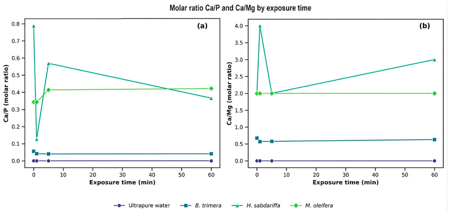
The analysis of the Ca/P and Ca/Mg molar ratios in the infusions during the erosive challenge revealed distinct patterns among the evaluated herbal infusions (Figure 2). In B. trimera, the Ca/P ratio remained well below the stoichiometric value of hydroxyapatite (1.67), decreasing from 0.056 (t = 0) to 0.041 (t = 60 min). In contrast, the Ca/Mg ratio remained relatively stable throughout the experimental period, ranging from 0.676 (t = 0) to 0.630 (t = 60 min).
Similarly, H. sabdariffa exhibited a decrease in the Ca/P ratio from 0.788 (t = 0) to 0.366 (t = 60 min). The increase in Ca concentration (from 16,888.864 to 29,404.660 mg/L) was accompanied by a proportionally greater increase in P (from 16,562.263 to 62,092.311 mg/L), resulting in a progressive reduction in the Ca/P ratio. The Ca/Mg ratio increased from 2.039 (t = 0) to 4.400 (t = 1 min), followed by a decrease to 2.131 (t = 5 min) and 2.814 (t = 60 min).
Unlike the other infusions, M. oleifera showed only minor variation in both molar ratios throughout the experimental period. The Ca/P ratio ranged from 0.343 to 0.422, whereas the Ca/Mg ratio varied from 2.002 to 1.839.

4. Discussion

The present study demonstrated that the erosive potential of B. trimera, H. sabdariffa, and M. oleifera infusions is determined by the combined influence of pH, titratable acidity, and the mineral composition of the beverages. Although pH is recognized as one of the primary factors governing dental demineralization, the present findings support the concept that pH alone is insufficient to predict erosive potential and should be interpreted together with buffering capacity and the chelating effects of organic acids present in the infusions [27,28,29]. This interpretation is consistent with the distinct Ca, P, and Mg release profiles determined by ICP-OES, indicating that the chemical composition of each infusion modulates the ion-exchange processes occurring during the erosive challenge.
The magnitude of the elemental changes observed during the erosive challenge was closely associated with the acidity of the infusions. H. sabdariffa and M. oleifera, both of which maintained pH values consistently below the critical threshold of approximately 5.5 for hydroxyapatite dissolution [19,30], produced more pronounced changes in elemental composition than B. trimera. Although the pH of B. trimera remained close to this critical value, a condition generally associated with mildly erosive beverages [31], prolonged exposure to this moderately acidic infusion still promoted measurable mineral mobilization [27,28,32]. Accordingly, B. trimera exhibited the smallest changes in elemental composition among the evaluated infusions, supporting its comparatively lower erosive potential under the experimental conditions of this study.
An interesting finding was the initial behavior of Ca concentrations in the B. trimera and H. sabdariffa infusions, characterized by an initial decrease after the first minute of exposure followed by a progressive increase throughout the experimental period. This pattern suggests that the early stages of the erosive challenge involve complex interactions between the infusion and the enamel surface before the establishment of a chemical equilibrium that favors the continuous release of mineral ions into the surrounding solution [8,33]. This behavior differs from that reported by Melo et al. [8], Jager et al. [28] and Fujii et al. [34], who observed an immediate and continuous release of mineral ions during the exposure of enamel to commercially available beverages. The different response observed in the present study may be related to the distinct physicochemical characteristics of herbal infusions, including their organic acid composition, buffering capacity, and mineral content, which can influence the kinetics of enamel dissolution and ion exchange.
Among the evaluated infusions, H. sabdariffa exhibited the greatest erosive potential, a finding consistent with its low pH and the presence of hibiscus, malic, and citric acids [35,36]. These organic acids promote hydroxyapatite dissolution through both direct acid attack and calcium chelation, thereby enhancing the release of Ca2+ and PO43− from the mineral matrix [19,37,38]. This interpretation is supported by the progressive mobilization of Ca and P observed throughout the erosive challenge and by previous studies demonstrating that, under acidic conditions, ion release exceeds mineral redeposition, thereby impairing enamel remineralization [8,19]. Although this infusion contained relatively high concentrations of minerals potentially associated with remineralization, particularly Ca and Mg, which are essential constituents of the enamel structure [37,39], these components were insufficient to counterbalance the acidic challenge. These findings indicate that, under the experimental conditions evaluated, the acidity of the infusion predominated over its potential protective mineral content, thereby favoring mineral mobilization from the dental substrate [19,40].
M. oleifera also exhibited a high availability of minerals commonly associated with enamel remineralization; however, the acidity of the infusion similarly outweighed this potentially protective effect. In contrast to the pronounced changes observed in H. sabdariffa, Ca and Mg concentrations remained relatively stable throughout the experimental period, whereas P decreased progressively and Na, Mn, and Zn exhibited significant temporal variations. These findings indicate that the interaction between the infusion and the dental surface continued to modify the ionic composition of the solution during the erosive challenge [8,33]. M. oleifera leaves are naturally rich in Ca, P, and Mg [41,42] and contain proteins and amino acids that have attracted considerable interest for applications in dental remineralization [43]. Furthermore, M. oleifera extracts and bioactive formulations containing calcium-phosphate complexes have demonstrated remineralizing potential under experimental conditions [44,45]. Nevertheless, under the conditions employed in the present study, this mineral-rich composition was insufficient to overcome the effects of acidity, resulting in continued ionic mobilization from the dental mineral matrix [19,29].
The distinct responses observed among the evaluated infusions highlight the importance of enamel mineral composition in determining its response to acidic challenges. During exposure to low-pH solutions, hydroxyapatite acts as a reservoir of inorganic elements, releasing ions into the surrounding medium as demineralization progresses [19,21,39]. In addition to their structural role, elements such as Fe, Mg, and Zn participate in the regulation of enamel matrix proteins, including amelogenin and enamelin, which are essential for enamel formation and mineral organization [46]. Likewise, the continuous availability of Ca and P is fundamental for maintaining the dynamic balance between demineralization and remineralization throughout life [19]. Changes in enamel crystallinity and carbonate content, such as those associated with aging, may further increase susceptibility to erosive challenges [47]. In this context, the ICP-OES results demonstrated that the erosive process was not limited to the release of Ca and P. Significant changes in Mg, Fe, Mn, and Zn concentrations indicate a broader redistribution of inorganic elements, reflecting the dynamic ion-exchange processes established between the infusions and the dental surface during the erosive challenge [8].
Among the elements quantified in the present study (Ca, P, Mg, Fe, Mn, and Zn), Mg deserves particular attention because of its important role in the structure and physicochemical properties of hydroxyapatite. Partial substitution of Ca by Mg within the apatite lattice can reduce crystal size and crystallinity while increasing mineral solubility [39]. However, depending on the surrounding chemical environment and the mode of Mg incorporation, this element may also enhance enamel microhardness and improve resistance to acid dissolution [33,37]. Similarly, Fe and Zn may contribute to mineral stability through the formation of protective surface complexes and by influencing crystal growth [37,39]. Nevertheless, the protective effects of these elements are limited under strongly acidic conditions, where hydroxyapatite dissolution becomes the predominant process [19,48].
The variations in elemental composition were also reflected in the Ca/P and Ca/Mg molar ratios determined in the infusions. Although these ratios were calculated from the solutions rather than directly from the enamel surface, they provide an indirect indicator of the chemical environment established during the erosive challenge and of the ion-exchange processes occurring between the infusions and the dental substrate. Consequently, deviations from the stoichiometric Ca/P ratio of hydroxyapatite (Ca/P = 1.67) [25,26,40] indicate conditions that are less favorable for mineral stability, in agreement with the progressive ionic mobilization observed throughout the experiment [19,48].
Although H. sabdariffa and M. oleifera exhibited higher Ca/P ratios than B. trimera, all values remained well below the stoichiometric ratio of hydroxyapatite, indicating chemical conditions unfavorable for maintaining mineral stability. These findings further demonstrate that the relatively high Ca concentrations present in the infusions were insufficient to offset either the excess relative amount of P or the effects of the acidic environment. Likewise, the Ca/Mg ratio reflected the relative availability of these elements within each infusion. Because Mg can exert both stabilizing and destabilizing effects on hydroxyapatite depending on the physicochemical conditions of the surrounding medium [33,37,49], the combined evaluation of the Ca/P and Ca/Mg molar ratios provided a more comprehensive interpretation of the chemical conditions governing the erosive challenge than the analysis of individual elemental concentrations alone [25,33].
Collectively, the present findings demonstrate that the erosive potential of the evaluated infusions was primarily determined by the interaction between solution acidity and its physicochemical characteristics rather than by mineral composition alone. Although the infusions contained elements that are potentially beneficial for maintaining enamel mineral stability, the persistence of pH values below the critical threshold created chemical conditions that favored demineralization, as evidenced by the progressive ionic mobilization and the Ca/P and Ca/Mg molar ratios determined throughout the experimental period. These findings emphasize that the erosive potential of beverages should be assessed by considering both their elemental composition and their acid–base characteristics, rather than relying exclusively on pH or mineral content.
The analytical approach employed in this study, based on ICP-OES quantification of the elements released into the immersion solutions, provided valuable insight into the ion-exchange dynamics associated with the erosive process. Future investigations integrating this approach with complementary techniques, such as surface microhardness measurements, profilometry, and surface morphological analyses, could further elucidate the relationship between the chemical changes occurring in the solutions and the structural alterations developed in dental enamel under clinically relevant conditions.

5. Conclusions

The results of this study indicate that the acidic pH of H. sabdariffa and M. oleifera infusions was associated with significant changes in the elemental composition of the solutions during the erosive challenge. H. sabdariffa showed the greatest changes in Ca and P concentrations, accompanied by marked variations in the Ca/P molar ratio. M. oleifera exhibited a distinct elemental profile, with relatively stable Ca and Mg concentrations and significant changes in P, Na, Mn, and Zn. In contrast, B. trimera, whose pH remained closer to the critical threshold for hydroxyapatite dissolution, produced less extensive elemental changes, although significant variations were still observed after exposure.
Despite their high mineral content, the infusions presented Ca/P and Ca/Mg molar ratios that indicated chemical conditions unfavorable to mineral stability during the erosive challenge. Overall, the findings suggest that the acidity of the infusions had a greater influence on ionic mobilization than their mineral content under the controlled laboratory parameters evaluated. Although the standardized in vitro model adopted in this study was appropriate for comparing the chemical behavior of the different infusions, the use of bovine teeth and the experimental conditions should be considered when interpreting the clinical applicability of the present findings. These results support the need for further studies integrating elemental analysis with complementary methods and more clinically representative scenarios to clarify the effects of frequent exposure to medicinal plant infusions on dental enamel.

Author Contributions

Conceptualization, E.S.d.P.M. (Eliane Silva de Pádua Melo); methodology, E.S.d.P.M. (Eliane Silva de Pádua Melo); investigation, E.S.d.P.M. (Eliane Silva de Pádua Melo); writing—original draft preparation, E.S.d.P.M. (Elaine Silva de Pádua Melo); Formal analysis, E.S.d.P.M. (Eliane Silva de Pádua Melo) and E.S.d.P.M. (Elaine Silva de Pádua Melo); Data curation, E.S.d.P.M. (Eliane Silva de Pádua Melo) and E.S.d.P.M. (Elaine Silva de Pádua Melo); writing, V.A.d.N.; Project administration, V.A.d.N.; writing—review, E.S.d.P.M. (Eliane Silva de Pádua Melo) and E.S.d.P.M. (Elaine Silva de Pádua Melo) and editing, V.A.d.N.; visualization, E.S.d.P.M. (Eliane Silva de Pádua Melo) and E.S.d.P.M. (Elaine Silva de Pádua Melo); supervision, V.A.d.N. All authors have read and agreed to the published version of the manuscript.

Funding

This research was financially supported by the Brazilian Research Council (CNPq) (CNPq: Process No. 314551/2023-9) and Coordenação de Aperfeiçoamento de Pessoal de Nível Superior-Brasil (CAPES)-Finance Code 001.

Institutional Review Board Statement

The methods and use of biological materials (bovine teeth) were evaluated in accordance with the criteria of the Ethics Committee on Animal Use (CEUA). However, formal protocol approval was not required, as this study did not involve experimental procedures with live animals or any other actions demanding such authorization. The animals were not slaughtered for experimental purposes, and the teeth were obtained from biological material classified as slaughter by-products, without commercial value.

Informed Consent Statement

Not applicable.

Data Availability Statement

The data used to support the findings of this study are available from the corresponding author upon request.

Conflicts of Interest

The authors declare no conflicts of interest.

Abbreviations

CEUAInstitutional Animal Care and Use Committee
CONCEANational Council for the Control of Animal Experimentation
ICP OESInductively Coupled Plasma Optical Emission Spectrometry
WHOWorld Health Organization

References

  1. World Health Organization. Global Oral Health Status Report: Towards Universal Health Coverage for Oral Health by 2030. 2022. Available online: https://www.who.int/publications/i/item/9789240061484 (accessed on 14 September 2025).
  2. Barbour, M.E.; Lussi, A.; Shellis, R.P. Screening and prediction of erosive potential of dietary substances. Eur. J. Oral Sci. 2011, 119, 234–241. [Google Scholar] [CrossRef] [Scilit] [PubMed]
  3. Steiger-Ronay, V.; Steingruber, A.; Becker, K.; Aykut-Yetkiner, A.; Wiedemeier, D.B.; Attin, T. Temperature-dependent erosivity of drinks in a model simulating oral fluid dynamics. J. Dent. 2018, 70, 118–123. [Google Scholar] [CrossRef] [Scilit] [PubMed]
  4. Jameel, R.A.; Zaidi, S.J.A.; Siddiqui, S.; Rehman, A.; Gul, J.; Saquib, M.; Rahim, Z.A. The effects of beverage erosion on enamel: Evaluating surface characteristics and loss of calcium and phosphate ions. Discov. Appl. Sci. 2024, 6, 439. [Google Scholar] [CrossRef] [Scilit]
  5. West, N.X.; Hughes, J.A.; Addy, M. The effect of pH on the erosion of dentine and enamel by dietary acids in vitro. J. Oral Rehabil. 2001, 27, 860–864. [Google Scholar] [CrossRef] [Scilit]
  6. Larnani, S.; Song, Y.; Kim, S.; Park, Y.S. Examining enamel-surface demineralization upon exposure to acidic solutions and the remineralization potential of milk and artificial saliva. Odontology 2024, 113, 201–212. [Google Scholar] [CrossRef] [Scilit] [PubMed]
  7. Vertuan, M.; Silva, J.F.; Souza, B.M.; Braga, A.S.; Magalhães, A.C. Effect of an experimental TiF4/NaF solution in preventing tooth erosion. Arch. Oral Biol. 2024, 157, 105823. [Google Scholar] [CrossRef] [Scilit] [PubMed]
  8. Melo, E.S.P.; Melo, E.; Arakaki, D.; Michels, F.; Nascimento, V.A. Methodology to quantify and screen the demineralization of teeth by immersing them in acidic drinks orange juice, Coca-Cola™, and grape juice: Evaluation by ICP OES. Molecules 2021, 26, 3337. [Google Scholar] [CrossRef] [Scilit] [PubMed]
  9. Niemeyer, S.H.; Baumann, T.; Lussi, A.; Meyer-Lueckel, H.; Scaramucci, T.; Carvalho, T.S. Salivary pellicle modification with polyphenol-rich teas and natural extracts to improve protection against dental erosion. J. Dent. 2021, 105, 103567. [Google Scholar] [CrossRef] [Scilit] [PubMed]
  10. Arafa, A.; Filfilan, S.S.; Fansa, H.A. Erosive effect of beverages on surface hardness and ultra-structure of deciduous teeth enamel. Pediatr. Dent. J. 2022, 32, 186–192. [Google Scholar] [CrossRef] [Scilit]
  11. Koffi-Gnagne, Y.; Aimé, K.W.; Thiémélé, S.Y.; Gboho, L.; Aké, M.D. Is homemade hibiscus drink dental lesions risk factors? Analysis of physicochemical parameters after oral cavity contact. IP Indian J. Conserv. Endod. 2020, 5, 71–74. [Google Scholar] [CrossRef] [Scilit]
  12. Najim, S.S.; Adnan, M.A. Dental erosion by beverages and determination of trace elements in teeth by atomic absorption spectrometry. Am. J. Anal. Chem. 2016, 7, 548–555. [Google Scholar] [CrossRef]
  13. Eliwa, M.E.; Mohamed, Y.; Hossam, E. Enamel remineralisation prospect of Moringa oleifera hydrogel, eggshell hydrogel versus sodium fluoride varnish on artificially demineralised primary teeth: In vitro study. Acta Odontol. Scand. 2024, 83, 264–272. [Google Scholar] [CrossRef] [Scilit] [PubMed]
  14. Elgamily, H.; Moussa, A.; Elboraey, A.; El-Sayed, H.; Al-Moghazy, M.; Abdalla, A. Microbiological assessment of Moringa oleifera extracts and itsiIncorporationin novel dental remedies against some oral pathogens. J. Med. Sci. 2016, 4, 585–590. [Google Scholar] [CrossRef] [Scilit] [PubMed]
  15. Silva, A.R.H.; Lopes, L.Q.S.; Cassanego, G.B.; Jesus, P.R.; Figueredo, K.C.; Santos, R.C.V.; Lopes, G.H.H.; Bauermann, L.F. Acute toxicity and antimicrobial activity of leaf tincture Baccharis trimera (Less). Biomed. J. 2018, 41, 194–201. [Google Scholar] [CrossRef] [Scilit] [PubMed]
  16. Dayel, M.F.A.; Sherif, F.E. Evaluation of the effects of Chlorella vulgaris, Nannochloropsis salina, and Enterobacter cloacae on growth, yield and active compound compositions of Moringa oleifera under salinity stress. Saudi J. Biol. Sci. 2021, 28, 1687–1696. [Google Scholar] [CrossRef] [Scilit] [PubMed]
  17. Borijan, A.; Ferrari, C.C.F.; Anouf, A.; Touyz, L.Z.G. Pop colas acids and tooth erosion: An in vivo, in vitro, electron-microscope, and clinical repost. Int. J. Dent. 2010, 2010, 957842. [Google Scholar] [CrossRef] [Scilit] [PubMed]
  18. Puy, C.L. The role of saliva in maintaining oral health and as an aid to diagnosis. Med. Oral Patol. Oral Cir. Bucal. 2006, 11, E449–E455. [Google Scholar]
  19. Abou Neel, E.A.; Aljabo, A.; Strange, A.; Ibrahim, S.; Coathup, M.; Young, A.M.; Bozec, L.; Mudera, V. Demineralization-remineralization dynamics in teeth and bone. Int. J. Nanomed. 2016, 11, 4743–4763. [Google Scholar] [CrossRef] [Scilit] [PubMed]
  20. LeGeros, R.Z.; Kijkowska, R.; Bautista, C.; LeGeros, J.P. Synergistice effects of magnesium and carbonate on properties of biological and synthetic apatites. Connect. Tissue Res. 1995, 33, 203–209. [Google Scholar] [CrossRef] [Scilit] [PubMed]
  21. Riyat, M.; Sharma, D.C. Analysis of 35 inorganic elements in teeth in relation to caries formation. Biol. Trace Elem. Res. 2009, 129, 126–129. [Google Scholar] [CrossRef] [Scilit] [PubMed]
  22. Beltrame, A.P.C.A.; Noschang, R.A.T.; Lacerda, D.P.; Souza, L.C.; Almeida, I.C.S. Are grape juices more erosive than orange juices? Eur. Arch. Paediatr. Dent. 2017, 18, 263–270. [Google Scholar] [CrossRef] [Scilit] [PubMed]
  23. Matumoto, M.S.S.; Terada, R.S.S.; Higashi, D.T.; Fujimaki, M.; Suga, S.S.; Guedes-Pinto, A.C. In vitro effect of energy drinks on human enamel surface. Ver. Odontol. Unesp. 2018, 47, 57–62. [Google Scholar] [CrossRef] [Scilit]
  24. Long, G.L.; Winefordner, J.D. Limit of detection: A closer look at the IUPAC definition. Anal. Chem. 1983, 55, 712A–724A. [Google Scholar] [CrossRef] [Scilit]
  25. Moriya, T. In vitro remineralization of bovine enamel with various Ca/P ratios. J. Dent. Health 1999, 49, 40–54. [Google Scholar] [CrossRef] [PubMed]
  26. Möhring, S.; Cieplik, F.; Hiller, K.A.; Ebensberger, H.; Ferstl, G.; Hermens, J.; Zaparty, M.; Witzgall, R.; Mansfeld, U.; Buchalla, W.; et al. Elemental compositions of enamel or dentin in human and bovine teeth differ from murine teeth. Materials 2023, 16, 1514. [Google Scholar] [CrossRef] [Scilit] [PubMed]
  27. Zimmer, S.; Kirchner, G. Influence of various acidic beverages on tooth erosion. PLoS ONE 2015, 10, e0129462. [Google Scholar] [CrossRef] [Scilit] [PubMed]
  28. Jager, D.H.J.; Vieira, A.M.; Ruben, J.L.; Huysmans, M.C.D.N.J.M. Estimated erosive potential depends on exposure time. J. Dent. 2012, 40, 1103–1108. [Google Scholar] [CrossRef] [Scilit] [PubMed][Green Version]
  29. Lussi, A.; Jaeggi, T. Erosion—Diagnosis and risk factors. Clin. Oral Investig. 2008, 12, S5–S13. [Google Scholar] [CrossRef] [Scilit] [PubMed]
  30. Zhao, D.; Chen, J.; Wong, G.H.M.; Tsoi, J.K.H. Erosion effects of OTC pediatric oral liquids and traditional chinese medicine on primary teeth. Int. Dent. J. 2025, 75, 1088–1096. [Google Scholar] [CrossRef] [Scilit] [PubMed]
  31. Shaik, M.I.; Hamdi, I.H.; Sarbon, N.M. A comprehensive review on traditional herbal drinks: Physicochemical, phytochemicals and pharmacology properties. Food Chem. Adv. 2023, 3, 100460. [Google Scholar] [CrossRef] [Scilit]
  32. Brunton, P.A.; Hussain, A. The erosive effect of herbal tea on dental enamel. J. Dent. 2001, 29, 517–520. [Google Scholar] [CrossRef] [Scilit] [PubMed]
  33. Abdallah, M.N.; Eimar, H.; Bassett, D.C.; Schnabel, M.; Ciobanu, O.; Nelea, V.; McKee, M.D.; Cerruti, M.; Tamimi, F. Diagenesis-inspired reaction of magnesium ions with surface enamel mineral modifies proper-ties of human teeth. Acta Biomater. 2016, 37, 174–183. [Google Scholar] [CrossRef] [Scilit] [PubMed]
  34. Fujii, M.; Kitasako, Y.; Sadr, A.; TagamI, J. Roughness and pH changes of enamel surface induced by soft drinks in vitro applications of stylus profilometry, focus variation 3D scanning microscopy and micro pH sensor. Dent. Mater. J. 2011, 30, 404–410. [Google Scholar] [CrossRef] [Scilit] [PubMed]
  35. Izquierdo-Vega, J.A.; Arteaga- Badillo, D.A.; Sánches-Gutiérres, M.; Morales-González, J.A.; Vargas-Mendonza, N.; Gómez-Aldapa, C.A.; Castro-Rosas, J.; Delgado-Olivares, L.; Madrigal-Bujaidar, E.; Madrigal-Santillán, E. Organic acids from Roselle (Hibiscus sabdariffa L.): A brief review of its pharmacological effects. Biomedicines 2020, 8, 100. [Google Scholar] [CrossRef] [Scilit] [PubMed]
  36. Jabeur, I.; Pereira, E.; Barros, L.; Calhelha, R.C.; Sokovic, M.; Oliveira, M.B.P.P.; Ferreira, I.C.F.R. Hibiscus sabdariffa L. as a source of nutrients, bioactive compounds and colouring agents. Food Res. Int. 2017, 100, 717–723. [Google Scholar] [CrossRef] [Scilit] [PubMed]
  37. Doğan, M.S. Relation of trace elements on dental health. In Trace Elements—Human Health and Environment; IntechOpen: London, UK, 2018. [Google Scholar] [CrossRef] [Scilit]
  38. Edo, G.I.; Samuel, P.O.; Jikah, A.N.; Oloni, G.O.; Ifejika, M.N.; Oghenegueke, O.; Ossai, S.; Ajokpaoghene, M.O.; Asaah, E.U.; Uloho, P.O.; et al. Proximate composition and health benefit of Roselle leaf (Hibiscus sabdariffa). Insight on food and health benefits. Food Chem. Adv. 2023, 3, 100437. [Google Scholar] [CrossRef] [Scilit]
  39. Nedoklan, S.; Knezovic, Z.; Knezovic, N.; Sutlovic, D. Nutrition and mineral content in human teeth through the centuries. Arch. Oral Biol. 2021, 124, 105075. [Google Scholar] [CrossRef] [Scilit] [PubMed]
  40. Ramesh, S.; Tanb, C.Y.; Hamdib, M.; Sopyanc, I.; Teng, W.D. The influence of Ca/P ratio on the properties of hydroxyapatite bioceramics. In Proceedings of the International Conference on Smart Materials and Nanotechnology in Engineering, Harbin, China, 1–4 July 2007; p. 6423. [Google Scholar] [CrossRef] [Scilit]
  41. Okafor, G.I.; Ogbobe, N.M. Production and quality evaluation of green and black herbal teas from Moringa oleifera Leaf. J. Food Resour. Sci. 2015, 4, 62–72. [Google Scholar] [CrossRef] [Scilit]
  42. Stohs, S.J.; Hartman, M.J. Review of the safety and efficacy of Moringa oleifera. Phytother. Res. 2015, 29, 796–804. [Google Scholar] [CrossRef] [Scilit] [PubMed]
  43. Amin, M.F.; Ariwibowo, T.; Putri, S.A.; Kurnia, D. Moringa oleifera: A review of the pharmacology, chemical constituents, and application for dental health. Pharmaceuticals 2024, 17, 142. [Google Scholar] [CrossRef] [Scilit] [PubMed]
  44. Younis, S.H.; Obeid, R.F.; Ammar, M.M. Subsurface enamel remineralization by lyophilized Moringa leaf extract loaded varnish. Heliyon 2020, 6, e05054. [Google Scholar] [CrossRef] [Scilit] [PubMed]
  45. Gonçalves, F.M.C.; Quinteiro, J.P.; Hannig, C.; Almeida, E.M.F.C.; Delbem, A.C.B.; Cannon, M.L.; Danelon, M. In situ remineralization of enamel caries lesions with a toothpaste supplemented with fluoride, amorphous calcium phosphate casein phosphopeptide and trimetaphosphate. J. Dent. 2025, 155, 105618. [Google Scholar] [CrossRef] [Scilit] [PubMed]
  46. Athimoolam, S. Influence of Elements on Gene Expression in Human Teeth. In Human Tooth and Developmental Dental Defects—Compositional and Genetic Implications; IntechOpen: London, UK, 2022. [Google Scholar] [CrossRef] [Scilit]
  47. Kranz, S.; Heyder, M.; Mueller, S.; Guellmar, A.; Krafft, C.; Nietzsche, S.; Tschirpke; Herold, C.; Sigusch, V.; Reise, B. Remineralization of Artificially Demineralized Human Enamel and Dentin Samples by Zinc-Carbonate Hydroxyapatite Nanocrystals. Materials 2022, 15, 7173. [Google Scholar] [CrossRef] [Scilit] [PubMed]
  48. Shellis, R.P.; Featherstone, J.D.B.; Lussi, A. Understanding the chemistry of dental erosion. Monogr. Oral Sci. 2014, 25, 163–179. [Google Scholar] [CrossRef] [Scilit] [PubMed]
  49. Alkattan, R.; Lippert, F.; Tang, Q.; Eckert, G.J.; Ando, M. The influence of hardness and chemical composition on enamel demineralization and subsequent remineralization. J. Dent. 2018, 75, 34–40. [Google Scholar] [CrossRef] [Scilit] [PubMed]
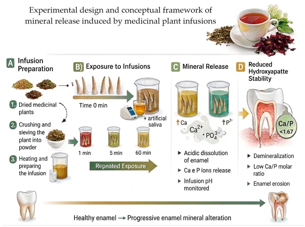
Figure 1. Experimental design and conceptual framework of mineral release induced by medicinal plant infusions. (A) Preparation of the medicinal plant infusions (Baccharis trimera, Hibiscus sabdariffa, and Moringa oleifera), including drying, grinding and sieving of the plant material, followed by infusion preparation according to the experimental protocol described in the manuscript. (B) Experimental workflow showing bovine teeth exposed to the infusions in the presence of artificial saliva at different exposure times (1, 5, and 60 min), simulating repeated consumption of acidic herbal beverages while monitoring pH throughout the experiment. (C) Conceptual representation of the ionic exchange between enamel and the infusion medium during repeated acidic exposure, promoting the release of calcium (Ca) and phosphorus (P) and altering the mineral composition of enamel. (D) Proposed mechanism of enamel demineralization, in which mineral loss results in deviations from the ideal hydroxyapatite Ca/P molar ratio (1.67), reduced hydroxyapatite stability, and increased susceptibility to enamel erosion. The arrow at the bottom illustrates the conceptual progression from healthy enamel to progressive enamel mineral alteration during repeated exposure to the medicinal plant infusions. The figure was created using BioRender.com (BioRender, Toronto, ON, Canada; https://www.biorender.com).
Sci 08 00278 g001
Figure 2. Molar ratio of Ca/P (a) and Ca/Mg (b) in the infusions of B. trimera, H. sabdariffa, and M. oleifera during the erosive challenge.
Sci 08 00278 g002
Table 1. Analytical data obtained by the ICP OES: wavelength, limit of detection (LOD), limit of quantification (LOQ), correlation coefficient (R2) and recovery (%).
ElementsWavelength (nm)LD (mg/L) LQ (mg/L)Correlation Coefficient (R2)Recovery (%)
Ca393.3660.00410.01380.998198
Fe259.9400.00360.01200.999496
K766.4900.03490.11630.9963104
Mg279.5530.00150.00490.998799
Mn257.6100.00040.00140.999394
Na588.9950.00000.00000.9960110
P177.4950.18660.62200.9876103
Zn213.8560.00080.00260.999498
Table 2. Infusions, time before (t = 0) and after the erosive challenge (0, 1, 5, and 60 min), pH values and temperatures according to each exposure.
InfusionExposure Time (min)pHTemperature
B. trimerat = 05.61 ± 0.01 a24.2
t = 15.40 ± 0.01 b24.2
t = 55.42 ± 0.01 b24.2
t = 605.76 ± 0.01 c24.4
H. sabdariffat = 03.72 ± 0.01 a24.5
t = 13.84 ± 0.01 b24.5
t = 53.82 ± 0.01 b24.5
t = 603.66 ± 0.01 c24.6
M. oleiferat = 03.46 ± 0.01 a24.6
t = 13.00 ± 0.01 c24.6
t = 53.22 ± 0.01 b24.7
t = 603.78 ± 0.01 d24.9
Values are expressed as mean ± standard deviation (n = 3). Different superscript letters within the same infusion indicate significant differences among exposure times for pH values (Tukey’s test, p < 0.05).
Table 3. Concentration of Ca, Fe, K, Mg, Mn, Na, P and Zn (mean ± SD) quantified before (t = 0) and after the erosive challenge in vitro (t = 1, 5, and 60 min).
InfusionsExposure Time (min) Elements (mg/L)
CaFeKMgMnNaPZn
Ultrapure water (control)t = 0<LOD a<LOD a<LOD a<LOD a<LOD a0.630 ± 0.002 a<LOD a<LOD a
t = 1<LOD a<LOD a0.141 ± 0.020 b<LOD a<LOD a4.730 ± 0.032 b0.540 ± 0.050 b<LOD a
t = 50.156 ± 0.004 b<LOD a0.283 ± 0.021 c<LOD a<LOD a10.921 ± 0.045 c1.760 ± 0.080 c<LOD a
t = 600.808 ± 0.008 c<LOD a0.671 ± 0.005 c<LOD a<LOD a26.997 ± 0.40 d4.700 ± 0.073 d<LOD a
B. trimerat = 01004.1 ± 7.6 a12.54 ± 0.35 b29,807 ± 1300 a901.0 ± 29 a42.07 ± 0.58 a1481 ± 31 b13,835 ± 320 b14.26 ± 0.20 a
t = 1722.7 ± 16 c10.69 ± 0.28 c28,693 ± 950 a766.2 ± 13 c30.23 ± 0.48 b841.1 ± 12 d13,363 ± 77 b7.673 ± 0.073 b
t = 5757.6 ± 18 b c14.02 ± 0.43 a29,401 ± 600 a796.5 ± 15 b c29.27 ± 0.60 b1183 ± 52 c14,499 ± 170 c7.874 ± 0.18 b
t = 60852.6 ± 22 b11.27 ± 0.20 b c28,478 ± 740 a820.7 ± 20 b24.99 ± 0.66 c2064 ± 120 a15,994 ± 220 a5.90 ± 0.21 c
H. sabdariffat = 016,889 ± 64 c151.93 ± 1.5 b35,249 ± 93 b5025 ± 16 b295.7 ± 3.1 b47,558 ± 270 c16,562 ± 330 c41.17 ± 0.59 b
t = 14042 ± 200 d221.91 ± 2.8 a16,180 ± 180 c557.2 ± 11 c369.2 ± 4.1 a38,176 ± 840 d24,804 ± 460 b46.12 ± 0.38 a
t = 521,393 ± 140 b226.65 ± 2.3 a43,383 ± 350 a6089 ± 33 a366.7 ± 3.3 a42,878 ± 1000 b29,059 ± 160 b46.54 ± 0.31 a
t = 6029,405 ± 980 a222.57 ± 0.67 a42,228 ± 390 a6338 ± 39 a361.65 ± 0.98 a92,230 ± 1500 a62,092 ± 660 a50.36 ± 0.16 a
M. oleiferat = 013,956 ± 230 a4.00 ± 0.31 c27,555 ± 150 b4227 ± 20 b80.64 ± 0.16 a69,537 ± 2000 b31,460 ± 420 a13.57 ± 0.20 a
t = 114,099 ± 97 a7.08 ± 0.30 b31,324 ± 310 a4723 ± 38 a75.67 ± 1.1 b53,130 ± 270 c31,826 ± 42 a11.14 ± 0.11 b
t = 514,200 ± 190 a8.3 ± 1.1 a30,355 ± 470 a4637 ± 61 a57.0 ± 6.1 c63,644 ± 530 b26,526 ± 520 b8.44 ± 0.35 c
t = 6014,159 ± 250 a5.46 ± 0.44 b30,508 ± 420 a4670 ± 40 a49.95 ± 3.9 c81,134 ± 160 a25,913 ± 590 b7.79 ± 0.36 c
Different superscript lowercase letters within the same column and infusion indicate statistically significant differences among exposure times (p < 0.05), according to Tukey’s HSD following one-way ANOVA or Dunn’s post hoc test following Kruskal–Wallis analysis when assumptions of normality and homogeneity of variances were not met. Means sharing the same letter are not significantly different.
Disclaimer/Publisher’s Note: The statements, opinions and data contained in all publications are solely those of the individual author(s) and contributor(s) and not of MDPI and/or the editor(s). MDPI and/or the editor(s) disclaim responsibility for any injury to people or property resulting from any ideas, methods, instructions or products referred to in the content.