Next Issue
Volume 5, March
Previous Issue
Volume 4, September


Vision, Volume 4, Issue 4 (December 2020) – 6 articles

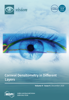
Cover Story (view full-size image): A tomographic image of the cornea obtained with a technique based on the Scheimpflug principle, where it is possible to accurately assess differences in densitometry in the layers of the cornea, with the anterior layers being denser than the deeper layers, identified with the warmest colors. Corneal density changes due to refractive surgery, scarring, inflammation, and aging. View this paper
  • Issues are regarded as officially published after their release is announced to the table of contents alert mailing list.
  • You may sign up for e-mail alerts to receive table of contents of newly released issues.
  • PDF is the official format for papers published in both, html and pdf forms. To view the papers in pdf format, click on the "PDF Full-text" link, and use the free Adobe Reader to open them.
Order results
Result details
Section
Select all
Export citation of selected articles as:
15 pages, 4841 KiB  
Concept Paper
Attention as a Unitary Concept
by Adam Reeves
Vision 2020, 4(4), 48; https://doi.org/10.3390/vision4040048 - 09 Nov 2020
Cited by 4 | Viewed by 2450
Abstract
In this paper, I discuss attention in terms of selecting visual information and acting on it. Selection has been taken as a bedrock concept in attention research since James (1890). Selective attention guides action by privileging some things at the expense of others. [...] Read more.
In this paper, I discuss attention in terms of selecting visual information and acting on it. Selection has been taken as a bedrock concept in attention research since James (1890). Selective attention guides action by privileging some things at the expense of others. I formalize this notion with models which capture the relationship between input and output under the control of spatial and temporal attention, by attenuating or discarding certain inputs and by weighing energetic costs, speed, and accuracy in meeting pre-chosen goals. Examples are given from everyday visually guided actions, and from modeling data obtained from visual searches through temporal and spatial arrays and related research. The relation between selection, as defined here, and other forms of attention is discussed at the end. Full article
Show Figures

Figure 1

6 pages, 1146 KiB  
Review
Contrasting a Misinterpretation of the Reverse Contrast
by Tiziano Agostini, Mauro Murgia, Fabrizio Sors, Valter Prpic and Alessandra Galmonte
Vision 2020, 4(4), 47; https://doi.org/10.3390/vision4040047 - 02 Nov 2020
Cited by 1 | Viewed by 2275
Abstract
The reverse contrast is a perceptual phenomenon in which the effect of the classical simultaneous lightness contrast is reversed. In classic simultaneous lightness contrast configurations, a gray surrounded by black is perceived lighter than an identical gray surrounded by white, but in the [...] Read more.
The reverse contrast is a perceptual phenomenon in which the effect of the classical simultaneous lightness contrast is reversed. In classic simultaneous lightness contrast configurations, a gray surrounded by black is perceived lighter than an identical gray surrounded by white, but in the reverse contrast configurations, the perceptual outcome is the opposite: a gray surrounded by black appears darker than the same gray surrounded by white. The explanation provided for the reverse contrast (by different authors) is the belongingness of the gray targets to a more complex configuration. Different configurations show the occurrence of these phenomena; however, the factors determining this effect are not always the same. In particular, some configurations are based on both belongingness and assimilation, while one configuration is based only on belongingness. The evidence that different factors determine the reverse contrast is crucial for future research dealing with achromatic color perception and, in particular, with lightness induction phenomena. Full article
Show Figures

Figure 1

11 pages, 399 KiB  
Review
The Evolving Role of Ophthalmology Clinics in Screening for Early Alzheimer’s Disease: A Review
by Paris Dickens and Kanna Ramaesh
Vision 2020, 4(4), 46; https://doi.org/10.3390/vision4040046 - 29 Oct 2020
Cited by 6 | Viewed by 2393
Abstract
Alzheimer’s disease (AD) is the leading cause of dementia, which is a growing public health concern. Although there is no curative treatment for established AD, early recognition and modification of the known risk factors can reduce both severity and the rate of progression. [...] Read more.
Alzheimer’s disease (AD) is the leading cause of dementia, which is a growing public health concern. Although there is no curative treatment for established AD, early recognition and modification of the known risk factors can reduce both severity and the rate of progression. Currently, an early diagnosis of AD is rarely achieved, as there is no screening for AD. The cognitive decline in AD is gradual and often goes unnoticed by patients and caregivers, resulting in patients presenting at later stages of the disease. Primary care physicians (general practitioners in the UK) can administer a battery of tests for patients presenting with memory problems and cognitive impairment, however final diagnosis of AD is usually made by specialised tertiary level clinics. Recent studies suggest that in AD, visuospatial difficulties develop prior to the development of memory problems and screening for visuospatial difficulties may offer a tool to screen for early stage AD. AD and cataracts share common risk and predisposing factors, and the stage of cataract presentation for intervention has shifted dramatically with early cataract referral and surgical intervention becoming the norm. This presentation offers an ideal opportunity to administer a screening test for AD, and visuospatial tools can be administered at post-operative visits by eye clinics. Abnormal findings can be communicated to primary care physicians for further follow up and assessment, or possible interventions which modify risk factors such as diabetes, hypertension and obesity can be undertaken. We propose that eye clinics and ophthalmology facilities have a role to play in the early diagnosis of AD and reducing the burdens arising from severe dementia. Full article
Show Figures

Figure 1

16 pages, 1320 KiB  
Article
The Effect of Corneal Thickness, Densitometry and Curvature on Intraocular Pressure Measurements Obtained by Applanation, Rebound and Dynamic Contour Tonometry
by Marco Antonio de Castro Olyntho Junior, Lucas Bertazzi Augusto, Carolina P. B. Gracitelli and Andrew J. Tatham
Vision 2020, 4(4), 45; https://doi.org/10.3390/vision4040045 - 21 Oct 2020
Cited by 10 | Viewed by 3551
Abstract
Evaluate the effect of corneal thickness, densitometry and curvature on intraocular pressure (IOP) measurements obtained by Goldmann applanation tonometry (GAT), non-contact tonometry (NCT), rebound tonometry (RT), and dynamic contour tonometry (DCT). A cross-sectional prospective study involving 40 participants was performed. Corneal measurements were [...] Read more.
Evaluate the effect of corneal thickness, densitometry and curvature on intraocular pressure (IOP) measurements obtained by Goldmann applanation tonometry (GAT), non-contact tonometry (NCT), rebound tonometry (RT), and dynamic contour tonometry (DCT). A cross-sectional prospective study involving 40 participants was performed. Corneal measurements were obtained using Pentacam (Oculus GMbH, Wetzlar, Germany), densitometry was measured at annuli of 0–2, 2–6, 6–10 and 10–12 mm. The relationship between corneal thickness (central, 4 and 6 mm), corneal astigmatism and corneal densitometry and IOP was examined. There was a significant relationship between corneal thickness (central, 4 and 6 mm) and GAT180, GAT90, RT, and NCT (P < 0.001 for all comparisons) but not for DCT. Higher corneal densitometry (6–10 mm and 10–12 mm zones) was associated with higher IOP from GAT180 and GAT90, and higher densitometry in the 6–10 mm zone correlated with higher IOP from NCT, however corneal densitometry increased with age. Accounting for age, the relationship between corneal densitometry and IOP measurements was not significant. In eyes with greater corneal astigmatism there was a greater difference between GAT90 and GAT180 measurements. IOP measurements may be affected by corneal thickness, densitometry and curvature. DCT was less affected by properties of the cornea compared to other devices. Full article
Show Figures

Figure 1

17 pages, 7198 KiB  
Review
Current Choroidal Imaging Findings in Central Serous Chorioretinopathy
by Gideon Nkrumah, Dmitrii S. Maltsev, Paez-Escamilla A. Manuel, Mohammed A. Rasheed, Marianno Cozzi, Alessandro Ivernizzi, Marco Lupidi, Sumit Randhir Singh and Jay Chhablani
Vision 2020, 4(4), 44; https://doi.org/10.3390/vision4040044 - 16 Oct 2020
Cited by 10 | Viewed by 5627
Abstract
Background: Central serous chorioretinopathy (CSCR) is a chorioretinal disease affecting mostly middle age males. It is marked by the serous detachment of the neurosensory layer at the macula. This review of the literature provides a framework of the current characteristic/relevant imaging findings of [...] Read more.
Background: Central serous chorioretinopathy (CSCR) is a chorioretinal disease affecting mostly middle age males. It is marked by the serous detachment of the neurosensory layer at the macula. This review of the literature provides a framework of the current characteristic/relevant imaging findings of CSCR. Although the pathogenesis of CSCR is unclear, the choroid plays a major role and its changes are fundamental to the diagnosis and treatment of CSCR. Methods: A systematic literature search focusing on current multimodal imaging for CSCR was performed. Only articles reporting on original clinical data were selected, studies in a language other than English were included only if an English abstract was provided. Additional sources included articles cited in the references list of the first selected articles. We deduced imaging findings based on current and relevant literature on the topic. Results: We found that sub foveal choroidal thickness (SFCT) and choroidal vascularity index (CVI) were greater in eyes with acute CSCR than in eyes with chronic CSCR or normal eyes. There was increased choroidal thickness (CT) in the macula compared to peripapillary region. In healthy eyes, the highest CVI was found in the nasal region followed by the inferior, temporal, and superior quadrant. The area with the least CVI was the macula. In eyes with CSCR, 100% had asymmetric dominant vortex veins compared to 38% in normal eyes. Conclusion: Choroidal imaging has advanced the diagnosis of CSCR. This has led to numerous imaging biomarkers like CVI, CT, and hyper-reflective dots for early detection and possible prognostication of CSCR. More techniques like wide field scans and en face imaging are being employed to characterize the choroid in CSCR. Full article
(This article belongs to the Special Issue Imaging the Choroid)
Show Figures

Figure 1

10 pages, 256 KiB  
Review
Autoimmune Dry Eye without Significant Ocular Surface Co-Morbidities and Mental Health
by Zahra Ashena, Radhika Dashputra and Mayank A. Nanavaty
Vision 2020, 4(4), 43; https://doi.org/10.3390/vision4040043 - 10 Oct 2020
Cited by 2 | Viewed by 3939
Abstract
Dry eye symptoms can negatively affect the psychological, physical, and social functioning, which can potentially impair the health-related quality of life. This review evaluated the association between autoimmune related dry eye in the absence of significant ocular surface co-morbidities and mental health. This [...] Read more.
Dry eye symptoms can negatively affect the psychological, physical, and social functioning, which can potentially impair the health-related quality of life. This review evaluated the association between autoimmune related dry eye in the absence of significant ocular surface co-morbidities and mental health. This review found a significantly higher prevalence of mental health disorders (such as depression and anxiety) in systemic lupus erythematous, rheumatoid arthritis, systemic sclerosis, Behcet’s disease, and primary Sjogren’s syndrome patients when compared to the general population. Moreover, patients with depression and anxiety interpret ocular sensations differently than healthy controls and the perception of dry eye symptoms can be influenced by their mood. Somatization is common in depression, and this could influence the perception of ocular discomfort. Anti-depressants and anxiolytics with their potential side effects on the tear film status may also contribute or aggravate the dry eye symptoms in these patients. Although ophthalmologists manage the dry eye disease, as per standardized algorithms, they should be mindful of different ocular sensation interpretation and coexistent mental health issues in a large number of this patient group and initiate a multidisciplinary management plan in certain cases. While rheumatologists look after their autoimmune condition, it may be worth liaising with GP and/or psychiatrist colleagues in order to address their neuropathic type pain and mental health co-morbidities. Full article
Previous Issue
Next Issue
Back to TopTop