Review

Integrating Additive Manufacturing into Dental Production: Innovations, Applications and Challenges


by
Maryna Yeromina
,
Jan Duplak
,
Jozef Torok
*,
Darina Duplakova
and
Monika Torokova
Faculty of Manufacturing Technologies with a Seat in Presov, Technical University of Kosice, Bayerova 1, 080 01 Presov, Slovakia
*
Author to whom correspondence should be addressed.
Inventions 2026, 11(1), 7; https://doi.org/10.3390/inventions11010007
Submission received: 31 October 2025 / Revised: 14 December 2025 / Accepted: 29 December 2025 / Published: 7 January 2026
(This article belongs to the Section Inventions and Innovation in Advanced Manufacturing)

Abstract

Additive manufacturing (AM) has emerged as a key enabling technology in contemporary dental manufacturing, driven by its capacity for customization, geometric complexity, and seamless integration with digital design workflows. This article presents a technology-oriented narrative review of additive manufacturing in dental implant production, focusing on dominant processing routes, material systems, and emerging research trends rather than a systematic or critical appraisal of the literature. An indicative descriptive analysis of publications indexed in the Web of Science and Scopus databases between 2014 and 2024 was used to contextualize the technological development of the field and identify major research directions. Emphasis was placed on metal powder bed fusion technologies, specifically Selective Laser Melting (SLM) and Direct Metal Laser Sintering (DMLS), which enable the fabrication of titanium implants with controlled porosity and enhanced osseointegration. Ceramic AM approaches, including SLA, DLP, and PBF, are discussed in relation to their potential for aesthetic dental restorations and customized prosthetic components. The publication trend overview indicates a growing interest in ceramic AM after 2020, an increasing focus on hybrid and functionally graded materials, and persistent challenges related to standardization and the availability of long-term clinical evidence. Key technological limitations—including manufacturing accuracy, material stability, validated metrology, and process reproducibility—are highlighted alongside emerging directions such as artificial intelligence-assisted workflows, nanostructured surface modifications, and concepts enabling accelerated or immediate clinical use of additively manufactured dental restorations.

1. Introduction

The evolution of additive manufacturing technologies in dental implant production has witnessed significant advancements over the past few decades, reflecting a growing intersection of innovation and application within the dental field. Early studies highlighted the potential of 3D printing in prosthodontics, emphasizing its ability to create complex geometries that traditional methods could not achieve [1]. Subsequent research began to unravel the advantages of customized implants, which were made possible by advancements in digital scanning and designing technologies. As noted, personalized dental implants not only enhance fit and patient comfort but also reduce the risk of complications [2,3].
In the mid-2010s, the introduction of biocompatible materials for additive manufacturing further revolutionized dental implant production. These materials, such as titanium and newer polymers, allowed for implants that better mimicked natural bone properties, promoting osseointegration [4,5]. Consequently, studies reported improved patient outcomes and faster recovery times, establishing a strong correlation between material choice and clinical success [6].
As the decade progressed, attention shifted toward refining printing techniques—specifically, the use of techniques such as fused deposition modeling and selective laser sintering. Such innovations not only enhanced the precision of implant production but also reduced production times, ultimately making these technologies more accessible to dental practitioners [7]. Contemporary literature acknowledges a trend toward integrating artificial intelligence and machine learning algorithms to facilitate better design and predict patient outcomes, further underscoring the rapid pace of development in this area [8,9]. Collectively, these findings reflect a dynamic transition in the field, marking additive manufacturing as a pivotal element in the future of dental implantology.
Recent investigations into AM technologies have increasingly highlighted their transformative potential in dental implant production. A central theme emerging from the literature is the improvement of customization and precision in implant design. For instance, researchers have demonstrated that AM allows patient-specific implants, significantly enhancing biocompatibility and reducing surgery time [10]. This capability has been acknowledged as a critical advancement in restorative dental practices, where individual anatomical variations are paramount [11].
Another prominent theme involves the materials used in AM processes. Innovations in biocompatible materials such as titanium and ceramics have been explored, with studies indicating that these materials not only support osseointegration but also exhibit favorable mechanical properties [12]. This enhancement in materials science contributes to the overall longevity and success of dental implants, establishing a robust foundation for their application in clinical settings.
Furthermore, advancements in printing technologies, including selective laser melting and digital light processing, have garnered attention for their impact on both accuracy and efficiency. These technologies have been reported to minimize material waste while maximizing the mechanical strength of the produced implants, lending credence to their role in sustainable dental practices [13].
Lastly, the integration of software tools for dental design and production has streamlined workflows and reduced the learning curve for practitioners, making these technologies more accessible [14]. Overall, the literature reflects a significant shift towards more efficient, customized, and sustainable approaches in dental implant production, highlighting the vital role of additive manufacturing technologies in modern dental practices.
Advanced software platforms based on artificial intelligence have emerged, enabling the automated detection of printing defects, prediction of implant mechanical properties, and generation of anatomically optimized geometries. At the same time, the use of nanotechnologies has expanded, particularly nanoporous ceramics, functional surface coatings with controlled release of bioactive substances, and laser-induced nanostructures that enhance osseointegration. These innovations substantially broaden the clinical and technological capabilities of AM beyond what was documented in earlier reviews.
The exploration of additive manufacturing technologies in the realm of dental implant production reveals diverse methodological approaches that shape both practical applications and theoretical understandings. For instance, studies employing qualitative methodologies emphasize user experiences and satisfaction, highlighting the transformative impact of personalized implants produced through three-dimensional printing techniques [15]. These investigations underscore how the integration of patient-specific data into the manufacturing process can significantly enhance outcomes, as detailed by recent analyses that utilized case studies and in-depth interviews [16].
Accordingly, this manuscript does not aim to perform a systematic or critical appraisal of the literature, but rather to provide a structured technological overview highlighting dominant approaches, quantitative ranges, and emerging research directions.

2. Additive Manufacturing Technologies in Dentistry

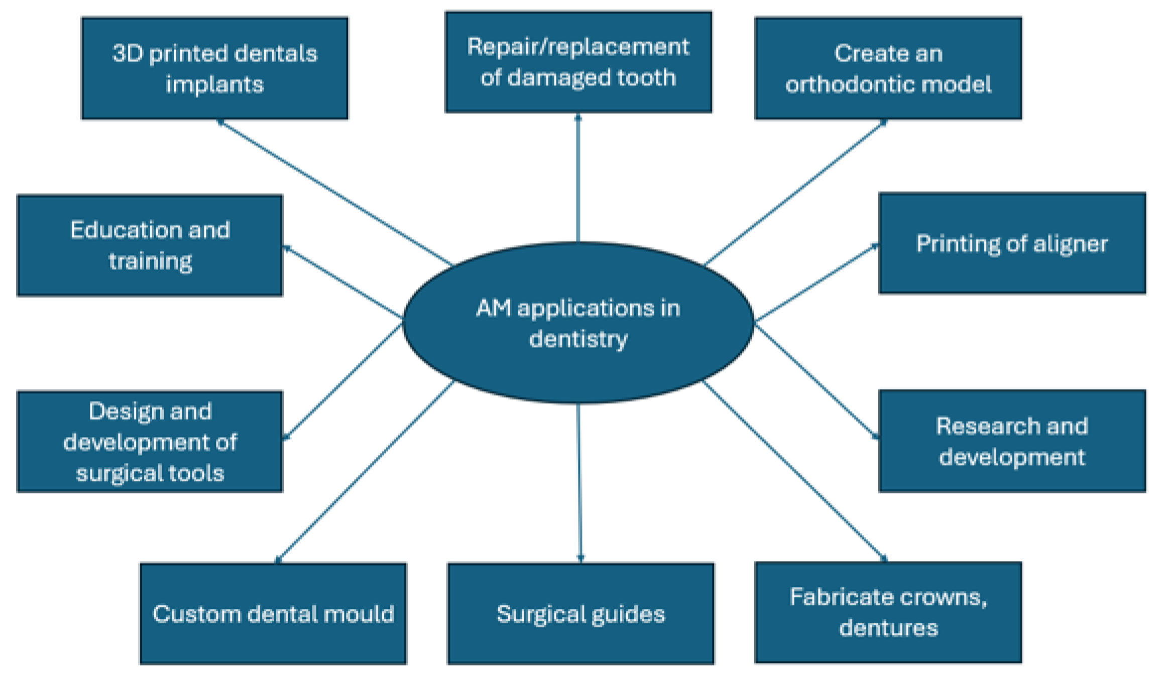
The integration of additive manufacturing, commonly known as 3D printing, has profoundly impacted various sectors, with its application in dentistry catalyzing significant advancements in both restorative and reconstructive procedures [17]. This technology, driven by digital workflows from design to production, has enabled the fabrication of customized dental implants, crowns, bridges, and orthodontic appliances with unprecedented accuracy and efficiency [18,19]. The rapid evolution of digital dentistry, encompassing CAD/CAM systems and intraoral scanners, has synergistically accelerated the adoption of additive manufacturing, creating a fully digital process for dental product processing [12,20]. This integration is part of a broader trend towards Industry 4.0, characterized by automation and customization, revolutionizing dental production systems [21]. This review comprehensively examines the current state and emerging trends of additive manufacturing in dentistry, highlighting its diverse applications, underlying technological principles, and associated challenges [18,22]. It further delineates how these technologies enable the direct fabrication of complex, customized physical objects based on computer-aided design models, offering a significant alternative to traditional subtractive manufacturing methods in various dental applications [23,24]. Specifically, these technologies are transforming oral implantology by facilitating the creation of patient-specific surgical guides, custom titanium meshes for bone augmentation, and personalized dental implants and frameworks [12]. The ability to rapidly fabricate complex dental prostheses using a layer-by-layer approach with 3D printing represents a revolutionary shift from conventional methods, offering the potential for tailored mechanical properties and intricate structures [25]. Preoperative assessment and planning may be conducted on a 3D model before the actual operation. Figure 1 highlights ten primary areas where additive manufacturing is applied in dental practice.

2.1. Metal Additive Manufacturing Dentistry (SML/DMLS Applications)

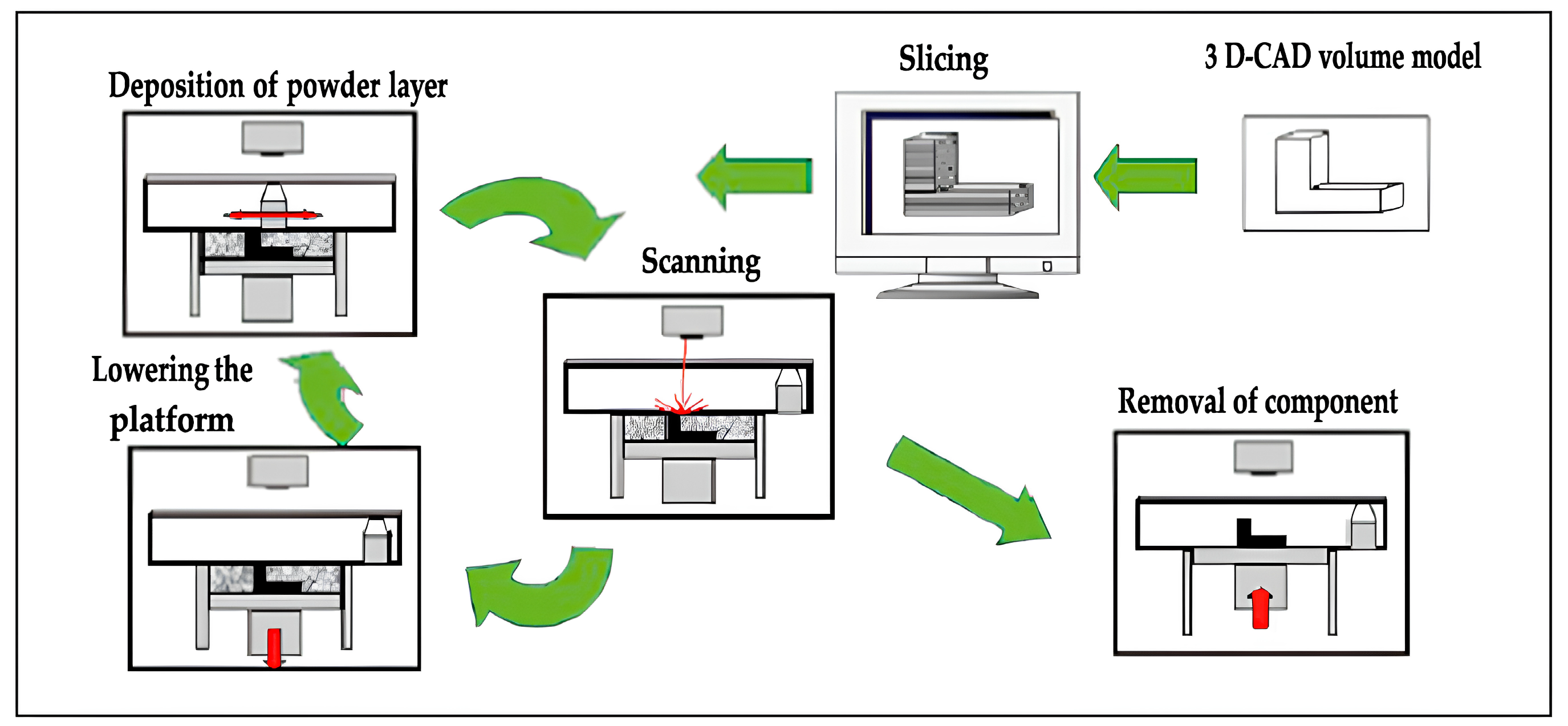
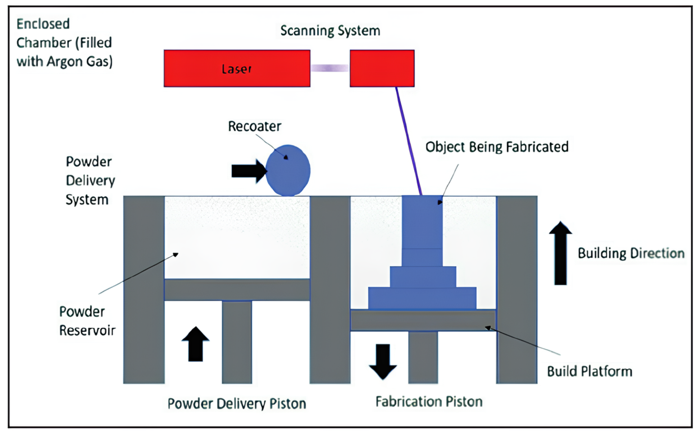
Metal additive manufacturing based on powder bed fusion (PBF) technologies plays a central role in the fabrication of dental implants and load-bearing components. Among these technologies, Selective Laser Melting (SLM) and Direct Metal Laser Sintering (DMLS) are the most widely applied due to their ability to process titanium and titanium alloys with high dimensional accuracy and favorable mechanical performance [26]. Both techniques rely on the layer-by-layer consolidation of fine metal powder using a high-energy laser source under an inert atmosphere, enabling the fabrication of complex geometries without the need for tooling. A schematic representation of the SLM process is shown in Figure 2.
Figure 2 illustrates the SLM process with key parameters, including a layer thickness of 20–40 µm, metal powder with a particle size of 10–45 µm, and melting performed by a fiber laser operating at 100–400 W with a wavelength of 1060–1080 nm in an inert argon atmosphere (<0.1% O2). These specifications determine the energy input into the powder bed and enable the reproducible fabrication of nearly 100% dense parts [27].
In SLM, complete melting of the powder particles results in components with very high relative density, typically exceeding 99%, and mechanical properties comparable to wrought titanium [27]. Process performance is governed by a combination of laser parameters (power, scanning speed, hatch spacing, beam diameter, scanning strategy) and build parameters (layer thickness, part orientation) [28,29]. Experimental studies consistently identify laser power and energy density as dominant factors influencing melt-track stability, porosity formation, and final density [30,31]. Reported layer thicknesses typically range from 20 to 40 µm, while powder particle sizes commonly fall within 10–45 µm [32]. When properly optimized, SLM enables the fabrication of dense titanium implants with reproducible mechanical behavior [33,34,35].
DMLS follows a similar processing principle but is often associated with a slightly lower degree of densification, with reported relative density values commonly in the range of 96–99% [36,37]. In the context of dental implantology, this characteristic can be advantageous, as DMLS offers enhanced control over the designed porosity and pore interconnectivity. Studies indicate that macropore sizes in the range of approximately 300–600 µm are particularly favorable for bone ingrowth and vascularization, contributing to improved osseointegration [38,39]. Relative porosity levels between 40% and 80%, depending on lattice architecture, have been reported to balance biological performance with sufficient mechanical stability [40]. A standard DMLS system (see Figure 3) consists of the following components [41]:
  • Feed piston—supplies unsintered metal powder.
  • Recoater—spreads a uniform thin powder layer onto the build platform.
  • Build piston—area where the part is formed layer by layer.
  • Laser + scanning system—melts powder in predefined regions based on the sliced CAD data.
  • Inert atmosphere (argon)—protects the melt pool from oxidation.
  • Heating elements—ensure thermal stability and consistent layer formation.
Comparative investigations highlight that SLM typically exhibits a lower powder consumption due to complete melting, whereas DMLS allows for more precise tuning of porous architectures relevant for clinical applications [42]. Typical PBF-related defects, such as lack-of-fusion pores, remain a critical concern for both technologies and are strongly dependent on process parameter selection and post-processing strategies [43].
Overall, the reviewed literature indicates that SLM and DMLS represent technologically mature solutions for the fabrication of titanium dental implants, offering a favorable combination of mechanical strength and biologically relevant porosity [44,45]. However, their clinical reliability remains strongly linked to process control, reproducibility, and post-processing [46,47]. This highlights a persistent translational challenge: while laboratory-scale optimization demonstrates excellent performance, standardized workflows and long-term clinical validation are still required for broader routine adoption.

2.2. Ceramic Additive Manufacturing for Dental Components

Additive manufacturing of dental ceramics offers several advantages over conventional subtractive processing, including reduced material waste, increased design freedom, and the ability to fabricate complex geometries with minimized internal stresses [48,49,50]. Despite these advantages, the direct additive fabrication of ceramic dental components remains less mature than metal AM, and its clinical implementation is still limited by processing complexity and material-related challenges [51].
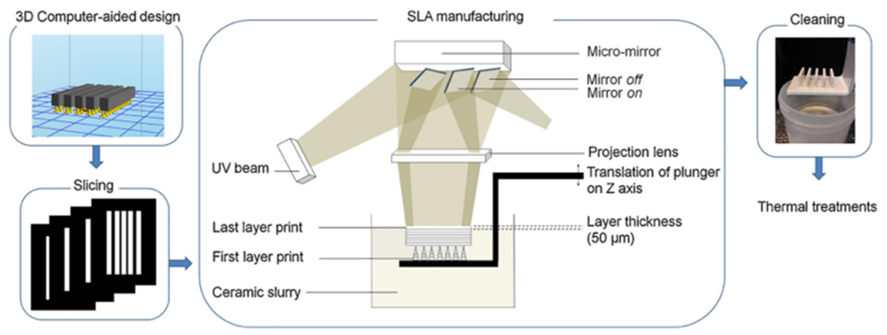
Among the available approaches, stereolithography (SLA), digital light processing (DLP), binder jetting (BJ), and powder bed fusion (PBF) have demonstrated the highest potential for dental ceramic applications [52,53,54,55]. SLA and DLP are currently the most widely investigated techniques due to their high resolution and suitability for fabricating patient-specific restorations [53,56]. In these processes, ceramic particles are dispersed in a photopolymerizable resin, which is selectively cured layer by layer using a laser or projected light (see Figure 4). Subsequent debinding and high-temperature sintering are required to remove the organic phase and densify the ceramic structure [57].
The properties of ceramic suspensions used in SLA depend on solid loading, particle size, and chemical composition [58,59,60]. A solid content above 50% is typically required to prevent deformation during thermal processing and minimize shrinkage during sintering [61,62]. Achieving high density in the final structure is essential for clinical-grade ceramic prostheses [62].
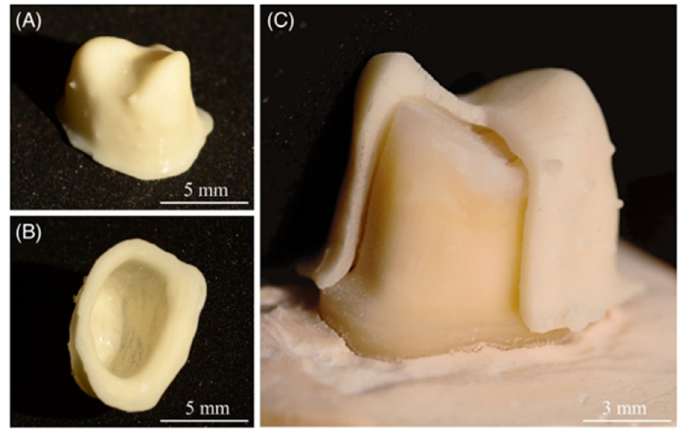
Dehurtevent et al. [63] evaluated alumina (Al2O3) parts fabricated by SLA using fine (0.46 μm) and coarse (1.56 μm) particles with different solid loadings (70%, 75%, 80%). Except for fine-particle suspensions with higher solid loading (75–80%), most formulations showed suitable viscosity for SLA processing. Coarse-particle suspensions with 75–80% solid loading achieved the highest sintered density, comparable to that of standard manufactured (SM) alumina. While SM samples exhibited homogeneous shrinkage, SLA-produced samples showed anisotropic shrinkage. The results indicate that alumina with coarse particle size and 80% solid loading is suitable for the fabrication of upper premolar ceramic crowns, confirming the clinical applicability of SLA-based ceramic AM (see Figure 5) [57].
In summary, ceramic additive manufacturing offers strong potential for aesthetic dental restorations and customized prosthetic components, but its clinical translation remains constrained by processing variability, shrinkage control, and high post-processing demands [51,58]. Compared to metal AM, ceramic technologies are at an earlier stage of technological maturity, suggesting that future progress will depend less on new printing concepts and more on improved material formulations, sintering strategies, and standardization of processing protocols. The limitations associated with ceramic additive manufacturing—particularly processing complexity, shrinkage control, and limited mechanical reliability—have motivated increasing interest in hybrid and composite material systems. These approaches aim to combine the aesthetic and biological advantages of ceramics with the processability, toughness, and functional adaptability of polymers. As a result, resin-based composites and polymer–ceramic hybrids have become a key focus of current research in dental additive manufacturing, as discussed in the following section [64].

2.3. Hybrid and Composite Materials in Dental 3D Printing

Resin-based composite (RBC) hybrids, which combine organic and inorganic components, are among the most widely used dental restorative materials in clinical practice. They are employed in a broad range of treatments, from small anterior restorations to extensive posterior reconstructions and even fixed partial prostheses, due to their excellent aesthetics, ease of clinical handling, strong adhesion to dental tissues, and low risk of adverse biological effects [65,66]. Most RBC materials consist of an organic matrix and an inorganic filler. The matrix is typically composed of a monomeric system that includes a free-radical polymerization initiator, while the filler serves to mechanically reinforce the resin matrix [67,68]. Dental RBCs reinforced with various fillers—such as silicon dioxide (SiO2), tetrapod whiskers, halloysite nanotubes, or inorganic fibers—demonstrate significantly enhanced mechanical properties due to the reinforcement and strengthening mechanisms of the composite structure [68,69]. In clinical applications, these composites are used to restore damaged or carious tooth structures. The procedure involves applying an adhesive system between the resin and the tooth surface, followed by light-curing polymerization, which restores both the function and aesthetics of the tooth [70]. Despite continuous advances, restorative dentistry still faces challenges such as secondary caries formation and pulpal pathology, both of which are associated with polymerization shrinkage occurring during the curing process of RBC materials [71]. During manual application, exposure of the oral cavity to ultraviolet (UV) radiation may pose potential safety concerns [72]. To minimize polymerization shrinkage, ensure complete curing, and enhance bond strength, the incremental layering technique is commonly employed. However, this method also presents certain limitations, including the entrapment of air bubbles during application, increased procedural complexity, and longer clinical operation times [73,74]. Therefore, when working with resin-based composites (RBCs), it is crucial to reduce errors associated with manual handling, control volumetric shrinkage during polymerization, and improve the mechanical performance of the material through innovative processing and fabrication techniques.
Biomaterials are also extensively used in dental manufacturing, particularly in the development of implants, prosthetic components, and structural frameworks that facilitate hard tissue regeneration. In this context, the material and biochemical properties of dental biomaterials and their influence on cell proliferation and tissue integration were analyzed. This study focuses on the use of scaffolds fabricated from a composite material composed of poly(lactic acid) (PLA) reinforced with calcium hydroxyapatite (cHA) particles. PLA is a biocompatible and biodegradable aliphatic polyester with thermoplastic and semi-crystalline characteristics, synthesized from renewable lactic acid sources [75,76]. Using Fused Deposition Modeling (FDM) technology, it is possible to manufacture complex three-dimensional (3D) scaffold structures with variable pore sizes and porosity through computer-aided design, layer-by-layer printing, and controlled solidification. This process enables the precise fabrication of dental implants and support structures from PLA/cHA composites by means of additive manufacturing (3D printing) [77].
The mechanical properties of PLA composites reinforced with hydroxyapatite (cHAP) have been significantly improved in several experimental studies. For example, Omigbodun et al. (2023) [77] reported that the addition of 10 wt% cHAP together with rGO enhanced the strength and structural stability of the polymer matrix. Custódio et al. (2021) [78] achieved a tensile strength of approximately 47.3 MPa and a Young’s modulus of up to 3.5 GPa with a 15% HAp content. Other studies have documented strength values approaching 64 MPa and a modulus of around 2.9 GPa (Tazibt et al., 2023) [79]. Collectively, these findings indicate that the mechanical performance of PLA/cHAP composites is comparable to that of trabecular bone and substantially exceeds the performance of several conventional bioresorbable polymers, although the exact values depend on composition and processing conditions.
Poly(lactic acid) (PLA) is a polymer widely used in dental manufacturing as a biocompatible and biodegradable material suitable for the additive fabrication of implants, prosthetic components, and scaffolds. The polymer can be synthesized either through a polycondensation reaction or by the ring-opening polymerization of lactide. The fundamental structural unit of PLA is lactic acid, an α-hydroxy acid containing a chiral carbon atom and existing in two optical isomers: L-lactic acid (PLLA) and D-lactic acid (PDLA). The L-isomer occurs naturally in humans and other mammals, whereas both enantiomers (D and L) can be produced via microbial or bacterial synthesis [80,81]. Lactic acid, serving as the precursor for PLA synthesis, can be derived from renewable resources such as ethanol or acetaldehyde, as well as from coal or petroleum-based sources. However, the majority of commercially available lactic acid is currently obtained through the bacterial fermentation of simple sugars, making PLA an environmentally sustainable material for advanced dental manufacturing [77].
Taken together, the reviewed additive manufacturing technologies illustrate a progression from material- and process-driven development toward application-oriented design, where mechanical performance, biological response, and manufacturing reliability must be considered simultaneously.

2.4. Design and Manufacturing of Porous Dental Implants

Dental implants represent an effective and widely accepted solution for replacing the root portion of missing teeth [82], with the primary objective of restoring aesthetics, speech function, and overall oral health [83]. The implants are surgically placed directly into the jawbone, where they act as a stable anchorage for prosthetic restorations [84]. From a technical perspective, dental implants are classified according to their geometry, surface roughness, surface treatment, and the type of connection with the prosthetic superstructure. Their dimensions (length and diameter) are adjusted to meet specific clinical requirements. For instance, commercially available cylindrical threaded implants typically range from 3.25 to 6.0 mm in diameter and 5 to 18 mm in length [82]. In the past decade, there has been a marked global increase in the demand for dental implants, with estimates suggesting their use in the treatment of approximately one million patients annually [85]. This growing demand can be attributed primarily to their high clinical success rate in full dental restoration, the aging of the global population, the increasing proportion of elderly individuals, and the greater public awareness of implant-based treatments [85]. Conventional prosthetic options, such as removable dentures, fixed crowns, and bridges, are often associated with certain limitations—notably progressive bone resorption of the residual alveolar ridge following tooth extraction, which reduces prosthesis stability and patient comfort. Endosseous titanium implants, therefore, provide a more functional, comfortable, and long-term stable alternative to traditional dental prostheses [86].
The high modulus of elasticity of titanium implants can result in the so-called stress-shielding effect, characterized by insufficient mechanical loading of the surrounding bone tissue [87]. Because bone is a living, dynamically remodeling tissue that responds to mechanical stimuli through the activity of bone cells, reduced loading may lead to bone resorption, subsequent implant loosening, and ultimately implant failure—a complication that has been particularly evident in the past with orthopedic implants [88]. In contrast, excessive mechanical loading can cause localized stress concentrations in the bone, also triggering resorptive processes in these regions [89]. Consequently, extensive research has focused on the development of biomaterials with mechanical properties more closely matching those of natural bone, thereby mitigating the risk of undesirable bone remodeling. Most studies in this area aim to optimize the interfacial interactions between the implant surface and the bone tissue, seeking to enhance osseointegration and mechanical compatibility. Recent advances in the design of orthopedic and dental implants, as well as in the engineering of bone scaffolds, have facilitated the creation of novel porous titanium structures that integrate insights from both fields and leverage the synergistic potential of orthopedic and dental implant technologies.
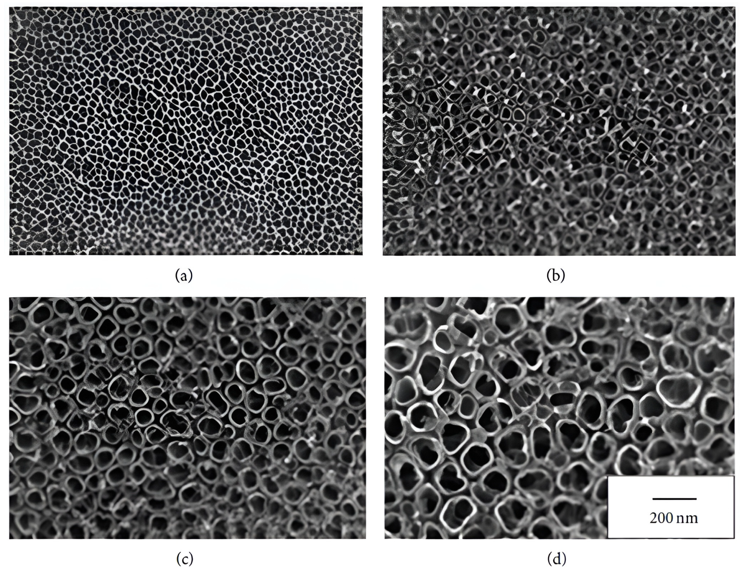
The morphological characteristics of dental implants play a crucial role in determining the quality of contact between the implant and bone tissue, thereby significantly influencing the osseointegration process. To enhance the stability and functionality of titanium implants, a variety of surface modification techniques have been developed, enabling the adjustment of their physical and chemical properties [90]. Surface modifications can markedly improve the interaction between the implant and surrounding bone, although the mechanisms underlying these improvements are not always fully understood. Such effects may arise from both morphological and chemical alterations—for example, surface roughening, which can simultaneously modify the chemical composition of the implant surface [91]. Among the commonly applied techniques, plasma spraying using powdered materials such as titanium dioxide (TiO2), calcium phosphate, and hydroxyapatite is frequently employed to improve biocompatibility and bone-implant bonding [92]. Another widely used method is sandblasting with hard particles (e.g., aluminum oxide, TiO2, or ceramic abrasives) to increase microscopic surface roughness [85]. In 2012, Çelen and Özden proposed an alternative and more precisely controlled technique of laser micro-machining for commercially pure titanium implants, allowing for enhanced control over surface topography [92]. More recently, nanoporous surface structures, such as TiO2 nanotubes, have been introduced as a promising approach for implant surface modification. These nanostructures can be fabricated through controlled anodization of titanium surfaces, offering improved biological response and enhanced osseointegration potential [93,94]. A layer of TiO2 nanotubes was formed on the implant surface using an anodization process carried out in a mixture of hydrofluoric and acetic acids at a 7:1 ratio. The anodization was performed at voltages of 5, 10, 15, and 20 V for one hour at room temperature, producing nanotubes with diameters of approximately 30, 50, 70, and 100 nm. Following anodization, the samples were rinsed with distilled water, dried for 24 h at 60 °C, and subsequently heat-treated in air at 500 °C for 2 h with a controlled heating and cooling rate of 1 °C/min to ensure stabilization of the formed oxide layer (Figure 6) [95].
The issue of pore size and distribution in porous implants has been extensively examined in the literature, and the findings consistently indicate that the optimal pore architecture depends on the clinical context and the intended biological or mechanical function. Preclinical studies have shown that effective pore sizes generally fall within ~100–600 μm, with specific recommended intervals varying according to the parameter assessed (e.g., vascularization versus mechanical strength) [96]. In several experiments involving SLM-produced titanium scaffolds, three standard pore sizes (300, 600, and 900 μm) were compared. These studies demonstrated that pores of approximately 600 μm provided the most favorable combination of bone ingrowth and mechanical fixation, while smaller pores (~300 μm) exhibited higher local bone filling in certain internal regions of the scaffold. These observations, derived from rabbit in vivo models, suggest that larger pores promote vascularization, whereas smaller pores support early stage bone contact [94]. Research on functionally graded Ti-6Al-4V structures has similarly shown that regions containing smaller pores (~300–400 μm) display locally higher bone volume fraction (BV/TV), while larger exterior pores facilitate vascularization and tissue invasion. Consequently, several authors recommend graded porosity as a strategy to enhance overall biological integration compared to homogeneous lattice architectures. However, the precise percentage increase in bone–implant contact (BIC) varies with the model, time point, and evaluation method, and reported improvement values are not uniform across studies [96]. Based on the combined evidence from reviews and experimental studies, graded architectures are therefore suggested for dental implant design using smaller pores (on the order of a few hundred micrometers) near the bone–implant interface to support higher BIC, and larger pores (~300–600 μm) in the outer regions to promote vascularization and nutrient transport (see Table 1). Accordingly, this manuscript recommends presenting pore-size guidelines as flexible ranges (~200–600 μm), with an emphasis on validating the final parameters within the appropriate clinical or preclinical model [97].

2.5. Comparison of Additive Manufacturing Methods in Dental Applications

Additive technologies used in dentistry and implantology differ significantly in material processing, resolution, surface morphology, and mechanical properties. These differences directly affect the clinical accuracy, reliability, and suitability of individual methods for specific types of dental and biomedical applications. The overview in the Table 2 summarizes the key parameters of major AM technologies and enables a quick comparison of their advantages, limitations, and areas of use. At the same time, it highlights that no technology is universal and that its selection must be tailored to the required accuracy, material properties, and clinical demands. This information is essential for making informed decisions when manufacturing implants, prosthetic restorations, or specialized dental devices [101,102,103,104].
A comparison of individual additive technologies revealed fundamental differences in precision, material properties, and clinical suitability. SLM/DMLS and EBM metal technologies offer the highest mechanical performance and are therefore best suited for implantology and permanently loaded structures. SLM/DMLS provides higher precision and a smoother surface than EBM, which is particularly important for superstructures. EBM has the advantage of lower internal stresses, but at the cost of a coarser surface texture.
SLA/DLP and PolyJet achieved the highest accuracy and the smoothest surface quality, making them ideal for the production of models, surgical templates, and aesthetic prototypes. Their fundamental limitation is low strength and limited long-term stability, which makes them unsuitable for permanent functional restorations. PolyJet provides exceptionally high surface quality, but its mechanical limitations remain significant.
Binder Jetting and FDM/MEX achieved lower accuracy and exhibited highly variable mechanical properties. Binder Jetting is limited by porosity and dimensional changes during post-processing, while FDM suffers from low resolution and material anisotropy. These technologies are therefore most appropriate for prototyping rather than clinically loaded components.
Overall, it is clear that the choice of technology must be strictly tailored to the intended clinical or technical application. Metal AM technologies (SLM/DMLS, EBM) are the clear choice for implants and permanent metallic superstructures, while SLA/DLP and PolyJet dominate in terms of accuracy and detail, but only for short-term or non-structural applications. Binder Jetting and FDM/MEX have their specific roles, particularly in prototyping, research applications, and the fabrication of non-functional devices, but not in the production of permanent dental prostheses.

3. Descriptive Overview of Publication Trends

The following section provides a descriptive and indicative overview of publication trends related to additive manufacturing in dentistry over the last decade. This analysis is not intended to represent a formal bibliometric or systematic review, but rather to contextualize the technological and material developments discussed in this manuscript within the broader research landscape. The primary aim is to illustrate general growth patterns, disciplinary distribution, and document types associated with additive manufacturing in dental applications, thereby supporting the technology-oriented focus of this review. The overview is based on two major scientific databases: Web of Science (WoS) and Scopus. Web of Science offers a curated selection of journals with a strong emphasis on engineering, materials science, and biomedical research, while Scopus provides broader interdisciplinary coverage, including applied and emerging research areas. Together, these databases enable an indicative comparison of publication dynamics rather than a comprehensive quantitative mapping of the literature.
The search strategy employed three core terms—“additive manufacturing”, “dental implants”, and “production”—combined using Boolean AND logic. The time window was defined as 2014–2024 in order to capture recent technological developments associated with the rapid adoption of digital dentistry and advanced manufacturing methods. No language or document-type restrictions were applied. Inclusion criteria focused on publications with a clear technological, material, or manufacturing orientation (e.g., SLM, DMLS, SLA, DLP, PBF), while articles with a predominantly clinical or restorative focus and limited relevance to manufacturing processes were excluded. Given the descriptive nature of this overview, formal deduplication procedures, network analysis, or thematic clustering were not performed, and the results should be interpreted as illustrative rather than exhaustive. Within these constraints, the analysis identified 143 relevant publications indexed in the Web of Science database and 6568 records in Scopus. The substantial difference between the two databases reflects their distinct indexing strategies and disciplinary scopes rather than inconsistencies in the underlying research activity. The observed publication growth primarily serves to demonstrate the increasing scientific and technological interest in additive manufacturing for dental applications, particularly in implant production and advanced prosthetic components.
Overall, this descriptive trend overview supports the central objective of the manuscript: to frame current additive manufacturing technologies in dentistry within their evolving research context, without claiming statistical completeness or critical evaluation of individual studies. The results therefore complement the subsequent technology- and material-focused discussion rather than constituting an independent analytical contribution.

3.1. Web of Science

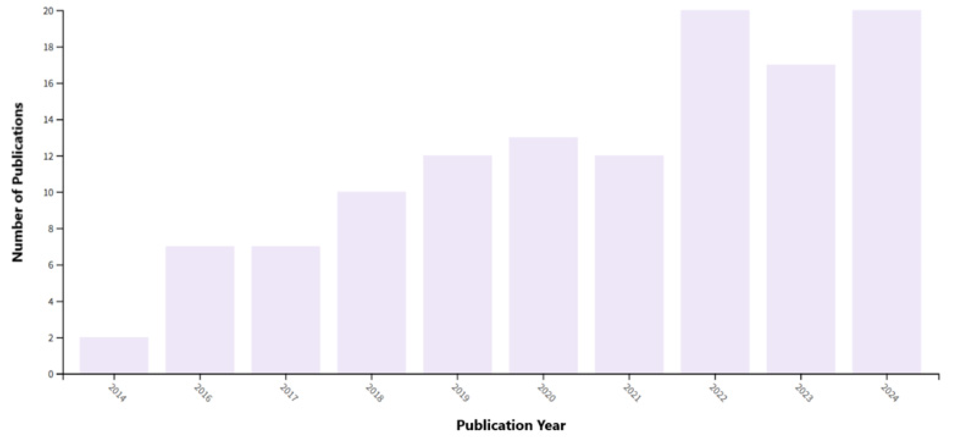
Figure 7 illustrates the number of scientific publications on additive manufacturing in dentistry indexed in the Web of Science database between 2014 and 2024. The number of papers has shown a steady increase, indicating a growing scientific interest in the application of additive technologies within the field of dentistry. The highest number of publications was observed in 2022 (16.7%) and 2024 (16.7%), reaching approximately 20 articles per year.
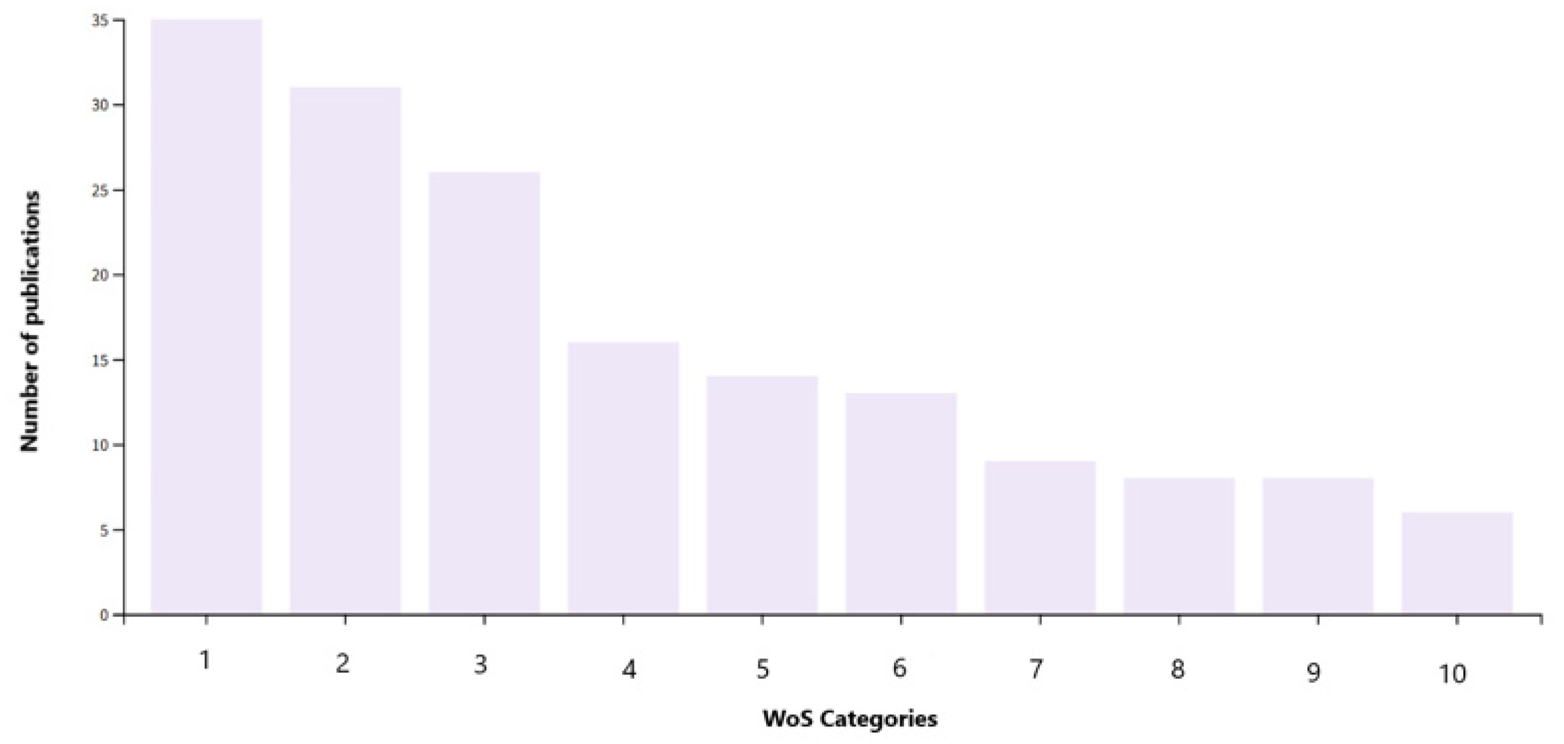
Figure 8 illustrates the distribution of scientific publications across the Web of Science categories related to additive manufacturing in dentistry. The graph indicates that the highest number of publications fell under the category Materials Science Multidisciplinary (29.17%), followed by Engineering Biomedical (25.8%) and Materials Science Biomaterials (21.7%). A smaller number of studies were classified in categories such as Metallurgical Engineering Metallurgy (13.3%), Dentistry Oral Surgery Medicine (11.7%) and Physics Applied (10.8%). This overview demonstrates that research on additive manufacturing in dentistry is multidisciplinary in nature, with the strongest focus on materials science and biomedical engineering, which constitute the foundation for the development of advanced dental implants and prosthetic components. The selection of categories was guided by the technical focus of the manuscript. Clinically oriented categories, such as “Dentistry, Restorative,” were excluded because they did not contain contributions with sufficient technical relevance to AM processes.
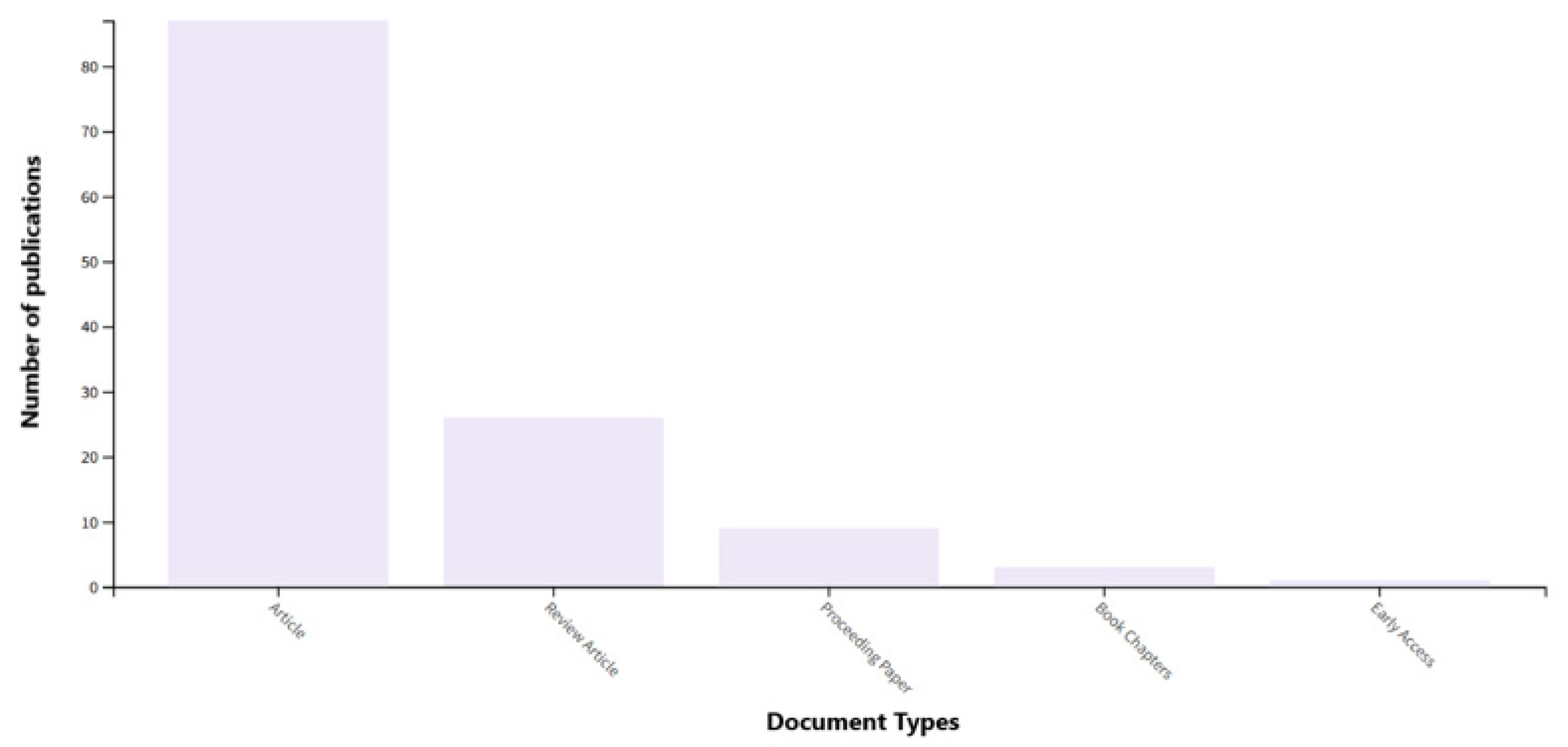
Figure 9 illustrates the distribution of scientific publications by document type in the Web of Science database related to the topic of additive manufacturing in dentistry. The graph indicates that Research Articles represented the largest share of publications (72.5%), significantly exceeding all other categories. These were followed by Review Articles (21.7%) and, to a lesser extent, Conference Proceedings (7.5%). Only a small number of publications fell under the categories of Book Chapters (2.5%) and Early Access papers (0.83%).

3.2. Scopus

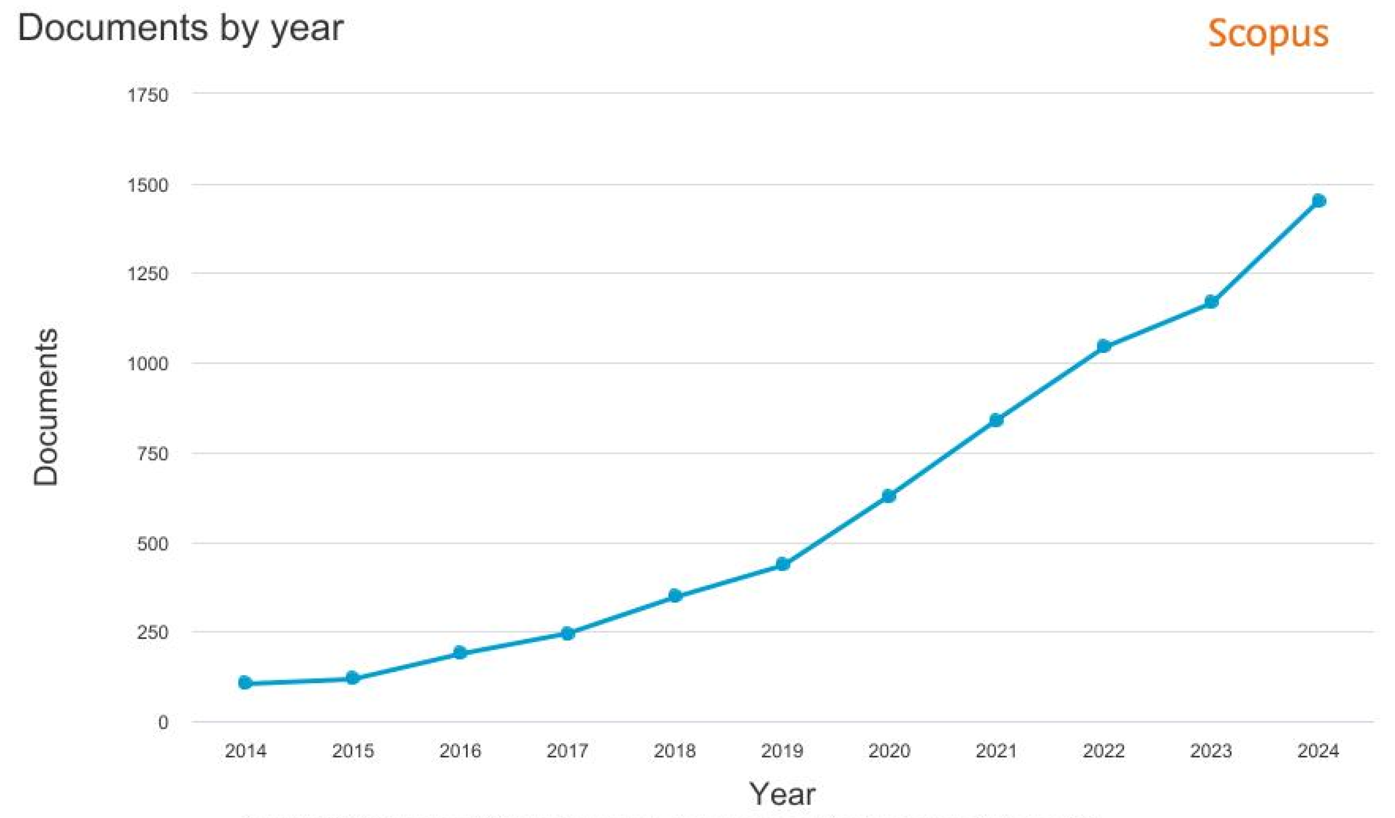
The graph (see Figure 10) shows that the number of publications increased steadily throughout the analyzed period, with a marked rise observed after 2019 (6.64%). This upward trend reflects the growing scientific and technological interest in the application of additive manufacturing technologies in dental production, driven by the rapid advancement of 3D printing, the development of new materials, and the ongoing digitization of manufacturing processes. The highest number of publications was recorded in 2024 (21.81%), confirming that research in this field represents a dynamically evolving trend within the contemporary scientific and technical community.
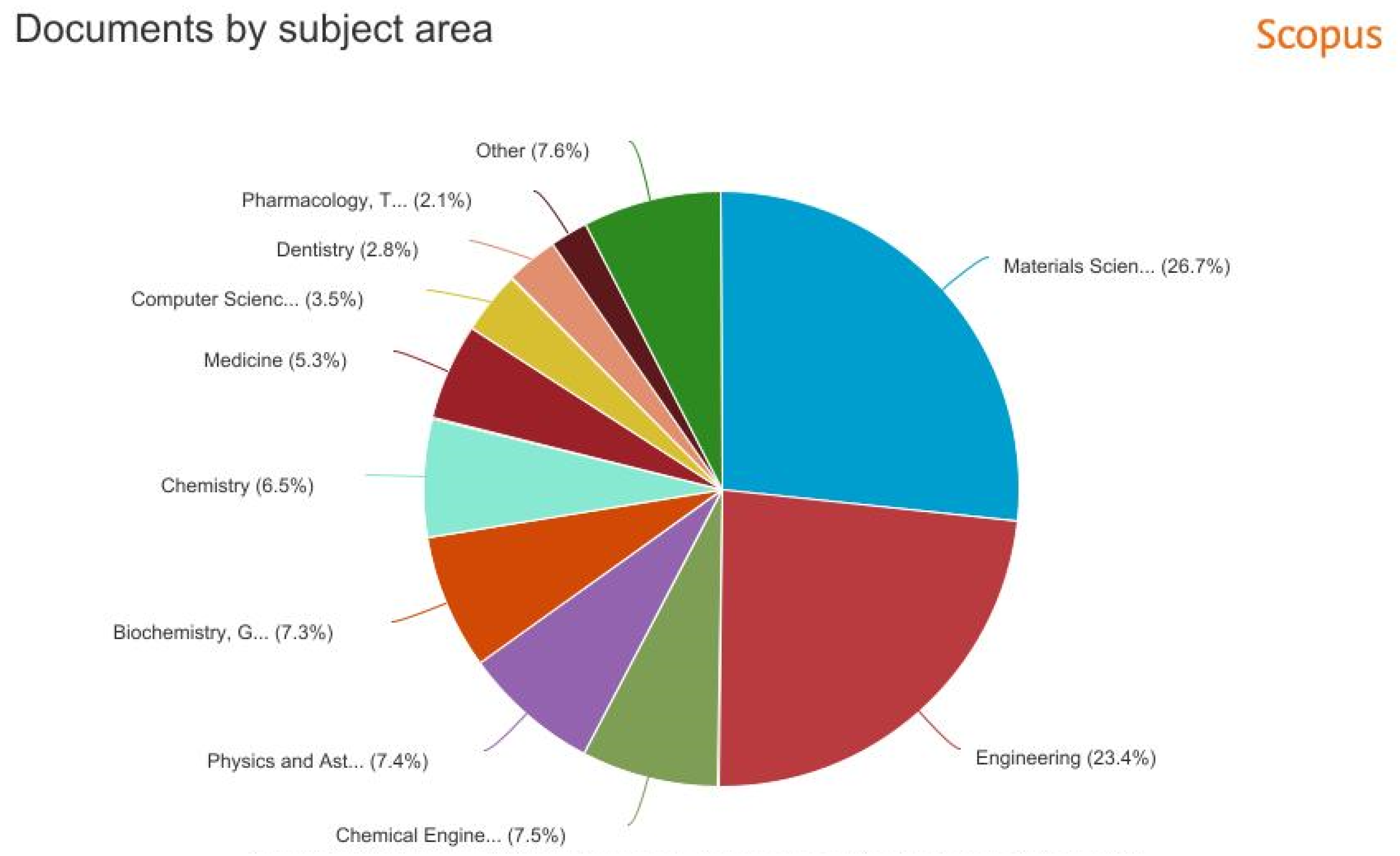
Figure 11 illustrates the distribution of scientific publications by research field in the Scopus database on additive manufacturing in dentistry. The largest share of publications belonged to materials science (26.7%) and engineering (23.4%), reflecting a strong focus on technological and material aspects. Other well-represented fields included chemical engineering, physics, biochemistry, and chemistry, while smaller shares appeared in medicine, computer science, and dentistry. The “other” (7.6%) category covered interdisciplinary studies. Overall, the research is multidisciplinary, with materials and engineering areas closely connected to biomedical applications.
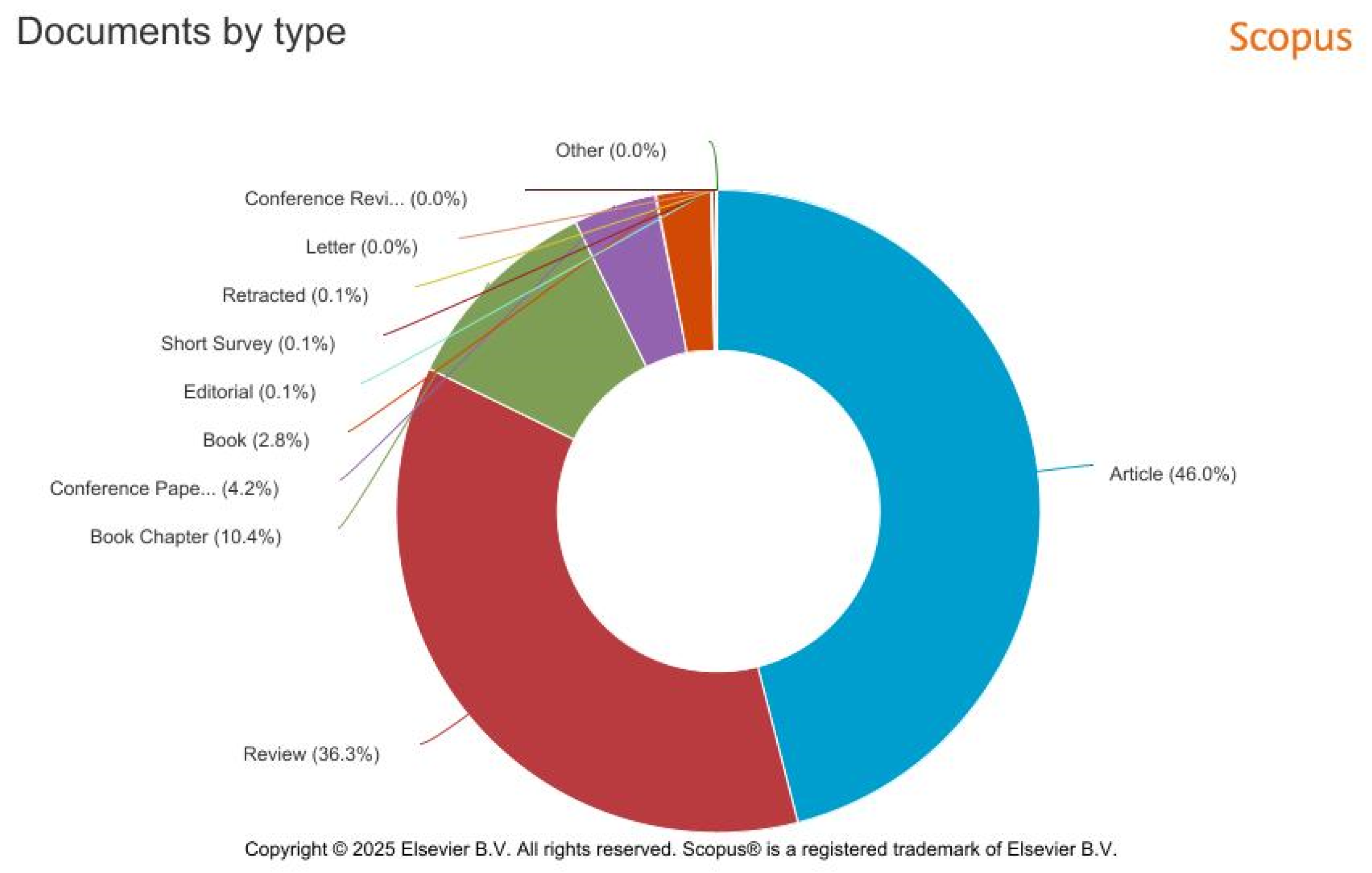
Figure 12 illustrates the distribution of scientific publications by document type in the Scopus database related to the topic of additive manufacturing in dentistry. The graph indicates that research articles represented the largest share of publications (46.0%), followed by review papers (36.3%). Book chapters (10.4%) and conference proceedings (4.2%) were less represented, while books (2.8%) and other document types, such as editorials, short surveys, and letters, accounted for only a negligible proportion. This analysis demonstrates that most research outputs in the field of additive manufacturing in dentistry are published in the form of research and review articles, reflecting a high level of academic activity and an increasing systematization of knowledge within this research domain.

4. Challenges and Future Directions in Additive Dental Manufacturing

Future developments in additive manufacturing for dental applications can be broadly structured into three interconnected domains: (i) material and surface innovations, (ii) digitalization and process intelligence, and (iii) regulatory and translational frameworks. Rather than representing isolated trends, these domains collectively determine the pace and scope of the clinical adoption of additively manufactured dental components. The following sections outline key developments within each domain and highlight their potential implications for implant performance, manufacturing reliability, and clinical workflows. In the context of dental additive manufacturing, technological performance alone is insufficient to ensure clinical adoption. Regulatory compliance, metrological reliability, and sustainability considerations play a decisive role in determining whether additively manufactured components can be safely and reproducibly translated into routine dental practice. These aspects therefore represent enabling constraints rather than peripheral considerations and must be addressed alongside materials and process development. Over the past decade, additive manufacturing (AM) has become one of the most promising technologies in modern dentistry, offering high product customization, efficient material use, and seamless integration with digital workflows [95]. Despite these advantages, its widespread clinical adoption remains limited by technological, material, and regulatory constraints. A major challenge is the insufficient long-term stability of polymer, metal, and ceramic materials used in dental AM. Photopolymer resins often exhibit low color stability, high water absorption, and mechanical degradation during aging [105], while ceramic materials face brittleness, non-uniform density, and microdefects that can reduce both mechanical strength and prosthetic fit accuracy [106]. This has led to the development of hybrid and composite ceramic systems aimed at improving mechanical strength, structural stability, and aesthetic properties [107].
Photopolymer (3D-printed) resins—water sorption, color stability, mechanical aging:
  • Water sorption: Modern 3D-printed dental resins typically show W_sp ≈ 24–26 µg/mm3, while modified formulations may reach ~28 µg/mm3; higher sorption increases the risk of internal stresses and microcracks during long-term exposure to oral fluids [108].
  • Color stability: Color change assessed by ΔE indicates that ΔE ≈ 3.3 is generally considered the clinically acceptable threshold. Some resin formulations exceed this value after accelerated aging, depending on composition and post-curing [109].
  • Mechanical changes during artificial aging: Thermocycling and water storage frequently reduce flexural strength by ~10–30%, with decreases up to ~27% documented in certain formulations [110].
Ceramics (3D-printed ZrO2 and other systems)—microdefects and their influence on strength:
  • Microdefects such as microcracks, insufficient compaction, weak intergranular bonding, and lack-of-fusion defects can significantly affect strength. In ZrO2 studies, flexural strength varied from 789 MPa to 423 MPa depending on sintering orientation, showing 30–50% reductions under suboptimal processing [111].
These quantitative data confirm that stability limitations in polymer and ceramic AM materials are measurable and clinically relevant: water sorption values typically range from ~24–28 µg/mm3; mechanical aging commonly reduces strength by ~10–30%; and sintering orientation alone can shift flexural strength in 3D-printed ZrO2 by several hundred MPa.
In metal AM (SLM and DMLS), key challenges remain in achieving uniform microstructure, controlled porosity, and a stable interface with the prosthetic superstructure, all of which are essential for osseointegration and long-term mechanical performance [112]. Surface modification techniques such as laser micromachining and TiO2 nanotube coatings have shown promising improvements in cell adhesion and biocompatibility [113]. Variability in process parameters—laser energy density, scanning speed, layer thickness, build orientation, and post-processing—can strongly influence dimensional accuracy and mechanical properties [114]. The absence of standardized workflows contributes to inconsistent results across equipment and facilities. In situ monitoring systems using optical sensors, thermal imaging, and spectral analysis, combined with closed-loop feedback, are emerging as promising solutions for improving process reproducibility [115].
Accuracy, metrology, and regulatory requirements remain critical issues. Fit accuracy, surface roughness, and dimensional tolerances vary significantly when AM-fabricated components are compared with traditional CAD/CAM techniques [116], emphasizing the need for standardized metrological protocols and validated reference models [117]. Long-term clinical evidence is still limited, especially for photopolymer and ceramic materials, and regulatory frameworks remain insufficiently harmonized across ISO 13485 [116], FDA guidance, and MDR requirements [117].
Future developments in AM include advances in ceramic technologies (SLA, DLP, PBF), which enable the precise control of porosity, microdefects, and optical behavior for aesthetic restorations [114,117]. Although several standards exist (ISO/ASTM 52900 [118], ASTM F42 [119], NIST guidelines, FDA technical guidance, MDR, ISO 13485), practical implementation still lacks harmonized digital traceability, material qualification criteria, monitoring protocols, and standardized microdefect metrics [120].
Taken together, current regulatory and standardization frameworks indicate that the primary bottleneck for dental additive manufacturing is not the absence of suitable technologies, but the lack of harmonized validation, traceability, and quality-control strategies across materials and manufacturing platforms.
Despite this, several critical weaknesses persist:
  • Digital thread and versioning: No unified machine-readable format exists for mandatory metadata such as print parameters, CAD versions, material batches, or machine settings.
  • Process validation and real-time monitoring: No harmonized acceptance criteria exist for in situ monitoring methods across technologies and manufacturers [121].
  • Metrics for microdefects and non-destructive evaluation: No uniform protocols exist for µCT/SEM-based quantification of porosity, cracks, or lack-of-fusion defects.
To improve consistency and regulatory readiness, several steps can be implemented:
  • Introduce a mandatory digital footprint for AM parts (metadata such as CAD version, slicer settings, material batch, machine parameters, timestamps) stored in a standardized JSON/XML format [120].
  • Establish harmonized material qualification criteria for input powders and resins (particle size, contamination, moisture, mechanical behavior) with standardized test methods [122].
  • Develop standardized protocols for process validation and in situ monitoring, including reference geometries and validated analytical software tools.
  • Define uniform µCT protocols (resolution, thresholding) and reporting metrics for defect quantification [123].
  • Clarify regulatory guidelines for documenting personalized and custom-made components within MDR requirements [124].
Hybrid and functionally graded materials that combine mechanical strength, bioactivity, and aesthetic properties are gaining attention [115]. Artificial intelligence (AI/ML) is being increasingly integrated into AM workflows, enabling automated defect detection, prediction of mechanical properties, and accelerated XCT reconstruction [116,118,119]. In implantology, nanostructured surface modifications such as TiO2 nanotubes improve osseointegration [109], while “print-and-place” workflows support immediate clinical installation of restorations with predictable outcomes [118]. Long-term progress will depend on coordinated advancements in materials science, process metrology, clinical validation, and regulatory harmonization.
Additive manufacturing technologies, particularly SLM and DMLS, enable the production of titanium dental implants with controlled microstructure and porosity that naturally promote osseointegration. Nevertheless, achieving rapid and stable integration remains challenging, which is why biomimetic surface treatments that mimic the natural extracellular matrix (ECM) are increasingly being used. Kim et al. [125] reported that the biomimetic approach is attractive because the implant surface can “mimic the structure of natural tissue” and enhance osteoinductivity.
The most commonly used biomimetic materials include ECM proteins such as type I collagen and fibronectin, which improve early adhesion, proliferation, and differentiation of osteoblasts; fibronectin in particular increases bone–implant contact by approximately 70% and enhances mechanical fixation of the implant up to fourfold. Composite ECM coatings (e.g., collagen with chondroitin sulfate) further strengthen the early cellular response [126].
SLM/DMLS implants also respond well to biomimetic peptides such as RGD sequences, GFOGER (a collagen-mimetic peptide), or multimodal peptides that promote specific integrin binding and accelerate surface mineralization. According to Kim et al. [125], GFOGER provides adhesion comparable to native collagen, which is advantageous for AM-produced surfaces with higher surface energy and roughness. Growth factors such as BMP and FGF-2 also play an important role, synergistically improving bone tissue regeneration and increasing the mechanical stability of the implant, with FN–FGF fusion proteins demonstrating significantly enhanced integration.
The combination of additive manufacturing with biomimetic surface strategies provides a synergistic effect: AM creates a microarchitecture suitable for cell growth, while biomimetic molecules increase the specificity of the biological response. Together, they lead to faster osteoblast adhesion, higher bone–implant contact and improved long-term implant stability. As a result, biomimetic surface treatments appear to be one of the most promising directions for the development of AM-manufactured dental implants.
In recent years, 4D printing has emerged as an extension of 3D additive manufacturing, with the “fourth dimension” referring to time-dependent or stimulus-responsive changes in material behavior. This approach enables the fabrication of dynamic, adaptive structures whose properties can transform after implantation, offering new possibilities for dental and craniofacial applications. In dental implantology, 4D printing is particularly relevant for bone regeneration and the production of scaffolds that support osseointegration. Shape-memory polymers such as PLA, PCL, and PLGA, together with hydrogels and natural biopolymers like hyaluronic acid, collagen, and alginate, can mimic the extracellular matrix and promote cell growth and tissue formation [127].
A major advantage of 4D printing is the ability to incorporate bioactive molecules, including BMP and VEGF, which stimulate osteogenesis and angiogenesis and enhance the regeneration of periodontal and pulp tissues. Such adaptive scaffolds can modify their mechanical or structural properties over time, providing optimal conditions during critical phases of healing. By combining these responsive biomaterials with metal implants produced via SLM/DMLS, 4D bioprinting may contribute to the development of next-generation hybrid implant systems with improved biological function and personalized behavior over time [128].

Sustainability and Material Innovations in Additive Manufacturing of Dental Implants

Current publications emphasize that additive manufacturing (AM) has the potential to significantly improve the sustainability of dental practice. In 2025, it was reported that AM “fabricates dental items incrementally from digital models, minimizing material waste and production duration while enabling intricate geometries”, and that sustainability issues are actively being addressed, particularly through “recycled materials and circular economy models” in the dental sector (Monalisa et al. [129]). Localized production of prosthetic components directly in the clinic or laboratory significantly reduces the need for transportation and intermediate logistical steps, contributing to a lower carbon footprint and a more efficient supply chain.
At the same time, the development of new materials plays a key role in reducing the environmental impact of additive technologies. A review of polymers used in 3D and 4D printing states that “the development of composite materials for 3D printing is the main focus of future research, as combining multiple materials can improve the materials’ properties” (Cai et al. [130]). These composite materials pave the way for systems that can be partially recyclable or biodegradable, which is important for more environmentally friendly dental manufacturing. Hydrogels and natural biopolymers, which are increasingly used in 4D printing due to their bioactive and less environmentally burdensome characteristics, also contribute to this perspective.
Taken together, these findings indicate that integrating additive manufacturing with material innovations provides a real opportunity to reduce waste, optimize resources, and move toward a more sustainable dental industry.

5. Conclusions

Additive manufacturing (AM) has become an important enabling technology in contemporary dental manufacturing, offering a high degree of customization and integration with digital workflows. An indicative overview of publications indexed in the Web of Science and Scopus databases between 2014 and 2024 confirms a sustained increase in scientific and technological interest in AM for dental applications, particularly in implant production and advanced prosthetic components.
From a technological perspective, three key observations emerged. Metal additive manufacturing technologies, especially Selective Laser Melting (SLM) and Direct Metal Laser Sintering (DMLS), currently represent the most mature solutions for load-bearing dental implants, enabling controlled porosity and favorable conditions for osseointegration. Ceramic additive manufacturing methods (SLA, DLP, PBF) show strong potential for aesthetic restorations and customized components but remain constrained by processing variability and post-processing demands. Hybrid and composite material systems increasingly act as an intermediate strategy, aiming to balance mechanical performance, biological response, and manufacturing reliability.
Despite significant progress, several challenges continue to limit broader clinical adoption, including process reproducibility, validated metrology, material stability, and regulatory harmonization. Future advances are therefore expected to depend primarily on improved process control, digital traceability, and clinically relevant validation rather than on the introduction of fundamentally new printing principles. Overall, additive manufacturing should be regarded as a rapidly evolving but still maturing technology whose integration into routine dental practice is likely to proceed gradually. By integrating materials, manufacturing technologies, regulatory constraints, and future development pathways into a single technology-oriented framework, this review aims to clarify both the current capabilities and the remaining translational challenges of additive manufacturing in dentistry.

Author Contributions

Conceptualization, M.Y. and D.D.; methodology, J.T. and J.D.; validation, J.T., J.D. and D.D.; formal analysis, M.T.; resources, M.Y.; data curation, D.D.; writing—original draft preparation, J.T. and M.Y.; writing—review and editing, D.D. and M.T.; visualization, J.D. All authors have read and agreed to the published version of the manuscript.

Funding

This article was supported by the Scientific Grant Agency of the Ministry of Education, Research, Development and Youth of the Slovak Republic and Slovak Academy of Sciences, grant VEGA 1/0258/24. This work was supported by the Slovak Research and Development Agency under contract no. APVV-21-0293.

Institutional Review Board Statement

Not applicable.

Informed Consent Statement

Not applicable.

Data Availability Statement

No new data were created or analyzed in this study. Data sharing is not applicable to this article.

Acknowledgments

This article is supported by project DRP0200194, Moving Plastics and Machine Industry towards Circularity (PLAN-C) under the Interreg Danube Region Program, co-funded by the European Union.

Conflicts of Interest

The authors declare no conflicts of interest.

Abbreviations

The following abbreviations are used in this manuscript:
AIArtificial Intelligence
AMAdditive Manufacturing
BICBone–Implant Contact
CADComputer-Aided Design
CAMComputer-Aided Manufacturing
cHACalcium Hydroxyapatite
cHAPCalcium Hydroxyapatite Powder
CNCComputer Numerical Control
DLPDigital Light Processing
DMLSDirect Metal Laser Sintering
FDAFood and Drug Administration
FDMFused Deposition Modeling
ISOInternational Organization for Standardization
PBFPowder Bed Fusion
PDLAPoly(D-Lactic Acid)
PLAPolylactic Acid
PLLAPoly(L-Lactic Acid)
RBCResin-Based Composite
rGOReduce Graphene Oxide
SLAStereolithography
SLMSelective Laser Melting
UVUltraviolet
XCTX-ray Computed Tomography

References

  1. Alqutaibi, A.Y.; Al-Eryani, K.; Basudan, M.; Alhajj, M.N.; Lin, W.S.; Morton, D. Advanced Additive Manufacturing in Implant Dentistry: 3D Printing Technologies, Printable Materials, Current Applications and Future Requirements. Bioprinting 2024, 35, e00356. [Google Scholar] [CrossRef]
  2. Duarte, S., Jr.; Phark, J.H. A Comprehensive Review of Machinable and 3D-Printed Ceramic-Reinforced Composites for Dental Restorations. J. Esthet. Restor. Dent. 2024, 37, 257–276. [Google Scholar] [CrossRef] [PubMed]
  3. Prakash, J.; Shenoy, M.; Alhasmi, A.; Saleh, A.; Shivakumar, G.C.; Shivakumar, S. Biocompatibility of 3D-Printed Dental Resins: A Systematic Review. Cureus 2024, 16, e51721. [Google Scholar] [CrossRef]
  4. Mishra, R.; Rajiv, A.; Chaudhary, P.; Sharma, B. Evolution of Ceramic Additive Manufacturing: A Comprehensive Survey for High-Performance and Dental Applications. Multidiscip. Rev. 2024, 6, 2023ss054. [Google Scholar] [CrossRef]
  5. Abdulkareem, M.A.; Al-Shamma, A.M.W. Additive Manufacturing Technologies in Dentistry (A Review). Tikrit J. Dent. Sci. 2024, 2, 21–32. [Google Scholar] [CrossRef]
  6. Mafra, I.J.; Bordin, D.; Siroma, R.S.; Moraschini, V.; Faverani, L.P.; Souza, J.G.; Mourão, C.F.; Shibli, J.A. Additive Manufacturing Titanium Dental Implants Placed in Sinuses Grafted with 70HA:30-TCP: A One-Year Retrospective Study for Evaluation of Survival Rate. Dent. J. 2024, 12, 181. [Google Scholar] [CrossRef]
  7. Tigmeanu, C.V.; Ardelean, L.C.; Rusu, L.-C.; Negrutiu, M.-L. Additive Manufactured Polymers in Dentistry, Current State-of-the-Art and Future Perspectives-A Review. Polymers 2022, 14, 3658. [Google Scholar] [CrossRef]
  8. Lu, Z.; Miao, X.; Zhang, C.; Sun, B.; Skardal, A.; Atala, A.; Ai, S.; Gong, J.; Hao, Y.; Zhao, J.; et al. An osteosarcoma-on-a-chip model for studying osteosarcoma matrix-cell interactions and drug responses. Bioact. Mater. 2024, 34, 1–16. [Google Scholar] [CrossRef] [PubMed]
  9. Salmaz, S.; Bolat, Ç. A Versatile Additive Manufacturing Instrument: Photopolymerization-Based 3D Printing and Current Trends for Dentistry and Orthodontics Applications. Eur. J. Mater. Sci. Eng. 2024, 9, 151–172. [Google Scholar] [CrossRef]
  10. Tatia, C.I.; Iancu, M.; Robu, A.; Trante, O.; Antoniac, I.; Fratila, A. Current Trends in Surface Modification for Dental Implants. Eur. J. Mater. Sci. Eng. 2024, 9, 309–322. [Google Scholar] [CrossRef]
  11. Jain, R.; Supriya, B.S.; Gupta, K. Recent Trends of 3-D Printing in Dentistry—A Review. Ann. Prosthodont. Rest. Dent. 2017, 21, 101–104. [Google Scholar]
  12. Huang, S.; Wei, H.; Li, D. Additive Manufacturing Technologies in the Oral Implant Clinic: A Review of Current Applications and Progress. Front. Bioeng. Biotechnol. 2023, 11, 1100155. [Google Scholar] [CrossRef]
  13. Hopfensperger, L.J.; Talmazov, G.; Ammoun, R.; Brenes, C.; Bencharit, S. Accuracy of 3D Printed Scan Bodies for Dental Implants Using Two Additive Manufacturing Systems: An In Vitro Study. PLoS ONE 2023, 8, e0283305. [Google Scholar] [CrossRef]
  14. Chaudhary, R.; Fabbri, P.; Leoni, E.; Mazzanti, F.; Akbari, R.; Antonini, C. Additive Manufacturing by Digital Light Processing: A Review. Prog. Addit. Manuf. 2022, 8, 331–351. [Google Scholar] [CrossRef]
  15. Quanzhan, Y.; Guirong, Z. Additive Manufacturing Technologies of Porous Metal Implants. China Foundry 2014, 11, 322–331. [Google Scholar]
  16. Concheri, G.; Meneghello, R.; Rosso, S.; Savio, G. Geometric Modeling of Cellular Materials for Additive Manufacturing in Biomedical Field: A Review. Appl. Bionics Biomech. 2018, 2018, 1654782. [Google Scholar] [CrossRef] [PubMed]
  17. Jun, M.-K.; Kim, J.-W.; Ku, H.-M. Three-Dimensional Printing in Dentistry: A Scoping Review of Clinical Applications, Advantages, and Current Limitations. Oral 2025, 5, 24. [Google Scholar] [CrossRef]
  18. Javaid, M.; Haleem, A. Current status and applications of additive manufacturing in dentistry: A literature-based review. J. Oral Biol. Craniofac. Res. 2019, 9, 179–185. [Google Scholar] [CrossRef]
  19. Almoharib, B.K.; Alshammari, O.M.; Alonazi, R.S.; Alanazi, M.A.; Alotaibi, B.M.; Alshammari, A.F.; Alenezi, S.Z.; Alenizi, A.A.; Alshammari, A.K.; Alshammari, D.J. Advances in Digital Dentistry: Impact of Different Technologies. Int. J. Health Sci. 2020, 4, 47–66. [Google Scholar] [CrossRef]
  20. Figueiredo-Pina, C.G.; Serro, A.P. 3D Printing for Dental Applications. Materials 2023, 16, 4972. [Google Scholar] [CrossRef]
  21. Huang, G.; Wu, L.; Hu, J.; Zhou, X.; He, F.; Wan, L.; Pan, S.-T. Main applications and recent research progresses of additive manufacturing in dentistry. BioMed Res. Int. 2022, 2022, 5530188. [Google Scholar] [CrossRef]
  22. Chen, Y.; Wei, J. Application of 3D Printing Technology in Dentistry: A Review. Polymers 2025, 17, 886. [Google Scholar] [CrossRef]
  23. Özberk, T.; Karakaya, I. The use of additive manufacturing technologies in restorative dentistry. Cyprus J. Med. Sci. 2024, 9, 76–83. [Google Scholar] [CrossRef]
  24. Rezaie, F.; Farshbaf, M.; Dahri, M.; Masjedi, M.; Maleki, R.; Amini, F.; Wirth, J.; Moharamzadeh, K.; Weber, F.E.; Tayebi, L. 3D Printing of Dental Prostheses: Current and Emerging Applications. J. Compos. Sci. 2023, 7, 80. [Google Scholar] [CrossRef]
  25. Gao, B.; Zhao, H.; Peng, L.; Sun, Z. A Review of Research Progress in Selective Laser Melting (SLM). Micromachines 2023, 14, 57. [Google Scholar] [CrossRef] [PubMed]
  26. Bremen, S.; Meiners, W.; Diatlov, A. Selective laser melting: A manufacturing technology for the future? Laser Tech. J. 2012, 9, 33–38. [Google Scholar] [CrossRef]
  27. Elizabeth, H. Additive Manufacturing Demonstrator Engine. In Additive Manufacturing for Defense and Government Symposium; No. M16-5376; NASA: Washington, DC, USA, 2022. Available online: https://ntrs.nasa.gov/citations/20160008859 (accessed on 1 November 2022).
  28. Yadroitsev, I.; Bertrand, P.; Smurov, I. Parametric analysis of the selective laser melting process. Appl. Surf. Sci. 2007, 253, 8064–8069. [Google Scholar] [CrossRef]
  29. Yadroitsev, I.; Yadroitsava, I.; Bertrand, P.; Smurov, I. Factor analysis of selective laser melting process parameters and geometrical characteristics of synthesized single tracks. Rapid Prototyp. J. 2012, 18, 201–208. [Google Scholar] [CrossRef]
  30. Zhang, L.C.; Attar, H. Selective Laser Melting of Titanium Alloys and Titanium Matrix Composites for Biomedical Applications: A Review. Adv. Eng. Mater. 2016, 18, 463–475. [Google Scholar] [CrossRef]
  31. Hanzl, P.; Zetek, M.; Bakša, T. The influence of processing parameters on the mechanical properties of SLM parts. Procedia Eng. 2015, 100, 1405–1413. [Google Scholar] [CrossRef]
  32. Do, D.K.; Li, P. The effect of laser energy input on the microstructure, physical and mechanical properties of Ti-6Al-4V alloys by selective laser melting. Virtual Phys. Prototyp. 2016, 11, 41–47. [Google Scholar] [CrossRef]
  33. Yasa, E.; Deckers, J.; Kruth, J. The investigation of the influence of laser re-melting on density, surface quality and microstructure of selective laser melting parts. Rapid Prototyp. J. 2011, 17, 312–327. [Google Scholar] [CrossRef]
  34. Attar, H.; Calin, M.; Zhang, L.C.; Scudino, S.; Eckert, J. Manufacture by selective laser melting and mechanical behavior of commercially pure titanium. Mater. Sci. Eng. 2014, 593, 170–177. [Google Scholar] [CrossRef]
  35. Shaoki, A.; Eckert, S.E.R.; Van Humbeeck, J. Osseointegration of three-dimensional designed titanium implants manufactured by selective laser melting. J. Biomed. Mater. Res. A 2016, 104, 1444–1453. [Google Scholar] [CrossRef] [PubMed]
  36. Distefano, F.; Pasta, S.; Epasto, G. Titanium Lattice Structures Produced via Additive Manufacturing for a Bone Scaffold: A Review. J. Funct. Biomater. 2023, 14, 125. [Google Scholar] [CrossRef] [PubMed]
  37. Davoodi, E.; Montazerian, H.; Mirhakimi, A.S.; Zhianmanesh, M.; Ibhadode, O.; Shahabad, S.I.; Esmaeilizadeh, R.; Sarikhani, E.; Toorandaz, S.; Sarabi, S.A.; et al. Additively manufactured metallic biomaterials. Bioact. Mater. 2022, 15, 214–249. [Google Scholar] [CrossRef]
  38. Haribaskar, R.; Kumar, T.S. Defects in Metal Additive Manufacturing: Formation, Process Parameters, Postprocessing, Challenges, Economic Aspects, and Future Research Directions. 3D Print. Addit. Manuf. 2024, 11, e1629–e1655. [Google Scholar] [CrossRef]
  39. Rahmani, R.; Bashiri, B.; Lopes, S.I.; Hussain, A.; Maurya, H.S.; Vilu, R. Sustainable Additive Manufacturing: An Overview on Life Cycle Impacts and Cost Efficiency of Laser Powder Bed Fusion. J. Manuf. Mater. Process. 2025, 9, 18. [Google Scholar] [CrossRef]
  40. Mullen, L.; Stamp, R.C.; Brooks, W.K.; Jones, E.; Sutcliffe, C.J. Selective laser melting: A regular unit cell approach for the manufacture of porous, titanium, bone in-growth constructs, suitable for orthopedic applications. J. Biomed. Mater. Res. Part B Appl. Biomater. 2009, 89, 325–334. [Google Scholar] [CrossRef] [PubMed]
  41. Humnabad, P.S.; Tarun, R.; Das, I. An overview of direct metal laser sintering (DMLS) technology for metal 3D printing. J. Mines Met. Fuels. 2022, 70, 127–133. [Google Scholar] [CrossRef]
  42. Stamp, R.; Fox, P.; O’neill, W.; Jones, E.; Sutcliffe, C. The development of a scanning strategy for the manufacture of porous biomaterials by selective laser melting. J. Mater. Sci. Mater. Med. 2009, 20, 1839–1848. [Google Scholar] [CrossRef]
  43. Hollander, D.A.; Von Walter, M.; Wirtz, T.; Sellei, R.; Schmidt-Rohlfing, B.; Paar, O.; Erli, H.-J. Structural, mechanical and in vitro characterization of individually structured Ti-6Al-4V produced by direct laser forming. Biomaterials 2006, 27, 955–963. [Google Scholar] [CrossRef]
  44. Mangano, C.; Raspanti, M.; Traini, T.; Piattelli, A.; Sammons, R. Stereo imaging and cytocompatibility of a model dental implant surface formed by direct laser fabrication. J. Biomed. Mater. Res. Part A 2009, 88, 823–831. [Google Scholar] [CrossRef]
  45. Dabrowski, B.; Swieszkowski, W.; Godlinski, D.; Kurzydlowski, K.J. Highly porous titanium scaffolds for orthopaedic applications. J. Biomed. Mater. Res. Part B Appl. Biomater. 2010, 95, 53–61. [Google Scholar] [CrossRef]
  46. Savov, V.; Peev, S.; Yotsova, R.; Rogova, V.-V. Influence of SLM-, SLS-, and DMLS-Manufactured Titanium Meshes on Bone Gain Parameters and Complications: A Systematic Review. Dent. J. 2025, 13, 387. [Google Scholar] [CrossRef]
  47. Zhang, S.; Miyanaji, H.; Yang, L.; Zandinejad, A.A.; Dilip, J.J.S.; Stucker, B. An experimental study of ceramic dental porcelain materials using a 3D print (3DP) process. In Proceedings of the 2014 Annual International Solid Freeform Fabrication Symposium (SFF Symp 2014), Austin, TX, USA, 4–6 August 2014. [Google Scholar]
  48. Huang, H. Machining characteristics and surface integrity of yttria stabilized tetragonal zirconia in high speed deep grinding. Mater. Sci. Eng. A 2003, 345, 155–163. [Google Scholar] [CrossRef]
  49. Uçar, Y.; Aysan Meriç, I.; Ekren, O. Layered manufacturing of dental ceramics: Fracture mechanics, microstructure, and elemental composition of lithography-sintered ceramic. J. Prosthodont. 2019, 28, e310–e318. [Google Scholar] [CrossRef] [PubMed]
  50. Alghazzawi, T.F. Advancements in CAD/CAM technology: Options for practical implementation. J. Prosthodont. Res. 2016, 60, 72–84. [Google Scholar] [CrossRef]
  51. Wang, J.; Shaw, L.L.; Cameron, T.B. Solid freeform fabrication of permanent dental restorations via slurry micro-extrusion. J. Am. Ceram. Soc. 2006, 89, 346–349. [Google Scholar] [CrossRef]
  52. Doreau, F.; Chaput, C.; Chartier, T. Stereolithography for manufacturing ceramic parts. Adv. Eng. Mater. 2000, 2, 493–496. [Google Scholar] [CrossRef]
  53. Miyanaji, H.; Yang, L.; Zhang, S.; Zandinejad, A. A preliminary study of the graded dental porcelain ceramic structures fabricated via binder jetting 3D printing. In Proceedings of the Solid Freeform Fabrication Symposium, Austin, TX, USA, 4–6 August 2014. [Google Scholar]
  54. Hollander, D.A.; Wirtz, T.; von Walter, M.; Linker, R.; Schultheis, A.; Paar, O. Development of IndividualThree-dimensional bone substitutes using “selective LaserMelting”. Eur. J. Trauma 2003, 29, 228–234. [Google Scholar] [CrossRef]
  55. Zhao, X.; Evans, J.R.G.; Edirisinghe, M.J.; Song, J.-H. Direct ink-jet printing of vertical walls. J. Am. Ceram. Soc. 2002, 85, 2113–2115. [Google Scholar] [CrossRef]
  56. Silva, N.R.F.A.; Witek, L.; Coelho, P.G.; Thompson, V.P.; Rekow, E.D.; Smay, J. Additive CAD/CAM process for dental prostheses. J. Prosthodont. 2011, 20, 93–96. [Google Scholar] [CrossRef]
  57. Methani, M.M.; Revilla-León, M.; Zandinejad, A. The potential of additive manufacturing technologies and their processing parameters for the fabrication of all-ceramic crowns: A review. J. Esthet. Restor. Dent. 2020, 32, 182–192. [Google Scholar] [CrossRef]
  58. Halloran, J.W. Ceramic stereolithography: Additive manufacturing for ceramics by photopolymerization. Annu. Rev. Mater. Res. 2016, 46, 19–40. [Google Scholar] [CrossRef]
  59. Barazanchi, A.; Li, K.C.; Al-Amleh, B.; Lyons, K.; Waddell, J.N. Additive technology: Update on current materials and applications in dentistry. J. Prosthodont. 2017, 26, 156–163. [Google Scholar] [CrossRef] [PubMed]
  60. Chartier, T.; Badev, A.; Abouliatim, Y.; Lebaudy, P.; Lecamp, L. Stereolithography process: Influence of the rheology of silica suspensions and of the medium on polymerization kinetics—Cured depth and width. J. Eur. Ceram. Soc. 2012, 32, 1625–1634. [Google Scholar] [CrossRef]
  61. Chartier, T.; Dupas, C.; Lasgorceix, M.; Brie, J.; Delhote, N.; Chaput, C. Additive manufacturing to produce complex 3D ceramic parts. J. Ceram. Sci. Technol. 2015, 6, 95–104. [Google Scholar]
  62. Ebert, J.; Özkol, E.; Zeichner, A.; Uibel, K.; Weiss, Ö.; Koops, U.; Telle, R.; Fischer, H. Direct inkjet printing of dental prostheses made of zirconia. Dent. Res. 2009, 88, 673–676. [Google Scholar] [CrossRef] [PubMed]
  63. Dehurtevent, M.; Robberecht, L.; Hornez, J.-C.; Thuault, A.; Deveaux, E.; Béhin, P. Stereolithography: A new method for processing dental ceramics by additive computeraided manufacturing. Dent. Mater. 2017, 33, 477–485. [Google Scholar] [CrossRef]
  64. Chen, C.-Y.; Huang, C.-K.; Lin, S.-P.; Han, J.-L.; Hsieh, K.-H.; Lin, C.-P. Low-shrinkage visible-light-curable urethane-modified epoxy acrylate/SiO2 composites as dental restorative materials. Compos. Sci. Technol. 2008, 68, 2811–2817. [Google Scholar] [CrossRef]
  65. Yadav, R.; Kumar, M. Investigation of the physical, mechanical and thermal properties of nano and microsized particulate-filled dental composite material. J. Compos. Mater. 2020, 54, 2623–2633. [Google Scholar] [CrossRef]
  66. Wang, Y.; Hua, H.; Li, W.; Wang, R.; Jiang, X.; Zhu, M. Strong antibacterial dental resin composites containing cellulose nanocrystal/zinc oxide nanohybrids. J. Dent. 2019, 80, 23–29. [Google Scholar] [CrossRef] [PubMed]
  67. Fujihara, K.; Teo, K.; Gopal, R.; Loh, P.L.; Ganesh, V.K.; Ramakrishna, S.; Foong, K.W.C.; Chew, C.L. Fibrous composite materials in dentistry and orthopaedics: Review and applications. Compos. Sci. Technol. 2004, 64, 775–788. [Google Scholar] [CrossRef]
  68. Liu, F.; Jiang, X.; Zhang, Q.; Zhu, M. Strong and bioactive dental resin composite containing poly (Bis-GMA) grafted hydroxyapatite whiskers and silica nanoparticles. Compos. Sci. Technol. 2014, 101, 86–93. [Google Scholar] [CrossRef]
  69. Wang, R.; Zhang, M.; Liu, F.; Bao, S.; Wu, T.; Jiang, X.; Zhang, Q.; Zhu, M. Investigation on the physical–mechanical properties of dental resin composites reinforced with novel bimodal silica nanostructures. Mater. Sci. Eng. C 2015, 50, 266–273. [Google Scholar] [CrossRef]
  70. Politi, I.; McHugh, L.E.J.; Al-Fodeh, R.S.; Fleming, G.J.P. Modification of the restoration protocol for resin-based composite (RBC) restoratives (conventional and bulk fill) on cuspal movement and microleakage score in molar teeth. Dent. Mater. 2018, 34, 1271–1277. [Google Scholar] [CrossRef] [PubMed]
  71. Chiang, Y.-C.; Hickel, R.; Lin, C.-P.; Kunzelmann, K.-H. Shrinkage vector determination of dental composite by μCT images. Compos. Sci. Technol. 2010, 70, 989–994. [Google Scholar] [CrossRef]
  72. Braga, R.R.; Ballester, R.Y.; Ferracane, J.L. Factors involved in the development of polymerization shrinkage stress in resin-composites: A systematic review. Dent. Mater. 2005, 21, 962–970. [Google Scholar] [CrossRef]
  73. Loguercio, A.D.; Reis, A.; Schroeder, M.; Balducci, I.; Versluis, A.; Yagüe Ballester, R. Polymerization shrinkage: Effects of boundary conditions and filling technique of resin composite restorations. J. Dent. 2004, 32, 459–470. [Google Scholar] [CrossRef]
  74. Bhattacharyya, S.; Varga, J. Endogenous ligands of TLR4 promote unresolving tissue fibrosis: Implications for systemic sclerosis and its targeted therapy. Immunol. Lett. 2018, 195, 9–17. [Google Scholar] [CrossRef]
  75. Cai, S.-J.; Li, C.W.; Weihs, D.; Wang, G.-J. Control of cell proliferation by a porous chitosan scaffold with multiple releasing capabilities. Sci. Technol. Adv. Mater. 2017, 18, 987–996. [Google Scholar] [CrossRef]
  76. Oladapo, B.I.; Zahedi, S.A.; Adeoye, A.O.M. 3D printing of bone scaffolds with hybrid biomaterials. Compos. Part B Eng. 2019, 158, 428–436. [Google Scholar] [CrossRef]
  77. Omigbodun, F.T.; Oladapo, B.I. Enhanced Mechanical Properties and Degradation Control of Poly(Lactic) Acid/Hydroxyapatite/Reduced Graphene Oxide Composites for Advanced Bone Tissue Engineering Application. Biomimetics 2024, 9, 651. [Google Scholar] [CrossRef]
  78. Custodio, C.L.; Broñola, P.J.M.; Cayabyab, S.R.; Lagura, V.U.; Celorico, J.R.; Basilia, B.A. Powder Loading Effects on the Physicochemical and Mechanical Properties of 3D Printed Poly Lactic Acid/Hydroxyapatite Biocomposites. Int. J. Bioprint. 2021, 7, 326. [Google Scholar] [CrossRef]
  79. Tazibt, N.; Kaci, M.; Dehouche, N.; Ragoubi, M.; Atanase, L.I. Effect of Filler Content on the Morphology and Physical Properties of Poly(Lactic Acid)-Hydroxyapatite Composites. Materials 2023, 16, 809. [Google Scholar] [CrossRef] [PubMed]
  80. Raeisdasteh Hokmabad, V.; Davaran, S.; Ramazani, A.; Salehi, R. Fabrication of porous biodegradable scaffolds: A strategy for tissue engineering. J. Biomater. Sci. Polym. Ed. 2017, 28, 1797–1825. [Google Scholar] [CrossRef] [PubMed]
  81. Elias, C.N. Factors affecting the success of dental implants. In Implant Dentistry: A Rapidly Evolving Practice; InTech: Rijeka, Croatia, 2011; pp. 319–364. [Google Scholar]
  82. Esposito, M.; Hirsch, J.; Lekholm, U.; Thomsen, P. Differential Diagnosis and Treatment Strategies for Biologic Complications and Failing Oral Implants: A Review of the Literature. Int. J. Oral Maxillofac. Implant. 1999, 14, 473–490. [Google Scholar]
  83. Cheng, A.; Humayun, A.; Cohen, D.J.; Boyan, B.D.; Schwartz, Z. Additively manufactured 3D porous Ti–6Al–4V constructs mimic trabecular bone structure and regulate osteoblast proliferation, differentiation and local factor production in a porosity and surface roughness dependent manner. Biofabrication 2014, 6, 045007. [Google Scholar] [CrossRef]
  84. Le Guéhennec, L.; Soueidan, A.; Layrolle, P.; Amouriq, Y. Surface treatments of titanium dental implants for rapid osseointegration. Dent. Mater. 2007, 23, 844–854. [Google Scholar] [CrossRef]
  85. Boerrigter, E.M.; Geertman, M.E.; van Oort, R.P.; Bouma, J.; Raghoebar, G.M.; van Waas, M.A.J.; van’t Hof, M.A.; Boering, G.; Kalk, W. Patient satisfaction with implant-retained mandibular overdentures. A comparison with new complete dentures not retained by implants—A multicentre randomized clinical trial. Br. J. Oral Maxillofac. Surg. 1995, 33, 282–288. [Google Scholar] [CrossRef]
  86. Schiefer, H.; Bram, M.; Buchkremer, H.P.; Stöver, D. Mechanical examinations on dental implants with porous titanium coating. J. Mater. Sci. Mater. Med. 2009, 20, 1763–1770. [Google Scholar] [CrossRef]
  87. Krishna, B.V.; Bose, S.; Bandyopadhyay, A. Low stiffness porous Ti structures for load-bearing implants. Acta Biomater. 2007, 3, 997–1006. [Google Scholar] [CrossRef]
  88. Isidor, F. Influence of forces on peri-implant bone. Clin. Oral Implant. Res. 2006, 17, 8–18. [Google Scholar] [CrossRef] [PubMed]
  89. Mangano, F.; Chambrone, L.; van Noort, R.; Miller, C.; Hatton, P.; Mangano, C. Direct Metal Laser Sintering Titanium Dental Implants. Int. J. Biomater. 2014, 2014, 461534. [Google Scholar] [CrossRef] [PubMed]
  90. Junker, R.; Dimakis, A.; Thoneick, M.; Jansen, J.A. Effects of implant surface coatings and composition on bone integration: A systematic review. Clin. Oral Implant. Res. 2009, 20, 185–206. [Google Scholar] [CrossRef]
  91. Gaviria, L.; Salcido, J.P.; Guda, T.; Ong, J.L. Current trends in dental implants. J. Korean Assoc. Oral Maxillofac. Surg. 2014, 40, 50–60. [Google Scholar] [CrossRef]
  92. Çelen, S.; Özden, H. Laser-induced novel patterns: As smart strain actuators for new-age dental implant surfaces. Appl. Surf. Sci. 2012, 263, 579–585. [Google Scholar] [CrossRef]
  93. Li, G.; Wang, L.; Pan, W.; Yang, F.; Jiang, W.; Wu, X.; Kong, X.; Dai, K.; Hao, Y. In vitro and in vivo study of additive manufactured porous Ti6Al4V scaffolds for repairing bone defects. Sci. Rep. 2016, 6, 34072. [Google Scholar] [CrossRef]
  94. Taniguchi, N.; Fujibayashi, S.; Takemoto, M.; Sasaki, K.; Otsuki, B.; Nakamura, T.; Matsushita, T.; Kokubo, T.; Matsuda, S. Effect of pore size on bone ingrowth into porous titanium implants fabricated by additive manufacturing: An in vivo experiment. Mater. Sci. Eng. C 2016, 59, 690–701. [Google Scholar] [CrossRef] [PubMed]
  95. Yi, Y.; Park, Y.; Choi, H.; Lee, K.; Kim, S.; Kim, K.; Oh, S.; Shim, J. The Evaluation of Osseointegration of Dental Implant Surface with Different Size of TiO2 Nanotube in Rats. J. Nanomater. 2015, 2015, 581713. [Google Scholar] [CrossRef]
  96. Deering, J.; Mahmoud, D.; Rier, E.; Lin, Y.; do Nascimento Pereira, A.C.; Titotto, S.; Fang, Q.; Wohl, G.R.; Deng, F.; Grandfield, K.; et al. Osseointegration of functionally graded Ti6Al4V porous implants: Histology of the pore network. Biomater. Adv. 2023, 155, 213697. [Google Scholar] [CrossRef]
  97. He, S.; Zhu, J.; Jing, Y.; Long, S.; Tang, L.; Cheng, L.; Shi, Z. Effect of 3D-Printed Porous Titanium Alloy Pore Structure on Bone Ingrowth: A Review. Coatings 2024, 14, 253. [Google Scholar] [CrossRef]
  98. Alkentar, R.; Kladovasilakis, N.; Tzetzis, D.; Mankovits, T. Effects of pore size parameters of titanium additively manufactured lattice structures on the osseointegration process in orthopedic applications: A comprehensive review. Crystals 2023, 13, 113. [Google Scholar] [CrossRef]
  99. McGregor, M.; Patel, S.; McLachlin, S.; Vlasea, M. Architectural Bone Parameters and the Relationship to Titanium Lattice Design for Powder Bed Fusion Additive Manufacturing. Addit. Manuf. 2021, 47, 102273. [Google Scholar] [CrossRef]
  100. Carroll, A.H.; Rigor, P.; Wright, M.A.; Murthi, A.M. Fragility of Randomized Controlled Trials on Treatment of Proximal Humeral Fracture. J. Should. Elb. Surg. 2022, 31, 1610–1616. [Google Scholar] [CrossRef] [PubMed]
  101. Moraru, E.; Stoica, A.-M.; Donțu, O.; Cănănău, S.; Stoica, N.-A.; Constantin, V.; Cioboată, D.-D.; Bădiță-Voicu, L.-L. Mechanical and Surface Characteristics of Selective Laser Melting-Manufactured Dental Prostheses in Different Processing Stages. Materials 2023, 16, 6141. [Google Scholar] [CrossRef] [PubMed]
  102. Chen, P.; Yokoi, T.; Sun, Y.-S.; Yang, H.; Kanetaka, H. Emerging Metal Additive Manufacturing for Individualized Dental Therapies: A Narrative Review. Dent. J. 2025, 13, 424. [Google Scholar] [CrossRef] [PubMed]
  103. Dawood, A.; Marti, B.; Sauret-Jackson, V.; Darwood, A. 3D Printing in Dentistry. Br. Dent. J. 2015, 219, 521–529. [Google Scholar] [CrossRef]
  104. Revilla-León, M.; Özcan, M. Additive manufacturing technologies used for processing polymers: Current status and potential application in prosthetic dentistry. J. Prosthodont. 2019, 28, 146–158. [Google Scholar] [CrossRef]
  105. Van Noort, R. The future of dental devices is digital. Dent. Mater. 2012, 28, 3–12. [Google Scholar] [CrossRef] [PubMed]
  106. Kim, M.-C.; Byeon, D.-J.; Jeong, E.-J.; Go, H.-B.; Yang, S.-Y. Color stability, surface, and physicochemical properties of three-dimensional printed denture base resin reinforced with different nanofillers. Sci. Rep. 2024, 14, 1842. [Google Scholar] [CrossRef]
  107. Khashayar, G.; Bain, P.A.; Salari, S.; Dozic, A.; Kleverlaan, C.J.; Feilzer, A.J. Perceptibility and acceptability thresholds for colour differences in dentistry. J. Dent. 2014, 42, 637–644. [Google Scholar] [CrossRef] [PubMed]
  108. Al-Ameri, A.; Alothman, O.Y.; Alsadon, O.; Bangalore, D. An In-Vitro Evaluation of Strength, Hardness, and Color Stability of Heat-Polymerized and 3D-Printed Denture Base Polymers After Aging. Polymers 2025, 17, 288. [Google Scholar] [CrossRef]
  109. Jia, Q.; Kim, S.H.; Xu, Y.; Ma, C.; Kim, K.M.; Jiang, H.B.; Kwon, J.S. Mechanical Properties and Microstructure of 3D-Printed Zirconia Based on Sintering Placement Orientation. Int. Dent. J. 2025, 75, 103972. [Google Scholar] [CrossRef] [PubMed]
  110. Zocca, A.; Colombo, P.; Gomes, C.M.; Günster, J. Additive manufacturing of ceramics: Issues, potentialities, and opportunities. J. Am. Ceram. Soc. 2015, 98, 1983–2001. [Google Scholar] [CrossRef]
  111. Chen, Z.; Li, Z.; Li, J.; Liu, C.; Lao, C.; Fu, Y.; Liu, C.; Li, Y.; Wang, P.; He, Y. 3D printing of ceramics: A review. J. Eur. Ceram. Soc. 2019, 39, 661–687. [Google Scholar] [CrossRef]
  112. Narayanan, R.; Seshadri, S.K.; Kwon, T.Y.; Kim, K.H. Calcium phosphate-based coatings on titanium and its alloys. J. Biomed. Mater. Res. Part B Appl. Biomater. 2008, 85, 279–299. [Google Scholar] [CrossRef]
  113. Dash, A.; Bose, S.; Bandyopadhyay, A. Additively Manufactured 17–4 PH Stainless Steels for Fracture Management Devices. Virtual Phys. Prototyp. 2024, 19, e2397698. [Google Scholar] [CrossRef]
  114. Alharbi, N.; Osman, R.; Wismeijer, D. Effects of build direction on the mechanical properties of 3D-printed complete coverage interim dental restorations. J. Prosthet. Dent. 2016, 115, 760–767. [Google Scholar] [CrossRef]
  115. Hazeveld, A.; Slater, J.J.H.; Ren, Y. Accuracy and reproducibility of dental replica models reconstructed by different rapid prototyping techniques. Am. J. Orthod. Dentofac. Orthop. 2014, 145, 108–115. [Google Scholar] [CrossRef] [PubMed]
  116. ISO 13485; Medical Devices—Quality Management Systems—Requirements for Regulatory Purposes. International Organization for Standardization: Geneva, Switzerland, 2016.
  117. Van der Meer, W.J. Application of intra-oral dental scanners in the digital workflow of implantology. PLoS ONE 2012, 7, e43312. [Google Scholar] [CrossRef]
  118. ISO/ASTM52900; Additive Manufacturing—General Principles—Terminology. ASTM International: West Conshohocken, PA, USA, 2015. Available online: https://www.astm.org/Standards/ISOASTM52900.htm (accessed on 11 November 2025).
  119. ASTM F42; Committee F42 on Additive Manufacturing Technologies. ASTM International: West Conshohocken, PA, USA, 2023.
  120. Pettersson, A.B.V.; Ballardini, R.M.; Mimler, M.; Li, P.; Salmi, M.; Minssen, T.; Gibson, I.; Mäkitie, A. Legal Issues and Underexplored Data Protection in Medical 3D Printing: A Scoping Review. Front. Bioeng. Biotechnol. 2023, 11, 1102780. [Google Scholar] [CrossRef] [PubMed]
  121. Alexander, A.E.; Wake, N.; Chepelev, L.; Brantner, P.; Ryan, J.; Wang, K.C. A Guideline for 3D Printing Terminology in Biomedical Research Utilizing ISO/ASTM Standards. 3D Print. Med. 2021, 7, 8. [Google Scholar] [CrossRef]
  122. Linck, W.; Cannarozzo Tinoco, M.A.; Bonato, S.V.; Grochau, I.H.; de J. Pacheco, D.A.; Schwengber Ten Caten, C. A Maturity Assessment Methodology for ISO 13485 Implementation in the Medical Devices Industry. Int. J. Qual. Reliab. Manag. 2021, 42, 1411–1437. [Google Scholar] [CrossRef]
  123. Rafi, K.; Liu, A.Z.; Di Prima, M.; Bates, P.; Seifi, M. Regulatory and Standards Development in Medical Additive Manufacturing. MRS Bull. 2022, 47, 98–105. [Google Scholar] [CrossRef]
  124. Monzón, M.D.; Ortega, Z.; Martínez, A.; Ortega, F. Standardization in Additive Manufacturing: Activities Carried Out by International Organizations and Projects. Int. J. Adv. Manuf. Technol. 2015, 76, 1111–1121. [Google Scholar] [CrossRef]
  125. Kim, T.-I.; Jang, J.-H.; Kim, H.-W.; Knowles, J.; Ku, Y. Biomimetic Approach to Dental Implants. Curr. Pharm. Des. 2008, 14, 2201–2211. [Google Scholar] [CrossRef]
  126. Zubayer, M.H.; Zhang, C.; Wang, Y. Deep Learning-Based Automatic Defect Detection of Additive Manufactured Stainless Steel. Metals 2023, 13, 1987. [Google Scholar] [CrossRef]
  127. Ziabari, A.; Venkatakrishnan, S.V.; Snow, Z.; Lisovich, A.; Sprayberry, M.; Brackman, P.; Frederick, C.; Bhattad, P.; Graham, S.; Bingham, P.; et al. Enabling rapid X-ray CT characterisation for additive manufacturing using CAD models and deep learning-based reconstruction. Comput. Mater. 2023, 9, 91. [Google Scholar] [CrossRef]
  128. Schaffner, P.; Dard, M.M. Structure and Function of RGD Peptides Involved in Bone Biology. Cell. Mol. Life Sci. 2003, 60, 119–132. [Google Scholar] [CrossRef] [PubMed]
  129. Monalisa, S.; Alipuor, M.; Paul, D.; Rahman, M.A.; Siddika, N.; Apu, E.H.; Mostafiz, R.B. Transforming Dental Care, Practice and Education with Additive Manufacturing and 3D Printing: Innovations in Materials, Technologies, and Future Pathways. Dent. J. 2025, 13, 555. [Google Scholar] [CrossRef] [PubMed]
  130. Cai, H.; Xu, X.; Lu, X.; Zhao, M.; Jia, Q.; Jiang, H.-B.; Kwon, J.-S. Dental Materials Applied to 3D and 4D Printing Technologies: A Review. Polymers 2023, 15, 2405. [Google Scholar] [CrossRef] [PubMed]
Figure 1. Key areas of additive manufacturing used in dentistry. Reproduced with permission from Javaid, M.; Haleem, A. Current Status and Applications of Additive Manufacturing in Dentistry: A Literature-Based Review. J. Oral Biol. Craniofac. Res. 2019, 9, 179–185 [18].
Inventions 11 00007 g001
Figure 2. Selective Laser Melting process. Reproduced with permission from Bremen, S.; Meiners, W.; Diatlov, A. Selective Laser Melting: A Manufacturing Technology for the Future? Laser Tech. J. 2012, 9, 33–38 [26].
Inventions 11 00007 g002
Figure 3. DMLS technology process [41].
Inventions 11 00007 g003
Figure 4. Stereolithography printing process. Reproduced with permission from Methani, M.M.; Revilla-León, M.; Zandinejad, A. The Potential of Additive Manufacturing Technologies and Their Processing Parameters for the Fabrication of All-Ceramic Crowns: A Review. J. Esthet. Restor. Dent. 2020, 32, 182–192 [57].
Inventions 11 00007 g004
Figure 5. SLA printed alumina crown. (A) Exterior, (B) Intaglio, (C) Crown placed on a prepared premolar tooth. Reproduced with permission from Methani, M.M.; Revilla-León, M.; Zandinejad, A. The Potential of Additive Manufacturing Technologies and Their Processing Parameters for the Fabrication of All-Ceramic Crowns: A Review. J. Esthet. Restor. Dent. 2020, 32, 182–192 [57].
Inventions 11 00007 g005
Figure 6. SEM images of different diameter sizes of TiO2 nanotubes: (a) 30, (b) 50, (c) 70, and (d) 100 nm using 200 nm scale bar [95].
Inventions 11 00007 g006
Figure 7. Visualization of the number of publications indexed in the Web of Science database between 2014 and 2024.
Inventions 11 00007 g007
Figure 8. Visualization of the number of publications indexed in the Web of Science categories. (1) Materials Science Multidisciplinary; (2) Engineering Biomedical; (3) Materials Science Biomaterials; (4) Metallurgy Metallurgical Engineering; (5) Dentistry Oral Surgery Medicine; (6) Physics Applied; (7) Engineering Manufacturing; (8) Chemistry Physical; (9) Physics Condensed Matter; (10) Engineering Chemical.
Inventions 11 00007 g008
Figure 9. Visualization of the number of publications by document type indexed in the Web of Science database.
Inventions 11 00007 g009
Figure 10. Visualization of the number of publications indexed in the Scopus database between 2014 and 2024. Source: Scopus database. Copyright © 2025 Elsevier B.V.
Inventions 11 00007 g010
Figure 11. Visualization of the number of publications by Scopus categories. Source: Scopus database. Copyright © 2025 Elsevier B.V.
Inventions 11 00007 g011
Figure 12. Visualization of the number of publications by document types. Source: Scopus database. Copyright © 2025 Elsevier B.V.
Inventions 11 00007 g012
Table 1. An overview and comparison.
Study (Year)Material Type/TechnologyPore Size (µm)Porosity (%)Model (In Vivo/In Vitro)Main Results (BIC, BV/TV, Ingrowth)
Taniguchi et al., 2015 [94]Ti6Al4V, SLM300, 600, 900~65%in vivo (rabbit)600 µm showed the best fixation after 2 weeks; greater vascularization with larger pores
Li et al., 2016 [93]Porous Ti6Al4V, AM100–60060–75%in vivoBV/TV and BIC highest at 300–500 µm; small pores promote BIC, larger pores improve tissue penetration
Deering et al., 2023 [96]Ti6Al4V, functionally graded AM300–600 (gradient)50–70%in vivoHigher BV/TV at 300–400 µm inside the implant; larger pores promote vascularization
He et al., 2024 [97]3D-printed porous Ti100–60055–80%in vivo + reviewPores of 300–500 µm are considered optimal for bone growth and vascularization
Alkentar et al., 2023 [98]Ti6Al4V lattices, AM400–600 optimal50–90%ReviewPores of 400–600 µm provide a balance between mechanical strength and osseointegration
McGregor et al., 2021 [99]Ti6Al4V, PBF300–70050–70%in vitro + modelingCorrelation between grid architecture and natural bone parameters
Carroll et al., 2022 [100]Ti implant350–65050–75%in vivoImproved osteoid formation in larger pores of 500–650 µm
Table 2. Comparative overview of additive manufacturing technologies for dental application.
CriterionSLM/DMLSEBMSLA/DLPMaterial JettingBinder JettingFDM/MEX
Material typeMetals (Ti-6Al-4V, Co-Cr)Metals (Ti-6Al-4V)Photopolymers, biocompatible resinsMultimaterial photopolymersCeramics, metals (often without infiltration)Thermoplastics (PLA, PEEK)
Resolution30–80 µm50–150 µm20–50 µm16–32 µm50–200 µm150–400 µm
Surface roughnessHigh (Ra 8–25 µm)Very high (Ra 20–50+ µm)Low (Ra 2–5 µm)Very low (1–3 µm)Medium to highHigh (Ra 10–40 µm)
Mechanical propertiesExcellent-suitable for implants (high fatigue strength)Excellent-comparable to wrought metalMedium-dependent on resinLow-unsuitable for functional partsMedium (High after infiltration)Highly variable
Accuracy/Dimensional stabilityHigh, but dependent on thermal stressLower than SLM (electron beam scatter)Very highVery highLimited by porosityLow without post-processing
Disclaimer/Publisher’s Note: The statements, opinions and data contained in all publications are solely those of the individual author(s) and contributor(s) and not of MDPI and/or the editor(s). MDPI and/or the editor(s) disclaim responsibility for any injury to people or property resulting from any ideas, methods, instructions or products referred to in the content.