1. Introduction
Radiation therapy is one of the main modalities of cancer treatment, and well over half of patients receive it at some point in their care. From the first X-ray experiments in 1895 to the arrival of linear accelerators, conformal planning, and image-guided delivery, the field has grown through a long series of practical innovations rather than any single breakthrough. Conventional treatment is built around fractionation, in which doses are delivered over multiple sessions at standard dose rates. This allows healthy tissues to repair damage, while tumors—typically less capable of repair—accumulate it [
1]. Even so, finding the right balance between tumor control and normal-tissue safety is still challenging. Late toxicities remain a major barrier to dose escalation, and the therapeutic window stays narrow [
2].
This enduring trade-off has motivated the search for new approaches, including the emerging concept of FLASH-RT. This radiation modality delivers radiation at UHDRs (>40 Gy/s) over extremely short timescales.
Preclinical studies have demonstrated that such delivery can spare normal tissues while preserving tumor control, a phenomenon now referred to as the “FLASH effect” [
3,
4]. The underlying mechanisms are still under investigation, with hypotheses ranging from transient oxygen depletion to differential effects on DNA damage repair pathways [
5,
6].
If the FLASH effect can be reliably harnessed, radiotherapy could go beyond its traditional trade-off between efficacy and toxicity. The dosimetric requirements of FLASH-RT are particularly demanding. The delivery of high doses (>40 Gy/s mean dose rate) in total irradiation times typically below 200 ms results in instantaneous dose rates greater than
Gy/s during the microsecond-long pulses. Conventional dosimeters, specifically air-filled ionization chambers, suffer from drastically reduced collection efficiency and large uncertainties (∼15%) due to heavy ion recombination under these high-dose-per-pulse conditions [
7,
8]. To overcome these limitations, active, real-time solid-state detectors (SSDs) with excellent spatial and temporal resolution are being rapidly developed and characterized. Their role may not be merely supportive but foundational: without accurate measurement and monitoring, clinical translation of FLASH would not be achievable.
The rise of FLASH-RT has achieved more than encourage biological curiosity—it has pushed the field to rethink how accelerators and treatment systems are designed. Experimental UHDR beamlines are evolving quickly, from tweaked clinical linacs to compact cyclotrons and purpose-built research platforms. Such diversity has made it clear that we need detectors capable of performing reliably across very different beam structures and energies. Just as crucial is the creation of a solid metrological framework so that results from one institution can be meaningfully compared with those from another. With no standardized dosimetry protocols yet in place, data from different centers remain hard to align. In this landscape, solid-state detectors take on an especially important role: their speed, small footprint, and robustness position them as strong contenders for the foundation of future UHDR dosimetry standards. This review places SSD technologies within the broader technical and translational trajectory of FLASH radiotherapy and outlines the key gaps that must be closed before the technique can move into routine clinical use.
Several broad reviews have previously summarized the general challenges of UHDR dosimetry, most notably the comprehensive work of Romano et al. [
9], which discusses beam time structure, ion recombination, and the limitations of conventional detectors across accelerator types. However, that review treats solid-state detectors only briefly, as one class among many technologies.
In contrast, the present manuscript provides a focused and up-to-date synthesis of solid-state detector (SSD) performance—including SiC diodes, silicon diodes, LGADs, and pixel detectors. We emphasize saturation mechanisms, integrating-mode operation, and high-bandwidth readout electronics. By incorporating experimental results published after 2022 and by analyzing SSD behavior specifically under FLASH-relevant instantaneous dose rates, this review complements and extends the broader perspective of Romano et al., offering a detector-specific framework aimed at future UHDR dosimetry standards.
The rapid progress of solid-state dosimetry in FLASH-RT is also part of a broader lineage of detector innovation that originated in high-energy physics. Many of the technologies now central to UHDR beam characterization—including silicon diodes, wide-bandgap semiconductors, and finely segmented pixel architectures—trace their conceptual foundations to advances in particle-tracking instrumentation. Pioneering contributions by researchers such as Prof. Guido Barbiellini Amidei, who developed the first luminometer based on small-angle
scattering and co-designed the first double-tagging system for
studies [
10,
11], helped establish the precision-detector paradigm that underlies much of today’s radiation-sensing technology. His early proposal for axion searches at the CERN SPS [
12] further exemplifies the cross-disciplinary detector concepts that continue to migrate from fundamental physics into biomedical applications, shaping the evolution of FLASH dosimetry.
2. Defining Dosimetric Challenges in FLASH-RT
In FLASH-RT, the definition of dose rate is not unique, and several conventions are used in the literature:
Average dose rate: total dose divided by irradiation time, expressed in Gy/s over the full beam-on period.
Instantaneous dose rate: dose per pulse divided by pulse duration, which can exceed Gy/s in electron beams.
Pulse-averaged dose rate: mean dose per pulse averaged over the repetition period, relevant for pulsed beams.
Dose-averaged dose rate (DADR): definitions used in proton pencil-beam scanning to capture heterogeneity in delivery.
These distinctions matter because the FLASH effect may depend not only on the average dose rate but also on the temporal microstructure of the beam. In addition, the choice of dose rate definition directly affects detector calibration and complicates cross-institutional comparisons, since no standardized UHDR dosimetry protocol currently exists. This lack of standardization remains a major limitation in interpreting and generalizing published FLASH results.
2.1. Electron and Photon Beam Microstructure
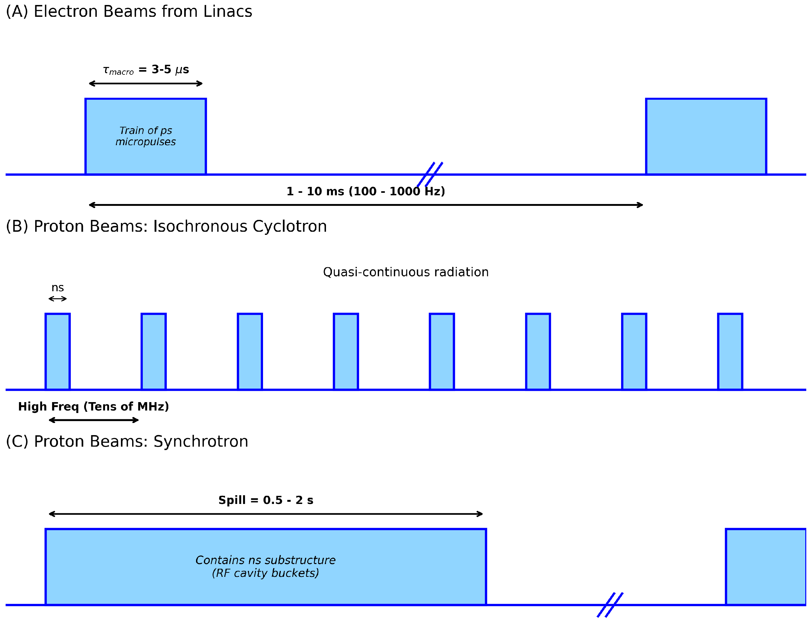
Electron FLASH beams from clinical linacs are delivered in macropulses of a few s at repetition rates of 100–1000 Hz, with instantaneous dose rates above Gy/s. Photon FLASH beams inherit this pulse structure. Beyond the macropulse, linacs also exhibit a radiofrequency (RF) substructure: ∼30 ps bunches separated by ∼350 ps at 2856 MHz. This ns-scale modulation was resolved experimentally using a low-gain avalanche detector. While the picosecond (ps) substructure is real, the subsequent chemical and biological stages—radical diffusion, oxygen depletion, and DNA damage fixation—unfold over nanoseconds to microseconds. Because tissue integrates dose deposition over these longer windows, the fine RF bunching is effectively averaged out. The consensus is that the parameters on the s scale (dose per pulse, pulse width, repetition rate, and mean dose rate) are biologically determinative factors.
Although the nanosecond-scale microstructure of electron and photon beams may not directly change the biological damage they produce, it may impact how detectors behave. Space-charge buildup, ion recombination, and charge-collection dynamics all hinge on the instantaneous current within a pulse. As a result, two FLASH beams with the same mean dose rate may look quite different from a dosimetric standpoint if their temporal structure is not comparable. Therefore, detectors may need not only high saturation limits but also enough bandwidth to resolve the beam on the time scales that matter for energy deposition. As a result, temporal characterization is not a secondary detail—it may be relevant to defining FLASH conditions in a way that can be reproduced across systems and institutions (see
Figure 1).
2.2. Proton Beam Microstructure
Proton delivery for radiotherapy is highly dependent on the type of accelerator used, which dictates the temporal microstructure of the beam. The two primary types of accelerators produce distinct time structures, both of which are relevant for UHDR dosimetry:
Synchrotrons: The beam extraction from a synchrotron occurs in long macro-pulses referred to as spills. These spills typically last between 0.5 and 2 s. Within each long spill, there is a fine temporal substructure, which consists of 10–20 ns bunches separated by approximately 200 ns due to the RF cavity bucket dynamics.
Cyclotrons (specifically, isochronous cyclotrons): These accelerators produce beams that are often described as quasi-continuous. The beam is inherently bunched at the accelerator’s RF frequencies, which are typically in the tens of MHz range, resulting in nanosecond (ns) pulses separated by approximately 20 ns. Resolving this fine temporal structure requires the high-bandwidth capabilities of solid-state detectors (SSDs) like LGADs [
13].
These distinctions in temporal structure are crucial because detector performance can be heavily influenced by the instantaneous current within a pulse, even if the mean dose rate is the same across different beam types. They also highlight the challenge of absolute dosimetry at UHDR, since dose-per-pulse and pulse width vary significantly across accelerator platforms and directly affect detector response.
2.3. Maximum Dose Rate in Counting Mode
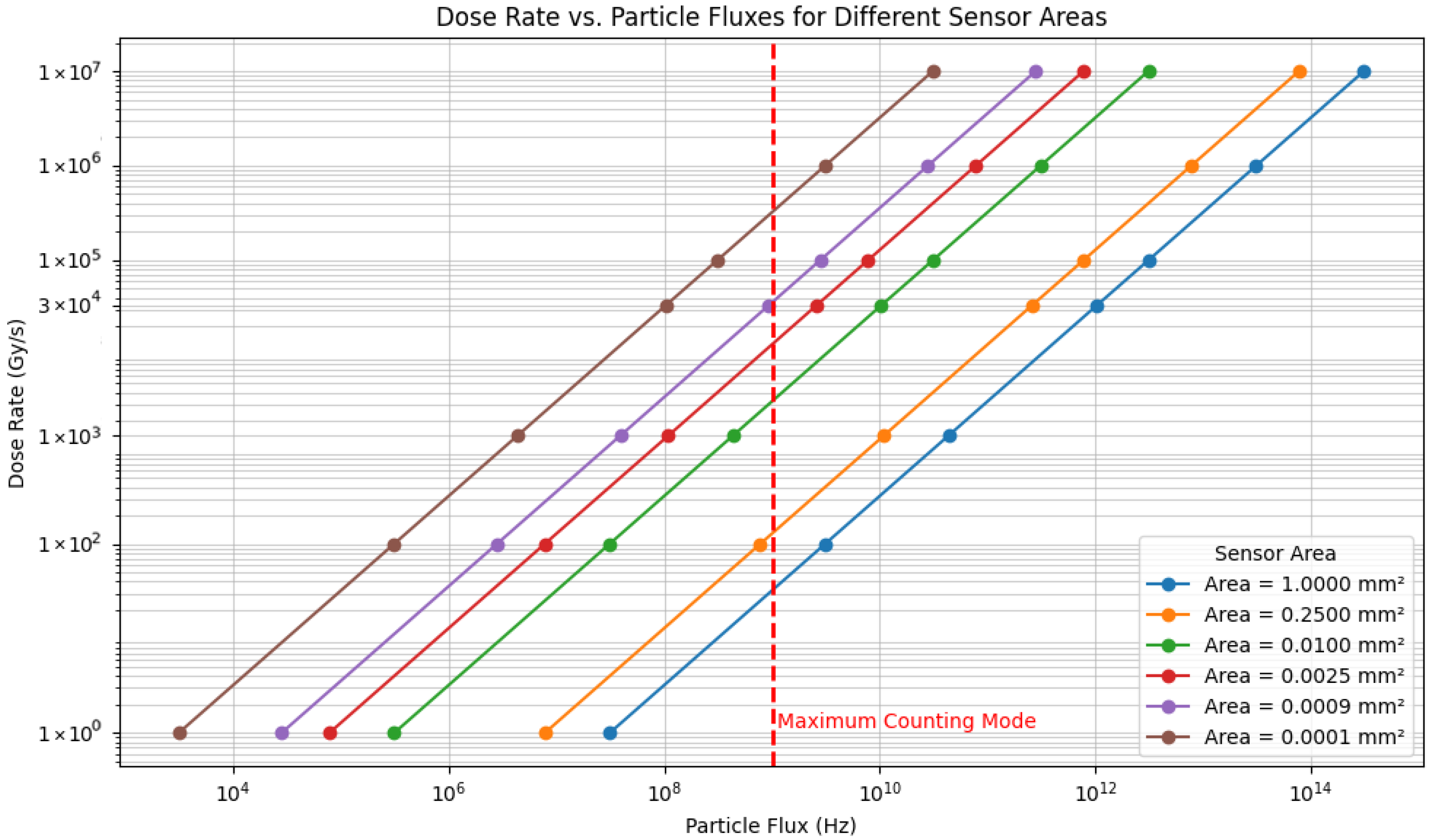
Figure 2 illustrates the relationship between the instantaneous dose rate and the particle flux (particles/ns) traversing the sensor with different transverse areas. The relation is a direct mathematical scaling derived from the fundamental definition of absorbed dose in water and the characteristics of the radiation beam:
The calculation uses a constant energy deposition value corresponding to 9 MeV electrons, treated as Minimum Ionizing Particles (MIPs).
Assuming a highly optimistic, theoretical upper bound for individual particle resolution of 1 particle/ns, the graph allows us to determine the maximum operational dose rate for a counting-mode detector based on its sensor area.
In UHDR dosimetry, this area-dependent scaling is critical when considering the particle counting approach. The ability of a solid-state detector (SSD) to resolve and count individual particles sets the fundamental upper limit on the dose rate measurable in this mode. This limit arises because the detector’s signal decay time and the bandwidth of the readout electronics restrict performance, causing signal merge (pile-up).
A large sensor, such as the 1.0 mm2 detector (relevant for SiC diodes), hits the 1 particle/ns limit at a very low rate of ∼3 Gy/s.
Conversely, the smallest pixel sensor, 10 m × 10 m (0.0001 mm2), can theoretically handle rates up to ∼ Gy/s while still operating within the 1 particle/ns counting limit.
This calculation can be readily scaled to other particle types or energies by substituting their respective energy deposition values. However, the 1 particle/ns limit represents an idealized upper bound. In practice, achievable resolution is constrained by the bandwidth of the front-end electronics, the amplifier slew rate, digitizer sampling frequency, and the intrinsic charge-collection time of the sensor. Real detectors therefore reach the pile-up limit well before the theoretical threshold, and
Figure 2 should be interpreted accordingly.
2.4. Temporal Resolution and the FLASH Debate
A central question is whether the FLASH effect depends primarily on mean dose rate (MDR) or on instantaneous dose rate within pulses. Detector physics studies, however, note that recombination and saturation in ion chambers depend on dose per pulse (DPP) and pulse width, which can be influenced by ns bunching. Mechanistic hypotheses involving radical chemistry and transient oxygen depletion also operate on ns-
s timescales. To avoid ambiguity, studies should at minimum report
s-scale parameters: MDR, DPP, pulse width, repetition rate, and total irradiation time [
9,
14]. Having established the temporal structure and dosimetric constraints of FLASH, we now turn to the detector technologies capable of operating under these conditions.
3. Solid-State Detectors (SSDs) and Their Principles
SSDs are crucial for FLASH dosimetry because their charge collection time is typically on the order of picoseconds to nanoseconds, circumventing the ion recombination issues that plague gas-filled ionization chambers at UHDRs. This section reviews the key SSD technologies being deployed in FLASH research.
3.1. Standard Silicon Diodes vs. Silicon Carbide (SiC)
Standard silicon PIN diodes have long been used in clinical dosimetry but face saturation challenges at the highest UHDRs. The primary comparison in the field is currently between traditional silicon (Si) and emerging silicon carbide (SiC) technologies.
Wide bandgap advantage: SiC has a wider bandgap (3.2 eV vs. 1.12 eV for Si) [
15]. This physical property results in significantly lower leakage currents. Furthermore, SiC exhibits greater radiation hardness, maintaining stability after cumulative doses that would degrade standard silicon performance [
16,
17].
Linearity and saturation: A key limitation of standard silicon diodes at FLASH dose rates is saturation due to the space-charge effect (where high charge density shields the electric field). SiC detectors demonstrate exceptional linearity up to much higher dose-per-pulse (DPP) values (e.g., 11 Gy/pulse) and instantaneous dose rates (up to 4 MGy/s) compared to standard Si diodes [
16].
Thermal stability: SiC offers superior thermal stability, which is advantageous for detectors placed close to high-power FLASH beam exits [
16].
A further advantage of SiC lies in its ability to operate without bias in certain high-flux regimes. Zero-bias operation reduces electronic noise, minimizes leakage current drift, and—importantly for clinical environments—simplifies the integration of the detector into compact dosimetry probes. Several groups have demonstrated that even under unbiased conditions, SiC maintains linearity at UHDR due to its high saturation velocity and reduced charge yield [
16,
18].
In contrast, standard silicon diodes typically benefit from applying reverse bias, which increases the depletion depth and sharpens the current pulse, but this does not prevent saturation at UHDR where space-charge effects dominate. Applying a modest reverse bias can increase the collected charge and slightly speed up carrier drift in SiC, but published UHDR studies report no significant extension of linearity or saturation limits compared with zero-bias operation.
In these studies, absolute dose calibration was performed using alanine dosimeters and prototype diamond detectors as reference standards, ensuring traceability of the SiC response under UHDR conditions.
3.2. Diamond Detectors for FLASH Dosimetry
Diamond detectors have become an important class of solid-state dosimeters for FLASH-RT due to their wide bandgap (5.5 eV), extremely low leakage current, and exceptional radiation hardness. Synthetic single-crystal diamond (SCD) devices exhibit stable operation under high instantaneous dose rates and have demonstrated excellent linearity across the UHDR conditions relevant to FLASH.
Recent studies have shown that diamond dosimeters maintain linear response over a broad range of dose-per-pulse values. Girolami et al. reported highly linear and reproducible behavior in electron FLASH beams, with no evidence of saturation up to the highest tested dose-per-pulse values [
19]. Di Martino et al. similarly demonstrated that diamond detectors preserve linearity and signal stability in UHDR proton beams, confirming their suitability across different particle modalities [
20].
Diamond detectors also exhibit outstanding radiation hardness. Additional studies have confirmed the long-term radiation hardness of single-crystal diamond under high cumulative doses, further supporting its suitability for routine UHDR use [
21]. Studies by Almaviva et al. showed minimal degradation in charge-collection efficiency after exposure to tens of kGy of accumulated dose, far exceeding the tolerance of conventional silicon devices [
22]. This robustness makes diamond particularly attractive for routine use in high-flux clinical or preclinical environments.
The fast charge-collection dynamics of diamond—enabled by high carrier mobility and saturation velocity—provide sub-nanosecond temporal resolution. This allows diamond sensors to resolve the microstructure of UHDR beams, including pulse shapes and instantaneous dose-rate variations. Time-resolved studies by Sato et al. demonstrated that diamond detectors can accurately capture the temporal characteristics of UHDR electron beams [
23,
24].
When compared with SiC, diamond offers several complementary advantages. Both materials exhibit wide bandgaps and strong radiation hardness, but diamond typically provides lower leakage current, higher thermal conductivity, and faster charge-collection times. SiC, on the other hand, benefits from lower fabrication cost, demonstrated zero-bias operation at UHDR, and greater commercial availability. Comparative studies indicate that both materials achieve excellent linearity at FLASH dose rates, with diamond offering superior timing performance, while SiC provides a practical pathway toward robust, clinically deployable dosimeters [
19,
20].
Overall, the combination of linearity, radiation hardness, and fast temporal response positions diamond detectors as a strong candidate for reference dosimetry in FLASH-RT. They complement SiC and other emerging solid-state technologies.
3.3. Low Gain Avalanche Detectors (LGADs)
Low-Gain Avalanche Detectors (LGADs) are thin silicon sensors with a doped gain layer that provides modest internal charge multiplication (typically 10–50) and enables picosecond-scale timing. Although developed for high-energy physics, their use in FLASH requires reduced-bias operation: at UHDR fluxes, the avalanche gain collapses and the readout electronics saturate. For this reason, LGADs are often run at low bias in FLASH settings, functioning as ultrafast thin diodes for temporal beam characterization.
The saturation challenge: In a counting mode at low flux (e.g., <10 Gy/s), the internal gain of an LGAD is highly beneficial for resolving single particles. However, at FLASH dose rates (> Gy/s), the particle flux is too high for counting. The simultaneous arrival of thousands of particles generates a massive charge cloud that can instantaneously saturate the gain layer and the readout electronics.
Operating as a “Fast Diode”: To utilize LGADs in UHDR environments, they can be operated in current-integrating mode, similar to a standard diode. Furthermore, they are often operated at lower bias voltages. Reducing the bias suppresses the avalanche gain mechanism, effectively turning the LGAD into a standard, albeit very thin (50 m), silicon diode.
Advantages in FLASH: Even without the gain, LGADs remain valuable because of their thin active volume and fast charge collection times (tens of picoseconds). This allows them to measure the temporal structure of the beam (e.g., pulse width and shape) with high fidelity, provided the gain is managed to prevent saturation [
13].
Another unique feature of LGADs is their segmentation. Strip or pixelated LGAD architectures can provide spatially resolved fluence measurements with sub-millimeter granularity while preserving nanosecond timing. This capability is particularly relevant for mapping the radial dependence of instantaneous dose rate within a macropulse—a quantity that cannot be inferred from average beam profiles. As FLASH beamlines evolve toward more complex delivery geometries, including scanned electron beams and prototype UHDR photon systems, segmented LGADs may enable novel methods of online beam monitoring and feedback control.
It is important to clarify that LGADs cannot be used in integrating mode while operating in their nominal high-gain regime. The avalanche multiplication process is known to collapse when the electric field in the gain layer is screened by high instantaneous charge density, a well-established feature of LGAD physics [
25,
26]. Under such conditions, the amplified current can exceed the dynamic range of typical front-end electronics. For this reason, integrating-mode operation is only feasible when the bias voltage is intentionally reduced to suppress internal gain, effectively allowing the LGAD to behave as a thin silicon diode.
Furthermore, several studies have shown that the timing and linearity performance of LGADs is often limited by the bandwidth and dynamic range of the readout electronics rather than by the sensor itself. Fast transient currents require GHz-class front-end electronics to avoid pulse distortion or saturation [
27,
28]. Even when the gain is suppressed, the current generated during short, intense pulses can exceed the slew rate or input range of standard preamplifiers, necessitating custom high-bandwidth readout solutions.
Segmented LGAD architectures have already been applied in FLASH-adjacent studies, demonstrating sub-millimeter spatial resolution in high-flux electron beams [
29].
3.4. Miniaturized Pixel Detectors (Timepix)
Pixel detectors such as Timepix are hybrid silicon sensors capable of providing high spatial resolution through finely segmented pixel matrices. Like LGADs, they must adapt to the extreme flux conditions encountered in FLASH-RT.
Counting vs. integrating: Timepix is typically designed for counting mode at low flux. At FLASH dose rates, the pile-up is instantaneous. To be useful, these detectors must utilize Time-over-Threshold (ToT) or integrating modes to handle the high flux [
30].
Application: Their primary value in FLASH lies in high-resolution, two-dimensional mapping of beam profiles and, when counting is possible (in beam tails), for particle track recognition and LET determination [
31].
Because of their fine pixelation (typically m pitch), Timepix detectors are particularly useful for characterizing the lateral structure of UHDR beams, including field uniformity, penumbra sharpness, and small-scale spatial modulations that single-point detectors cannot resolve.
Although Timepix cannot operate in true particle-counting mode at FLASH intensities, the ToT response provides a quasi-integrating signal that remains useful for relative dosimetry and beam diagnostics. This makes Timepix a complementary tool rather than a primary dosimeter. It provides detailed spatial information that can be combined with absolute dose measurements from SiC or diamond detectors.
Newer ASIC generations such as Timepix3 and Timepix4 introduce data-driven readout and nanosecond timestamping, enabling limited temporal information to be extracted even under high flux. However, their dynamic range and front-end electronics still impose constraints at UHDR, reinforcing their role as high-resolution beam monitors rather than standalone FLASH dosimeters.
4. Operational Modes and Electronic Readout
The extreme UHDR environment necessitates a shift from conventional counting-based readout to charge integration. This distinction is critical for all silicon-based sensors, including SiC diodes, LGADs, and pixel detectors.
4.1. High-Flux Integrating Mode
In the high-flux environment of a microsecond-long macropulse (∼ Gy/s instantaneous dose rate), individual particles cannot be resolved.
Principle: The detector operates in an integrating mode. The total charge generated by the pulse is collected and measured.
LGAD specifics: For LGADs, this means the “counting” capability is abandoned. The device is treated as a current source. If the bias voltage is high (nominal gain mode), the current spike can be large enough to damage readout electronics or induce non-linear space-charge effects. Therefore, operating at reduced bias (low gain) is often necessary to maintain linearity.
SiC specifics: SiC diodes are naturally suited for this mode due to their wide bandgap and resistance to saturation effects, as discussed in
Section 3.1.
A key engineering challenge is that these transient currents frequently exceed the slew rate or input range of conventional preamplifiers. To address this, several groups are developing custom front-ends based on current conveyors, fast transimpedance amplifiers, or passive integration networks with GHz bandwidth [
9,
13].
The design of such electronics cannot be decoupled from the detector physics: the biasing scheme, depletion depth, and carrier mobility directly shape the current waveform that the electronics must process.
At UHDR, the dominant limitations often arise not from the sensor but from the readout chain. Large transient currents can exceed the bandwidth or dynamic range of standard front-end electronics, leading to pulse distortion or saturation even when the detector itself remains linear. These constraints motivate the development of specialized acquisition schemes and high-bandwidth electronics, discussed in
Section 4.2 and
Section 4.3.
4.2. Low-Flux Counting Mode
Counting mode remains relevant only for the following aspects:
Beam setup/diagnostics: characterizing the beam at very low currents before switching to FLASH parameters.
Beam tails: measuring scatter or penumbra regions where the flux is sufficiently low (<10 Gy/s).
Single-particle timing: using LGADs in their high-gain mode to characterize the RF structure of the beam, but only under conditions where the total flux is heavily attenuated (e.g., single-electron mode).
Pixel detectors such as Timepix can also operate in counting mode, but only in the extreme low-flux regime. At FLASH intensities, pile-up saturates the discriminator almost immediately, requiring the use of Time-over-Threshold (ToT) or integrating modes instead.
4.3. Synchronous and High-Bandwidth Readout Architectures
Synchronous acquisition schemes have recently been developed to stabilize charge integration during microsecond-scale macropulses and to prevent saturation of the front-end electronics in UHDR environments [
12,
15,
31,
32]. These approaches complement advances in high-bandwidth amplifiers and digitizers, which improve the handling of large transient currents. Moreover, they are essential for preserving linearity at high instantaneous dose rates [
13]. Diamond detectors provide a clear illustration of this interplay: their sub-nanosecond charge-collection time produces intrinsically linear signals even at UHDR, but the front-end electronics can become the dominant source of distortion or saturation in both electron and proton beams.
5. Emerging Concepts and Future Directions
5.1. SSD Radiation Hardness Strategies
Radiation damage to silicon-based detectors is a major concern, as it changes the detector’s operational characteristics (increasing leakage current, changing depletion voltage). Several complementary strategies exist to improve radiation hardness:
Material selection: utilizing wide-bandgap materials like SiC significantly increases intrinsic radiation tolerance compared to Si.
Defect engineering: For silicon devices (like LGADs), strategies include introducing impurities (carbon/oxygen) to trap lattice defects. However, SiC remains the superior candidate for longevity in high-dose clinical environments.
5.2. Emerging Semiconductor Materials for UHDR Dosimetry
Beyond established materials such as SiC and diamond, several emerging semiconductors have been proposed for UHDR dosimetry. Although still at an early stage, their material properties suggest potential advantages for future FLASH applications.
Gallium nitride (GaN) combines a wide bandgap (3.4 eV), high breakdown field, and strong radiation hardness. Prototype GaN Schottky diodes have demonstrated fast charge-collection dynamics and stable response under high-dose-rate irradiation, indicating potential suitability for UHDR environments [
32,
33]. GaN’s commercial maturity in power electronics further supports its prospects as a next-generation detector material [
34].
Metal–halide perovskites represent an even more exploratory direction. Their high mobility–lifetime product and tunable bandgap have enabled ultrafast radiation detectors with sub-nanosecond response times [
35,
36]. Early studies also show tolerance to high instantaneous dose rates, though long-term stability and radiation hardness remain open challenges [
37]. Recent work has also demonstrated improved stability and charge-collection efficiency in perovskite-based detectors, highlighting their potential for future UHDR dosimetry applications [
38].
Overall, GaN and perovskite detectors illustrate the rapid diversification of solid-state materials being explored for FLASH dosimetry. While neither technology is yet mature enough for clinical deployment, their early performance suggests promising directions for future UHDR detector development.
6. Discussion
Taken together, the studies reviewed in this work indicate that no single detector class can meet all FLASH dosimetry needs. Instead, the emerging consensus is that hybrid systems—combining SiC diodes for absolute dosimetry, LGADs for temporal profiling, and pixel detectors for spatial mapping—are required to fully characterize UHDR beams. The major challenges remain standardization, calibration at UHDR, and cross-institutional reproducibility. In this section, we synthesize the performance of each SSD technology and evaluate its clinical translation potential.
The successful clinical translation of FLASH-RT is currently hindered by a “dosimetry gap.” As reviewed, the transition from conventional dose rates to ultra-high dose rates (UHDRs) exceeding Gy/s renders standard ionization chambers unreliable. The literature identifies solid-state detectors (SSDs) as the foundational technology to bridge this gap, but the choice of detector material and operating mode is critical.
6.1. Silicon (Si) vs. Silicon Carbide (SiC)
The most significant comparison for FLASH dosimetry is between standard silicon and silicon carbide. The data reviewed consistently positions SiC as the more robust candidate for absolute dosimetry. Its wide bandgap (3.2 eV) [
15] confers superior radiation hardness, maintaining stability after tens of kGy of accumulated dose [
16]. More importantly, SiC demonstrates superior linearity at UHDR, resisting saturation up to doses (21 Gy/pulse) where standard silicon diodes fail [
16,
18]. In addition to material selection, mechanical robustness and packaging are increasingly recognized as critical factors for UHDR detectors. Encapsulated SiC devices, for instance, demonstrate superior thermal and mechanical stability compared to bare die structures. That enables operation near beam exits or in environments with significant secondary radiation. Additionally, integrating detectors into fiber-coupled or water-equivalent housings may facilitate deployment in clinical settings.
6.2. Diamond Detectors
Diamond detectors now represent one of the strongest candidates for reference dosimetry in FLASH. Their wide bandgap (5.5 eV), extremely low leakage current, and exceptional radiation hardness allow for stable operation under high instantaneous dose rates. Experimental studies have demonstrated linearity in both electron and proton UHDR beams, with minimal degradation after tens of kGy of accumulated dose. Diamond also offers sub-nanosecond charge-collection times, enabling precise reconstruction of pulse shapes and instantaneous dose rate. Compared with SiC, diamond provides superior timing performance, while SiC offers advantages in cost, scalability, and zero-bias operation. Together, these materials form a complementary foundation for future UHDR dosimetry standards.
6.3. LGADs and Temporal Profiling
LGADs provide unmatched temporal resolution among SSDs, with intrinsic rise times in the tens of picoseconds. This makes them uniquely suited for resolving the microstructure of FLASH beams, including pulse width, substructure, and shot-to-shot stability. However, their internal gain mechanism saturates at high instantaneous charge densities, requiring operation at reduced bias (low-gain mode) for integrating-mode dosimetry. LGADs therefore serve primarily as beam monitors rather than absolute dosimeters in UHDR conditions.
6.4. Pixel Detectors and Spatial Mapping
Timepix and related hybrid pixel detectors offer high spatial resolution for mapping beam profiles, penumbra structure, and field uniformity. Their Time-over-Threshold (ToT) mode provides a quasi-integrating response that remains useful for relative dosimetry. However, front-end saturation prevents their use as primary dosimeters at FLASH intensities. Instead, they complement SiC and diamond detectors by providing detailed spatial information that single-point detectors cannot capture.
6.5. Electronics as a Limiting Factor
Across all detector classes, the readout electronics—not the sensor material—often determine the upper limit of performance at UHDR. The sub-microsecond duration of FLASH pulses produces current transients that can exceed the bandwidth, dynamic range, or slew rate of conventional front-end circuits. High-bandwidth transimpedance amplifiers, current conveyors, and synchronous digitizers (hundreds of MS/s to several GS/s) are increasingly required to preserve signal fidelity. This electronic bottleneck is now recognized as a central challenge in UHDR dosimetry and a key target for future standardization efforts.
6.6. Emerging Materials
Beyond SiC and diamond, emerging materials such as GaN and metal–halide perovskites have shown early promise for ultrafast radiation detection. GaN offers strong radiation hardness and fast charge collection, while perovskites provide high mobility–lifetime products and sub-nanosecond response. While both technologies are currently pre-clinical, their rapid evolution indicates that UHDR detection will soon move beyond conventional silicon and carbon-based materials.
6.7. Toward Integrated, Multi-Detector Systems
The combined evidence indicates that FLASH dosimetry will likely rely on multi-detector systems rather than a single universal device. SiC or diamond detectors provide absolute dose-per-pulse measurements; LGADs characterize temporal structure; pixel detectors map spatial distributions; and high-bandwidth electronics synchronize these measurements with accelerator output. Such integrated systems will be essential for establishing reproducible, cross-institutional UHDR dosimetry standards.
7. Conclusions
Solid-state detectors have reached a level of maturity that makes them indispensable for characterizing the extreme beam conditions required for FLASH-RT. The primary barriers to clinical translation no longer stem from the intrinsic behavior of the sensors. SiC and diamond show excellent linearity and radiation hardness, and LGADs and pixel detectors offer exceptional temporal and spatial resolution. However, the limiting factors now lie in the supporting metrological and electronic infrastructure.
Reliable UHDR dosimetry will require standardized dose rate definitions, cross-platform calibration strategies, and high-bandwidth front-end electronics capable of handling the large transient currents generated during microsecond-scale macropulses. Recent advances in synchronous acquisition and fast readout architectures represent important steps toward this goal. Nevertheless, further progress will require electronics that are explicitly designed to handle the large transient currents and bandwidth demands of UHDR beams. Additionally, shared performance benchmarks that allow different groups and accelerator platforms to compare results reliably will be required.