Article

Early Growth and Developmental Characteristics of Chinese Bahaba (Bahaba taipingensis)

1
Guangdong Beluga Whale Marine Biotechnology Co., Ltd., Huizhou 516300, China
2
Key Laboratory of East China Sea Fishery Resources Exploitation, Ministry of Agriculture and Rural Affairs, East China Sea Fisheries Research Institute, Chinese Academy of Fishery Sciences, Shanghai 200090, China
3
School of Life Science, South China Normal University, Guangzhou 510631, China
*
Authors to whom correspondence should be addressed.
These authors have contributed equally to this work.
Fishes 2024, 9(8), 329; https://doi.org/10.3390/fishes9080329
Submission received: 29 July 2024 / Revised: 18 August 2024 / Accepted: 19 August 2024 / Published: 21 August 2024
(This article belongs to the Special Issue Reproductive Biology and Breeding of Fish)

Abstract

:
The Chinese bahaba (Bahaba taipingensis), belonging to the Sciaenidae family, is one of the largest croakers with a limited geographical distribution. It is a critically endangered fish species according to the IUCN and a protected animal in China. In this study, the morphological characteristics of Chinese bahaba were observed and analyzed across different developmental stages, namely, the embryonic, larval, juvenile, and young fish stages. The results demonstrated that the mature eggs had a terminal yolk and a single oil globule. The eggs remained floating, and the mean diameters of the fertilized egg and oil globules were 1.14 ± 0.09 mm and 0.35 ± 0.07 mm, respectively. The findings revealed that the embryonic development of Chinese bahaba occurs broadly in seven stages, including the blastogenesis, cleavage, blastocyst, gastrula, neuro embryonic, organ differentiation, and membrane emergence stages, which lasted approximately 27 h and 10 min until hatching under 22.5 ± 0.5 °C. After 70 d, the larvae developed into young fish with a mean total length and body length of 97.75 ± 12.61 and 75.27 ± 13.27 mm, respectively. The digestive organs and the swim bladder began to differentiate, and the swim bladder, bladder duct, intestine, stomach, and mouth gradually formed at 2 d after hatching. Juvenile development occurred via six stages and there were certain differences in the morphological characteristics of Chinese bahaba across the different stages of growth and development. This study provides a theoretical reference for studying the growth, development, and artificial breeding of Chinese bahaba.
Key Contribution: This study observed and analyzed the growth and development characteristics of Chinese bahaba (Bahaba taipingensis) with many differences in the morphological characteristics during the period of embryonic, larval, juvenile, and young fish in Chinese bahaba.

1. Introduction

The Chinese bahaba (Bahaba taipingensis), belonging to the Bahaba genus under the Sciaenidae family of order Perciformes, is a rare species of fish that is unique to China. It is mainly distributed in the estuary of the Pearl River in Guangdong Province, in the Zhoushan Sea area located in Hangzhou Bay in Zhejiang Province, in the sea area of Quanzhou Bay at the estuary of the Minjiang River in Fujian Province, and in the estuarine regions of other rivers [1,2]. However, its numbers have reduced sharply in recent years owing to the deterioration of the water environment and illegal overfishing, and the Chinese bahaba is currently on the verge of extinction. Until now, it was only occasionally found in “Dragon’s Nest Island–Shajiao–Dahu Island” in the waters of Dongguan in the estuary of the Pearl River [1,2,3]. The Chinese bahaba was enlisted as a critically endangered species in the Red List of endangered species by the International Union for Conservation of Nature (IUCN) in 2006 and was listed as a national first-class key protected aquatic wildlife in 2021 [1,3]. These reports indicate that it is urgently necessary to improve the resource protection of Chinese bahaba and preserve this endangered species. Research studies on the artificial breeding of Chinese bahaba can provide important insights and guidelines for realizing the proliferation and release of Chinese bahaba, resource rescue, and protection of the marine ecological environment [4,5,6].
Artificial breeding serves as an important technical link in the expansion of rare species resources and the sustainable development of the breeding industry. In particular, the development of embryos, larvae (from the hatching to the formation of fin and the development of each motor organ), juveniles (from the period of complete motor organ development to the period of scales formed), and young fish (all the scales are formed, and the body color and habitats are the same as those of the adult fish) are key links in the process of artificial breeding. Few studies have reported the artificial breeding of large fishes in the waters of China, and successful artificial breeding efforts have mostly concentrated on breeding small fishes in offshore seas [4,5,6]. This is primarily attributed to the lack of an established breeding technology and adequate equipment for the artificial breeding of large fishes. Additionally, research studies on the breeding of large fishes in the Totoaba genus (such as Chinese bahaba) are primarily limited by the severe shortage of germplasm resources. Presently, few studies are investigating the breeding of Chinese bahaba, and only some preliminary experimental studies have been conducted on the natural environment of Totoaba macdonaldi in Mexico [7,8,9]. Therefore, research studies on the artificial breeding of Chinese bahaba can provide important insights and guidelines for the protection of its germplasm resources, the restoration of ecological resources, and the rational development and utilization of resources.

2. Materials and Methods

In the present study, for their rescue and protection, Chinese bahaba were obtained from Guangdong Beluga Whale Marine Biotechnology Co. Ltd. (Huizhou, China) with 8 months of protective cultivation. The eggs of Chinese bahaba were obtained via gonad enhancement cultivation in 2021. A batch of high-quality fertilized eggs was selected for cultivation, and the results obtained herein can serve as a reference for the restoring this rare and protected aquatic species. The experiments were conducted at Guangdong Beluga Whale Marine Biotechnology Co. Ltd. The spawners Chinese bahaba used for artificial reproduction were wild juveniles rescued erroneously from the estuary of the Pearl River or sexually mature individuals cultured in captivity. A total of three parental groups, including three females and six males (body weight, 22.0–38.3 kg; body length, 85.1–90.3 cm; body width, 20.3–25.1 cm), were selected in this study. All the Chinese bahaba spawners were reared at water salinity of 12 ppt, temperature of 4 deg C, pH of 7.6–8.0, 0.2 m/s of water flow velocity, and 7 mg/L of dissolved oxygen content. The breeding protocol was based on a patent of an artificial breeding method of Chinese bahaba in China (authorization announcement number: CN114651751B).

2.1. Conditions for Embryonic Development

The embryonic development of Chinese bahaba was divided into 24 periods according to the changes in the morphological characteristics at each stage of embryonic development. The collected fertilized eggs were incubated in 300 L conical incubation barrel under continuous aeration, and 50% of the water was changed every 6 h. The temperature of the water was adjusted to 22.5 ± 0.5 °C, with a salinity of 10 ± 1 ppt and pH of 7.6–7.8.

2.2. Conditions for Cultivation of Larvae and Juvenile Fishes

After hatching, the larvae were cultivated in a 30 m3 circular tank fitted with a closed recirculating water system. The seawater was disinfected under a high-intensity UV light and fed into the recirculating water system. The salinity of the water used for cultivation was 10 ± 1 ppt, the pH was 7.6–7.8, the temperature was 25 °C, the dissolved oxygen was ≥ 6 mg/L, and the intensity of incident light was 1500–2000 lx. The larvae were cultivated in static water in the early stage (7 d); the micro-circulation of the circulating water system was switched on from 8 d and the circulation was gradually increased on a daily basis with the development of the larvae and juvenile fishes. The amount of circulating water was increased from 20%/d to 300%/d, and the amount of water in the circulation was 500%/d or higher at the juvenile stage. After 10 d of cultivation, the circular tank was cleaned every morning using a suction system to remove the residual bait and fecal matter excreted by the larvae and juvenile fishes. The larvae and juvenile fishes were fed with Brachionus plicatilis, the brine shrimp (Artemia sp.), and dietary supplements (commercial feed) specially designed for the cultivation of marine young fish by Hayashikane Sangyo Co., Ltd. (Shimonoseki, Japan).

2.3. Observation and Statistics

Samples were collected multiple times and at different intervals during incubation, and more than 30 samples were collected at each time point. The onset of a new stage of egg development was indicated by the appearance of new characteristics in 50% of the eggs. The larvae collected at each developmental stage was fixed and preserved in 5% formaldehyde for further examination. For sample collection, the fishes were anesthetized with the MS-222 anesthetic (40 mg/L), and samples were collected on a daily basis until the larvae were 15 d old, following which sample collection was restricted to once every 2 d. The samples were collected at 5–7 d intervals during the juvenile stage and once every 10–15 d during the juvenile stage, and more than 10 larvae were sampled at each time point. The samples were similarly fixed in 5% formaldehyde for subsequent verification. The larval morphology was observed using a Leica S9I microscope, and the developmental sequence and characteristics of the embryos were described and recorded. For histological analysis, the larvae at days 1–7 were fixed in Bouin’s solution for 24 h at 4 °C and transferred to 70% ethanol. Subsequently, all the fixed samples were dehydrated in gradient ethanol concentration (70–100%), cleared in xylol, embedded in paraffin wax, and cut into 5 μm sections before staining with hematoxylin–eosin. All the sections were viewed under Panoramic DESK, P-MIDI, and P250 (Budapest, Hungary). The juveniles were photographed using an Olympus DP71 digital microscope or a Canon 50D digital camera. The data collected from the embryos and juveniles were analyzed using the Image-Pro express 5.1 software and presented as the mean ± standard deviation. The formulae used for calculating the volumes of the yolk sac and oil sphere are provided hereafter [10,11,12].
Volume of yolk sac (Ve) = 1/6лLH2 − 1/6лd3 or Ve = 1/6лLH2 − 1/6лl h2
Volume of oil sphere (Vo) = 1/6лd3 or Vo = 1/6лl h2
where L and H represent the long and short diameters of the yolk sac in mm, respectively; d represents the diameter of the oil globule (mm); and l and h represent the long and short diameters of the oil globule in mm, respectively.
The larvae and juveniles of Chinese bahaba were delineated based on previous studies [10,11,12].

3. Results

3.1. Embryonic Development

Chinese bahaba has typical telolecithal eggs: spherical, colorless, transparent, and containing an oil globule after fertilization. The average diameters of the eggs and oil globules were 1.14 mm and 0.35 mm, respectively. The embryos of Chinese bahaba underwent complete development within 27 h and 10 min at a water temperature of 22 ± 0.5 °C, and the eggs underwent disciform cleavage.

3.2. Blastoderm Formation

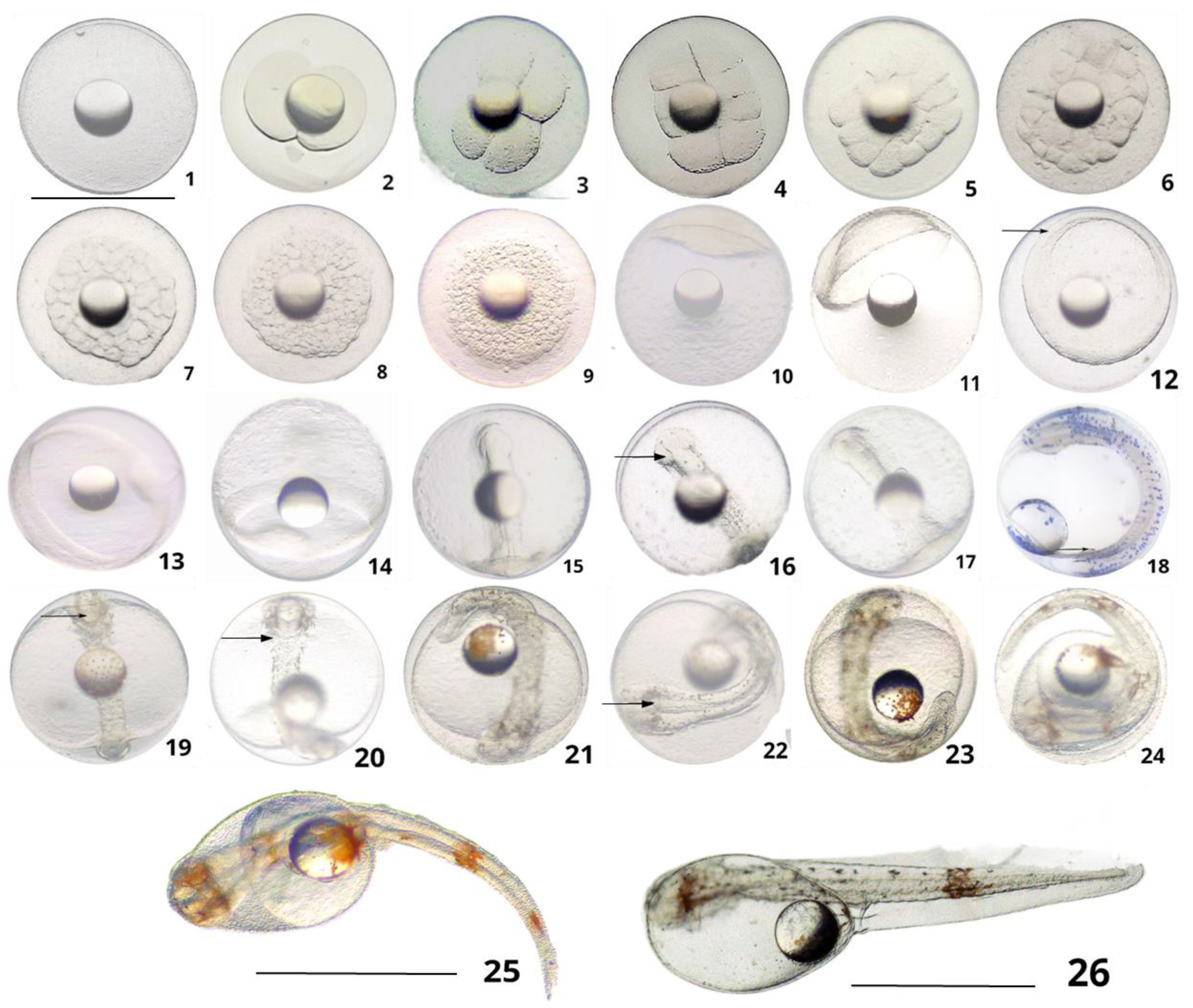
The fertilized eggs absorbed water and swelled up, which was followed by the gradual expansion of the periplasm. The placenta rose after ten minutes post-fertilization, and the protoplasm shifted and concentrated towards the animal pole of the egg to gradually form a raised embryonic disc on the surface of the yolk (Figure 1(1)).

3.3. Cleavage

Two-cell stage: The area of the embryonic disc underwent gradual expansion, and a longitudinal cleavage furrow began to form at the center of the upper region of the embryonic disc after approximately 15 min post-fertilization and extended to both sides to split the cell longitudinally into two cells of equal size (Figure 1(2)).
Four-cell stage: The second longitudinal division occurred after approximately 30 min post-fertilization and was marked by the appearance of a cleavage furrow at the center of the upper region of the two cells. The second furrow intersected the first cleavage furrow at right angles and divided the two cells into four cells (Figure 1(3)).
Eight-cell stage: The third longitudinal division occurred after approximately 45 min post-fertilization. Briefly, a third concave furrow, parallel to and perpendicular to the first cleavage plane, appeared on each side of the first cleavage plane to give rise to two rows of four cells each with different morphologies and sizes (Figure 1(4)).
Sixteen-cell stage: The fourth division occurred after approximately 1 h and 5 min post-fertilization and appeared as a concave furrow that was perpendicular to the first and third division surfaces but parallel to the second-division furrow. This fourth cleavage furrow divided the cell mass longitudinally to give rise to 16 cells of different sizes (Figure 1(5)).
Thirty-two-cell stage: The fifth division occurred after approximately 1 h and 15 min post-fertilization and gave rise to 32 irregularly arranged cells by meristematic cleavage (Figure 1(6)).
Sixty-four-cell stage: The sixth division occurred after approximately 1 h and 25 min post-fertilization and resulted in the generation of 64 cells of similar size (Figure 1(7)).
Multicellular stage: This stage appeared after approximately 1 h and 50 min post-fertilization and was characterized by inconsistencies in cell size, a significant reduction in cell size compared to that in the previous stage, and irregular cellular arrangement (Figure 1(8)).
Morula stage: This stage appeared after approximately 2 h and 40 min post-fertilization. The morula was characterized by indistinct cell division boundaries and assumed a bulging mulberry-like shape (Figure 1(9)).

3.4. Blastocyst

High blastula stage: The high blastula stage appeared after 3 h and 20 min post-fertilization. In this stage, the cells divided to generate smaller cells that accumulated on the embryonic disc to form a cap-like structure protruding from the yolk, which represented the high blastula stage (Figure 1(10)).
Low blastula stage: This stage appeared after 4 h and 20 min post-fertilization and was characterized by the gradual reduction and flattening of the central bulge of the embryonic disc. Additionally, the surrounding layer of cells began to envelop and eventually cover the yolk to form a flat, low-cap-like shape to give rise to the low blastula stage (Figure 1(11)).

3.5. Gastrula

The main morphological characteristics of the gastrula stage are that the embryonic disc moves continually towards the vegetative pole and engages inwardly to form the embryonic ring. The embryonic shield forms on one side of the embryonic ring, which then serves as the prototype of the embryonic body.
Early gastrula stage: This stage appeared after 4 h and 45 min post-fertilization and was characterized by the enlargement and invagination of the cells at the edge of the embryonic disc to form a ring-like cell layer that enveloped the vegetal pole from all sides to form the embryonic ring (Figure 1(12)).
Mid-gastrula stage: This stage appeared after approximately 7 h and 5 min post-fertilization. The mid-gastrula stage was characterized by the expansion of the embryonic ring which began to underlie the yolk by a third and continued invaginating to form the embryonic shield rudiment (Figure 1(13)).
Telophase of gastrula stage: This stage appeared after approximately 8 h and 40 min post-fertilization. In this stage, the embryonic disc inferiorly enveloped the yolk by half, and the stage was characterized by the formation of the neural plate, the continued forward extension of the embryonic shield, and the emergence of the embryonic body (Figure 1(14)).

3.6. Neuroembryo Stage

This stage began with the thickening of the embryonic body to form the neural plate and then the neural tube. This was followed by the emergence of the eye sac in the head, the formation of the protoportal from the subgerminal envelope, and finally, the complete closure of the blastopore.
Neural embryo stage: This stage appeared after approximately 9 h and 25 min post-fertilization and was characterized by the continued lengthening of the embryonic body and the thickening of the center of the embryonic body. Additionally, the center of the embryonic body appeared tubular under the microscope, indicating the formation of the neural tube (Figure 1(15)).
Formation of the optic vesicle: This stage was observed after approximately 9 h and 50 min post-fertilization and was characterized by the appearance of garden-shaped protuberances on both sides of the head, indicating the appearance of 4–5 pairs of myotomes on the embryonic body (Figure 1(16)).
Formation of the yolk plug: This stage was observed after approximately 10 h and 40 min post-fertilization. In this stage, the embryonic body enveloped the yolk inferiorly by approximately 5/6th, leaving only the original mouth area. The structure resembled an embolus, and the muscle joint pairs increased to 8–9 (Figure 1(17)).
Blastopore closure: This stage appeared after approximately 13 h and 50 min post-fertilization. The blastopore is a completely closed structure, and the muscle segments increased to 9–10 pairs at the blastopore stage. A bright vesicular structure, namely, Kupffer’s vesicle, appeared on the inner side of the tail, and the movement of the protocorm ended at this stage (Figure 1(18)).

3.7. Organ Differentiation

Formation of the olfactory vesicle: This stage appeared after 16 h and 5 min post-fertilization and was characterized by the appearance of a round structure at the tip of the head, which corresponded to the emergence of the olfactory sac (Figure 1(19)).
Formation of the auditory capsule: This stage appeared after 18 h and 35 min post-fertilization and was characterized by the appearance of a pair of rounded structures on the posterior side of the otic vesicle, which corresponded to the formation of the auditory capsule (Figure 1(20)).
Formation of caudal bud: This stage appeared after 21 h and 5 min post-fertilization and was characterized by the elongation of the embryonic body, which encompassed 3/5th of the circumference of the yolk sac. The posterior end of the embryonic body gradually bulged into a caudal bud containing 16–18 pairs of muscular segments, which was accompanied by Kupffer’s vesicle (Figure 1(21)).
Initiation of heartbeat: This stage occurred after 22 h and 45 min post-fertilization and was characterized by the appearance of the heartbeat in the embryo, with a frequency of approximately 80–100 beats/min. A fluid was seen flowing in the embryo under microscopic examination, and crystals appeared in the eye sac (Figure 1(22)).
Initiation of muscular contraction: This stage was observed after 24 h and 50 min post-fertilization. In this stage, the embryonic body began showing signs of muscular contraction and the presence of clear crystals, and the tail began to twist from side to side (Figure 1(23)).

3.8. Membranous Stage

Pre-hatching stage: This stage appeared after 26 h and 45 min post-fertilization. In this stage, the embryonic body was violently twisted, and the egg membrane started thinning gradually and was less elastic (Figure 1(24)).
Hatching stage: This occurred after 27 h and 5 min post-fertilization, and the majority of embryos emerged headfirst while some individuals emerged caudally first (Figure 1(25)).
First hatchling: After emerging from the membrane, the larvae hung upside down in the water with their heads upwards. The mean length of the larvae, volume of yolk sac, and the volume of the oil globule were 2.65 ± 0.32 mm, 0.42 mm3, and 0.08 mm3, respectively (Figure 1(26)).

3.9. Developmental and Growth Characteristics of Larvae and Juveniles

3.9.1. Yolk-Sac Larval Stage (1–7 DAH)

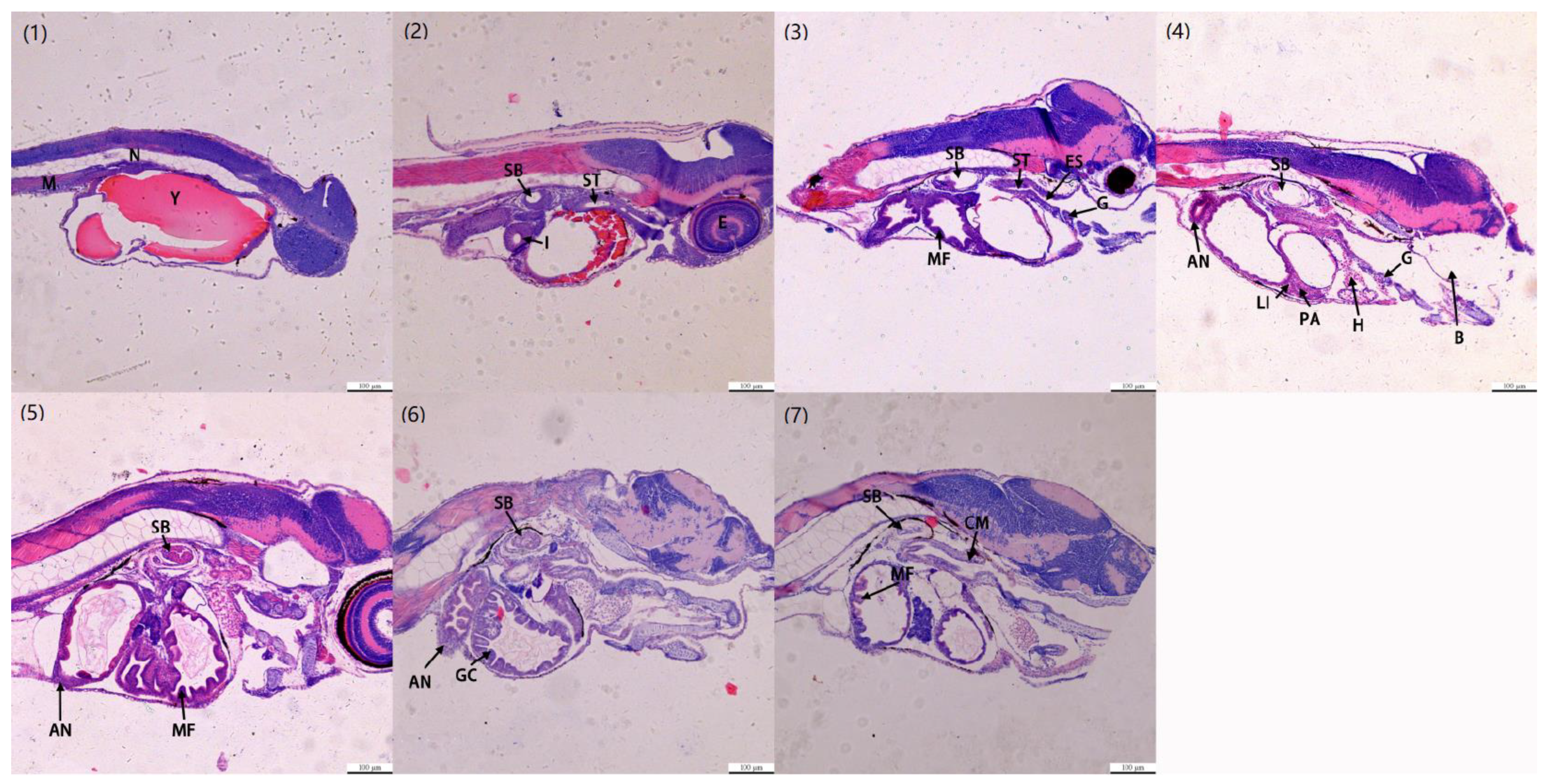
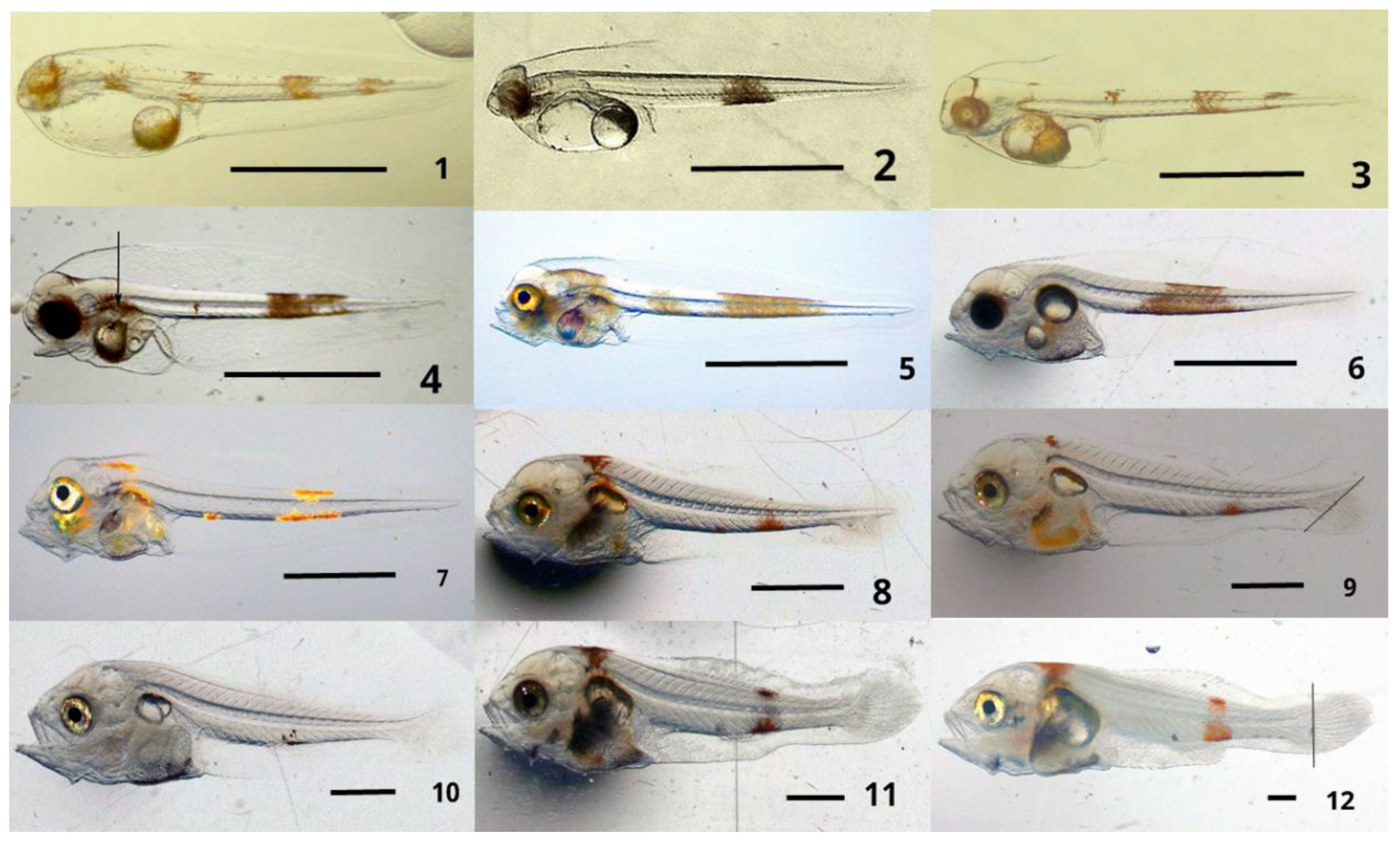
The total length, volume of yolk sac, and volume of oil globule of the newly hatched larvae at 1 DAH were 2.65 mm, 0.23 mm3, and 0.07 mm3, respectively. At 1 DAH, the carapace remained adhered to the yolk sac, and the rounded oil globule was situated in the posterior part of oval yolk sac. The larvae remained in the upper layer of the water in an inverted position most of the time with occasional intermittent scuttling, and the internal blood was firstly observed in the larvae (Figure 2(1)). The results of H.E staining showed the presence of homogeneous eosinophilic yolk sacs in the abdominal cavity, undifferentiated digestive tract, and closed mouth and anus (Figure 3(1)).
The total length, volume of yolk sac, and volume of the oil globule of the newly hatched larvae at 2 DAH were 2.84 mm, 0.09 mm3, and 0.07 mm3, respectively. The yolk sac appeared smaller, but the volume of the oil globule remained largely unaltered. The rudimentary gill arch was visible behind and below the eye, the pectoral fin protoconch was also visible, and internal blood flow was evident in the larvae at 2 DAH (Figure 2(2)). The results of H.E staining showed the reduced eosinophilic yolk sac, the presence of air glands and the swim bladder, the differentiation of the digestive tract, the presence of some undifferentiated cord cells (original hepatopancreas), and the closure of the mouth and anus (Figure 3(2)).
The total length, volume of the yolk sac, and volume of the oil globule of the newly hatched larvae at 3 DAH were 2.77 mm, 0.02 mm3, and 0.06 mm3, respectively. At this stage, the size of the yolk sac continued to decrease, the digestive tract extended backward and began taking shape, and the pectoral fins began growing membranous folds. Additionally, the accumulation of melanin was visible in the eyes, which were insensitive to light, and the flow of reddish blood was evident in the heart (Figure 2(3)). The results of H.E staining showed that yolk was heavily absorbed; the length of 65.55 ± 3.16 μm and 36.24 ± 2.75 μm of the long axis and wide axis of the swim bladder, oropharyngeal cavity, esophagus, and stomach appeared; folds of esophageal mucosa appeared; gill cartilage and primitive gill filaments appeared; and the mouth and anus were closed (Figure 3(3)).
The total length, volume of the yolk sac, and volume of the oil globule of the newly hatched larvae at 4 DAH were 2.74 mm, 0.01 mm3, and 0.03 mm3, respectively. The yolk sac became smaller and oval-shaped, and the volume of the oil globule decreased. Additionally, the protoconid of the swim bladder appeared behind the lateral view of the auditory bladder, which continued to show growth and development. The mouth formed at 4 DAH, and the larvae could occasionally move the lower jaws, but the anus remained closed. The larvae were unable to ingest food and primarily used the yolk sac as the nutritional material (Figure 2(4)). The results of H.E staining showed that the yolk sac was almost completely absorbed; the long axis and wide axis of the swim bladder were 89.47 ± 4.26 μm and 57.24 ± 3.61 μm, respectively; the cord cells were further differentiated into hepatocytes and pancreatic cells; blood cells were visible; the upper branchial vessels and branchial lobules were formed; the mouth was formed; and the anus was closed (Figure 3(4)).
The total length and volume of the oil globule of the newly hatched larvae at 5 DAH were 2.97 mm and 0.01 mm3, respectively. The yolk sac was completely depleted at this stage and no longer visible, and the volume of the oil globule reduced to 1/5th of that at the larval stage. The pigmentation on the posterior half of the body formed a single patch, and the pectoral fins nearly reached 1/3rd of the total length and width of the body. The anus opened at this stage, and the remains of the ingested rotifers were visible in the digestive tract. Although the larvae could ingest food at 4 DAH, feeding behavior was not observed in any of the active individuals in the nursery pool, and active robbing behavior was not observed either (Figure 2(5)). The results of H.E staining showed that the long axis and wide axis of the swim bladder were 119.26 ± 5.78 μm and 83.15 ± 7.53 μm, respectively, and the folds of the digestive tract were more obvious. It could be observed that the intestine was composed of single columnar cells, the nucleus was located at the base, and the anus was opened (Figure 3(5)).
The total length of the larvae at 6 DAH was 3.31 mm. The volume of the oil globule reduced by 2/3rd compared to that on the previous day, and only traces of the oil globule were visible at this stage. Peristalsis was obvious in the digestive tract, which was completely filled with ingested food. The swim bladder was inflated and appeared subglobular in shape. The swim bladder appeared crystalline under a dissecting microscope. An otolithic structure was observed at the site of the auditory capsule that shifted upward to the posterior superiority of the eye in the lateral view. The eye was developing rapidly at this stage and was sensitive to reflected light. The individuals in the brood pool were able to actively and aggressively search for and snatch food, and the spinal cord ended in a straight line (Figure 2(6)). The results of H.E staining showed that the swim bladder developed continuously, the bladder duct thickened, and the lumen expanded continuously; the long axis and wide axis were 131.26 ± 7.48 μm and 91.95 ± 3.03 μm, respectively; and goblet cells appeared in the digestive tract cells (Figure 3(6)).
At 7 DAH, the larvae attained a total length of 3.88 mm and represented a stage between the yolk sac fish and pre-flexed larvae. Only traces of the oil globule were visible at this stage. The gill morphology was obvious, and red gill filaments could be seen under the dissecting microscope. The volume of inflation of the swim bladder varied according to the stress produced by fishing. At this stage, traces of gill rakers were observed on the gill arches, the liver already covered the first half of the digestive tract, and the frequency of intestinal peristalsis was higher. The eyes were well-developed at this stage, and the larvae could swim continually when chasing food (Figure 2(7)). The results of H.E staining showed that the long axis and wide axis of the swim bladder were 135.71 ± 8.24 μm and 92.47 ± 8.19 μm, respectively; the outer layer of gastric mucosa showed loose connective tissue and circumocular layer; and the digestive tract showed obvious mucosal folds (Figure 3(7)).

3.9.2. Pre-Flexing Larval Stage (8–10 DAH)

The total length of the larvae was 4.58 ± 0.51 mm at 10 DAH. The hypocaudal bone was visible below the end of the spine, and the spine tended to be upturned at the end of the tail. The base of the anal fins started appearing at 10 DAH, and the pigmentation on the posterior half of the body was markedly reduced. The pigmentation on the dorsal side disappeared completely, and only the abdominal pigmentation remained in the shape of a dendritic tree (Figure 2(8)).

3.9.3. Flexing Larval Stage (11–19 DAH)

The total length of the larvae was 5.75 ± 0.85 mm at 13 DAH. The pectoral fins tended to be well-developed at this stage and served as the main organs of locomotion and balance. The hypocaudal bone below the end of the spine continued to lengthen, and the end of the spine continued to upturn. The anal fin buds protruded from the protozoal base, and the dendritic pigmentation of the abdomen in the posterior half of the body reduced at 13 DAH. The digestive tract was filled with ingested food, the young fish began to congregate, and positive-phototaxis was evident in the broodstock ponds (Figure 2(9)).
The total length of the larvae was 6.35 ± 1.01 mm at 16 DAH. The end of the spine upturned to an angle of 45 ° with respect to the long axis of the body. Two bends appeared in the digestive tract, and the pigmentation in the posterior half of the abdomen narrowed to a stellate pattern. The young fishes were clustered in groups and began cruising and were also seen grabbing for food (Figure 2(10)).

3.9.4. Post-Flexing Larval Stage (20–29 DAH)

The total length of the larvae was 7.93 ± 0.97 mm at 20 DAH. At this stage, the base of the dorsal fin differentiated into fin spines and filiform fin rays. The fin rays of the anal fin underwent extension, and the unturned end of the spine tended to remain perpendicular to the long axis of the body. The membranes of the upper and lower fins began to constrict inwardly at the base of caudal peduncle, and the caudal fin membrane started assuming a rounded shape. Brown pigmentation appeared in the form of pine needles at the upper and lower ends of the body, posterior to the anal fin (Figure 2(11)).
The total length of the larvae was 12.75 ± 2.19 mm at 25 DAH. At this stage, the swim bladder assumed an oval shape and its inflation varied with stress. The upward curvature of the spine was complete, and the end of the spine remained perpendicular to the long axis of the body. The change in caudal flexion was complete, and the fin membranes were distinctly differentiated at this stage. The caudal fin membranes continued to assume a rounded shape, and the dorsal and anal fin membranes also differentiated in rudimentary form (Figure 2(12)).

3.9.5. Larvae (30–55 DAH)

The development of the fins was completed at the juvenile stage; additionally, the shape of the body tended to be similar to that of adult fishes, and the scale coverage was complete. This stage of rapid growth corresponded to the phase of greatest changes in external morphology and also represented an important period for the growth and development of internal organs.
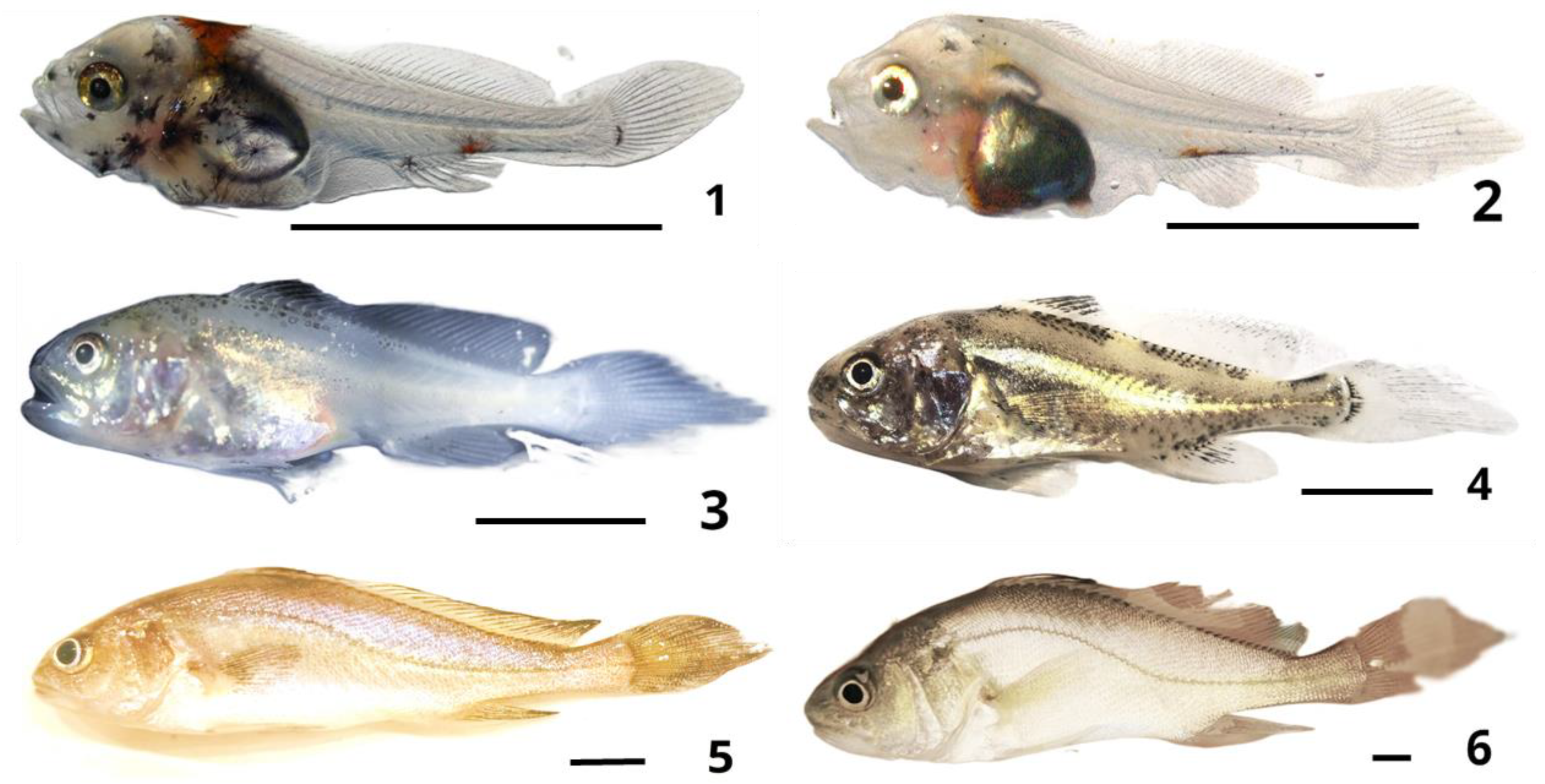
The total length and body length of the 30 DAH juvenile fishes were 18.13 ± 4.61 mm and 14.67 ± 3.87 mm, respectively. The differentiation of the two anterior and posterior dorsal fins was complete; the position of the first dorsal fin was VII, and that of the second dorsal fin was I 23–25. The spines on the first dorsal fin were smaller than those of the second dorsal fin. The pigmentation of the head and carapace was obvious, and a black fork-shaped pigmentation was visible on the periostracum. A dark red pigmentation was observed on the occipital bone of the head, and a scattering of small, pigmented dots was visible the carapace. The visceral organs could be clearly observed under microscopic examination (Figure 4(1)).
The total length and body length of 35 DAH juvenile fishes were 25.75 ± 4.61 mm and 21.27 ± 3.77 mm, respectively. All the fins were well-developed at this stage, and the number of fin spines and fin rays were the same as those of adult fishes. The pigmentation of the carapace appeared faded and was visible as black dots. Additionally, fine refractive materials, suspected to be scales, appeared on the surface of the carapace. The visceral organs were still visible, and the gills showed the presence of red blood. The heartbeat was clearly observable, and intestinal peristalsis was obvious (Figure 4(2)).
The total length and body length of 40 DAH juvenile fishes were 42.75 ± 8.66 mm and 37.73 ± 7.27 mm, respectively. The scale coverage expanded to the carapace, which was accompanied by an increase in pigmentation. The gills, heart, and part of the digestive tissues were also visible under a dissecting microscope, and there were differences in the size of the individuals in the nursery pool. The fishes exhibited cannibalism, in which the larger fishes devoured the smaller fishes when the feed was insufficient (Figure 4(3)).
The total length and body length of 45 DAH juvenile fishes were 57.25 ± 12.01 mm and 43.33 ± 9.03 mm, respectively. The scales on the head and carapace were intact, and the internal organs were no longer visible with the naked eye. The caudal fin assumed a wedge-like shape at this stage. The stress response of the individuals in the pool was strong, and it was very difficult to catch them with a plunge net in the broodstock pool (Figure 4(4)).

3.9.6. Juveniles

The total length and body length of the 65 DAH juveniles were 97.75 ± 12.61 mm and 75.27 ± 13.27 mm, respectively. The juveniles were scaly all over, and the color of their bodies was darker at the top and lighter at the bottom. Black blotches appeared medially at the base of the pectoral fins, and the caudal fins remained wedge shaped. Anal fins II-7; the second spines were approximately 2/3rd the length of the anal fins and were thick and hard. The shape of the body was similar to that of adult fishes (Figure 4(5)).
The total length and body length of the 120 DAH juveniles were 177.33 ± 26.71 mm and 144.81 ± 25.05 mm, respectively. At this stage, black spots were evident at the base of the pectoral fins, and the shape of the caudal fin changed from wedge-shaped to acutely rounded. The scales were fully grown, and the scales on the lateral line assumed a 54–579/11 scaling style. The external morphology of the 120 DAH juveniles was almost identical to that of adult fishes (Figure 4(6)).

4. Discussion

Previous studies on the diameter of eggs and oil globules of Sciaenidae fishes have reported that the diameter of the eggs and oil globules of Chinese bahaba are smaller than those of the large yellow croaker (Larimichthys crocea) [5] and the small yellow croaker (Larimichthys polyactis) [6] in the genus Bahaba, and the big-head croaker (Collichthys lucidus) [13] in the genus Collichthys. However, the diameter of the eggs and oil globules of Chinese bahaba are reported to be significantly larger than those of the yellow drum (Nibea albiflora) [4], blackspotted croaker (Nibea diacanthus) [14], amoy croaker (Nibea miichthioides) [15], and dusky meagre (Nibea japonica) [16] under the Nibea genus; those of the brown croaker (Miichthys miiuy) [17] under the Miichthys genus; and those of Sciaenops ocellatus [18] under the Sciaenpos genus (Table 1). As indicated in Table 1, the total length of the larvae corresponds to the diameter of the egg; however, there is no direct correlation between the diameter of the egg and the size of the parent fish or the first sexually mature individuals, habitat, and their ecological habits. It can be assumed that the diameter of the eggs and oil globules is species-specific.
The fertilized eggs also exhibit species-specific developmental features. For instance, Kupffer’s vesicle generally assumes a peculiar structure during the embryonic development of Osteichthyes that appears during the closure of the blastopore and disappears during the appearance of the caudal bud. The early and late appearance of Kupffer’s vesicle varies slightly in Sciaenidae; the time of appearance of Kupffer’s vesicle is usually determined by the number of myotomes, and the early and late appearance of myotomes has been shown to have a positive significance on embryonic development [20]. It has been reported that the closure of the embryonic pore in the small yellow croaker is accompanied by the appearance of 12 pairs of myotomes and Kupffer’s vesicle; however, 17 pairs of myotomes appear when the embryo develops into the tail-bud stage, during the disappearance of Kupffer’s vesicle [6]. It has been reported that 8–10 pairs of muscle segments appear during the appearance of Kupffer’s vesicle, while 17–20 pairs of muscle segments appear during the disappearance of Kupffer’s vesicle at the caudal bud stage of the small yellow croaker [5]. Another study observed that 9–10 pairs of muscle segments appear during the closure of the embryonic foramen of the croaker, which corresponds to the appearance of Kupffer’s vesicle, while 16–17 pairs of muscle segments appear during the disappearance of Kupffer’s vesicle at the caudal bud stage [13]. The results of the present study demonstrated that 9–10 pairs of muscle segments were present in Chinese bahaba at the time of appearance of Kupffer’s vesicle during embryonic development, while 16–18 pairs of muscle segments were observed during the disappearance of Kupffer’s vesicle at the tail bud stage. There was no significant difference in the number of muscle segment pairs between the Chinese bahaba and other species under this family. The precise role of Kupffer′s vesicle in the embryonic development of Chinese bahaba could not be confirmed or explained based on the observations of this study. Some studies suggest that Kupffer’s vesicle regulates the flow of fluid in the vesicle to promote symmetry of the right and left axes in the embryo [21]. It has been additionally suggested that Kirschner’s vesicle can promote the uptake of yolk by the embryo [22]. Contrarily, some studies have suggested that Kupffer’s vesicle may not be necessary for growth and development, but rather its appearance is merely an initiator repertoire phenomenon of organ development during the early stages of growth [19,23]. This may also confirm the species–specific appearance of Kupffer’s vesicles.
The diameter of the eggs and oil globules is directly related to the nutrient base for the postembryonic development of the young fishes. It has been reported that certain species under the Sciaenidae genus exhibit a prolonged mixed nutritional stage during which they partake in endogenous to exogenous nutrients in the middle of pre-larval development. For instance, little yellow croakers begin ingesting exogenous substances through their mouths at 4 DAH, but the yolk sac disappears at 6 DAH and the oil globules are not absorbed until 10 DAH [6]. Large yellow croakers also have large yolk sacs and oil globules, and although the larvae start ingesting exogenous materials via their mouths at 4 DAH, the endogenous nutrients such as oil globules remain visible until 7 DAH to provide the larvae with the energy and nutrients necessary for growth and development [5]. The present study also exhibited a prolonged mixed-nutrient phase that lasted for 3 DAH from 5 DAH when the digestive tract was open to feeding to 8 DAH when the oil globules disappeared completely. It suggested that the 3–5 DAH larvae were at the key stage of digestive system development in Chinese bahaba. At this stage, the digestive organs of Chinese bahaba larvae were mainly developed for the subsequent active feeding. The developmental characteristics of the Chinese bahaba with the prolonged mixed-nutrient phase in the early stage of development provided sufficient endogenous nutrients for growth and development during the early stage of larval development, which effectively guaranteed the high survival rate of the larvae.
A previous study reported the growth and developmental characteristics of the early larvae of Argyrosomus regius; for example, the total length, volume of the yolk sac, and volume of the oil globule of the first hatched larvae were 2.621 ± 0.037 mm, 0.616 mm3, and 0.010 mm3, respectively; the full length was 3.492 ± 0.051 mm when the larvae started ingesting exogenous food via the mouth; the yolk sac disappeared completely after 60 h of hatching of the larvae; and the oil globules were consumed after 156 h of hatching [24]. Notably, comparing these reported findings with the results obtained in the present study revealed that the growth rates of the two species were similar from the time of hatching to the time of ingestion of exogenous food materials; however, the yolk sac of Chinese bahaba disappeared after 120 h of hatching, which was approximately twofold that of Argyrosomus regius (60 h). This revealed that Chinese bahaba larvae were better able to obtain nutrients from their parents during the pre-developmental period to ensure the continuity of the developmental process. It was additionally observed that the oil globules in the larvae were depleted after 158–160 h of hatching in both species, and there was no significant difference in the rate of depletion of oil globules, which provide lipids for larval development. It is commonly assumed that the larvae of fish still require some lipid reserves to meet the energy requirements of predation even when they enter the exogenous trophic stage [25]. Chambers et al. suggested that the volume of the oil globules of the larvae at hatching contributes significantly to the prolongation of the temporal threshold for starvation tolerance (the PNR time) and that the lipids stored around the organs contribute significantly to fasting tolerance [26]. This is also reflected in the weight-for-weight ratio of the oil globule which is mainly attributed to the increased deposition of lipids around the intestine [27]. It suggests that oil globules are biologically important for starvation tolerance and the prolongation of PNR time in larvae, as well as for improving the ability to feed on exogenous food. Notably, the Chinese bahaba larvae and juveniles exhibited different growth rates at different DAHs, which demonstrated that the length of Chinese bahaba larvae was very slowly increased; thus, the growth rate began to increase at 8 DAH. In particular, the length of Chinese bahaba was significantly increased at the post-flexing larval stage (25 DAH); subsequently, the length was rapidly increased. Combined with the development characteristics of H.E staining in 1–7 DAH larvae, this suggested that the internal organs’ development mainly consumed the endogenous nutrients such as oil globules during DAH 1–5 and then began to actively feed with the complete digestive system that consumed exogenous nutrients. After 25 d, the basic organ development of the Chinese bahaba larvae was completed, which began to initiate rapid growth.
Certain species exhibit transitions from the endogenous mode of nutrition to a mixed endogenous–exogenous mode of nutrition for only 1 day or less than 1 day, such as most species of groupers. A previous study on the growth and development of the orange-spotted grouper (Epinephelus malabaricu) revealed that the larvae were able to ingest exogenous food materials that passed into the digestive tract at 4 DAH, while traces of the yolk sac were visible in the body, to an age of 5 DAH. However, both the yolk sac and oil globules disappeared completely from the digestive tract at 5 DAH, indicating that the mixed mode of nutrition lasted only 1 day [28]. Although the larvae and juveniles could locomote by swinging the folds of the pectoral fin and caudal membrane during this time, this swimming ability did not ensure active predation of exogenous food, which was responsible for the high mortality rate during this stage of cultivation. Another study reported that early litters of Epinephelus septemfasciatus are depleted of yolk sacs and oil globules at 4 DAH, and the ingestion of rotifers or oyster larvae can be observed at 5 DAH [29], indicating a more abrupt transition from the endogenous to exogenous mode of nutrition. In the present study, the digestive organs (intestine, stomach, and mouth) and swim bladder were gradually formed at 2 DAH larvae of Chinese bahaba. Furthermore, at 5 DAH, these larvae were capable of locomotion using the pectoral fins, and the anus began to open; however, these larvae were not capable of active feeding. The ability of active feeding was observed in the nursery tank when the larvae reached an age of 6 DAH, when unabsorbed oil globules were still visible in the digestive tract. These findings indicate that the larvae of Chinese bahaba exhibited a stage of mixed nutrition, indicated by the continued supply of endogenous nutrition and the continuous improvement of the locomotory morphology of the larvae, which suggests that Chinese bahaba can actively feed instead of using the yolk sac for energy. This suggests that the mortality rate of Chinese bahaba larvae can be effectively reduced in the early stage of development.

5. Conclusions

The present study elucidated the developmental and growth characteristics of the embryos, larvae, juveniles, and young fry of Chinese bahaba. The mature eggs had a terminally located yolk and a single oil globule. The eggs remained floating, and the mean diameters of the fertilized eggs and oil globules were 1.14 ± 0.09 mm and 0.35 ± 0.07 mm, respectively, during embryonic development, which could be divided into seven stages, including the blastogenesis, cleavage, blastocyst, gastrula, neuro embryo, organ differentiation, and membrane emergence stages. After 27 h and 10 min, the fertilized eggs were hatched; the digestive organs and the swim bladder, bladder duct, intestine, stomach, and mouth gradually formed at 2 DAH; and the newly hatched larval required 70 d to develop into young fish with a mean total length and body length of 97.75 ± 12.61 cm and 75.27 ± 13.27 cm, respectively. The findings obtained in this study provide novel insights into the reproductive biology and artificial breeding of Chinese bahaba.
Author Contributions

Conceptualization, K.J.; Data curation, T.A.; Formal analysis, J.W.; Investigation, J.S.; Methodology, J.S. and J.W.; Software, J.S. and K.Y.; Supervision, K.Y. and K.J.; Validation, K.J.; Visualization, J.S. and J.W.; Writing—original draft, L.Y. and Y.R.; Writing—review and editing, K.Y. and K.J. All authors have read and agreed to the published version of the manuscript.

Funding

This study was supported by grants from Special fund of Guangdong Provincial Department of Natural Resources (GDNRC [2022] 38), and Research on breeding technology of candidate species for Guangdong modern marine ranching (2024-MRB-00-001).

Institutional Review Board Statement

This study was approved by the Shanghai Ocean University’s Institutional Animal Care and Use Committee (IACUS) (Shanghai, China) (Approval code: SHOU-DW-2024-101).

Informed Consent Statement

Not applicable.

Data Availability Statement

The data presented in this study are available on request from the corresponding author.

Acknowledgments

We gratefully acknowledge the help of our colleagues in the Lab of East China Sea Fisheries Research Institute, Chinese Academy of Fishery Science and the Guangdong Beluga Whale Marine Biotechnology Co., Ltd. We would like to thank all the reviewers for their valuable comments and advice.

Conflicts of Interest

Authors Lin Yan, Jianshe Shi and Kuoqiu Yan were employed by the company Guangdong Beluga Whale Marine Biotechnology Co., Ltd. The remaining authors declare that the research was conducted in the absence of any commercial or financial relationships that could be construed as a potential conflict of interest.

References

  1. Wang, Y.; Hu, M.; Sadovy, Y.; Cheung, S.; Shin, P.K. Threatened fishes of the world: Bahaba taipingensis Herre, 1932 (Sciaenidae). Environ. Biol. Fish. 2009, 85, 335–336. [Google Scholar] [CrossRef]
  2. He, W.; Lu, W.; Li, X.; Lu, N.; Sun, D.; Li, Y. Taxonomic status of Chinese bahaba (Bahaba taipingensis) and its phylogenetic relationship with other species in the family Sciaenidae. Mitochondrial DNA 2012, 23, 53–61. [Google Scholar] [CrossRef] [PubMed]
  3. Sadovy, Y.; Cheung, W.L. Near extinction of a highly fecund fish: The one that nearly got away. Fish Fish. 2003, 4, 86–99. [Google Scholar] [CrossRef]
  4. Geng, Z.; Xu, D.; Shi, H.; Lou, B.; Mao, G.; Li, S. Study on development and growth of early life stages of Nibea albiflora (Richardson). Adv. Mar. Sci. 2012, 30, 77–86. [Google Scholar]
  5. Chen, S.; Du, C.; Zhu, J. The development of Embryo, Larva, Juvenile and Young Pseudosciaena crocea in Daiquyang. Adv. Mar. Sci. 2014, 32, 551–559. [Google Scholar]
  6. Zhan, W.; Lou, B.; Chen, R.Y.; Mao, G.M.; Liu, F.; Xu, D.D.; Wang, L.G.; Ma, T.; Xu, Q.X. Observation of embryonic, larva and juvenile development of small yellow croaker, Larimichthys polyactis. Oceanol. Limnol. Sin. 2016, 47, 1033–1039. [Google Scholar]
  7. Mata-Sotres, J.A.; Lazo, J.P.; Baron-Sevilla, B. Effect of age on weaning success in totoaba (Totoaba macdonaldi) larval culture. Aquaculture 2015, 437, 292–296. [Google Scholar] [CrossRef]
  8. Escuredo-Vielba, R.; Del Río-Portilla, M.A.; Mata-Sotres, J.A.; Barón-Sevilla, B.; Guerrero-Rentería, Y.; Paniagua-Chávez, C.G. Characterization and localization of primordial germ cells in Totoaba macdonaldi. Comp. Biochem. Physiol. Part B Biochem. Mol. Biol. 2018, 225, 29–37. [Google Scholar] [CrossRef]
  9. Garcés-Rodríguez, Y.; Sánchez-Velasco, L.; Díaz-Viloria, N.; Jiménez-Rosenberg, S.; Godínez, V.; Montes-Arechiga, J.; Munguia-Vega, A. Larval distribution and connectivity of the endemic Sciaenidae species in the Upper Gulf of California. J. Plankton Res. 2018, 40, 606–618. [Google Scholar] [CrossRef]
  10. Kendall, A.W., Jr.; Ahlstrom, E.H.; Moser, H.G. Early life history stage of fishes and their characters. In Ontogeny and Systematics of Fishes; Special Publication 1; Moser, H.G., Richards, W.J., Cohen, D.M., Fahay, M.P., Kendall, A.W., Jr., Richardson, S.L., Eds.; American Society of Ichthyologists and Herpetologists: Lawrence, KS, USA, 1984; pp. 11–22. [Google Scholar]
  11. Leis, J.M.; Carson-Ewart, B.M. The Larvae of Indo-Pacific Coastal Fishes: An Identification Guide to Marine Fish Larvae; Brill: Leiden, The Netherlands, 2000; Volume 2. [Google Scholar]
  12. Zhong, J.S.; Lou, B.; Yuan, J.F. Study on the early development in larvae and juveniles of Miichthys miiuy. J. Shanghai Fish. Univ. 2005, 14, 231–237. [Google Scholar]
  13. Liu, J.G.; Song, W.; Jiang, K.J.; Liang, S.Z.; Zhang, F.Y.; Chen, W.; Ma, L.B. Observation of embryonic development and larval morpholoy of Collichthys lucidus. Mar. Fish. 2018, 40, 691–702. [Google Scholar]
  14. Zhang, Y.Z.; Hu, S.L.; Xu, S.; Xie, A.J.; Hu, J.C. Study on early development of Nibea diacanthus. J. Jimei Univ. 2006, 1, 13–17. [Google Scholar]
  15. Huang, Y.; Hu, S.; Zhou, Z. Study on the early development and adaptability to temperature and salinity of Nibea miichthioides. Period. Ocean. Univ. China 2006, 36, 101–106. [Google Scholar]
  16. Xu, Y.; Sun, M.; Chai, X. Embryo development of Nibea japonica and its relationship with incubation temperature. J. Zhejiang Ocean. Univ. (Nat. Sci.) 2010, 29, 544–550. [Google Scholar]
  17. Sun, Q.H.; Shi, W.D.; Sun, J.Z. Morphological studies on the early development of Miichthys miiuy. South China Fish. Sci. 2005, 1, 8B17. [Google Scholar]
  18. Wang, B.; Fang, H.; Zhang, X.; Zhang, J.; Guo, W.; Liu, H. A preliminary observation on the development of embryonic, larval and juvenile Red drum (Sciaenops ocellalus). J. Ocean. Huanghai Bohai Sea 1999, 17, 52–57. [Google Scholar]
  19. Xu, Y.; Zhang, Z.; Liu, X.; Wang, B.; Shi, B.; Liu, Y. Morphometric characteristics of the embryonic and postembryonic development of yellowtail kingfish, Seriola aureovittata. J. Fish. Sci. China 2019, 26, 172–182. [Google Scholar]
  20. Hall, T.E.; Smith, P.; Johnston, I.A. Stages of embryonic development in the Atlantic cod Gadus morhua. J. Morphol. 2004, 259, 255–270. [Google Scholar] [CrossRef] [PubMed]
  21. Kreiling, J.A.; Prabhat; Williams, G.; Creton, R. Analysis of Kupffer’s vesicle in zebrafish embryos using a cave automated virtual environment. Dev. Dyn. Off. Publ. Am. Assoc. Anat. 2007, 236, 1963–1969. [Google Scholar] [CrossRef]
  22. Brummett, A.R.; Dumont, J.N. Kupffer’s vesicle in Fundulus heteroclitus: A scanning and transmission electron microscope study. Tissue Cell 1978, 10, 11–22. [Google Scholar] [CrossRef]
  23. Masuma, S.; Kanematu, M.; Teruya, K. Embryonic and morphological development of larvae and juveniles of the amberjack, Seriola dumerili. Jpn. J. Ichthyol. 1990, 37, 164–169. [Google Scholar] [CrossRef]
  24. Klimogianni, A.; Pagoulatou, M.; Trageli, M.; Hotos, G.N. Investigation on early development, the feeding ability and larval survival under starvation in common meagre, Argyrosomus regius (Asso 1801). J. Aquat. Sci. 2013, 1, 1–6. [Google Scholar]
  25. Moteki, M.; Yoseda, K.; Sahin, T.; Üstündağ, C.; Kohno, H. Transition from endogenous to exogenous nutritional sources in larval Black Sea turbot Psetta maxima. Fish. Sci. 2001, 67, 571–578. [Google Scholar] [CrossRef]
  26. Chambers, R.C.; Leggett, W.C.; Brown, J.A. Egg size, female effects, and the correlations between early life history traits of capelin, Mallotus villosus: An appraisal at the individual level. Fish. Bull. 1989, 87, 515–523. [Google Scholar]
  27. Huang, W.B.; Chiu, T.S.; Shih, C.T. Effects of maternal conditions on early life history traits of black porgy Acanthopagrus schlegeli. J. Appl. Ichthyol. 1999, 15, 87–92. [Google Scholar] [CrossRef]
  28. Zou, J.; Xiang, W.; Hu, C.; Lin, J.; Zhang, Z. Growth and development of the larva, juvenile and young fish of Epinephelus malabaricus (Bloch & Schneider). Chin. High Technol. Lett. 2003, 13, 77–84. [Google Scholar]
  29. Chen, C.; Zhao, M.; Wang, L. Development and growth of embryos and early larvae of Epinephelus septem fasciatus. Prog. Fish. Sci. 2011, 32, 24–31. [Google Scholar]
Figure 1. Embryonic developmental stages of Bahaba taipingensis. (1) Formation of blastoderm; (2) 2-cell stage; (3) 4-cell stage; (4) 8-cell stage; (5) 16-cell stage; (6) 32-cell stage; (7) 64-cell stage; (8) multicellular stage; (9) morula stage; (10) high blastula stage; (11) low blastula stage; and (12) early gastrula stage. The arrow indicates the germ layer. (13) Mid-gastrula stage; (14) telophase of gastrula stage; (15) neural embryo stage; and (16) optic vesicle stage. The arrow indicates the eye sac. (17) Formation of yolk plug; (18) blastopore closure; and (19) formation of olfactory vesicle. The arrow indicates the olfactory sac. (20) Formation of auditory capsule; the arrowhead indicates the auditory sac. (21) Formation of caudal bud. (22) Initiation of heartbeat. The heart is indicated by the arrow. (23) Initiation of muscular contraction. (24) Pre-hatching and (25) hatching stages. (26) The larvae. Bar = 1 mm.
Fishes 09 00329 g001
Figure 2. Morphological characteristics of the larval stage of Bahaba taipingensis. Newly hatched larvae at (1) 1 DAH, (2) 2 DAH, (3) 3 DAH, and (4) 4 DAH; the primary swim bladder is indicated by the arrow. Newly hatched larvae at (5) 5 DAH, (6) 6 DAH, (7) 7 DAH, (8) 10 DAH, and (9) 13 DAH; the black line indicates the upward flexion of the end of the notochord. Newly hatched larvae at (10) 16 DAH, (11) 20 DAH, and (12) 25 DAH; the black line indicates that the bend at the end of the notochord to complete the upper curvature. Bar = 1 mm.
Fishes 09 00329 g002
Figure 3. Intraperitoneal histological features of 1–7 DAH larva of Bahaba taipingensis. (1) 1 DAH; (2) 2 DAH; (3) 3 DAH; (4) 4DAH; (5) 5 DAH; (6) 6 DAH; (7) 7 DAH. Bar = 100 μm. M, muscle; Y, yolk sac; N, notochord; SB, swim bladder; ST, stomach; I, intestine; ES, esophagus; G, gill; B, buccopharyngeal cavity; E, eye; MF, mucosal fold; H, heart; LI, liver; PA, pancreas; AN, anus; GC, goblet cell; CM, circular muscle. Bar = 100 μm.
Fishes 09 00329 g003
Figure 4. Morphological characteristics of the larval stage of Bahaba taipingensis. Morphological characteristics of juvenile fishes (1) 30 DAH; (2) 35 DAH; (3) 40 DAH; (4) 45 DAH; (5) 65 DAH; and (6) 120 DAH. Bar = 10 mm.
Fishes 09 00329 g004
Table 1. Comparison of the diameters of the eggs and oil globules, full lengths of newly hatched larvae and adults, and sizes of different species under the Sciaenops genus.
Scientific NameDiameter of Egg (mm)Diameter of Oil Globule (mm)Larval Length (mm)Size aReferences
Sciaenops ocellatus0.950.281.73Medium[18]
Bahaba taipingensis1.14 ± 0.090.35 ± 0.072.65 ± 0.32LargePresent study
Nibea albiflora0.890.251.95Medium[4]
Nibea diacanthus0.830.201.88Medium to large[14]
Nibea miichthioides0.79–0.820.24–0.261.72–1.98Medium to large[15]
Nibea japonica0.93–1.100.23–0.282.27Medium to large[19]
Miichthys miiuy1.080.332.47Medium to large[17]
Larimichthys crocea1.19–1.370.33–0.462.76Small to medium[5]
L. polyactis1.410.473.26Small[6]
Collichthys lucidus1.18 ± 0.040.46 ± 0.022.54 ± 0.06Small[13]
a The size corresponds to the length of the parental fish; small < 300 mm, medium 300–1000 mm, and large > 1000 mm.
Disclaimer/Publisher’s Note: The statements, opinions and data contained in all publications are solely those of the individual author(s) and contributor(s) and not of MDPI and/or the editor(s). MDPI and/or the editor(s) disclaim responsibility for any injury to people or property resulting from any ideas, methods, instructions or products referred to in the content.