Article

Aurantiochytrium sp. Meal as Feed Additive for Pacific White Shrimp Reared under Low Temperature and Challenged by WSSV in Association with Thermal Stress

by
Flávia Banderó Hoffling
1,
Alex Silva Marquezi
2,
Isabela Pinheiro
1,
Cedric Simon
3,
Artur Nishioka Rombenso
3,
Walter Quadros Seiffert
1,
Felipe do Nascimento Vieira
1 and
Delano Dias Schleder
2,*
1
Laboratório de Camarões Marinhos, Federal University of Santa Catarina, Florianópolis 88061-600, Brazil
2
Instituto Federal Catarinense, Campus Araquari, Araquari 89245-000, Brazil
3
Commonwealth Scientific and Industrial Research Organisation, CSIRO, 306 Carmody Road, St Lucia 4067, Australia
*
Author to whom correspondence should be addressed.
Fishes 2024, 9(3), 108; https://doi.org/10.3390/fishes9030108
Submission received: 8 February 2024 / Revised: 15 March 2024 / Accepted: 15 March 2024 / Published: 18 March 2024
(This article belongs to the Special Issue Enhancing Shrimp Growth and Immunity through Feed Additives)

Abstract

:
A study was conducted to test the inclusion of Aurantiochytrium sp. meal in the rearing of Penaeus vannamei grown in a clear water system and at a suboptimal temperature of 22 °C. The doses tested were 0 (control), 1, 2, 3, and 4% at a stocking density of 100 shrimp/m3. Rearing was carried out with aeration and individual heaters, and seawater temperature was controlled with a chiller. After nine weeks, shrimp were weighed and hemolymph was collected for hemato-immunological tests, and growth performance were calculated. Shrimp raised at 22 °C and fed a 1% of dietary supplementation of Aurantiochytrium sp. meal were superior in immunological parameters. After the dietary assay, a total of 42 shrimps (10.9 ± 0.06 g) per treatment were infected with White Spot Syndrome Virus (WSSV). They were orally infected with 2.6 ± 106 virus particles per g of the animal, maintained at a suboptimal temperature of 22 °C for 108 h, and acclimated to an optimal temperature of 28 °C for an additional 48 h. At 7 days post infection, surviving shrimp were collected for hemato-immunological analysis. Cumulative mortality results showed that shrimp fed diets containing 3% and 4% Aurantiochytrium sp. meal had higher survival than other treatments when challenged with WSSV.
Key Contribution: The inclusion of Aurantiochytrium sp. meal as a feed additive increased the resistance of challenged P. vannamei to the White Spot Syndrome Virus associated with thermal stress.

1. Introduction

Temperature is one of the main environmental factors in Pacific white shrimp (P. vannamei) farming [1]. Low or suboptimal temperatures due to cold fronts, during autumn and winter seasons, are challenging for shrimp, as they are ectothermic organisms that thermoregulate inefficiently and at a high energy cost. The ideal temperature range for the species is between 27–30 °C [2,3] and values outside this optimal range can increase susceptibility to diseases. Tropical and subtropical regions go through periods of autumn and winter with occurrence of low temperatures for the species P. vannamei, causing damages in the performance of the animals and productive cycles of the farms, such as the appearance of viral and bacterial diseases. The disease that causes the most damage in the shrimp industry is known as White Spot Syndrome Virus (WSSV); a viral disease reported in shrimp farming worldwide [4,5] which has a negative impact on the industry, capable of causing widespread mortalities in crops in a few days [6]. Historical data suggest that sudden fluctuations in water temperature and low temperatures are associated with disease outbreaks [7]. A sudden drop of 4 degrees in optimal temperature (29–32 °C) in P. vannamei ponds was observed to trigger the effects of WSSV after tropical storms and showed a significant increase in viral load after typhoon passage [8]. Vidal and collaborators [9] observed that low temperatures (25.8 ± 0.7 °C) were critical for the overall mortality of shrimp infected with WSSV compared to infected animals kept at temperatures around 30–32 °C.
The general definition of optimal temperature range is that in which shrimp demonstrate fast and efficient growth. Wyban et al. [2] reported that 23 °C is a sub-optimal temperature for all three size classes of P. vannamei, as it impacts the growth rates and feed conversion ratio (FCR). In addition, shrimp grow less at temperatures of 20–24 °C when compared to rearing at temperatures of 28–32 °C [1,3,10,11]. P. vannamei is the most widespread shrimp species in aquaculture around the world due to its plasticity to different rearing conditions, being farmed in several subtropical regions and countries. Particularly in ectotherms, cold can change the dynamics of the shrimp cell membrane, reducing its fluidity and impacting its functioning, impairing the transport of nutrients and ions [12].
One of the known biochemical strategies to increase cell membrane fluidity under cold stress is to increase the proportion of long-chain polyunsaturated fatty acids (LC-PUFA) in the animal’s body composition [12,13]. The process for the synthesis of fatty acids through chain elongation from 18:0 to 24:0 requires specific substrates and mitochondrial and microsomal enzymatic synthesis. However, the synthesis of certain fatty acids such as linoleic acid (18:2 n-6, LOA) and α-linolenic acid (18:3 n-3, ALA) by the desaturase enzyme system is not possible, and it is this limitation that leads to the frequent consideration of fatty acids as essential nutrients in diets for aquatic organisms [14]. For marine shrimp (penaeidae), requirement levels of about 1% linoleic acid (18:2 n-6) and about 1.5% α-linolenic acid (18:3 n-3) are suggested [15].
Regarding LC-PUFA, crustaceans are incapable of synthesizing them from monounsaturated fatty acids, so there is a dietary requirement for some n-3 and n-6 -LC-PUFA fatty acids [15]. Generally, the most essential LC-PUFA for various physiological processes are 20:4 n-6 (ARA—Arachidonic acid), 20:5 n-3 (EPA—Eicosapentaenoic acid), and 22:6 n-3 (DHA—Docosahexaenoic acid) [16]. For P. vannamei, the requirement is 0.50% of EPA or DHA while the requirement for P. monodon is 1.2% of each [15]. The inclusion of LC-PUFA in their diet may have beneficial physiological effects, particularly under low temperatures. These LC-PUFA perform important functions directly linked to the immune system and inflammatory responses [17,18,19].
Polyunsaturated fatty acids come from trophic organisms in marine environments, such as heterotrophic microorganisms, macroalgae, and microalgae [17,20]. Protists of the Order Thraustochytrids, Family Thraustochytriidae, like the Aurantiochytrium sp., are capable of synthesizing PUFA and LC-PUFA and display high production capability of LC-PUFA, especially DHA [21]. The genus Aurantiochytrium is classified as marine heterotrophic protists, non-photosynthetic, without plastid, with a wide geographic distribution, native to mangroves and marine environments [22,23,24], and can be a promising biotechnological proposal for aquafeeds [25].
Species classified as major producers of fatty acids such as Aurantiochytrium sp., A. limacinum (Schyzochytrium limacinum), and A. mangrovei (Schyzochytrium mangrovei) have been widely used in diets for aquatic organisms in recent years [22,26,27,28,29,30]. The classical pathway occurs with elongation and desaturation and is present in bacteria, plants, fungi, and microalgae, and polyketide synthase (PKS) has been identified in bacteria and algae [31]. The production of fatty acids in Aurantiochytrium sp. occurs through the alternative pathway, catalysed by the PUFA synthase, homologous to PKS [32]. The two forms can complement each other.
Thus, the inclusion of Aurantiochytrium sp. as a feed additive in practical diets for Pacific white shrimp could be the key to better maintenance of the animals in periods of cold stress, helping to deal with suboptimal temperatures, reducing their deleterious effects and causing a protective effect against diseases. The present study aimed to evaluate the performance of P. vannamei juveniles receiving five dietary treatments with 0, 1, 2, 3, and 4% of Aurantiochytrium sp. meal in a clean water system at a suboptimal temperature (22 °C) regarding zootechnical, immunological, microbiological parameters, and the WSSV challenge.

2. Materials and Methods

2.1. Shrimps, Feeding and Maintance

The shrimp used for the study were obtained from the company Aquatec (Post-larvae aged 10 days, Rio Grande do Norte state, Brazil), stored and kept in a greenhouse, in a BFT system, and fed commercial feed 40% CP (Guabi) until reaching the desired weight. When reaching an average weight of 3.8 ± 0.02 g, the temperature of the matrix tank was adjusted from 24 °C to 22 °C, a week before transferring the shrimp. The diets (Table 1) were formulated according to the nutritional requirements of P. vannamei [15] to be isonitrogenous and isoenergetic (Table 2), undergoing a pelleting process. The control diet contained soy lecithin and fish oil to meet the nutritional requirements of P. vannamei for phospholipids. The amount of soy lecithin decreased as the amount of Aurantiochytrium meal increased, and the amount of soy and fish oils together was 3% for all diets, with minor fluctuations aiming to increase the n-3:n-6 ratio, as the level of Aurantiochytrium meal increase. The feeding rate was calculated according to the methodology of Van Wyk and Scarpa [33] and the feed was provided four times a day (08:00, 11:00, 14:00, and 17:00). The animals (total or partial) were weighted weekly for adjustments in feed supply.

2.2. Proximal Analysis of Aurantiochytrium sp.

The additive used in the experimental feeds was analysed for dry matter (DM), ash, crude protein (N), gross energy, total lipids, and minerals following methodologies described in more detail by Simon et al. [34]. The DM content was determined after drying at 105 °C for 6 h using the gravimetric method. Gross energy was determined on a Parr 6200 oxygen bomb calorimeter (Par Instrument Company®, Moline, IL, USA). The ash content was determined by muffle combustion at 550 °C for 6 h. The crude lipid was extracted in chloroform–methanol–water (2:1:0.4 ratio) and measured gravimetrically. For crude protein contents, the total nitrogen content was measured and multiplied by 6.25. (CHNS autoanalyzer®—Leco Corp., St. Joseph, MI, USA).
The analysis of the composition of fatty acids followed the method of Coutteau et al. [35]. Briefly, after crude lipid analysis, the lipids were esterified via an acid-catalysed methylation and 0.3 mg of an internal standard was added to each sample (21:0 Supelco Analytical Products®, Bellefonte, PA, USA). Fatty acids were identified against the internal standard after separation by gas chromatography (GC). An Agilent Technologies 6890 N GC system (Agilent Technologies®, Santa Clara, CA, USA) equipped with a DB-23 capillary column and flame ionization detection was used. The temperature program was 50–175 °C at 25 °C min−1, then 175–230 °C at 2.5 °C min−1. The injector and detector temperatures were set at 250 °C and 320 °C, respectively. The carrier gas was hydrogen at a flow rate of 40 mL min−1. The peaks were identified by comparing the retention times with the internal standard and later referenced with known standards (37 Comp. FAME mix, Supelco Analytical Products®, Bellefonte, PA, USA). The chemical composition of Aurantiochytrium sp. meal used in the present study is presented in Table 3. The total lipid values make up most of the chemical composition of the meal and are comprised mainly of palmitic acid (16:0) and DHA (22:6 n-3).

2.3. Experimental System

A 9-week growth trial was conducted in an indoor clean water system with 80–100% daily water change. Six hundred shrimps with average initial weight of 3.8 ± 0.02 g were transferred to the clean water system at 22 °C, at high storage density (100 shrimp/m3) in polyethylene boxes with 400 L of usable volume (experimental unit). Forty animals were randomly divided into 15 experimental units equipped with microporous tubes for aeration. The experiment was made with five dietary treatments with 0, 1, 2, 3, and 4% of Aurantiochytrium sp. meal, and treatments were run in triplicate. Room temperature was controlled using air conditioning and water was cooled using a central chiller (22 °C) to replace static water. Monitoring of temperature and dissolved oxygen was performed twice a day with the aid of a YSI multiparameter probe (Pro20). Alkalinity, pH, salinity, ammonia, and nitrite were measured once a week using methodologies described by the APHA [36].

2.4. Growth Performance

At the end of the experimental period, according to methodologies described by Van-Wyk and Scarpa [33] and Bagenal and Tesch [37], the following variables were measured: final mean weight, feed conversion ratio (FCR), weekly weight gain, yield, and survival. These variables were calculated to evaluate feeding parameters in the two phases of the study, in terms of wet weight, as follows:
Final mean weight (g−1) = final biomass/number of shrimp.
Feed conversion ratio (g−1) = feed intake (g)/(final biomass − initial biomass).
Weekly weight gain (g week−1) = final weight − initial weight/weeks of rearing.
Total weight gain (g−1) = average of final biomass − average of initial biomass.
Productivity (kg m−3) = final biomass/tank volume.
Survival (%) = (number final of shrimp/number of shrimp stored) × 100.

2.5. Microbiological Parameters

For microbiological analysis, at the end of the experiment, the intestines of five shrimp per experimental unit (15 per treatment) were collected. The intestines were homogenized and serially diluted in concentrations of 1:10 in sterile 3% saline (NaCl). After dilution, they were seeded in petri dishes with Soy Triptone Agar (TSA) culture for total heterotrophic bacteria count and in Thiosulfate Citrate Bile Sucrose Agar (TCBS) for total vibrionaceous bacteria count. The counts were performed using the total colony-forming units (CFU mL−1 g−1 of intestine) after 24 h of incubation in an oven at 30 °C.

2.6. White Spot Syndrome Virus (WSSV) Challenge with Thermal Stress

After the experimental rearing, a challenge trial with WSSV in association with thermal stress (Figure 1) was conducted in the Aquaculture Laboratory of the Instituto Federal Catarinense, campus Araquari (LAq/IFCCA), using 42 shrimp per treatment (14 per tank, triplicate). The animals (10.9 ± 0.06 g) were acclimatized for 48 h to the new tanks (50 L); after that, they were infected orally, via gavage, all at under 22 °C.
The presence of the virus in the experimental inoculum and the animals (three shrimp per treatment) was confirmed using PCR (Hi-PCR® WSSV Detection Kit—HiMedia®). The inoculum had a concentration of 3.7 × 10⁸ viral particles per µL. The shrimps were infected with 70 µL per g animal, totaling 2.6 × 10¹⁰ viral particles per g of animal. The viral dose was defined based on previous studies [38,39] and a pilot 50% lethal dose trial (LD 50) performed under the same temperature regime of the present study.
The WSSV inoculum was produced as described by Rezende et al. [38]. Briefly, 50 µL of infected muscle samples, homogenized (1:10 w/v) in buffer solution (330 mM NaCl, 10 mM Tris, pH 7.4), was inoculated intramuscularly into healthy SPF animals. At the first signs of disease, the animals were sacrificed, mechanically minced and homogenized (1:10 w/v) in the same buffer solution and then centrifuged at 2000× g for 20 min at 4 °C. The supernatant was centrifuged again at 9000× g for 10 min at 4 °C, filtered (0.45 µm), and stored in liquid nitrogen. The quantification of the viral dose in the WSSV inoculum was carried out in a partner laboratory with RT-qPCR, using primer pairs WS1F (5′-CGTGCTGGCGATGACATTCCAATA-3′) and WS1R (5′-TTGAACCATCAAGACTCGCCCTCT-3′), as described by Valente et al. [40].
During the infection, a concomitant thermal stress was performed aiming to replicate the temperature variations that occur in subtropical regions, particularly in late spring and early summer. The thermal stress was initiated 108 h post infection (h.p.i; 4.5 days post infection) by increasing the water temperature from 22 to 28 °C for 12 h (0.5 °C h−1), then kept at 28 °C for a further 48 h, meaning a total of 168 h (seven days) of challenge trial. Mortality was monitored every 3 h for the entire challenge. A negative control group (NC) was performed by inoculating shrimp WSSV-free inoculum and submitting them only to thermal stress.
The daily water renewal rate was 50% and the following parameters were monitored: temperature (21.8 ± 0.7 °C and 28.4 ± 0.6 °C), measured every 3 h; dissolved oxygen (7.0 ± 0.5 mg L−1) measured twice a day, using an oximeter (Ecosense® D0200A, YSI, Yellow Springs, OH 45387 USA); pH (8.0 ± 0.2), measured once a day using a pHmeter (AK59 AKSO®, São Leopoldo, RS, Brazil); salinity (30 ± 2 g L−1), measured once a day using a portable refractometer; ammonia (<1 mg L−1), measured every 48 h, according to APHA [36].

2.7. Hemato-Immunological Assessment

At the end of the rearing period, the hemolymph of five shrimp per experimental unit (15 per treatment) was collected from the ventral sinus to form a pool, using sterile 1 mL syringes, cooled at 4 °C. For total hemocyte count (THC), 50 µL of the collected hemolymph was fixed in modified Alsever’s anticoagulant solution (MAS) (27 mM sodium citrate, 9 mM EDTA, 115 mM glucose, 336 mM NaCl, pH 7.2) with 4% formaldehyde. The remaining portion was coagulated at 4 °C and centrifuged at 6000× g for 10 min, for two times, to obtain serum, which was aliquoted and stored at −20 °C for further analysis. The hemolymph of four shrimp per experimental unit (12 per treatment) was collected after 108 h (at the end of sub-optimal temperature period) and 168 h of challenge (after thermal increase), following the process described above, and the same hemato-immunological parameters were performed. The detailed methodologies of the hemato-immunological parameters were described by Bolívar-Ramirez et al. [41].

2.8. Statistical Analysis

All data were tested for normality (Shapiro-Wilk test) and homogeneity (Levene test) to meet the requirements of ANOVA. Gut bacteria count data were transformed to log10 (x + 1) before being submitted for analysis. Those data were subjected to one-way analysis of variance (ANOVA) (α = 0.05) and a post-hoc Tukey test with a 5% confidence level using the Statistica 10 program (StatSoft) [42]. Mortality data from the viral challenge associated with thermal stress was analysed using the Kaplan–Meier test with Statistica 10 software (StatSoft). To evaluate the effect of the different levels of dietary supplementation of Aurantiochytrium sp. on the hemato-immunological parameters, before and after the infection and thermal stress, a two-way ANOVA was performed followed by a Tukey test. All data were tested for normality and homogeneity to meet the requirements of ANOVA and MANOVA. For all tests, the significance level adopted was 5% in all the statistical analyses.

3. Results

3.1. Experimental System and Growth Performance

The temperature of the experimental period remained within the suboptimal range stipulated for the experiment (22.1 ± 0.2 °C), without statistically differing between treatments. Dissolved oxygen was within the recommended range for the species (7.1 ± 0.2 mg L−1). Nitrogen compounds remained within the recommended range for the marine shrimp [43] and did not differ significantly, as did alkalinity (121.7 ± 4.0 mg CaCO3 L−1) and pH. The source of water used is marine water, with constant salinity (32.7 ± 0.9 mg L−1) throughout the experiment. No evaluated parameter showed statistical difference (Table 4). The average weekly weight gain of shrimps was around 1.02 g per week, and the productivity was around 1.28 kg−1 m3. Survival remained at values above 96.7% in all treatments. Feed conversion was above 2:1 kg in all treatments.

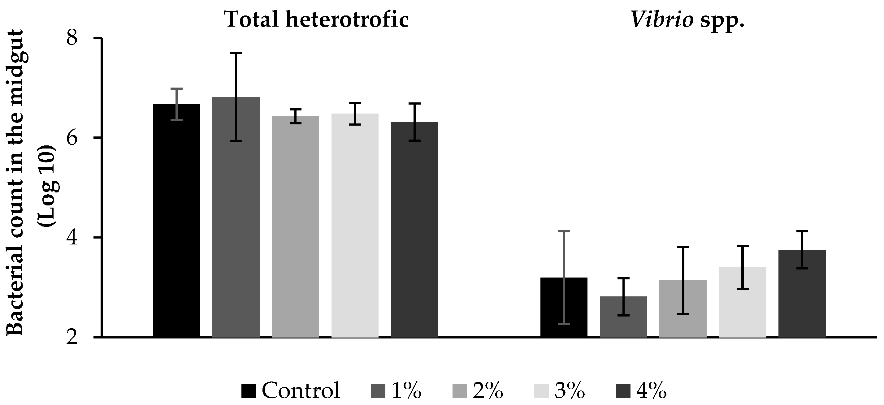
3.2. Microbiological Analysis

Regarding the concentration of bacteria per gram of intestine, in Log 10 colony-forming units (CFU), no statistical differences were identified between treatments in bacterial counts (Figure 2).

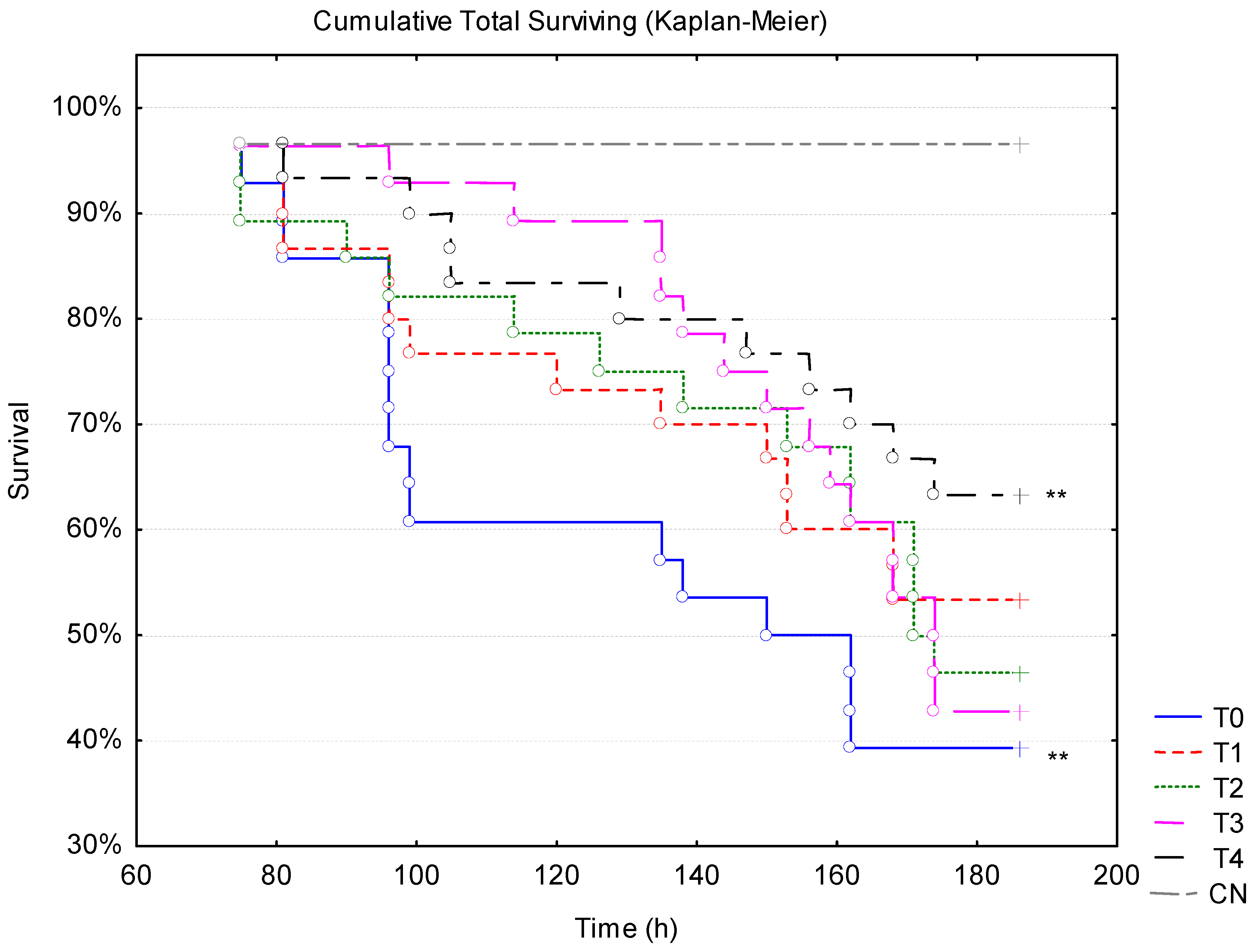
3.3. WSSV Challenge Associated with Thermal Stress

At the end of the challenge, animals from the 4% treatment showed the lowest mortality at 36.7% (p < 0.01) (Table 5). The non-supplemented treatment presented the worst resistance, with the highest mortality to the viral challenge associated with thermal stress (61.7%), and during the phase under temperature of 22 °C (40.8%). During the phase under temperature of 28 °C, these non-supplemented animals showed the third highest mortality (20.8%). The thermal variation alone was not capable of causing any mortality, as observed in the negative control (NC), since this group was not inoculated with WSSV.
In the temperature phase of 22 °C, all treatments with Aurantiochytrium sp. meal inclusion showed the lowest absolute mortality; however, only 3 and 4% treatments were significantly lower compared to the control (p < 0.05). On the other hand, animals from the 3% treatment were the ones that presented the highest mortality after the increase in temperature, so that at the end of the challenge, only 4% treatment presented lower total cumulative mortality compared to the control (0%) (p < 0.05) (Figure 3).

3.4. Hemato-Immunological Parameters

Before infection, there were no differences in hemato-immunological parameters among the treatments, except for THC, which was higher in the 1%-supplemented treatment (Table 6). After the challenge, hemocyte count significantly reduced after WSSV infection in all groups when at 22 °C, and even more after thermal stress (p < 0.01). Thermal stress alone also caused a significant reduction in THC in the NC group in comparison to first sample (Table 6). In general, the average values in the agglutination test showed a reduction throughout the challenge, differing only before and after the infection (p < 0.05). Protein concentration and specific PO also showed a significant reduction after WSSV infection, with no difference among treatments (p > 0.05), differing only before and after infection (p < 0.05).
Regarding the negative control group, although the thermal stress was not able to cause any mortality, it caused significant disturbances in the hemato-immunological parameters. The variation profile of hemato-immunological parameters evaluated highlights that supplementation can mitigate the effects of low temperatures, with or without infection, depending on the level of supplementation.

4. Discussion

4.1. Growth Performance and Diet Composition

During the entire rearing period, the animals from the different dietary treatments were kept under a suboptimal water temperature of 22 °C. However, their final survival was not affected by this temperature, remaining above 96.7% in shrimp from all dietary treatments. The survival demonstrates the adaptability of the species to the temperature range of 22 °C and the importance of not having fluctuations in temperature, despite being at a temperature below the ideal recommended for the species. No statistical differences were identified regarding the growth performance parameters among the treatments.
Considering the low temperature and the high density (100 shrimp/m3) used in the experiment, with a high survival rate, the weekly growth was higher than that found by other authors. Walker et al. [1] found values below those found in the present study for the weekly growth of shrimp grown at 20 and 24 °C (0.14 to 0.49 g week–1) in clear water culture conditions with constant renewals and lower density. Prates et al. [11] subjected shrimp to treatments at low temperatures (20 and 24 °C) in a biofloc system with super-intensive density (300 shrimp/m3) and found weekly growth rates of 0.33 to 0.75 g week−1, values below those found in the present study, despite the use of flocs as a dietary feed supplement. These better results from the present study compared to those mentioned may be related to the better fatty acid profile of the diets.
It is known that different factors such as rearing temperature, life stage, lipid source, and so on can affect the lipid dynamic and requirements in aquatic animals [29,44]. In the present study, the control diet had 2.2% soy lecithin inclusion and a total lipid content of 8.1%; meanwhile, the soy lecithin inclusions and total lipid content of the supplemented diets varied from 1.5 to 1.1% and from 8.1 to 10.8% DM, with increasing levels of Aurantiochytrium sp. meal, respectively. These inclusion ranges did not significantly increase the growth performance of the animals. However, they maintained them at levels similar to those of animals raised at optimal temperatures. This fact demonstrates that the dietary inclusion of Aurantiochytrium sp. meal is probably safe for the cultivation of P. vannamei under suboptimal temperature (22 °C), since they resulted in a weekly growth of around 1.02 g.
At suboptimal temperatures, the addition of phospholipids (4.22% of soy lecithin) improved the weight gain of Nile tilapia (Oreochromis niloticus) reared at 22 °C [45]. For shrimps, a previous study by Zhang et al. [46] found better overall health outcomes for P. vannamei on diets containing 10–12% lipids and only 1% dietary lecithin at 28 ± 2 °C. Liou et al. [47] observed the best growth and immune responses of P. vannamei at 29.0 ± 1.0 °C with 11% lipids and 1.8% soy lecithin. In another study, diets including 5% fish oil supplemented with 3.1% soy lecithin (97.6% phospholipids) resulted in effective improvement in shrimp growth compared to other sources of plant oil, at 32.8 ± 1.0 °C [48]. The best performance of shrimp grown at optimal temperatures in comparison with the present study may be related to the use of lipid sources and fatty acids in the diet for growth, and not just for maintaining membrane fluidity and the immune system.
Shrimp can activate some lipid mediators and related metabolic pathways to regulate the balance of the organism itself when in a situation of temperature fluctuations [49]. The presence of phospholipids may have contributed to the shrimp response to cold. González-Félix et al. [48] noted that sources of LC-PUFA are as important as phospholipid intake. Shrimp that were fed diets including plant oils associated with soy lecithin did not improve performance when compared to animals fed diets including fish oil without lecithin. This may be since fish oil has high levels of n-3 fatty acids, which are less significant in plant oils. The isolated inclusion of EPA or DHA in the diet of P. monodon increased the growth of the animals, with the level of 4% of both EPA and DHA separately, within diets with 7.5% of total lipid, promoting maximum growth [50]. In the present study, shrimp fed diets with the inclusion of 1, 2, 3, and 4% Aurantiochytrium sp. meal had DHA contents of 0.48, 0.73, 0.91, and 1.11%, respectively, while shrimp fed the control diet had about 0.25% DHA corroborating the results of Gonzalez-Felix et al. [48], where the inclusion of soy lecithin in all diets may have reduced the DHA requirement of shrimp.
In a study conducted by Nobrega et al. [29] with Nile tilapia (Oreochromis niloticus) at a suboptimal temperature (22 °C), it was reported that the dietary supplementation with Aurantiochytrium sp. meal in the range of 0.45 to 1.42%, as a source of DHA, resulted in better growth, body fat composition, and n-3:n-6 PUFA ratio in muscle than the dietary supplementation with cod liver oil. In this range of Aurantiochytrium sp. intake (diets with 0.5, 1, and 2%), a total of about 0.15 to 0.58% LC-PUFA was obtained. In our diets, the total range of Aurantiochytrium sp. uptake resulted in 0.33 to 1.23% of LC-PUFA and showed no difference in growth performance. However, it is worth noting that there are significant differences between the nutrient requirements of P. vannamei and tilapia for phospholipids and LC-PUFA.
Shrimp, as marine organisms, have a greater affinity and requirement for these compounds than Nile tilapia [15], which can be cultured without an n-3 source when the growth temperature is within the optimal range [51]. In the present study, the tested suboptimal temperature with the increase of the dietary n-3:n-6 ratio and the addition of the ingredient rich in DHA and PUFA beyond the known requirement had no adverse effects on shrimp survival and growth. On the contrary, the shrimp appeared to adapt well to the low temperature in a system without fluctuations, and no differences in growth performance were observed even when the n-3:n-6 ratio was increased with the increase in feed additive dose.
Farmed shrimp tend to have their feeding behaviour affected by low temperature, which may reduce feed intake and remain immobile inside the tank [52]. However, this was not the case in our study, in which the animals showed active feeding behaviour and consumption, with low feed waste. This may be related to the restrict water temperature control, stabilizing the temperature along the experiment, and the adequate temperature acclimatization of the animals prior to the experiment.
Still, the suboptimal temperature affected the feeding rate, which was less efficient when compared to studies carried out at ideal temperatures [53,54]. Similar values of feed conversion and survival were found in a study using increasing levels of the same Aurantiochytrium sp. meal in replacement of fish oil for P. vannamei reared at an optimal temperature of 28 °C [55].

4.2. Microbiological and Hemato-Immunological and Analyses and Challenge with WSSV in Association with Thermal Stress

There were no significant differences in the quantification of total heterotrophic and total vibrionaceous bacteria. The presence of vibrio bacteria is natural in the marine environment and intestinal tract of shrimp. The experiment’s water changes were performed daily at rates of 80–100% of the total volume, a factor that influenced the maintenance of low levels of nitrogenous compounds in the system, possibly controlling the proliferation of vibrionaceous bacteria. Vibrio spp. can grow over a wide range of temperatures (0.5 °C to 48 °C), but there is evidence that its growth rate is accelerated at higher temperatures, such as in the species of V. parahaemolyticus and V. alginolyticus at 37 to 42 ºC [56]. The total bacteria count is in line with results found in previous studies with similar methodology, as the Vibrio spp. represents lower values than those found in the same studies, at optimal temperatures [41,57,58]. Suboptimal temperatures may slow Vibrio spp. growth [56].
Regarding the challenge trial, at low temperature phase (22 °C), supplementation with Aurantiochytrium sp. meal at the concentrations of 3% and 4% (Table 5) increased the resistance of the animals to viral infections, being even statistically significant when compared to the control group (0%). However, when the temperature increased to 28 °C during the challenge, the group supplemented with 4% Aurantiochytrium sp. meal was the only one that resulted in greater survival and therefore greater resistance to WSSV.
This finding may be associated with the composition of Aurantiochytrium sp., which has β-1,3-glycans in the cell wall [59]. This polysaccharide has properties that promote animal health, such as antioxidant, anti-inflammatory, and immunostimulant action [60,61,62]. In marine shrimp, different studies have shown that dietary supplementation of β-1,3-glycans promoted greater resistance against WSSV infection [63,64]. These results are in line with what was observed in the present study.
In parallel, Aurantiochytrium sp. can produce LC-PUFA such as eicosapentaenoic acid (EPA), docosahexaenoic acid (DHA), and others [65], but is particularly rich in DHA [22]. In addition, it is able to synthesize other bioactive compounds such as carotenoids, especially squalene [66]. These compounds can be essentially useful in increasing the cold resistance of ectothermic aquatic animals and minimizing their deleterious effects, as temperature reduction can lead to loss of membrane fluidity, transmembrane protein dysfunction, protein degradation and aggregation, oxidative damage, and others [12,13,67].
Variation in the fatty acid composition of phospholipids is particularly important during the process of adaptation to temperature changes. Generally, there is an increase in the proportion of PUFA and monounsaturated fatty acids (MUFA) and a reduction of saturated fatty acids (SFA) in membrane phospholipids [68,69]. The increase in PUFA maintains the liquid-crystal state of the lipid matrix, which allows the membrane to continue functioning properly, to compensate for the cold-induced solidification of the lipid bilayers [70,71].
In this sense, this rich constitution of bioactive compounds of Aurantiochytrium sp. may be associated with the beneficial effects observed in the present study, particularly the increase in shrimp resistance against viral infection at low temperature, and more specifically, the supplementation levels of 3 and 4%. However, when the shrimp were submitted to thermal stress by raising the temperature, the animals supplemented with 3% of Aurantiochytrium sp. showed high mortality, matching the values of the control (0%). This low tolerance to thermal oscillation of animals from 3% treatment was not fully understood, requiring further investigations.
Hemato-immunological parameters showed a significant reduction throughout the bioassay, demonstrating the immunosuppressive effect of viral infection. This effect has already been demonstrated in several studies with WSSV infection [72,73,74,75,76]. The decrease in immune parameters was also observed in the NC group (uninfected), which highlights that heat stress can have deleterious effects with immune system disorders, as reported by Qiu et al. [77] and Zhou et al. [78]. These results reinforce the importance of developing new technologies to minimize the effects of heat stress, especially in breeding farms in subtropical regions.
Prior to the virus challenge, animals raised at suboptimal temperature and fed a 1% feed supplement with Aurantiochytrium sp. meal were generally superior in immune parameters. However, this was not reflected in the mortality and immune response of the animals after virus infection, which had only the second lowest mortality at the end of the challenge.

5. Conclusions

The dietary inclusion of Aurantiochytrium sp. meal had no effect on the growth performance of P. vannamei cultivated at suboptimal temperatures. However, the 4% dose resulted in lower mortality of shrimp after the viral challenge with WSSV associated with heat stress.

Author Contributions

Conceptualization, F.d.N.V., D.D.S. and W.Q.S.; methodology, F.d.N.V., D.D.S. and F.B.H.; validation, F.d.N.V., F.B.H., I.P., A.S.M. and D.D.S.; formal analysis: F.B.H., I.P., A.S.M. and D.D.S.; investigation, F.B.H., A.S.M. and I.P.; resources: F.d.N.V., D.D.S., W.Q.S., A.N.R. and C.S.; data curation: F.d.N.V., D.D.S. and F.B.H.; writing—original draft preparation, F.B.H. and A.S.M.; writing—review and editing, F.B.H., D.D.S., A.N.R. and F.d.N.V.; visualization, F.B.H. and I.P.; supervision, F.d.N.V. and D.D.S.; project administration, F.d.N.V. and D.D.S.; funding acquisition, F.d.N.V., D.D.S., W.Q.S., A.N.R. and C.S. All authors have read and agreed to the published version of the manuscript.

Funding

This project has received funding from FAPESC (2021TR100876) and CNPQ (402501/2022-5) and F.N.V is researcher fellow from CNPQ (310250/2020-0). W. Q. S. is researcher fellow from CNPQ (303684/2021-6). F.B.H received a scholarship from FAPESC (48/2021).

Institutional Review Board Statement

All applicable international and national guidelines for the care and use of animals were followed by the authors. An Institutional Review Board Statement is not necessary for studies with invertebrates other than cephalopods.

Informed Consent Statement

Not applicable.

Data Availability Statement

Data are contained within the article, and are available from the corresponding author upon reasonable request.

Acknowledgments

The authors acknowledge Carlos Miranda, Ivanilson Santos, Ramon Carneiro, Mateus Aranha Martins, Isabela Gomes, Yuri Gatto, and Norha Constanza Bolívar for their invaluable support during the experiment.

Conflicts of Interest

The authors declare no conflicts of interest.

References

  1. Walker, S.J.; Neill, W.H.; Lawrence, A.L.; Gatlin, D.M. Effects of Temperature and Starvation on Ecophysiological Performance of the Pacific White Shrimp (Litopenaeus Vannamei). Aquaculture 2011, 319, 439–445. [Google Scholar] [CrossRef]
  2. Wyban, J.; Walsh, W.A.; Godin, D.M. Temperature Effects on Growth, Feeding Rate and Feed Conversion of the Pacific White Shrimp (Penaeus Vannamei). Aquaculture 1995, 138, 267–279. [Google Scholar] [CrossRef]
  3. Ponce-Palafox, J.; Martinez-Palacios, C.A.; Ross, L.G. The Effects of Salinity and Temperature on the Growth and Survival Rates of Juvenile White Shrimp, Penaeus Vannamei, Boone, 1931. Aquaculture 1997, 157, 107–115. [Google Scholar] [CrossRef]
  4. Flegel, T.W. Review of Disease Transmission Risks from Prawn Products Exported for Human Consumption. Aquaculture 2009, 290, 179–189. [Google Scholar] [CrossRef]
  5. Peruzza, L.; Shekhar, M.S.; Kumar, K.V.; Swathi, A.; Karthic, K.; Hauton, C.; Vijayan, K.K. Temporal Changes in Transcriptome Profile Provide Insights of White Spot Syndrome Virus Infection in Litopenaeus Vannamei. Sci. Rep. 2019, 9, 13509. [Google Scholar] [CrossRef] [PubMed]
  6. Lightner, D. V Virus Diseases of Farmed Shrimp in the Western Hemisphere (the Americas): A Review. J. Invertebr. Pathol. 2011, 106, 110–130. [Google Scholar] [CrossRef] [PubMed]
  7. Millard, R.S.; Ellis, R.P.; Bateman, K.S.; Bickley, L.K.; Tyler, C.R.; van Aerle, R.; Santos, E.M. How Do Abiotic Environmental Conditions Influence Shrimp Susceptibility to Disease? A Critical Analysis Focussed on White Spot Disease. J. Invertebr. Pathol. 2021, 186, 107369. [Google Scholar] [CrossRef]
  8. Zhang, J.S.; Li, Z.J.; Wen, G.L.; Wang, Y.L.; Luo, L.; Zhang, H.J.; Dong, H.B. Relationship between White Spot Syndrome Virus (WSSV) Loads and Characterizations of Water Quality in Litopenaeus Vannamei Culture Ponds during the Tropical Storm. Iran. J. Vet. Res. 2016, 17, 210. [Google Scholar]
  9. Vidal, O.M.; Granja, C.B.; Aranguren, F.; Brock, J.A.; Salazar, M. A Profound Effect of Hyperthermia on Survival of Litopenaeus Vannamei Juveniles Infected with White Spot Syndrome Virus. J. World Aquac. Soc. 2001, 32, 364–372. [Google Scholar] [CrossRef]
  10. Abdelrahman, H.A.; Abebe, A.; Boyd, C.E. Influence of Variation in Water Temperature on Survival, Growth and Yield of Pacific White Shrimp Litopenaeus Vannamei in Inland Ponds for Low-salinity Culture. Aquac. Res. 2019, 50, 658–672. [Google Scholar] [CrossRef]
  11. Prates, E.; Holanda, M.; Pedrosa, V.F.; Monserrat, J.M.; Wasielesky, W. Compensatory Growth and Energy Reserves Changes in the Pacific White Shrimp (Litopenaeus Vannamei) Reared in Different Temperatures and under Feed Restriction in Biofloc Technology System (BFT). Aquaculture 2023, 562, 738821. [Google Scholar] [CrossRef]
  12. Hayward, S.A.L.; Manso, B.; Cossins, A.R. Molecular Basis of Chill Resistance Adaptations in Poikilothermic Animals. J. Exp. Biol. 2014, 217, 6–15. [Google Scholar] [CrossRef]
  13. Chowanski, S.; Lubawy, J.; Spochacz, M.; Ewelina, P.; Grzegorz, S.; Rosinski, G.; Slocinska, M. Cold Induced Changes in Lipid, Protein and Carbohydrate Levels in the Tropical Insect Gromphadorhina Coquereliana. Comp. Biochem. Physiol. A Mol. Integr. Physiol. 2015, 183, 57–63. [Google Scholar] [CrossRef] [PubMed]
  14. Glencross, B.D. Exploring the Nutritional Demand for Essential Fatty Acids by Aquaculture Species. Rev. Aquac. 2009, 1, 71–124. [Google Scholar] [CrossRef]
  15. COUNCIL, N.R. Nutrient Requirements of Fish and Shrimp; National Academies Press: Washington, DC, USA, 2011. [Google Scholar]
  16. Das, U. Essential Fatty Acids—A Review. Curr. Pharm. Biotechnol. 2006, 7, 467–482. [Google Scholar] [CrossRef]
  17. Harwood, J.L.; Guschina, I.A. The Versatility of Algae and Their Lipid Metabolism. Biochimie 2009, 91, 679–684. [Google Scholar] [CrossRef] [PubMed]
  18. Perini, J.Â.D.L.; Stevanato, F.B.; Sargi, S.C.; Visentainer, J.E.L.; Dalalio, M.M.D.O.; Matshushita, M.; De Souza, N.E.; Visentainer, J.V. Ácidos Graxos Poli-Insaturados n-3 e n-6: Metabolismo Em Mamíferos e Resposta Imune. Rev. Nutr. 2010, 23, 1075–1086. [Google Scholar] [CrossRef]
  19. Qiu, X. Biosynthesis of Docosahexaenoic Acid (DHA, 22:6-4, 7,10,13,16,19): Two Distinct Pathways. Prostaglandins Leukot. Essent. Fatty Acids 2003, 68, 181–186. [Google Scholar] [CrossRef] [PubMed]
  20. Pereira, H.; Barreira, L.; Figueiredo, F.; Custódio, L.; Vizetto-Duarte, C.; Polo, C.; Rešek, E.; Engelen, A.; Varela, J. Polyunsaturated Fatty Acids of Marine Macroalgae: Potential for Nutritional and Pharmaceutical Applications. Mar. Drugs 2012, 10, 1920–1935. [Google Scholar] [CrossRef]
  21. Wang, K.; Sun, T.; Cui, J.; Liu, L.; Bi, Y.; Pei, G.; Chen, L.; Zhang, W. Screening of Chemical Modulators for Lipid Accumulation in Schizochytrium Sp. S31. Bioresour. Technol. 2018, 260, 124–129. [Google Scholar] [CrossRef] [PubMed]
  22. Marchan, L.F.; Chang, K.J.L.; Nichols, P.D.; Mitchell, W.J.; Polglase, J.L.; Gutierrez, T. Taxonomy, Ecology and Biotechnological Applications of Thraustochytrids: A Review. Biotechnol. Adv. 2018, 36, 26–46. [Google Scholar] [CrossRef]
  23. Jaritkhuan, S.; Suanjit, S. Species Diversity and Polyunsaturated Fatty Acid Content of Thraustochytrids from Fallen Mangrove Leaves in Chon Buri Province, Thailand. Agric. Nat. Resour. 2018, 52, 24–32. [Google Scholar] [CrossRef]
  24. Shene, C.; Paredes, P.; Flores, L.; Leyton, A.; Asenjo, J.A.; Chisti, Y. Dynamic Flux Balance Analysis of Biomass and Lipid Production by Antarctic Thraustochytrid Oblongichytrium Sp. RT2316-13. Biotechnol. Bioeng. 2020, 117, 3006–3017. [Google Scholar] [CrossRef] [PubMed]
  25. Dawood, M.A.O.; Koshio, S.; Esteban, M.Á. Beneficial Roles of Feed Additives as Immunostimulants in Aquaculture: A Review. Rev. Aquac. 2018, 10, 950–974. [Google Scholar] [CrossRef]
  26. Samocha, T.M.; Patnaik, S.; Davis, D.A.; Bullis, R.A.; Browdy, C.L. Use of Commercial Fermentation Products as a Highly Unsaturated Fatty Acid Source in Practical Diets for the Pacific White Shrimp Litopenaeus Vannamei. Aquac. Res. 2009, 41, 961–967. [Google Scholar] [CrossRef]
  27. Wang, Y.; Li, M.; Filer, K.; Xue, Y.; Ai, Q.; Mai, K. Replacement of Fish Oil with a DHA-Rich Schizochytrium Meal on Growth Performance, Activities of Digestive Enzyme and Fatty Acid Profile of Pacific White Shrimp (Litopenaeus Vannamei) Larvae. Aquac. Nutr. 2017, 23, 1113–1120. [Google Scholar] [CrossRef]
  28. Kumar, V.; Habte-Tsion, H.; Allen, K.M.; Bowman, B.A.; Thompson, K.R.; El-Haroun, E.; Filer, K.; Tidwell, J.H. Replacement of Fish Oil with Schizochytrium Meal and Its Impacts on the Growth and Lipid Metabolism of Pacific White Shrimp (Litopenaeus Vannamei). Aquac. Nutr. 2018, 24, 1769–1781. [Google Scholar] [CrossRef]
  29. Nobrega, R.O.; Batista, R.O.; Corrêa, C.F.; Mattioni, B.; Filer, K.; Pettigrew, J.E.; Fracalossi, D.M. Dietary Supplementation of Aurantiochytrium Sp. Meal, a Docosahexaenoic-Acid Source, Promotes Growth of Nile Tilapia at a Suboptimal Low Temperature. Aquaculture 2019, 507, 500–509. [Google Scholar] [CrossRef]
  30. Chang, K.J.L.; Parrish, C.C.; Simon, C.J.; Revill, A.T.; Nichols, P.D. Feeding Whole Thraustochytrid Biomass to Cultured Atlantic Salmon (Salmo Salar) Fingerlings: Culture Performance and Fatty Acid Incorporation. J. Mar. Sci. Eng. 2020, 8, 207. [Google Scholar] [CrossRef]
  31. Moi, I.M.; Leow, A.T.C.; Ali, M.S.M.; Rahman, R.N.Z.R.A.; Salleh, A.B.; Sabri, S. Polyunsaturated Fatty Acids in Marine Bacteria and Strategies to Enhance Their Production. Appl. Microbiol. Biotechnol. 2018, 102, 5811–5826. [Google Scholar] [CrossRef]
  32. Nagano, N.; Sakaguchi, K.; Taoka, Y.; Okita, Y.; Honda, D.; Ito, M.; Hayashi, M. Detection of Genes Involved in Fatty Acid Elongation and Desaturation in Thraustochytrid Marine Eukaryotes. J. Oleo Sci. 2011, 60, 475–481. [Google Scholar] [CrossRef] [PubMed]
  33. Van Wyk, P.; Scarpa, J. Water Quality Requirements and Management. In Farming Marine Shrimp in Recirculating Freshwater Systems; Van Wyk, P., Scarpa, J., Eds.; Harbor Branch Oceanographic Institution: Fort Pierce, FL, USA, 1999; Volume 13, pp. 141–162. [Google Scholar]
  34. Simon, C.J.; Truong, H.H.; Noble, T.H.; Osborne, S.A.; Wynne, J.W.; Wade, N.M. Microbial Biomass, Marine Invertebrate Meals and Feed Restriction Influence the Biological and Gut Microbiota Response of Shrimp Penaeus Monodon. Aquaculture 2020, 520, 734679. [Google Scholar] [CrossRef]
  35. Coutteau, P.; Sorgeloos, P. Intercalibration Exercise on the Qualitative and Quantitative Analysis of Fatty Acids in Artemia and Marine Samples Used in Mariculture. ICES Coop. Res. Rep. 1995, 211, 34. [Google Scholar]
  36. APHA American Public Health Association; American Water Works Association; Water Environment Federation. Standard Methods for the Examination of Water and Wastewater, Eaton, A.D., Clesceri, L.S., Rice, E.W., Greenberg, A.E., Eds.; 21st ed.; American Public Health Association: Washington DC, USA, 2005. [Google Scholar]
  37. Bagenal, T.B.; Tesch, F.W. Methods for Assessment of Fish Production in Fresh Waters, 3rd ed.; Bagenal, T., Ed.; Blackwell Scientific: Hoboken, NJ, USA, 1978. [Google Scholar]
  38. Rezende, P.C.; Miranda, C.; Fracalossi, D.M.; Hayashi, L.; Seiffert, W.Q.; do Nascimento Vieira, F.; Schleder, D.D. Brown Seaweeds as a Feed Additive for Litopenaeus Vannamei Reared in a Biofloc System Improved Resistance to Thermal Stress and White Spot Disease. J. Appl. Phycol. 2022, 34, 2603–2614. [Google Scholar] [CrossRef]
  39. Schleder, D.D.; Blank, M.; Peruch, L.G.B.; Poli, M.A.; Gonçalves, P.; Rosa, K.V.; Fracalossi, D.M.; do Nascimento Vieira, F.; Andreatta, E.R.; Hayashi, L. Impact of Combinations of Brown Seaweeds on Shrimp Gut Microbiota and Response to Thermal Shock and White Spot Disease. Aquaculture 2020, 519, 734779. [Google Scholar] [CrossRef]
  40. de Souza Valente, C.; Oliveira Ortiz, K.; Depperschmidt, R.; de Medeiros Fraga, A.P.; do Nascimento Vieira, F.; Freire Marques, M.R. Transcription of Defense Related Genes in Pacific White Shrimp, Litopenaeus Vannamei, Kept in Biofloc and in Clear Seawater and Challenged with the White Spot Syndrome Virus. Aquac. Int. 2020, 28, 293–307. [Google Scholar] [CrossRef]
  41. Bolívar-Ramírez, N.C.; Mallmann, A.S.; Schleder, D.D.; Machado, C.; Seiffert, W.Q.; do Nascimento Vieira, F. Tannins as a Food Additive in Pacific White Shrimp Diet. Aquaculture 2022, 556, 738232. [Google Scholar] [CrossRef]
  42. Zar, J.H. Biostatistical Analysis, 5th ed.; Prentice-Hall/Pearson: Upper Saddle River, NJ, USA, 2010. [Google Scholar]
  43. Boyd, C.E.; Gautier, D. GAA’S, Implementing. Effluent Composition &Water Quality Standards. Glob. Aquac. Advocate 2000, 3, 61–66. [Google Scholar]
  44. Glencross, B.D.; Smith, D.M.; Thomas, M.R.; Williams, K.C. Optimising the Essential Fatty Acids in the Diet for Weight Gain of the Prawn, Penaeus Monodon. Aquaculture 2002, 204, 85–99. [Google Scholar] [CrossRef]
  45. Batista, R.O.; Richter, B.L.; Banze, J.F.; Schleder, D.D.; Salhi, M.; Nobrega, R.O.; da Silva, M.F.O.; Mattioni, B.; Pettigrew, J.E.; Fracalossi, D.M. Soy Lecithin Supplementation Promotes Growth and Increases Lipid Digestibility in GIFT Nile Tilapia Raised at Suboptimal Temperature. Fishes 2023, 8, 404. [Google Scholar] [CrossRef]
  46. Zhang, S.; Li, J.; Wu, X.; Zhong, W.; Xian, J.; Liao, S.; Miao, Y.; Wang, A. Effects of Different Dietary Lipid Level on the Growth, Survival and Immune-Relating Genes Expression in Pacific White Shrimp, Litopenaeus Vannamei. Fish. Shellfish. Immunol. 2013, 34, 1131–1138. [Google Scholar] [CrossRef] [PubMed]
  47. Liou, C.-H.; To, V.-A.; Zhang, Z.-F.; Lin, Y.-H. The Effect of Dietary Lecithin and Lipid Levels on the Growth Performance, Body Composition, Hemolymph Parameters, Immune Responses, Body Texture, and Gene Expression of Juvenile White Shrimp (Litopenaeus Vannamei). Aquaculture 2023, 567, 739260. [Google Scholar] [CrossRef]
  48. González-Félix, M.L.; Lawrence, A.L.; Gatlin, D.M.; Perez-Velazquez, M. Growth, Survival and Fatty Acid Composition of Juvenile Litopenaeus Vannamei Fed Different Oils in the Presence and Absence of Phospholipids. Aquaculture 2002, 205, 325–343. [Google Scholar] [CrossRef]
  49. Wang, Z.; Zhou, J.; Li, J.; Lv, W.; Zou, J.; Fan, L. A New Insight into the Intestine of Pacific White Shrimp: Regulation of Intestinal Homeostasis and Regeneration in Litopenaeus Vannamei during Temperature Fluctuation. Comp. Biochem. Physiol. Part. D Genom. Proteom. 2020, 35, 100687. [Google Scholar] [CrossRef]
  50. Glencross, B.D.; Smith, D.M. Optimizing the Essential Fatty Acids, Eicosapentaenoic and Docosahexaenoic Acid, in the Diet of the Prawn, Penaeus Monodon. Aquac. Nutr. 2001, 7, 101–112. [Google Scholar] [CrossRef]
  51. Corrêa, C.F.; Nobrega, R.O.; Mattioni, B.; Block, J.M.; Fracalossi, D.M. Dietary Lipid Sources Affect the Performance of Nile Tilapia at Optimal and Cold, Suboptimal Temperatures. Aquac. Nutr. 2017, 23, 1016–1026. [Google Scholar] [CrossRef]
  52. Zhou, M.; Wang, A.-L.; Xian, J.-A. Variation of Free Amino Acid and Carbohydrate Concentrations in White Shrimp, Litopenaeus Vannamei: Effects of Continuous Cold Stress. Aquaculture 2011, 317, 182–186. [Google Scholar] [CrossRef]
  53. González-Félix, M.L.; Gatlin, D.M.; Lawrence, A.L.; Perez-Velazquez, M. Effect of Various Dietary Lipid Levels on Quantitative Essential Fatty Acid Requirements of Juvenile Pacific White Shrimp Litopenaeus Vannamei. J. World Aquac. Soc. 2002, 33, 330–340. [Google Scholar] [CrossRef]
  54. Sánchez-Paz, A. White Spot Syndrome Virus: An Overview on an Emergent Concern. Vet. Res. 2010, 41, 43. [Google Scholar] [CrossRef] [PubMed]
  55. Guimarães, A.M.; Schleder, D.D.; Nagata, M.; Nóbrega, R.O.; Fracalossi, D.M.; Seiffert, W.Q.; Vieira, F.N. Aurantiochytrium sp. Meal Can Replace Fish Oil in Practical Diets for the Juvenile Pacific White Shrimp. Aquac. Nutr. 2019, 25, 798–807. [Google Scholar] [CrossRef]
  56. Sheikh, H.I.; Najiah, M.; Fadhlina, A.; Laith, A.A.; Nor, M.M.; Jalal, K.C.A.; Kasan, N.A. Temperature Upshift Mostly but Not Always Enhances the Growth of Vibrio Species: A Systematic Review. Front. Mar. Sci. 2022, 9, 959830. [Google Scholar] [CrossRef]
  57. Bolívar-Ramírez, N.C.; Rodrigues, M.S.; Guimarães, A.M.; Guertler, C.; Rosa, J.R.; Seiffert, W.Q.; Andreatta, E.R.; do Nascimento Vieira, F. Effect of Dietary Supplementation with Butyrate and Probiotic on the Survival of Pacific White Shrimp after Challenge with Vibrio Alginolyticus. Rev. Bras. Zootec. 2017, 46, 471–477. [Google Scholar] [CrossRef]
  58. da Rosa Coelho, J.; Rosa, K.V.; Rocha, J.S.; Ramírez, N.C.B.; Maraschin, M.; do Nascimento Vieira, F. In Vitro Antimicrobial Activity Of Carvacrol against Shrimp Pathogens and Its Use As Feed Additive for the Pacific White Shrimp. Bol. Inst. Pesca 2021, 47, e645. [Google Scholar] [CrossRef]
  59. Rojo, A.; Ibarra, L.; Brown, J.M.M.; Velasco, G.; Martínez-Téllez, M.Á.; Medina-Jasso, M.A.; Nieves-Soto, M.; Quintana-Zavala, D. Potential of Nannochloropsis in Beta Glucan Production. In Nannochloropsis: Biology, Biotechnological, Potential and Challenges; Jan, M., Kazik, P., Eds.; Nova Sciences: Hauppauge, NY, USA, 2017; pp. 181–225. [Google Scholar]
  60. Li, C.; Weng, S.; He, J. WSSV–Host Interaction: Host Response and Immune Evasion. Fish. Shellfish. Immunol. 2019, 84, 558–571. [Google Scholar] [CrossRef] [PubMed]
  61. Wu, Y.-S.; Liau, S.-Y.; Huang, C.-T.; Nan, F.-H. Beta 1,3/1,6-Glucan and Vitamin C Immunostimulate the Non-Specific Immune Response of White Shrimp (Litopenaeus Vannamei). Fish. Shellfish. Immunol. 2016, 57, 269–277. [Google Scholar] [CrossRef] [PubMed]
  62. Przybylska-Diaz, D.A.; Schmidt, J.G.; Vera-Jiménez, N.I.; Steinhagen, D.; Nielsen, M.E. β-Glucan Enriched Bath Directly Stimulates the Wound Healing Process in Common Carp (Cyprinus Carpio L.). Fish. Shellfish. Immunol. 2013, 35, 998–1006. [Google Scholar] [CrossRef] [PubMed]
  63. Bai, N.; Gu, M.; Zhang, W.; Xu, W.; Mai, K. Effects of β-Glucan Derivatives on the Immunity of White Shrimp Litopenaeus Vannamei and Its Resistance against White Spot Syndrome Virus Infection. Aquaculture 2014, 426–427, 66–73. [Google Scholar] [CrossRef]
  64. Ochoa-Álvarez, N.A.; Casillas-Hernández, R.; Magallón-Barajas, F.J.; Ramirez-Orozco, J.M.; Carbajal-Millán, E. Protector Effect of Beta-Glucans from Shrimp Pond-Related Yeasts in Penaeus Vannamei Rearing under White Spot Syndrome Virus Presence. Lat. Am. J. Aquat. Res. 2021, 49, 18–28. [Google Scholar] [CrossRef]
  65. Yang, H.; Huang, Y.; Li, Z.; Guo, Y.; Li, S.; Huang, H.; Yang, X.; Li, G.; Chen, H. Effects of Dietary Supplementation with Aurantiochytrium Sp. on Zebrafish Growth as Determined by Transcriptomics. Animals 2022, 12, 2794. [Google Scholar] [CrossRef]
  66. Watanabe, K.; Arafiles, K.H.V.; Higashi, R.; Okamura, Y.; Tajima, T.; Matsumura, Y.; Nakashimada, Y.; Matsuyama, K.; Aki, T. Isolation of High Carotenoid-producing Aurantiochytrium sp. Mutants and Improvement of Astaxanthin Productivity Using Metabolic Information. J. Oleo Sci. 2018, 67, 571–578. [Google Scholar] [CrossRef]
  67. TEETS, N.M.; DENLINGER, D.L. Physiological Mechanisms of Seasonal and Rapid Cold-hardening in Insects. Physiol. Entomol. 2013, 38, 105–116. [Google Scholar] [CrossRef]
  68. Tocher, D.R. Metabolism and Functions of Lipids and Fatty Acids in Teleosts Fish. Rev. Fish. Sci. 2003, 11, 107–184. [Google Scholar] [CrossRef]
  69. Liu, J.; Wang, K.; Wang, Y.; Chen, W.; Jin, Z.; Yao, Z.; Zhang, D. Strain-Specific Changes in the Gut Microbiota Profiles of the White Shrimp Litopenaeus Vannamei in Response to Cold Stress. Aquaculture 2019, 503, 357–366. [Google Scholar] [CrossRef]
  70. Abdel-Ghany, H.M.; El-Sayed, A.-F.M.; Ezzat, A.A.; Essa, M.A.; Helal, A.M. Dietary Lipid Sources Affect Cold Tolerance of Nile Tilapia (Oreochromis Niloticus). J. Therm. Biol. 2019, 79, 50–55. [Google Scholar] [CrossRef]
  71. Kostetsky, E.Y.; Velansky, P.V.; Sanina, N.M. Phase Transitions of Phospholipids as a Criterion for Assessing the Capacity for Thermal Adaptation in Fish. Russ. J. Mar. Biol. 2013, 39, 214–222. [Google Scholar] [CrossRef]
  72. Mai, W.; Wang, W. Protection of Blue Shrimp (Litopenaeus Stylirostris) against the White Spot Syndrome Virus (WSSV) When Injected with Shrimp Lysozyme. Fish. Shellfish. Immunol. 2010, 28, 727–733. [Google Scholar] [CrossRef]
  73. Ji, P.F.; Yao, C.L.; Wang, Z.Y. Reactive Oxygen System Plays an Important Role in Shrimp Litopenaeus Vannamei Defense against Vibrio Parahaemolyticus and WSSV Infection. Dis. Aquat. Organ. 2011, 96, 9–20. [Google Scholar] [CrossRef]
  74. Immanuel, G.; Sivagnanavelmurugan, M.; Marudhupandi, T.; Radhakrishnan, S.; Palavesam, A. The Effect of Fucoidan from Brown Seaweed Sargassum Wightii on WSSV Resistance and Immune Activity in Shrimp Penaeus Monodon (Fab). Fish. Shellfish. Immunol. 2012, 32, 551–564. [Google Scholar] [CrossRef] [PubMed]
  75. Lehmann, M.; Schleder, D.D.; Guertler, C.; Perazzolo, L.M.; Vinatea, L. Hypoxia Increases Susceptibility of Pacific White Shrimp to Whitespot Syndrome Virus (WSSV). Arq. Bras. Med. Vet. Zootec. 2016, 68, 397–403. [Google Scholar] [CrossRef]
  76. Abbaszadeh, A.; Yavari, V.; Hoseini, S.J.; Nafisi, M.; Mozanzadeh, M.T. Effects of Different Carbon Sources and Dietary Protein Levels in a Biofloc System on Growth Performance, Immune Response against White Spot Syndrome Virus Infection and Cathepsin L Gene Expression of Litopenaeus Vannamei. Aquac. Res. 2019, 50, 1162–1176. [Google Scholar] [CrossRef]
  77. Qiu, J.; Wang, W.-N.; Wang, L.; Liu, Y.-F.; Wang, A.-L. Oxidative Stress, DNA Damage and Osmolality in the Pacific White Shrimp, Litopenaeus Vannamei Exposed to Acute Low Temperature Stress. Comp. Biochem. Physiol. Part. C Toxicol. Pharmacol. 2011, 154, 36–41. [Google Scholar] [CrossRef]
  78. Zhou, M.; Wu, Z.; Liang, R.; Gu, N. Effects of Dietary Taurine, Carnitine and Cholesterol Supplementation on Growth Performance and Immunological Status of Litopenaeus Vannamei under Cold Exposure. Aquac. Res. 2017, 48, 1279–1290. [Google Scholar] [CrossRef]
Figure 1. Timeline of the challenge with WSSV associated with thermal stress, representing the challenge sequence and sample collection.
Fishes 09 00108 g001
Figure 2. Concentration of bacteria (g intestine in Log 10 CFU), of P. vannamei rearing in clear water and fed diet containing 0 (control), 1, 2, 3, and 4% of Aurantiochytrium sp. meal inclusion offered after nine weeks. The concentrations of bacteria shown are total heterotrophic bacteria (p value = 0.7056) and total Vibrio spp. (p value = 0.4458). Data are presented as mean ± error deviation.
Fishes 09 00108 g002
Figure 3. Cumulative mortality (%) of P. vannamei rearing in clear water and fed diet containing 0 (control), 1, 2, 3, and 4% of Aurantiochytrium sp. meal inclusion offered after nine weeks, after infection with WSSV associated with thermal stress via Kaplan–Meier analysis p value = 0.00069). All dietary treatments were significantly different from the negative control (p value < 0.01). ** Treatment T0 × T4 (p value = 0.0259). Subtitle: T0 (control), T1 (1% Aurantiochytrium sp. meal), T2 (2% Aurantiochytrium sp. meal), T3 (3% Aurantiochytrium sp. meal), and T4 (4% Aurantiochytrium sp. meal).
Fishes 09 00108 g003
Table 1. Experimental diets with inclusion of Aurantiochytrium sp. meal for P. vannamei rearing in a clear water system under suboptimal temperature (22 °C).
Ingredient (g/kg as Fed)% Inclusion of Aurantiochytrium sp. Meal
Control 1234
Soybean meal300290290290290
Fishmeal160160160160160
Wheat meal159166165165165
Poultry meal140140140140140
Kaolin10010091.58375.5
Vitamin and mineral premix a2525252525
Soy lecithin22151513.511
Fish oil202527.53030
Soybean oil1052.500
Magnesium sulphate1515151515
Monocalcium phosphate1010101010
Sodium chloride1515151515
Potassium chloride1515151515
Carboxymethylcellulose55555
DL-Methionine2.52.52.52.52.5
Vitamin C11111
Choline hydrochloride0.10.10.10.10.1
Aurantiochytrium sp meal. b010203040
a Vitamin and Mineral Premix (In Vivo Mix, Paulínea, Brazil) compound by 900 mg kg−1 vit. A, 25 mg kg−1 vit. D, 46,900 mg kg−1 vit. E, 14,000 mg kg−1 vit. K, 20,000 mg kg−1 vit. B2, 40,000 mg kg−1 pantothenic acid, 70,000 mg kg−1 niacin, 50 mg kg−1 vit. B12, 750 mg kg−1 biotin, 3000 mg kg−1 folic acid, 30,000 mg kg−1 vit. B1, 33,000 mg kg−1 vit B6, 20 mg kg−1 magnesium, 6.1 mg kg−1 potassium, 23,330 mg kg−1 copper, 1000 mg kg−1 iodine, 6500 mg kg−1 manganese, 100 g kg−1 zinc, 125 mg kg−1 selenium (In Vivo Nutrição e Saúde Animal Ltd.a—São Paulo, SP, Brazil). b Aurantiochytrium sp. meal—ALL-G-RICH® produced and supplied by Alltech Inc. (Nicholasville, KY, USA) and imported by Alltech do Brasil Agroindustrial Ltd.a (Araucaria, Paraná, Brazil).
Table 2. Proximal composition and fatty acid composition of experimental diets with inclusion of Aurantiochytrium sp. meal for P. vannamei.
Chemical Composition (%)% Inclusion of Aurantiochytrium sp. Meal
Control1234
Moisture content9.59.59.58.59.6
Crude protein40.840.040.440.140.3
Crude lipid8.18.99.710.510.8
Carbohydrate25.325.525.724.725.8
Ash16.316.114.716.213.5
Dry matter (%)90.590.590.591.590.4
Gross energy (MJ/kg) a16.616.316.616.616.7
Fatty acids (% DM)
14:00.120.170.220.270.30
16:01.892.422.993.53.85
18:00.600.620.640.690.66
18:1 n-92.142.232.242.312.19
18:2 n-62.221.951.791.661.63
18:3 n-30.200.170.150.150.14
ARA b0.060.060.070.070.07
EPA c0.080.100.110.120.12
DHA d0.250.480.730.911.11
SFA2.773.44.054.685.04
MUFA2.472.622.652.782.63
PUFA2.882.862.953.033.17
LC-PUFA0.330.580.841.031.23
n-30.530.761.01.191.37
n-62.312.051.91.781.74
n-3:n-60.230.370.530.670.79
DHA:EPA3.134.806.647.589.25
PUFA:SFA1.040.840.730.650.63
a Gross Energy are express in MJ kg−1; b Arachidonic acid (20:4 n-6); c Eicosapentaenoic acid (20:5 n-3); d Docosahexaenoic acid (22:6 n-3). All data of chemical composition are in terms of dry matter. Fatty acid group: SFA = saturated fatty acids, MUFA = monounsaturated fatty acids, PUFA = polyunsaturated fatty acids, LC-PUFA = long chain PUFA (with 20 to 22 carbons).
Table 3. Chemical composition of Aurantiochytrium sp.
Aurantiochytrium sp. ALL-G-RICH
Chemical Composition g kg−1Minerals mg kg−1
Moisture content24.65P1798.83Na1015.46
Crude protein108.68Ca2898.55Fe13.89
Lipid688.73Mg2496.27Al7.87
Ash30.64K1519.93Zn12.43
Carbohydrate147.30S4964.37Si208.24
Dry matter975.35 --
Gross energy a31.90 --
Fatty acids composition %
14:005.3418:4 n-30.26MUFA 0.85
16:0062.98ARA b0.82PUFA 28.39
16:1 n-70.11EPA c0.12LC-PUFA 27.13
18:1 n-90.33DHA d 26.62n-3 27.39
18:2 n-60.29SFA 68.61n-6 1.26
a Gross Energy are express in MJ kg−1. b Arachidonic acid (20:4 n-6). c Eicosapentaenoic acid (20:5 n-3) d Docosahexaenoic acid (22:6 n-3). All data of chemical composition are in terms of dry matter. Fatty acid group: SFA = saturated fatty acids, MUFA = monounsaturated fatty acids, PUFA = polyunsaturated fatty acids, LC-PUFA = long chain PUFA (with 20 to 22 carbons).
Table 4. Growth performance results of P. vannamei rearing in clear water and fed diet containing 0 (control), 1, 2, 3, and 4% of Aurantiochytrium sp. meal inclusion offered after nine weeks.
TreatmentInitial Weight (g−1)Final Weight (g−1)Weekly Weight Gain (g−1)Yield
(kg−1/m3)
Survival (%)Feed Conversion Ratio (kg−1)
Control3.8 ± 0.0113.1 ± 0.671.04 ± 0.071.35 ± 0.10100.0 ± 0.02.16 ± 0.15
1%3.8 ± 0.0212.6 ± 0.780.98 ± 0.081.24 ± 0.0497.5 ± 2.52.32 ± 0.07
2%3.8 ± 0.0312.7 ± 0.780.99 ± 0.091.25 ± 0.0898.3 ± 1.42.26 ± 0.13
3%3.8 ± 0.0413.9 ± 0.071.10 ± 0.031.34 ± 0.0296.7 ± 1.42.16 ± 0.07
4%3.8 ± 0.0212.9 ± 0.411.01 ± 0.041.25 ± 0.0297.5 ± 2.52.26 ± 0.03
p value0.99310.24110.23640.13640.52510.3025
Data presented as mean ± standard deviation. p-value for one-way ANOVA.
Table 5. Cumulative mortality (%) of P. vannamei, rearing in clear water and fed diet containing 0 (control), 1, 2, 3, and 4% of Aurantiochytrium sp. meal inclusion offered after nine weeks, after infection with WSSV associated with heat stress.
TreatmentsSuboptimal Temperature Phase (22 °C)Thermal Increment PhaseOptimal Temperature Phase (28 °C)Cumulative Total Mortality
Negative control *0000
Control40.8 ± 21.3 a020.8 ± 16.6 a61.7 ± 12.6 a
1%26.7 ± 23 a020.0 ± 17.3 a46.7 ± 40.4 a
2%21.9 ± 11.7 a3.7 ± 6.428.5 ± 5.7 a54.1 ± 13.4 a
3%10.7 ± 1.0 b046.3 ± 3.2 b57.0 ± 2.6 a
4%16.7 ± 15.3 b3.3 ± 5.716.7 ± 20.8 a36.7 ± 35.1 b
Time (h)1081248168
p value0.0184-0.01700.0006
Mean data ± standard deviation via Kaplan–Meier analysis. * The NC (negative control) was not infected with WSSV but was subjected to the same temperature conditions. The NC data were not statistically analyzed and served as a standard to visualize the infection compared to other treatments. All treatments are statistically different from the NC (p < 0.01). Superscript letters show the differences between the treatments compared to the control treatment between the infection periods (22 °C, 28 °C and at the end of the challenge).
Table 6. Total hemocytes count (THC), protein concentration, phenoloxidase (PO), enzyme activity and serum agglutinating activity of P. vannamei, rearing in clear water and fed diet containing 0 (control), 1, 2, 3, and 4% of Aurantiochytrium sp. inclusion offered after nine weeks.
TreatmentsTHC (106)Protein
Concentration (mg mL−1)
PO activity
(U min−1 mg−1 Protein)
Agglutination Titer (Log2)
1 st sample: after 9 weeks of trial, before infection WSSV (22 °C)Control53.90 ± 5.77 B167.9 ± 43.860.9 ± 43.69.33 ± 0.58 ABC
1%100.78 ± 19.44 A163.4 ± 48.987.7 ± 37.810.33 ± 0.58 A
2%53.53 ± 20.43 B161.6 ± 10.059.4 ± 21.69.00 ± 1.00 ABC
3%55.76 ± 12.47 B164.7 ± 37.749.6 ± 26.59.91 ± 0.87 AB
4%57.13 ± 30.52 B199.4 ± 63.247.0 ± 19.99.77 ± 0.88 AB
2nd sample: 4.5 days after infection WSSV (22 °C)Negative control27.58 ± 13.14143.6 ± 9.330.0 ± 11.28.74 ± 1.08
Control23.21 ± 7.12 B135.4 ± 17.738.9 ± 21.010.02 ± 0.58 AB
1%25.89 ± 12.90 B139.1 ± 13.925.4 ± 13.18.02 ± 0.73 BC
2%21.00 ± 8.77 B131.1 ± 12.519.8 ± 1.88.45 ± 0.56 ABC
3%27.73 ± 15.22 B109.5 ± 24.943.3 ± 16.58.42 ± 0.38 ABC
4%16.83 ± 4.90 BC99.4 ± 11.532.3 ± 2.98.06 ± 0.93 BC
3 rd sample: 7 days after infection WSSV (28 °C)Negative control14.71 ± 5.68115.2 ± 6.015.4 ± 4.58.63 ± 0.40
Control3.76 ± 3.54 C117.1 ± 14.514.8 ± 8.08.4 ± 0.65 ABC
1%6.03 ± 6.88 C114.2 ± 44.018.4 ± 11.67.37 ± 0.48 C
2%8.28 ± 4.40 C115.9 ± 5.821.3 ± 9.38.09 ± 0.36 BC
3%4.70 ± 7.08 C134.1 ± 24.322.4 ± 11.38.0 ± 1.00 BC
4%9.07 ± 7.11 C118.4 ± 23.913.3 ± 3.59.24 ± 0.54 ABC
Factor 1: Feed0.02070.95380.70440.1715
Factor 2: Time infection/thermal stress<0.01<0.01<0.01<0.01
Interaction between 1 and 20.03090.84500.4169<0.01
Data presented as mean ± standard deviation. p-value for Factorial ANOVA. Different letters in each column indicate significant differences by Tukey’s test at a significance level of 5% for the interaction between factor 1 and 2. The negative control data were not statistically analyzed and served only as a standard to visualize the infection compared to other treatments.
Disclaimer/Publisher’s Note: The statements, opinions and data contained in all publications are solely those of the individual author(s) and contributor(s) and not of MDPI and/or the editor(s). MDPI and/or the editor(s) disclaim responsibility for any injury to people or property resulting from any ideas, methods, instructions or products referred to in the content.

