Article

Preliminary Trial of Male to Female Sex Reversal by 17β-Estradiol in Combination with Trilostane in Spotted Scat (Scatophagus argus)

Guangdong Research Center on Reproductive Control and Breeding Technology of Indigenous Valuable Fish Species, Key Laboratory of Marine Ecology and Aquaculture Environment of Zhanjiang, Guangdong Provincial Key Laboratory of Aquatic Animal Disease Control and Healthy Culture, Fisheries College, Guangdong Ocean University, Zhanjiang 524088, China
*
Author to whom correspondence should be addressed.
These authors contributed equally to this work.
Submission received: 13 November 2023 / Revised: 4 December 2023 / Accepted: 14 December 2023 / Published: 19 December 2023
(This article belongs to the Special Issue Natural and Environmental Sex Reversal in Aquatic Animals)

Abstract

:
The spotted scat (Scatophagus argus) is an important ornamental species with sexually biased ornamental values that favor males. Therefore, it makes sense to breed mono-male fingerlings as ornamental fish. The spotted scat has an XX/XY sex determination system; therefore, the first step in producing genetically all-male offspring should be the induction of a fertile sex-reversed XY pseudo-female, which would then be mated with a normal XY male to produce a YY super-male. However, the XY pseudo-female produced by estradiol (E2) treatment failed due to ovarian malformations. Here, male to female sex reversal was induced in spotted scat through a combination of E2 and an androgen inhibitor (trilostane, TR). Spotted scat fingerlings of approximately 2.8–3.5 cm were fed a diet containing both E2 (300 µg/g) and TR (300 µg/g). The fish were treated for 90 days and then fed a normal diet until they reached one year of age, when they were sampled. Twenty-eight treated XY individuals were identified using sex-linked markers. According to their gonadal histological characteristics, these treated XY fish could be divided into three groups: males with testes (n = 21), intersex individuals with ovaries–testes (n = 3), and fully sex-reversed individuals with ovaries (n = 4). All treated XX fish (n = 8) developed into normal females. There were no obvious abnormalities in the ovaries of the XY sex-reversed fish compared to the treated XX fish. Serum 11-ketotestosterone (11-KT) levels were significantly lower in XY-reversed females and treated XX females than in XY males. Compared with XY male fish, the expression of female (42sp50, foxl2, figla, zar1, and zp2) and male (dmrt1, gsdf, amh and cyp11b2) biased genes was up- and down-regulated, respectively, in the gonads of XY-completely sex-reversed fish. Immunohistochemical results indicate that 42Sp50 was expressed in oocytes of XY-partially and completely sex-reversed fish, while strong Gsdf signals were mainly detectable in testicular somatic cells of XY-partially sex-reversed fish and XY male fish. XY females were successfully produced by the combined treatment of E2 and TR in the spotted scat. Their fertility will be tested in the future.
Key Contribution: For the first time, we induced XY-completely sex-reversed males by combining E2 with TR and the ovarian appearance of XY males was normal.

1. Introduction

Sex development in fish is highly plastic. In some species, genetic factors play a crucial role in sex determination, while environmental factors such as temperature and hormones can also influence sex determination and differentiation processes in some species, sometimes even causing sex reversal [1,2]. It is well established that hormone therapy can induce gonadal sex reversal in fish, with estrogens such as 17β-estradiol (E2) and estrone acting as female inducers, and androgens (including methyltestosterone) acting as male inducers [3,4]. Studies have shown that administering sex hormones to juvenile fish during the critical period of gonadal differentiation can result in mono-sexual or near-mono-sexual populations. This has been observed in various fish species, including Nile tilapia (Oreochromis nilotica), Mossambicus tilapia (O. mossambicus), killifish (Oryzias latipes), chinook salmon (Oncorhynchus mykiss), common carp (Cyprinus carpio), and pejerrey (Odontesthes bonariensis) [3,5]. Sex-reversed individuals can be used for genetic sex control breeding in aquaculture, as some economic traits favor one type of sex. In species with an XX/XY sex determination system, XY individuals can be converted to pseudo-females by estrogen. These XY pseudo-females can then be crossed with normal XY males to produce YY super-males, which can then be mated with normal XX females to produce XY all-male offspring [6]. In aquaculture, many commercial ornamental species display differences in body shape and color between males and females. Selective breeding of mono-sex progeny with higher ornamental economic value can increase the economic benefits.
The spotted scat (Scatophagus argus) belongs to the family Scatophagidae of the order Perciformes. It is the only species of this family found in China. The spotted scat is widely cultured in Southeast Asian countries for its edible and ornamental value [7,8]. The male spotted scat develops brighter body color under the same cultural conditions, suggesting the potential for breeding an ornamental all-male spotted scat [9]. Therefore, it is reasonable to breed all-male spotted scat for ornamental purposes. The spotted scat has an XX/XY sex determination system [10]. There are no reports of natural sex change during gonadal development in spotted scat [11,12,13]. In order to breed all-male spotted scat, an XY female spotted scat should be induced to produce the YY super-male fish according to the above procedure. E2 has been reported to influence the direction of early gonadal differentiation in fish and has been effectively used to induce feminization in aquatic animals [14]. Currently, in our laboratory, Mustapha et al. [15] achieved varying degrees of sex reversal in XY fish (approximately 60 days old) by treatment with E2 (300 µg/g) for 3 months. Based on gonadal histology, XY sex-reversed individuals can be classified as either XY-partially sex-reversed or XY-completely sex-reversed. However, based on gonadal morphology and breeding observations, we found that induction was ineffective, causing the gonads of XY pseudo-females to become malformed and sterile [15]. As a result, it was not subsequently possible to breed an all-male population.
During the estrogen treatment, the XY male fish could also synthesis androgens, which could affect gonadal differentiation by repressing the expression of female pathway genes [16]. We hypothesize that the failure to induce normal ovaries in the XY spotted scat could be partly due to the intrinsic androgen synthesized in the E2-treated fish. Based on this hypothesis, we reasoned that the addition of androgen inhibitors together with E2 might produce better sex reversal results than treatment with estrogen alone. The present study investigated for the first time the effects of a dietary treatment combining E2 and an androgenic inhibitor (TR) on sex differentiation in spotted scat. We obtained normal ovaries from XY female spotted scat, which has provided essential knowledge for sex control breeding.

2. Materials and Methods

2.1. Experimental Fish

The spotted scat fries were harvested from the marine waters surrounding Dianbai (Maoming, China) using nets. The juvenile fish (0.8–1.2 cm) were raised in the Marine Biology Research Base of Guangdong Ocean University (Zhanjiang City, Guangdong Province, China). All experiments were carried out in accordance with the Laboratory Animals Regulations of the Bureau of Science and Technology (Ministry of Science and Technology, 1988). The juvenile fish were initially 0.8–1.2 cm in total length and were reared in 2000 L drums with a water temperature of 28–33 °C and dissolved oxygen levels of 5–8 mg/L, and fed twice daily at 9:00 and 16:00. They were fed 3# food (0.36 mm–0.58 mm) until their total length reached ~1.8 cm, then 4# food (0.58 mm–0.84 mm) until their total length reached ~2.3 cm, then 5# food (0.84 mm–1.1 mm) until their total length reached ~2.8 cm, and 6# food (1.1 mm–1.4 mm) until their total length reached ~3.5 cm. All diets were purchased from Santong Bio-engineering (Weifang) Co., Ltd. (Weifang, China)

2.2. Experimental Feed Preparation and Culturing Process

The feeding treatment was started when the fish reached 2.8–3.5 cm in length (approximately two months after hatching). E2 and TR were purchased from MCE (MedChemExpress, Monmouth Junction, NJ, USA) and dissolved together in ethanol at concentrations of 4 mg/mL each. The E2/TR mixture was then added to the commercial diet (6# food) mentioned above. After thorough mixing, the diet was dried at 60 °C and stored in a sealed bag for future use. The experimental group consisted of 300 fish. The fish were fed with E2 and TR additives, both at 300 µg/g. The particle size of the diet was modified according to the length of the fish. The fish were treated for three months and then transferred to an outdoor concrete tank (width: length: depth, 5 m × 5 m × 1 m). They were then fed with commercial feed without hormones from Guangdong Yuequn Biotechnology Co., Ltd. (Guangdong, China). The fish were fed twice a day (at 9:00 and 16:00). Each feeding lasted approximately 30 min to ensue all the fish were fed to capacity. Residual feed and feces were then removed. Fish were treated for three months and then fed a normal diet until sampling.

2.3. Sampling

Fish were being reared to approximately one year of age and fasted for 24 h prior to sampling. Fish were anaesthetized using 1% eugenol (E809010, Macklin Biochemical, Shanghai, China). A small section of the caudal fin was removed and stored in ethanol at −20 °C for DNA extraction. Genomic DNA (gDNA) was extracted using a DNA extraction kit (AD1173, Guangzhou Aitier Biotechnology Co., Ltd., Guangzhou, China). The gDNA was used to accurately identify the sex of the collected spotted scat using sex identification markers previously obtained in our laboratory [17]. Blood was collected from the caudal vein and kept at 4 °C overnight. It was then centrifuged at 3000× g for 5 min, and the supernatant was collected and stored at −80 °C for serum hormone determination. After dissection, gonadal samples were carefully excised for RNA extraction and histological studies. A portion of the gonads was quickly frozen in liquid nitrogen and stored at −80 °C for total RNA extraction, while another portion was fixed in Bouin’s solution for 24 h at RT and preserved in 70% ethanol for histological sectioning.

2.3.1. Histology Observation

Phenotypic sex was determined based on histologic characteristics. Briefly, gonadal tissue samples were dehydrated through a graded alcohol series, followed by immersion in xylene for transparency, and embedded in paraffin. The samples were then serially sectioned, with each section being 5–8 μm thick. Sections were stained with hematoxylin-eosin (HE) and observed and photographed using a Nikon Eclipse Ti-E microscope (Tokyo, Japan).

2.3.2. Measurement of Serum Hormone Levels

Serum 11-ketotestosterone (11-KT) and testosterone (T) were detected using enzyme immunoassay detection kits (Shanghai Jianglai Biotechnology Co., Shanghai, China). Samples, standards, and biotin-labelled antibodies were added sequentially to microtiter wells precoated with fish hormone antigen. Warm bath and water washes were then performed. Color was developed using the substrate TMB, which turns blue in the presence of peroxidase and then to a final yellow color in the presence of acid. Sample concentrations were determined by measuring the absorbance (OD) at 450 nm using a Multiskan FC Enzyme Labeler (Thermo Fisher Scientific Inc., Waltham, MA, USA).

2.3.3. Gene Expression Analysis

Total gonadal RNA was extracted using Trizol (Life Technologies, Carlsbad, CA, USA), and the quality of extracted RNA samples was evaluated by 1% agarose gel electrophoresis, while the concentration was measured using a NanoDrop2000 spectrophotometer (Thermo Fisher Scientific Inc., Waltham, MA, USA). The RNA was then processed for cDNA synthesis using a commercial kit (PrimeScript RT Reagent Kit with gDNA Eraser, Takara Bio Inc., Beijing, China). According to the kit instructions, 1 µg of total RNA was reverse transcribed into cDNA. The reverse transcription conditions were 42 °C for 2 min, 37 °C for 15 min, and 85 °C for 5 s. The cDNA was then diluted 10-fold for real-time PCR. The real-time PCR reaction system (20 µL) contained 2 µL cDNA, 10 µL enzyme and dye mixture (PerfectStart® Green real-time PCR Mater Mix plus kit, TransGen Biotech Co., Beijing, China), 7.2 µL ddH2O, and 0.4 µL forward and reverse primers. Primer sequences are listed in Table 1. Real-time PCR was performed using a real-time fluorescence quantitative PCR analyzer (Roche, Basel, Switzerland). The reaction conditions were: predenaturation at 94 °C for 300 s; followed by denaturation at 94 °C for 30 s, annealing at 60 °C for 20 s, and extension at 72 °C for 20 s (fluorescence data acquisition) for 40 cycles. Primer efficiencies were verified in our previous study and ranged from 90% to 105% [18]. Melting curves were checked to confirm the specificity of the PCR amplification. The internal reference gene b2m was used to calculate the relative expression level using the formula R = 2−ΔΔCt. All primers were synthesized by Guangzhou Aiki Biotechnology Co. (Guangzhou, China).

2.3.4. Immunohistochemical (IHC) Analysis

To localize Gsdf and 42Sp50, immunohistochemistry was performed on the gonads of treated spotted scat. The specific spotted scat Gsdf and 42Sp50 polyclonal antibodies were produced in our laboratory [17,19]. Antibody against Gsdf from goat and antibody against 42Sp50 from rabbit were both diluted to 1:500. Rabbit anti-goat antibody conjugated with horseradish peroxidase (1:500 dilution, YKCP-204-01, Youke, Shanghai, China) and goat anti-rabbit antibody conjugated with horseradish peroxidase (1:1000 dilution, YKCP-202-01, Youke, Shanghai, China) were used as secondary antibodies. IHC was carried out as previously described [20]. Briefly, a portion of the gonads was removed, fixed in Bouin’s solution overnight at room temperature, and then embedded in paraffin. Gonads were sectioned at 5 μm thickness, with a minimum of 5 sections per tissue block. Tissue sections were incubated with 3% hydrogen peroxide (H2O2) for 10 min at room temperature to quench endogenous peroxidase activity. The sections were then washed three times, for 5 min each, with phosphate-buffered saline (PBS). After washing, sections were blocked with 5% fetal bovine serum (FBS) dissolved in PBS at 37 °C for 1 h, followed by incubation with primary rabbit anti-42Sp50/Gsdf antibodies (1:500) at 37 °C for 1 h. After five washes with PBS times of 5 min each, sections were incubated with secondary goat anti-rabbit antibodies at 37 °C for 1 h. After rinsing with PBS, the sections were stained with 3,3′-diaminobenzidine (DAB) (Sigma-Aldrich, St. Louis, MO, USA) and finally with hematoxylin.

2.4. Data Statistics

All data were presented as mean ± standard deviation (X ± SD). The results were statistically analyzed using SPSS 19.0 software (IBM, Chicago, IL, USA). Student’s t-test and one-way ANOVA were used to analyze the significance of differences at p < 0.05.

3. Results

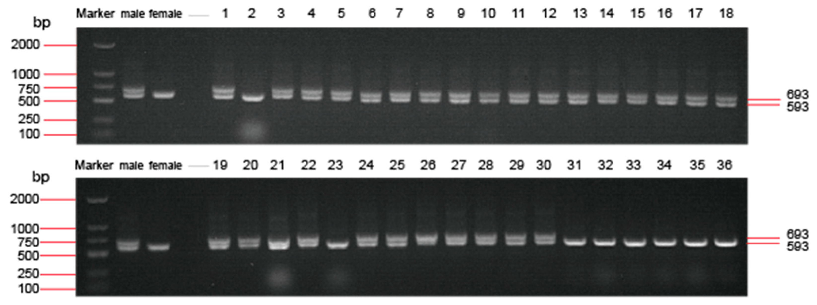
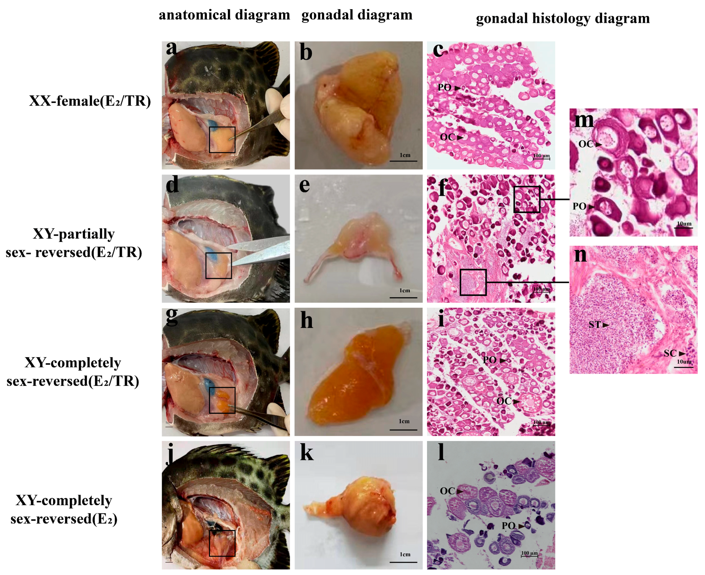
3.1. Genetic and Phenotype Sex Identification of the Treated Spotted Scats

The 28 XY fish were sampled from the treatment group using sex-linked markers. The electrophoresis results, shown in Figure 1, showed two bands of 593 and 693 bp in XY fish and only one band of 593 bp in XX fish. The gonads of the 28 XY individuals, XY-completely sex-reversed males (4/28, 15%), had fully developed into ovaries and were morphologically highly similar in appearance to the ovaries of treated XX females (Figure 2). In XY-partially sex-reversed fish (3/28, 11%) the gonads contained both testis and ovary tissue and could be considered an ovotestis. Morphological abnormalities were found in XY sex-reversed females obtained in our laboratory by E2 treatment alone (300 mg/g), indicating that E2/TR resulted in better-developed ovaries (Figure 2j,k) [15].

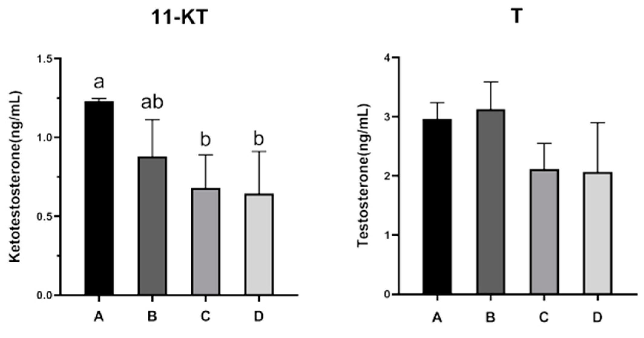
3.2. Changes in Serum Sex Steroid Hormone Levels

Serum levels of 11-KT were significantly lower in XY-completely sex-reversed, XY-partially sex-reversed, and XX females than those in XY-males. However, the 11-KT levels were found to be similar in XY-completely sex-reversed individuals and XX females. In addition, serum T levels were comparable in all groups of fish and were only slightly lower in XY-completely sex-reversed and female individuals compared to males and XY-partially sex-reversed individuals (Figure 3).

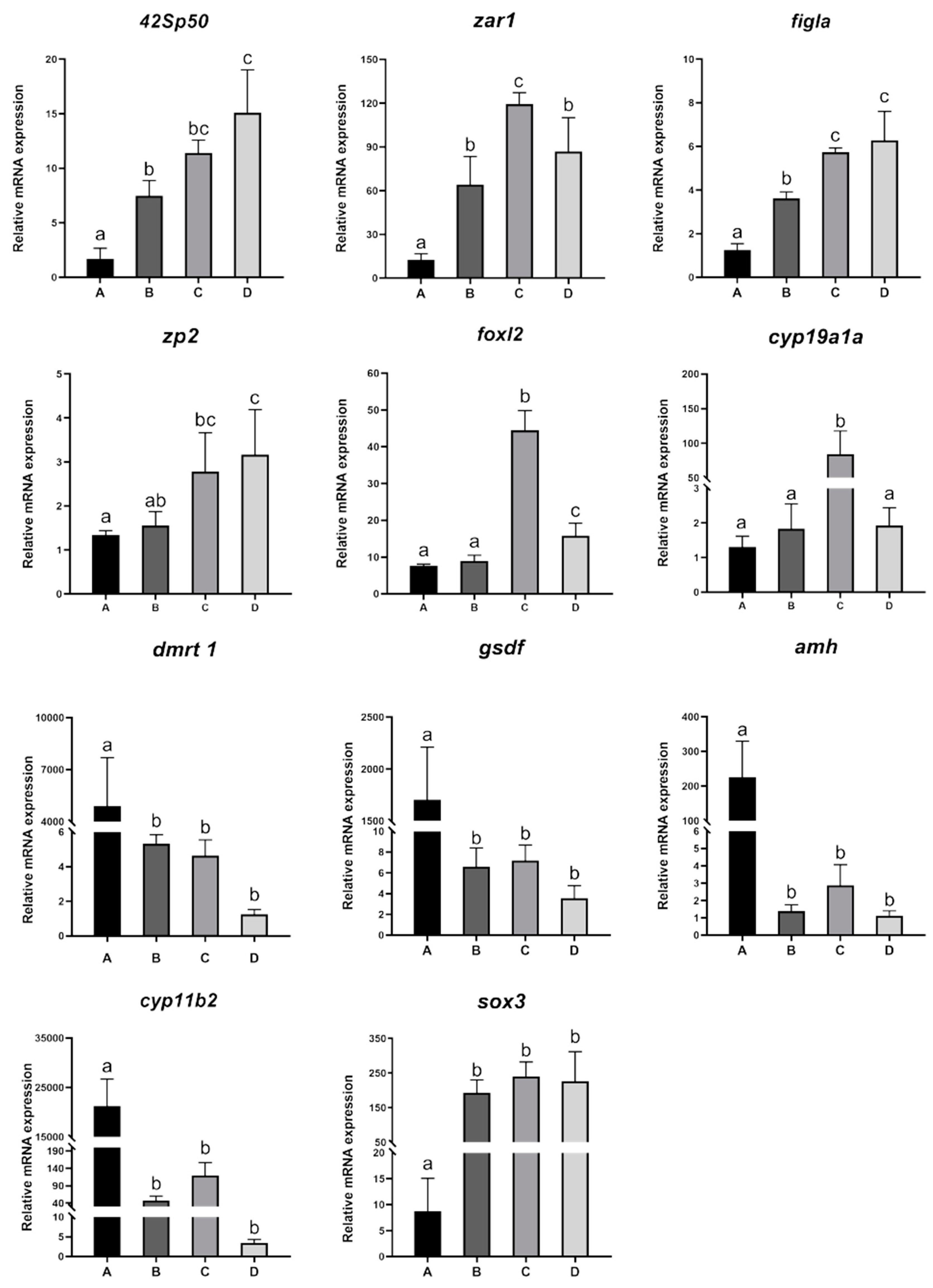
3.3. Expression Changes in Sex Differentiation-Related Genes

The female-related genes 42sp50, foxl2, figla, zar1, and zp2 were significantly more highly expressed in XX females and XY-completely sex-reversed fish than in XY-partially sex-reversed fish and XY males, with the lowest expression in XY males. The male-related genes dmrt1, gsdf, amh, and cyp11b2 were significantly more highly expressed in XY males than in XY-partially sex-reversed fish, XY-completely sex-reversed fish, and XX females, and there were no significant differences between them, although expression was lowest in XX females (Figure 4).

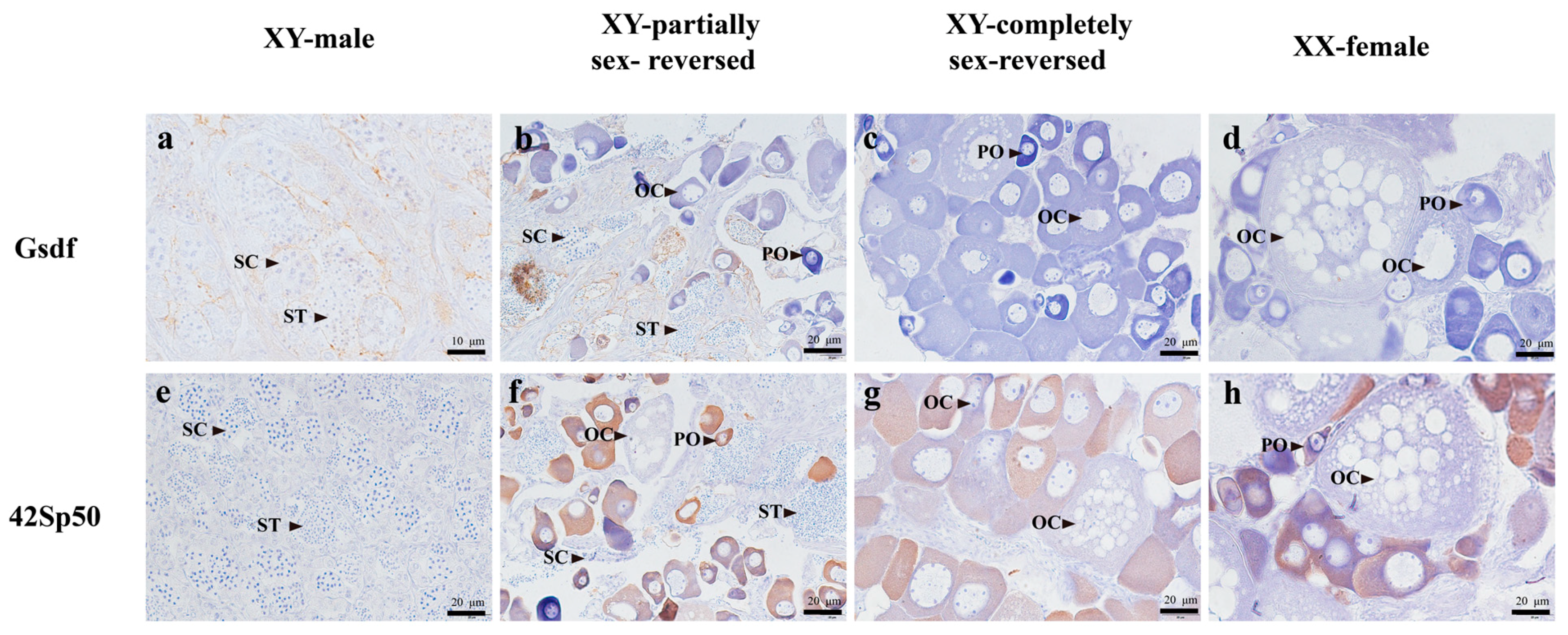
3.4. Localization Analysis of Key Gene Expression by Immunohistochemical (IHC) Assay

Gsdf staining signals were detected only in somatic cells surrounding spermatocytes of XY-partially sex-reversed fish and XY male fish, with no signals in XY-completely sex-reversed and female fish. While 42Sp50 signals were detected in oocytes of the ovarian tissue in XY-partially sex-reversed, XY-completely sex-reversed, and XX female fish, no signals were detected in males.

4. Discussion

Estrogens play an essential role in female sex determination and gonadal differentiation and maintenance in fish. Long-term exposure to exogenous estrogens can affect gonadal development in fish, resulting in the feminization of gonadal morphology and histological structure or even male-to-female reversal [21,22,23,24,25]. E2 is a natural inducer of female sex differentiation in fish, and numerous studies have investigated exogenous E2-induced male sex reversal in various fish species [26,27]. However, it is important to note that E2 has toxic properties and prolonged exposure can cause gonadal developmental abnormalities and other adverse effects. In addition, the toxicity of estrogen varies depending on the treatment method, dosage, and biological growth stage. Therefore, a crucial aspect of fish sex control breeding technology is to determine how to use estrogen treatment to obtain sex-reversed, fertile individuals. Fuzzen et al. [28] demonstrated that E2 interferes with the normal functioning of the endocrine system in zebrafish. Zhou et al. [29] found that E2 had a negative effect on the growth of small yellow croaker (Larimichthys polyactis). Cravalho et al. [30] showed that 50 and 100 mg∙kg−1 E2 had an inhibitory effect on the body length and body mass of common snook (Centropomus undecimalis). Blazquez et al. [31] found that E2 inhibited the growth of body mass in sea bass (Dicentrarchus labrax). Teal Chad et al. [32] showed that in green sunfish (Lepomis cyanellus), the survival rate of the E2-treated group was lower than that of the control group and growth was suppressed. In addition, hormone-induced sex reversal in fish has also been linked to its concentration. At concentrations above a certain threshold, toxicity increases and may cause developmental impairment, gonadal malformation, and decreased survival rate in fish. It is therefore advisable to explore a variety of induction methods in order to achieve a balanced and practical approach. Previous studies in our laboratory have shown that no sex reversal was observed in either XX or XY individuals fed a normal diet, whereas 2-month-old XY spotted fish treated with E2 alone could be reversed to females with ovarian malformations (Figure 2k) [15]. In this preliminary study, normally developing gonads were obtained applying the combined treatment of E2 and TR (Figure 2h). It is hypothesized that TR could potentially mitigate the detrimental effects of E2 to some extent and play an important role in the sexual reversal of spotted scat.
TR inhibits 3-β-hydroxysteroid dehydrogenase (3β-HSD), an enzyme that converts dehydroepiandrosterone (DHEA) to androstenedione (A4) [33], a key androgen precursor [34]. When administered alone to male Nile tilapia, TR significantly reduced serum concentrations of 11-KT and downregulated the expression of cyp11b2, the gene encoding androgen synthase, while showing no effect on the expression of foxl2 and cyp19a1a or serum E2 levels. In addition, immunohistochemistry failed to detect cyp19a1a expression in the gonads of XY fish within these treatment groups, indicating that androgen suppression alone did not result in sex reversal in XY fish [16]. In male Nile tilapia, serum levels of 11-KT and cyp11b2 expression were significantly lower in sex-reversed fish treated with TR and E2 than in sex-reversed fish treated with E2 alone. This indicates that concurrent treatment with TR and E2 inhibits androgen production more completely [35,36,37,38]. Gonadal differentiation in dioecious fish is initiated by the induction of sex-determining genes [39]. Dmrt1 is the candidate male sex-determining gene in spotted scat, a key transcription factor that contributes significantly to male gonadal determination and sex differentiation [15]. The results of this study showed that dmrt1 expression was significantly reduced in both XY-partially sex-reversed and XY-completely sex-reversed fish compared to XY males with testes. The same is true for male development-related members of the TGF-β superfamily, including gsdf [19] and amh [40]. gsdf acts as a downstream gene in the dmrt1 male sex determination pathway, contributing to testicular differentiation and early germ cell development in fishes [41,42,43,44]. In the spotted scat, gsdf expression in the testis significantly exceeds that in the ovary at the adult stages [19]. In Nile tilapia, mutations in gsdf cause male-to-female sex reversal, suggesting that gsdf acts as a male sex differentiation factor [45]. Amh, whose duplicates are also a sex-determining gene in some fish species (e.g., Hypoatherina tsurugae and Nile tilapia), where it mainly controls spermatogonial differentiation and proliferation in the early stages of the gonad and represses the oocyte development [40,46,47,48]. Based on the results of this preliminary study, it is hypothesized that the co-treatment with E2/TR has an effect on the expression of male-related genes, such as dmrt1, gsdf, and amh, during gonadal development in spotted scat. The gene cyp19a1a, a key gene for estrogen synthesis and regulated by the transcription factor foxl2 for ovarian differentiation and maintenance, showed dimorphic expression in the gonads, favoring the ovary [49,50,51,52]. In male fish, exogenous estrogen treatment promotes the upregulation of cyp19a1a expression, which promotes gonadal development towards the ovary [53]. The expression of cyp19a1a and foxl2 was significantly higher in XY-completely sex-reversed fish than in XY males in the spotted scat obtained in this study. In Nile tilapia, 42Sp50 expression is much higher in the ovary than in the testis and this is an important factor in early ovarian development. Its expression is only associated with phenotypic sex and not with genotypic sex. Mutations in this gene result in impaired folliculogenesis and infertility in female Nile tilapia [54]. Our IHC results showed that the location of 42Sp50 in the ovaries of XY-completely sex-reversed individuals was very similar to that in the ovaries of XX females. The location of 42Sp50 in the XY sex-reversed fish obtained by E2 treatment is also similar to that of the untreated XX females (Figure 5) [17]. Therefore, we speculate that the gonads of XY-completely sex-reversed spotted scat obtained through the preliminary combined treatment trial may have functional ovaries based on the ovarian gene expression profiles.
The estrogen concentration, treatment time, and size of treated fish used in the current study were similar to those used in previous studies [15]. The XY-completely sex-reversed fish obtained from the current preliminary study applying combined E2/TR treatment presented similarities to XX fish in terms of ovarian appearance, expression of most relevant gene and hormone levels, and are likely to develop into normal females. In contrast, ovaries obtained fish treated with E2 alone were deformed and infertile, and the appearance of the gonads was significantly different between fish treated with E2 alone and E2/TR (Figure 2). The appearance of ovaries obtained in the preliminary study of E2/TR treatment was similar to that of normal females, with a triangular shape [12]. This may be due to the addition of TR, which reduced the level of androgens in the fish’s body, thereby reducing the toxic effects at the time of treatment. However, the hypothesis that the combined E2/TR treatment reduced E2 toxicity is not sufficiently supported by our data. In terms of gene expression, some genes showed significant differences between XY-completely sex-reversed individuals and XX female fish (Figure 4). In addition, our previous studies have also shown that some genes are differentially expressed in the ovaries of sex-reversed XY fish treated with E2 alone compared to untreated XX fish [15]. Therefore, further comprehensive studies are needed to evaluate the efficacy of sex reversal induced by the combination of E2 and TR. In our future investigations, we will increase the blank control group and employ different combinations of E2/TR concentration to perform comparative treatment experiments. In addition, fertility tests of the completely sex-reversed XY pseudo-females will be performed.

5. Conclusions

In this study, we induced XY-completely sex-reversed spotted scat for the first time with the E2/TR combination. The normal appearance of the ovaries of the sex-reversed XY fish indicates that they may be fertile, providing an important basis for breeding all-male offspring in the spotted scat.

Author Contributions

Conceptualization, Z.-T.J. and G.-L.L.; methodology, Z.-T.J., G.S., and G.-L.L.; validation, D.-N.J. and H.-J.S.; formal analysis, Z.-T.J. and D.-N.J.; resources, G.-L.L. and G.S.; data curation, Z.-T.J. and Y.-Q.H.; writing—original draft preparation, Z.-T.J.; writing—review and editing, D.-N.J., Y.-Q.H., and Y.L.; visualization, Z.-T.J. and Y.L.; supervision, D.-N.J. and H.-J.S.; project administration, G.-L.L.; funding acquisition, G.-L.L. All authors have read and agreed to the published version of the manuscript.

Funding

This study was supported by grants from the National Natural Science Foundation of China [grant numbers: 32172971, 31972775, 32002368, and 32273131]; Guangdong Basic and Applied Basic Research Foundation [grant number: 2021A1515010430]; Guangdong Provincial Science and Technology Programme [grant number: 2023B0202010005]; and Zhanjiang Science and Technology Plan Project (grant number: 2022A01046).

Institutional Review Board Statement

The study was approved by the IACUC, Fisheries College of Guangdong Ocean University (approval number GDOU-IACUC-2021-A1225).

Data Availability Statement

Data are contained within the article.

Acknowledgments

We thank Youxing Xie of Guangdong Ocean University (Zhanjiang, China) for his help in breeding spotted scat.

Conflicts of Interest

The authors declare no conflict of interest.

References

  1. Zhu, Y.Y.; Zhang, M.; Ye, K.; Shen, W.L.; Wu, X.F.; Wang, Z.Y. Effects of 17β-estradiol on the Expression of Genes related to Sex Differentiation of the Large Yellow Croaker. J. Jimei Univ. 2019, 24, 6. [Google Scholar]
  2. Li, C.Y. The effects on sexual reversion of Xiphophrus helleri by temperature. Jilin Norm. Univ. J. 2001, 1, 70–72. [Google Scholar]
  3. Robert, H. Devlin and Yoshitaka Nagahama. Sex determination and sex differentiation in fish: An overview of genetic, physiological, and environmental influences. Aquaculture 2002, 208, 191–364. [Google Scholar]
  4. Ge, X.P.; Xia, D.Q.; Yu, J.H. Research progress of fish sex determination. J. Fish. Sci. China 2002, 9, 371–374. [Google Scholar]
  5. Strüssmann, C.A.; Takashima, F.; Toda, K. Sex differentiation and hormonal feminization in pejerrey Odontesthes bonariensis. Aquaculture 1996, 139, 31–45. [Google Scholar] [CrossRef]
  6. Mei, J.; Gui, J.F. Genetic basis and biotechnological manipulation of sexual dimorphism and sex determination in fish. Sci. China Life Sci. 2015, 58, 124–136. [Google Scholar] [CrossRef]
  7. Barry, T.P.; Fast, A.W. Biology of the spotted scat (Scatophagus argus) in the Philippines. Asian Fish. Sci. 1992, 5, 163–179. [Google Scholar] [CrossRef]
  8. Cai, Z.P.; Wang, Y.; Hu, J.W.; Zhang, J.B.; Lin, Y.G. Reproductive biology of Scatophagus argus and artificial induction of spawning. J. Trop. Oceanogr. 2010, 29, 180–185. [Google Scholar]
  9. Biswas, G.; Sundaray, J.K.; Bhattacharyya, S.B.; Kailasam, M. Evaluation of growth performance and survival of wild collected spotted scat, Scatophagus argus (Linnaeus, 1766) during rearing of fry to marketable size juveniles for aquarium trade at varied stocking densities. J. Indian Soc. Coast. Agric. Res. 2016, 34, 120–126. [Google Scholar]
  10. Mustapha, U.F.; Jiang, D.N.; Liang, Z.H.; Gu, H.T.; Yang, W.; Chen, H.P.; Deng, S.P.; Wu, T.L.; Tian, C.X.; Zhu, C.H.; et al. Male-specific Dmrt1 is a candidate sex determination gene in spotted scat (Scatophagus argus). Aquaculture 2018, 495, 351–358. [Google Scholar] [CrossRef]
  11. Mandal, B.; Kailasam, M.; Bera, A.; Sukumaran, K.; Hussain, T.; Makesh, M.; Thiagarajan, G.; Vijayan, K.K. Gonadal recrudescence and annual reproductive hormone pattern of captive female Spotted Scats (Scatophagus argus). Anim. Reprod. Sci. 2020, 213, 106273. [Google Scholar] [CrossRef] [PubMed]
  12. Cui, D.; Liu, Z.W.; Liu, N.X.; Zhang, Y.Y.; Zhang, J.B. Histological study on the gonadal development of Scatophagus Arg. J. Fish. China 2013, 37, 5. [Google Scholar] [CrossRef]
  13. Sun, X.N.; Liu, J.Y.; Feng, G.P.; Zhuang, P.; Wang, Y. Advances in reproductive biology of Scatophagus argus. Fish. Sci. 2022, 47, 283–288. [Google Scholar]
  14. Piferrer, F. Endocrine sex control strategies for the feminization of teleost fish. Aquaculture 2001, 197, 229–281. [Google Scholar] [CrossRef]
  15. Mustapha, U.F. Early Gonadal Development, Expression Profile and Regulation of Sex-Related Genes and Hormonal Induction of Sex-Reversal Mechanism in Scatophagus argus. Ph.D. Thesis, Guangdong Ocean University, Zhanjiang, China, 2022, (unpublished material). [Google Scholar]
  16. Shi, H.J. Functional Study of Dax1, Dax2, Foxj1a and Foxh1 in Gametogenesis in tilapia. Ph.D. Thesis, South-West University, Chongqing, China, 2018. [Google Scholar]
  17. Zhi, F.; Jiang, D.N.; Mustapha, U.F.; Li, S.X.; Shi, H.J.; Li, G.L.; Zhu, C.H. Expression and regulation of 42Sp50 in spotted scat (Scatophagus argus). Front. Genet. 2022, 13, 964150. [Google Scholar] [CrossRef] [PubMed]
  18. Mustapha, U.F.; Peng, Y.X.; Huang, Y.Q.; Assan, D.; Zhi, F.; Shi, G.; Huang, Y.; Li, G.L.; Jiang, D.N. Comparative transcriptome analysis of the differentiating gonads in Scatophagus argus. Front. Mar. Sci. 2022, 9, 962534. [Google Scholar] [CrossRef]
  19. Jiang, D.N.; Mustapha, U.F.; Shi, H.J.; Huang, Y.Q.; Si-Tu, J.X.; Wang, M.; Deng, S.P.; Chen, H.P.; Tian, C.X.; Zhu, C.H.; et al. Expression and transcriptional regulation of gsdf in spotted scat (Scatophagus argus). Comp. Biochem. Physiol. B 2019, 233, 35–45. [Google Scholar] [CrossRef]
  20. Li, M.; Sun, Y.; Zhao, J.; Shi, H.; Zeng, S.; Ye, K.; Jiang, D.; Zhou, L.; Sun, L.; Tao, W.; et al. A tandem duplicate of anti-mullerian hormone with a missense SNP on the y chromosome is essential for male sex determination in Nile tilapia, Oreochromis niloticus. PLoS. Genet. 2015, 11, e1005678. [Google Scholar] [CrossRef]
  21. Jiang, X.L. Effects of Sex Steroids on Sex Determination and Differentiation of Nile tilapia. Master’s Thesis, South-West University, Chongqing, China, 2014; pp. 21–27. [Google Scholar]
  22. Li, Y.J.; Wu, L.M.; Wang, L.; Ma, X.; Li, X.J. Molecular cloning and characterization of cyp19a1b gene and the effect of Letrozole on its expression in Carassius auratus. J. Fish. China 2018, 42, 1169–1180. [Google Scholar]
  23. Hu, P.; Liu, B.; Meng, Z.; Liu, X.F.; Jia, Y.D.; Yang, Z.; Lei, J.L. Recovery of gonadal development in tiger puffer Takifugu rubripes after exposure to 17β-estradiol during early life stages. Chin. J. Oceanol. Limnol. 2017, 35, 613–623. [Google Scholar] [CrossRef]
  24. Bhanda, R.K.; Nakamura, M.; Kobayashi, T.; Nagahama, Y. Suppression of steroidogenic enzyme expression during androgen-induced sex reversal in Nile tilapia (Oreochromis niloticus). Gen. Comp. Endocrinol. 2006, 145, 20–24. [Google Scholar] [CrossRef] [PubMed]
  25. Brion, F.; Tyler, C.R.; Palazzi, X.; Laillet, B.; Porcher, J.M.; Garric, J.; Flammarion, P. Impacts of 17β-estradiol, including environmentally relevant concentra-tions, on reproduction after exposure during embryo-larval-, juvenile- and adult-life stages in zebrafish (Danio rerio). Aquat. Toxicol. 2004, 68, 193–217. [Google Scholar] [CrossRef] [PubMed]
  26. Zhang, J.J.; Hu, Q.; Guan, X.M.; Li, F.; Wang, M.K.; Li, S.Z. Feminization induc-tion and hermaphroditism of northern pike (Esox lucius). J. Fish. China 2018, 42, 557–564. [Google Scholar]
  27. Wang, C.L.; Guan, W.Z.; Li, Y.Q.; Liu, F. Study on 17β-estradiol induced feminization of Pelteobagrus fulvidraco. South China Fish. Sci. 2020, 16, 25–30. [Google Scholar]
  28. Fuzzen, M.L.M.; Bernier, N.J.; Kraak, G. Differential effects of 17β-estradiol and 11-ketotestosterone on the endocrine stress response in zebrafish (Danio rerio). Gen. Comp. Endocrinol. 2001, 170, 365–373. [Google Scholar] [CrossRef]
  29. Zhou, F.Q.; Xie, Q.P.; Lou, B.; Meng, X.L.; He, X.; Sun, Y.; Zhan, W.; Chen, R.Y.; Liu, F.; Wang, L.G.; et al. Effect of 17β-estradiol bath on growth and gonadal development of early stage of Larimichthys polyactis. J. Zhejiang Ocean Univ. Nat. Sci. 2018, 37, 302–307. [Google Scholar]
  30. Carvalho, C.V.A.; Passini, G.; Costa, W.M.; Vieira, B.N.; Cerqueira, V.R. Effect of estradiol-17β on the sex ratio, growth and survival of juvenile common snook (Centropomus undecimalis). Acta. Sci. Anim. Sci. 2014, 36, 239–245. [Google Scholar] [CrossRef]
  31. Blazquez, M.; Zanuy, S.; Carrillo, M.; Piferrer, F. Structural and functional effects of early exposure to estradiol-17β and 17α-ethynylestradiol on the gonads of the gonochoristic teleost Dicentrarchus labrax. Fish. Physiol. Biochem. 1998, 18, 37–47. [Google Scholar] [CrossRef]
  32. Chad, N.T.; Daniel, J.S.; Susan, B.F.; Colby, M.R.; Kevin, F.; Javan, M.B.; William, T.S.; Scott, A.B. The effects of estradiol-17β on the sex reversal, survival, and growth of green sunfish Lepomis cyanellus. Aquaculture 2022, 562, 738853. [Google Scholar]
  33. Ouschan, C.; Lepschy, M.; Zeugswetter, F.; Möstl, E. The influence of trilostane on steroid hormone metabolism in canine adrenal glands and corpora lutea-an in vitro study. Vet. Res. Commun. 2012, 36, 35–40. [Google Scholar] [CrossRef]
  34. DeQuattro, Z.A.; Hemming, J.D.; Barry, T.P. Effects of androstenedione exposure on fathead minnow (Pimephales promelas) reproduction and embryonic development. Environ. Toxicol. Chem. 2015, 34, 2549–2554. [Google Scholar] [CrossRef]
  35. Kobayashi, T.; Kajiura-Kobayashi, H.; Nagahama, Y. Induction of XY sex reversal by estrogen involves altered gene expression in a teleost, tilapia. Cytogenet. Genome. Res. 2003, 101, 289–294. [Google Scholar] [CrossRef]
  36. Nakamura, M.; Bhandari, R.K.; Higa, M. The role estrogens play in sex differentiation and sex changes of fish. Fish. Physiol. Biochem. 2003, 28, 113–117. [Google Scholar] [CrossRef]
  37. Lee, Y.H.; Wu, G.C.; Du, J.L.; Chang, C.F. Estradiol-17beta induced a reversible sex change in the fingerlings of protandrous black porgy, Acanthopagrus schlegeli Bleeker: The possible roles of luteinizing hormone in sex change. Biol. Reprod. 2004, 71, 1270–1278. [Google Scholar] [CrossRef]
  38. Wu, G.C.; Tomy, S.; Nakamura, M.; Chang, C.F. Dual roles of cyp19a1a in gonadal sex differentiation and development in the protandrous black porgy, Acanthopagrus schlegeli. Biol. Reprod. 2008, 79, 1111–1120. [Google Scholar] [CrossRef]
  39. Zhang, X.; Guan, G.; Li, M.; Zhu, F.; Liu, Q.; Naruse, K.; Herpin, A.; Nagahama, Y.; Li, J.; Hong, Y. Autosomal gsdf acts as a male sex initiator in the fish medaka. Sci. Rep. 2016, 6, 19738. [Google Scholar] [CrossRef]
  40. Hattori, R.S.; Murai, Y.; Oura, M.; Masuda, S.; Majhi, S.K.; Sakamoto, T.; Fernandino, J.I.; Somoza, G.M.; Yokota, M.; Strussmann, C.A. A Y-linked anti-Mullerian hormone duplication takes over a critical role in sex determination. Proc. Natl. Acad. Sci. USA 2012, 109, 2955–2959. [Google Scholar] [CrossRef]
  41. Inaba, H.; Hara, S.; Horiuchi, M.; Ijiri, S.; Kitano, T. Gonadal expression profiles of sex-specific genes during early sexual differentiation in Japanese eel Anguilla japonica. Fish. Sci. 2021, 87, 203–209. [Google Scholar] [CrossRef]
  42. Smith, E.K.; Guzmán, J.M.; Luckenbach, J.A. Molecular cloning, characterization, and sexually dimorphic expression of five major sex differentiation-related genes in a Scorpaeniform fish, sablefish (Anoplopoma fimbria). Comp. Biochem. Physiol. B 2013, 165, 125–137. [Google Scholar] [CrossRef] [PubMed]
  43. Wang, W.; Zhu, H.; Dong, Y.; Dong, T.; Tian, Z.; Hu, H. Identification and dimorphic expression of sex-related genes during gonadal differentiation in sterlet Acipenser ruthenus, a primitive fish species. Fish. Physiol. Biochem. 2017, 43, 1557–1569. [Google Scholar] [CrossRef] [PubMed]
  44. Yan, Y.L.; Desvignes, T.; Bremiller, R.; Wilson, C.; Dillon, D.; High, S.; Draper, B.; Buck, C.L.; Postlethwait, J. Gonadal soma controls ovarian follicle proliferation through Gsdf in zebrafish. Dev. Dyn. 2017, 246, 925–945. [Google Scholar] [CrossRef]
  45. Kaneko, H.; Ijiri, S.; Kobayashi, T.; Izumi, H.; Kuramochi, Y.; Wang, D.S.; Mizuno, S.; Nagahama, Y. Gonadal soma-derived factor (gsdf), a TGF-beta superfamily gene, induces testis differentiation in the teleost fish Oreochromis niloticus. Mol. Cell. Endocrinol. 2015, 415, 87–99. [Google Scholar] [CrossRef]
  46. Zhang, Z.; Zhu, B.; Chen, W.; Ge, W. Anti-Müllerian hormone (AMH/amh) plays dual roles in maintaining gonadal homeostasis and gametogenesis in zebrafish. Mol. Cell. Endocrinol. 2020, 1, 517. [Google Scholar] [CrossRef]
  47. Bej, D.K.; Miyoshi, K.; Hattori, R.S.; Strüssmann, C.A.; Yamamoto, Y. A duplicated, truncated amh gene is involved in male sex determination in an Old World Silverside. G3 2017, 7, 2489–2495. [Google Scholar] [CrossRef]
  48. Zhang, X.; Li, M.; Ma, H.; Liu, X.; Shi, H.; Li, M.; Wang, D. Mutation of foxl2 or cyp19a1a results in female to male sex reversal in XX Nile tilapia. Endocrinology 2017, 158, 2634–2647. [Google Scholar] [CrossRef]
  49. Ijiri, S.; Kaneko, H.; Kobayashi, T.; Wang, D.S.; Sakai, F.; Paul-Prasanth, B.; Nakamura, M.; Nagahama, Y. Sexual dimorphic expression of genes in gonads during early differentiation of a teleost fish, the Nile tilapia, Oreochromis niloticus. Biol. Reprod. 2008, 78, 333–341. [Google Scholar] [CrossRef]
  50. Hirst, C.E.; Major, A.T.; Smith, C.A. Sex determination and gonadal sex differentiation in the chicken model. Int. J. Dev. Biol. 2018, 62, 149–162. [Google Scholar] [CrossRef]
  51. Jiang, M.; Jia, S.; Chen, J.; Chen, K.; Ma, W.; Wu, X.; Luo, H.; Li, Y.; Zhu, Z.; Hu, W. Timing of gonadal development and dimorphic expression of sex-related genes in gonads during early sex differentiation in the Yellow River carp. Aquaculture 2020, 518, 734825. [Google Scholar] [CrossRef]
  52. Wei, H.; Li, W.; Liu, T.; Li, Y.; Liu, L.; Shu, Y.; Zhang, L.J.; Wang, S.; Xing, Q.; Zhang, L.L.; et al. Sexual development of the hermaphroditic scallop Argopecten irradians revealed by morphological, endocrine and molecular analysis. Front. Cell Dev. Biol. 2021, 9, 646754. [Google Scholar] [CrossRef]
  53. Li, M.; Sun, L.; Wang, D. Roles of estrogens in fish sexual plasticity and sex differentiation. Gen. Comp. Endocrinol. 2019, 277, 9–16. [Google Scholar] [CrossRef]
  54. Chen, J.L. The Role of Elongation Factor eEF1A1b and 42Sp50 in Gametogenesis in Nile tilapia. Ph.D. Thesis, South-West University, Chongqing, China, 2018. [Google Scholar]
Figure 1. Genotype identification of the treated spotted scat using sex-linked markers. XY fish with two bands of 593 and 693 bp, and only one band 593 bp in XX fish.
Fishes 09 00001 g001
Figure 2. Morphological and histological observations of gonads from E2/TR or E2-treated spotted scats; (a,d,g,j) are gonadal anatomy of XX female (E2/TR), XY-partially sex-reversed (E2/TR), XY-completely sex-reversed fish (E2/TR) and XY-completely sex-reversed fish (E2), whereas XY-completely sex-reversed fish (E2) is a sample obtained in our previous studies [15]. (b) The appearance of the ovary of treated XX fish (E2/TR); (e,h) the appearance of the gonads of XY-partially sex-reversed and XY-completely sex-reversed fish after the combined treatment with E2/TR, respectively; (k) the appearance of the gonads of XY-completely sex-reversed fish after treatment with E2; (c,f,i,l) are the gonadal section diagrams of XX female (E2/TR), XY-partially sex-reversed (E2/TR), XY-completely sex-reversed (E2/TR) and XY-completely sex-reversed (E2) fish, respectively; (m,n) are localized magnified views of the ovary and spermatheca, respectively. Black arrowheads indicate germ cells. PO: primary oocyte; OC: oocyte; SC: spermatocyte; ST: spermatid.
Fishes 09 00001 g002
Figure 3. Serum 11-ketotestosterone and testosterone levels in E2/TR-treated spotted scat. A. male, B. XY-partially sex-reversed, C. XY-completely sex-reversed, D. Female. n = 3, x = ±SD. Different lowercase letters above the error bar indicate significant differences (p < 0.05).
Fishes 09 00001 g003
Figure 4. Expression levels of sex determination and differentiation related genes in the gonads of E2/TR-treated spotted scats determined by real-time PCR. A. XY male, B. XY-partially sex-reversed, C. XY-completely sex-reversed, D. XX female. n = 3, x = ±SD. Different lowercase letters above error bars indicate significant differences (p < 0.05).
Fishes 09 00001 g004
Figure 5. Localization of Gsdf and 42Sp50 in E2/TR-treated XX and XY spotted scat by immunohistochemistry (IHC); (ad) expression of Gsdf staining signals in XY-male, XY-partially sex-reversed, XY-completely sex-reversed and XX-female gonads, respectively; (eh) expression of 42Sp50 staining signals in XY-male, XY-partially sex-reversed, XY-completely sex-reversed and XX-female gonads, respectively Brownish color indicates a positive signal. Black arrowheads indicate germ cells, PO: primary oocyte, OC: oocyte, SC: spermatocyte, ST: spermatid.
Fishes 09 00001 g005
Table 1. Primer sequences used in this study.
Gene NamePrimer Sequences (5′-3′) ForwardPrimer Sequences (5′-3′) ReverseNCBI ID
42sp50GCCACCAGTGCGAACCATCAATTGCCTGTGCGACGGTCAAG124049430
foxl2GGCAGAACAGTATCAGACACCATCTCCTCCGAACAAG124058880
figlαGATACAGACAGCGATGATGGGTGCTACTTGAATGATGAA124057770
zar1ACCACAGAAGAGTGAAGATGCCTCAACACGATACGGATT124061181
zp2GAGGATTCAGTGTGGTTCACTCTAAGCATTCGGTGTCT124059932
cyp19a1aTGCATCGGCATGAACGAGAGGTTCCAGGTCATCCAGGTGAGTCT124061003
dmrt1GAAGGCAGCAAGATCAGGAGGACAGCAGCAGGTCAGATGGTTCC124057575
amhTTCCACAGAGACCAGAGATTTCAGAAGTTCCAGTCCATT124061215
gsdfGGTCTCTGGCTACTCTGTGCATCCTGGTCATTGGTC124051891
cyp11b2GTCTACTGCTCAACAAGGAGCCAATACGCTCACCATA124065384
sox3CCGTAGGAAGACCAAGACTGTTCATGCTGTGATGCT124068361
b2mGAACTTCCTGGCGCTAAGCATATGATGTCCCCATGAGTGACC124053526
Disclaimer/Publisher’s Note: The statements, opinions and data contained in all publications are solely those of the individual author(s) and contributor(s) and not of MDPI and/or the editor(s). MDPI and/or the editor(s) disclaim responsibility for any injury to people or property resulting from any ideas, methods, instructions or products referred to in the content.

