Article

Effect of the Flagellar Gene fliL on the Virulence of Pseudomonas plecoglossicida to Hybrid Grouper (Epinephelus fuscoguttatus ♀ × E. lanceolatus ♂)

1
Fisheries College, Jimei University, Xiamen 361021, China
2
College of Life Science, Xinjiang Agricultural University, Urumqi 830052, China
*
Author to whom correspondence should be addressed.
Fishes 2023, 8(8), 397; https://doi.org/10.3390/fishes8080397
Submission received: 9 June 2023 / Revised: 21 July 2023 / Accepted: 24 July 2023 / Published: 1 August 2023
(This article belongs to the Special Issue Interactions between Fish and Pathogens in Aquaculture)

Abstract

:
Pseudomonas plecoglossicida is the pathogen of visceral white spot disease in marine fish, which usually occurs at 16–19 °C and has resulted in heavy economic losses. Our previous RNA sequencing revealed that the expression of the fliL gene in P. plecoglossicida was significantly up-regulated during infection of the host. In order to study the influence of the fliL gene on the virulence of P. plecoglossicida, the fliL gene of the NZBD9 strain was knocked out by the homologous recombination method, the fliL gene-deleted strain (ΔfliL strain) constructed, and complemented the fliL gene to the ΔfliL strain to obtain the C-ΔfliL strain. The growth curves of the NZBD9 strain, ΔfliL strain, and C-ΔfliL strain did not show significant differences. Compared with the NZBD9 strain, the motility, adhesion, and biofilm formation ability were tendered in the ΔfliL strain (p < 0.05); the complement of the fliL gene enhanced these abilities to the level of the NZBD9 strain. The results of artificial infection experiments showed that the LD50 of NZBD9 strain, ΔfliL strain, and C-ΔfliL strain in hybrid grouper (Epinephelus fuscoguttatus ♀ × E. lanceolatus ♂) were 5.0 × 10³ CFU/fish, 6.3 × 10⁴ CFU/fish, and 1.3 × 10³ CFU/fish, respectively. RNA sequencing was performed on wild-type strains and ΔfliL strains. A total of 126 differentially expressed genes (DEGs) were screened (p < 0.05), of which 114 were downregulated and 12 were upcontrolled, among which several genes related to the six-type secretion system and transport activity were significantly downregulated. The DEGs were aligned to the GO and KEGG databases and enriched to 44 GO pathways and 39 KEGG pathways, respectively. The active pathways of ABC transporters were significantly enriched in both databases. These results indicate that the fliL gene is related to the movement, biofilm formation, and adhesion ability of P. plecoglossicida, and may reduce virulence by affecting substance transport and bacterial secretion.
Key Contribution: The fliL gene might be influencing the P. percoglossicida’s system of material transportation and flagellum assembly to reduce its virulence.

1. Introduction

With the vigorous development of the mariculture industry, especially the promotion of a high-density aquaculture model, the frequency and range of aquatic diseases caused by bacteria have gradually increased [1,2,3]. Marine and freshwater fish diseases caused by Vibrio, Pseudomonas, and Aeromonas are more common, causing serious losses to aquaculture [4,5,6,7]. Pathogenic Pseudomonas plecoglossicida was first isolated and identified as a new strain in ayu (Plecoglossus altivelis) suffering from bacterial haemorrhagic ascites from Japan in 2000 [8]. In recent years, reports have revealed that this pathogen can cause infection in a wide variety of fish, including crucian carp (Carassius auratus), rainbow trout (Oncorhynchus mykiss), large yellow croaker (Larimichthys crocea), and orange-spotted grouper (Epinephelus coioides), causing high mortality and posing a great threat to aquaculture [9,10,11]. Previous research in our laboratory found that many genes were involved in regulating the virulence of P. plecoglossicida. The rpoD (RNA polymerase sigma factor) gene was found to be a key virulence gene in the process of infecting grouper [12], and fliG (flagellar motor switch protein, fliG), flgC (flagellar basal-body rod protein, flgC), exbB (biopolymer transport gene), and other genes also made great contributions to the infection of orange-spotted grouper [13,14,15]. However, the pathogenesis of bacterial diseases is an extremely complex process, and the virulence of P. plecoglossicida is not well understood. Based on the early transcriptome data of our laboratory (NCBI, SRP115064), the results showed that the fliL gene was significantly overexpressed in the spleen of orange-spotted grouper infected with the wild-type P. plecoglossicida strain NZBD9. It is speculated that this gene is related to the virulence of P. plecoglossicida.
Most bacteria migrate to a favorable living environment by rotating flagella/flagella [16]. In this process, torque is generated by the flagella motor to provide rotational power for the flagella filament [17]. FliL (flagellar basal body-associated FliL family protein) is a single-transmembrane protein (IM protein) associated with flagellar motor function [18], close to the flagellar basal body [19]. Studies have shown that the interaction of FliL with the rotor (MS ring) and stator (MotA and MotB) enhances the flagellar motor torque, thereby maintaining the integrity and stability of the flagellar rod [19,20,21,22]. Motility mainly plays a role in the early stage of bacterial infection [23]. Studies have found that fliL is essential for the movement of Rhodobacter sphericum, and in the absence of the fliL gene, flagella rotation is seriously damaged [24]. In Vibrio alginolyticus, loss of fliL will not affect cell morphology or flagella, but it will lead to a significant decrease in swimming speed under a high load. Therefore, fliL is very important to generate torque under high-load conditions [16]. Moreover, it is reported that FliL is related to the swimming or adhesion mechanisms of Clostridium difficile, and the loss of the fliL gene may affect the cell’s ability to perceive the surface [19]. However, research on the function of the fliL gene in P. plecogerossicida has not been published yet.
In this study, a two-step allele exchange method was used to construct an unmarked deletion strain of the fliL gene (ΔfliL strain) and a complement strain of ΔfliL (C-ΔfliL strain) of P. plecoglossicida with reference to the previous method [25]. Meanwhile, a fliL gene complement strain was constructed. In order to study the effect of the fliL gene on the pathogenicity of bacteria, the biological characteristics and virulence tests were carried out on the deleted and supplemented strains. In addition, to further elucidate the potential mechanism of virulence of the fliL gene, the transcriptomes of the NZBD9 strain, the fliL gene deletion strain of P. plecoglossicida were compared and analyzed.

2. Materials and Methods

2.1. Bacterial Strains and Culture Conditions

The strains used in this experiment were all P. plecoglossicida, in which the wild-type strain NZBD9 was isolated from the spleen of diseased, large yellow croaker. The ΔfliL strain was obtained by the knockout fliL gene of the wild strain, and the C-ΔfliL strain was obtained by complementing the fliL gene to the ΔfliL strain. All three strains were grown in Luria-Bertani (LB) medium at 18 °C or 28 °C with 220 r/min (NZBD9, ΔfliL containing 100 μg/mL ampicillin, and C-ΔfliL adding 10 μg/mL tetracycline.). Escherichia coli Top10 and DH5α were obtained from TransGen Biotech (Beijing, China) and cultured in LB broth at 37 °C with 220 r/min. The plasmid pK18mobsacB and pCM130/tac located in DH5α were preserved in our laboratory and cultured in LB broth at 37 °C with 220 r/min.

2.2. Construction of Deletion and Complement Strains

2.2.1. Construct the ΔfliL Strain

According to the genome sequence of the NZBD9 strain (NCBI, PHNR00000000), primers P1P2 and P3P4 were designed (primers’ sequence is shown in Supplementary Table S1) to amplify the upstream and downstream homologous fragment (about 800 bp) of the fliL gene, respectively, and the fusion fragment (about 1600 bp) was obtained by running overlapping PCR (P1/P4 primers). The PCR amplification procedure was as follows: 95 °C for 4 min; 30 cycles of 95 °C for 30 s, 56 °C for 30 s, and 72 °C for 1 min; and 72 °C for 5 min. The fusion fragment (ΔfliL) and plasmid pK18mobsacB were digested with restriction enzymes EcoR Ⅰ and Xba I (Takara Biotech, Beijing, China), respectively, (water bath at 37 °C for 5 h), and T4 ligase (Takara Biotech, China) was added for connection (16 °C for 5 h) [25]. Then, the ligation product was transformed into Top10 receptive cells of E. coli by heat shock, and the recombinant plasmid pK18mobsacB-ΔfliL was screened with LB agar (50 μg/mL Kan). After enzyme digestion and sequencing (Shanghai Shengong Bioengineering Co., Ltd., Shanghai, China), the correct recombinant plasmid was transformed into the susceptible cells of the NZBD9 strain by electroporation with 2.5 kV (MicroPulserTM, BIO-RAD, Mountain View, CA, USA), and the successful transformants were screened out by Kan (25 μg/mL). After dilution, the transformant was spread onto LB agar plates containing 10% sucrose until a single colony grew [26]. Then, single colonies that could grow in ampicillin but could not grow in Kan were screened; P1/P4 primers were used to PCR isolated colonies, and the wild-type strain was used as a positive control. The PCR amplification procedure was the same as above. The strain that can amplify the fusion fragment (about 1600 bp) is the strain that has lost the fliL gene (ΔfliL strain).

2.2.2. Construction of the Complement Strain C-ΔfliL

According to the fliL gene sequence of the NZBD9 strain and the enzyme restriction site of plasmid pCM130/tac, the primers C-ΔfliL-F/R were designed (primers’ sequence is shown in Table S1). The complete fliL gene fragment was amplified by PCR with the DNA of the NZBD9 strain as a template. The PCR amplification procedure was as follows: 95 °C for 4 min; 30 cycles of 95 °C for 30 s, 58 °C for 30 s, and 72 °C for 30 s; and 72 °C for 5 min. The gene fragment and plasmid pCM130/tac were digested with BsrG I and Nsi I (New England Biolabs, Ipswich, MA, USA) enzymes (water bath at 37 °C for 5 h), and T4 ligase was added for connection (16 °C for 5 h). Then, the ligation product was transformed into DH5α receptive cells of E. coli by heat shock, and the recombinant plasmid pCM130/tac-fliL was screened with LB agar (10 μg/mL tetracycline). The receptor cells of the ΔfliL strain were prepared, and the recombinant plasmid was shocked (2.5 kV) into them. The ΔfliL strain cells with recombinant plasmid were inoculated on an LB agar plate containing 10 μg/mL tetracycline and incubated under 18 °C. A single colony was selected and cultured in LB broth (containing 10 μg/mL tetracycline) for colony PCR verification. The sequence of all primers used is shown in Supplementary Table S1.

2.3. Validation of mRNA Levels of Deletion and Complement Strains

The genomes of the NZBD9 strain, ΔfliL strain, and C-ΔfliL strain were extracted using the bacterial genome DNA kit (TransGen Biotech, Beijing, China). The internal primer P5/P6 (Table S1) of the fliL gene was designed as the verification primer and subjected to PCR. The PCR amplification procedure was as follows: 95 °C for 4 min; 30 cycles of 95 °C for 30 s, 52 °C for 30 s, and 72 °C for 30 s; and 72 °C for 5 min. To verify the expression of the fliL gene at the mRNA level, total bacterial RNA was extracted with TRIzol reagent. According to the manufacturer’s protocols, the cDNA was synthesized using TransScript All-in-One First-Strand cDNA Synthesis SuperMix for qRT-PCR (One-Step gDNA Removal) (Transgenic Biotechnology Company, Beijing, China). The temperature was set to 42 °C for 15 min and 85 °C for 5 s. Compared with the NZBD9 strain, the qRT-PCR based on the method of SYBR Green, was used to run the relative gene expression quantitatively, and the relative gene expression level was calculated by the 2−ΔΔCT method. The primer of the fliL gene and the endogenous gene 16S rDNA was designed by Primer Premier 5.0, and the sequence is shown in Supplementary Table S1. Three samples were in each group, and for each sample, three replicates were performed.

2.4. Determination of Growth Curve

The NZBD9 strain, ΔfliL strain, and C-ΔfliL strain of P. plecoglossicida were cultured in LB broth at 18 °C with shaking at 220 r/min overnight to OD600 = 0.5, and then diluted with sterile LB broth to OD600 = 0.2 ± 0.01. The bacterial suspension was diluted 100,000 times with sterile LB broth, and 200 μL was added into 96-well plates after mixing evenly. There were 12 replicates for each strain, and 200 μL LB broth with 12 wells used for control group. The OD value at 600 nm was measured at 18 °C with a multifunctional microtiter plate detector (every 30 min for 48 h).

2.5. Motility Measurement

The motility assay of P. plecoglossicida was carried out according to the method of Yang et al. [15] with some modifications. The NZBD9 strain, ΔfliL strain, and C-ΔfliL strain were cultured in LB broth, and overnight cultured at 18 °C with shaking at 220 r/min until OD600 = 0.2. One uL bacterial suspension was inoculated on an LB semi-solid plate containing 0.4% agar and cultured at 18 °C for 24 h. The colonies were then photographed and the diameter of each colony was determined. Three independent biological repeats were set up for the detection of each strain, and each repeat included three technical repeats.

2.6. Determination of Bacterial Adhesion Ability

2.6.1. Preparation of Surface Mucus

Vigorous pearl gentian grouper with no parasites and wounds on the body surface was obtained from a farm without a recent history of the epidemic in Xiamen (Fujian, China). The method of Jiao et al. [14] was followed and slightly modified. We rinsed the surface of the fish with sterile PBS, gently scraped off the mucus on the surface of the fish with clean slides, and mixed well in PBS. The mucus was placed overnight at 4 °C and centrifuged for 30 min twice at 4000× g and 4 °C. The supernatants were sterilized by filtration using filters with pore sizes of 0.45-μm and 0.22-μm successively. The mucus’ protein concentration was adjusted to 1 mg protein/mL with PBS according to the Bradford Protein Assay Kit (Solarbio®, Beijing, China).

2.6.2. In Vitro Adhesion Experiment

In vitro adhesion was determined following a previous study [11]. Some 20 μL of mucus (1 mg protein/mL) was evenly coated onto a 22 mm2 glass slide area and fixed with methanol for 20 min. A total of 200 μL of a bacterial suspension (OD600 = 0.3 ± 0.01) was spotted on a mucus-coated glass slide. Then, the glass slides were placed in a humidified chamber, incubated at 18 °C for 2 h, and then washed five times in PBS (0.01 mol/L, pH = 7.2). Finally, the bacteria were fixed with 4% methanol for 30 min, stained with 1% crystal violet for 3 min, and counted under a microscope (×1000) (Leica DM4000 B LED, Leica, Wetzlar, Germany). Five glass slides were performed per strain, and the adhered bacteria in 20 randomly selected fields were counted per slide.

2.7. Biofilm Determination

Biofilm formation was explored following previous research [11] with some modifications. Overnight cultures of P. plecoglossicida were adjusted to OD600 = 0.2 ± 0.01 in LB broth. The bacterial suspension (100 μL) was added to the wells of 96-well plates (ten wells per strain) and incubated at 18 °C for 24 h. Then, each well was washed gently twice with PBS (2 mL), dyed with 120 μL of crystal violet (0.1%) for 15 min, gently washed twice with sterile PBS, and air dried. Finally, 200 μL of acetic acid (33%) was added to each well to dissolve the stained biofilm for 30 min, and OD590 of each well was determined using a SYNERGY H1 microplate reader (BioTek, Winooski, VT, USA).

2.8. Measurement of the Chemotactic Response

The assay was accomplished in accordance with the previous method [12]. The P. plecoglossicida NZBD9, ΔfliL, and C-ΔfliL strains were cultured in sterile LB broth medium at 18 °C until an OD600 of about 0.5. The bacteria were adjusted to OD600 = 1.0 using sterile PBS. One end of the mucus-filled capillary tube (inner diameter 0.1 mm, the other end sealed) was immersed in a syringe containing bacterial suspension (250 μL) and incubated at 18 °C for 1 h. Sterile PBS was applied as a blank control. The bacteria−mucus mixtures in the capillary were diluted into 995 μL PBS for serial dilution, and 100 μL bacterial suspension per gradient was coated on an LB agar plate to calculate CFU. Three replicates were set up for each strain.

2.9. Half Lethal Dose (LD50) Test for P. plecoglossicida

Healthy weight-match pearl gentian grouper were temporarily cultured in clean and sterile seawater (temperature controlled at 18 ± 1 °C) for one week. After the fish adapted to recirculating aquaculture, the infection test was carried out. Grouping was set according to the reported method [27]. A total of 160 pearl gentian grouper were randomly divided into 16 groups; each group contained 10 fish, including 5 groups of NZBD9 strain, ΔfliL strain, and C-ΔfliL strain, plus a set of negative control (PBS). The intrabitoneal injection dose of the bacterial solution was set to 5 × 10⁶, 5 × 10⁵, 5 × 10⁴, 5 × 10³, and 5 × 10² CFU/fish. The fish injected with 0.2 mL PBS was used as the negative control. The death of the injected fish was observed every 12 h after injection, and the dead fish were removed every time.

2.10. Transcriptomic Analysis

2.10.1. Library Preparation and Sequencing

Overnight cultures of the NZBD9 strain and ΔfliL strain were centrifuged (4 °C, 4000× g, 15 min), and the bacteria were collected for RNA sequencing. Three samples were prepared for each strain as independent biological replicates. The concentration and purity of bacteria total RNA extracted from the sample were detected by Nanodrop2000. The TruSeqTM total RNA library preparation kit was used to construct a library with an initial total RNA volume of 2 μg.
In the synthesis of the second strand of cDNA, dTTP was replaced by dUTP, and the second strand was degraded by adding the UNG enzyme (Illumina, San Diego, CA, USA). The enriched library was amplified by PCR and quantified by TBS380 (Picogreen). The constructed RNA-seq libraries were sequenced on the Illumina NovaSeq 6000 sequencing platform [28] by Shanghai Majorbio Biomedical Technology Co., Ltd. (Shanghai, China).

2.10.2. Raw Data Statistics and Quality Control

Using statistical methods, the base distribution and quality fluctuation of all sequencing readings in each cycle were counted to obtain raw data. Illumina Hiseq original data contained junction sequences, reads of low quality, sequences with high N-rates, and excessively short sequences, which would affect subsequent sequence assembly. Therefore, the original sequencing data was filtered to obtain high-quality sequencing data (clean data) to ensure the smooth conduct of subsequent analyses. The method was as follows: remove the adapter sequence in reads and 5′-end contains non-A, G, C, T base. The ends of the reads with a quality value less than Q20 were trimmed, the reads with 10% N content were removed, and the small fragments with a length of less than 25 bp were discarded. The FASTX-Toolkit v0.0.14 (http://hannonlab.cshl.edu/fastx_toolkit/ accessed on 22 December 2022) was used to analyze the Illumina raw sequencing data as well as library construction and sequencing quality. SeqPrep (https://github.com/jstjohn/SeqPrep accessed on 22 December 2022) was used to perform quality control of the raw sequencing data to obtain high-quality clean data to ensure the accuracy of subsequent analyses.

2.10.3. Comparison with Reference Genome

Clean data (reads) was compared by software based on the Burrows−Wheeler method, and mapped data (reads) was obtained for subsequent analysis. The sequence was indexed by classification and transformation characteristic matrix and compared with the reference genome (NCBI, GCA_003391255-1) with the software of Bowtie (http://bowtie-bio.sourceforge.net/index.shtml, accessed on 22 December 2022).

2.10.4. Differentially Expressed Genes (DEGs) and Enrichment Analysis

The gene expression levels were quantitatively analyzed by RSEM software based on one million transcripts (TPM). DESeq2 software was used to analyze the differentially expressed genes (DEGs) between the groups, and the significance screening level was p-adjust < 0.05 and |log2FC| ≥ 1. GOATOOLS (https://files.pythonhosted.org/packages/, accessed on 24 December 2022) and R script were used for GO and KEGG Pathway enrichment analysis of DEGs, and the Fisher precise test was used for calculation. In GO enrichment analysis, the Bonferroni method was used to correct the p value, and the corrected p value (FDR) was <0.05, indicating that the GO function was significantly enriched. For KEGG Pathway enrichment analysis, BH (Benjamini and Hochberg) was selected to correct the p value. The corrected p value (FDR) was set to 0.05 as the threshold, and FDR < 0.05 was considered significant enrichment.

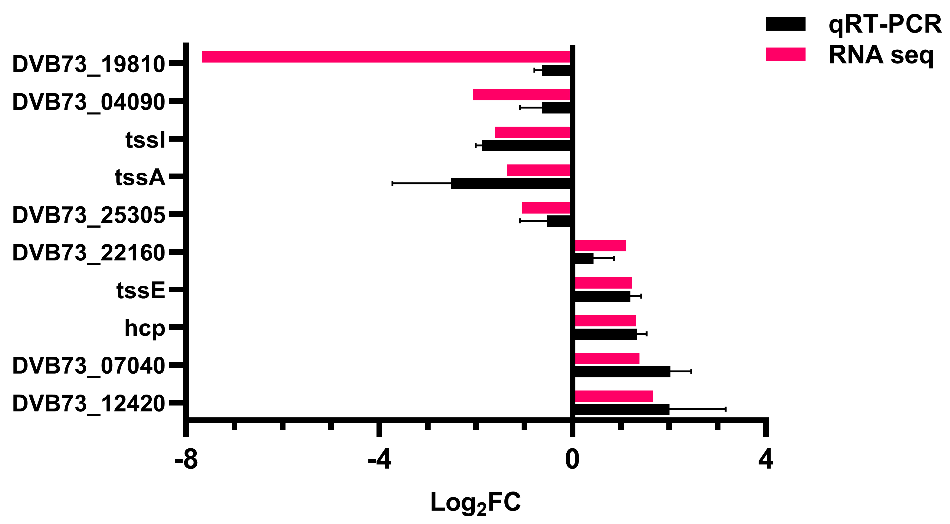
2.10.5. Validation of Transcriptome Data

To confirm the reliability of transcriptome data, several upregulated and downregulated genes were randomly selected from RNA-seq results for qRT-PCR validation. The sequence of gene primers is shown in Supplementary Table S2. The 16S rDNA (Table S1) was used as the internal reference gene, and the 2−ΔΔCT method was used for data processing. Three samples were in each group, and for each sample, three replicates were performed.

2.11. Data Analysis

All data were the average standard deviation (SD) of three independent experiments. The data were analyzed by IBM SPSS Statistics v 26.0, and the Dunnett test was used for one-way ANOVA. The p value < 0.05 was indicated as statistically significant.

2.12. Data Access

The RNA-seq data were stored in the GenBank SRA database with entry numbers SRP423148 (NZBD9 strain group) and SRP428021(ΔfliL strain group).

3. Results

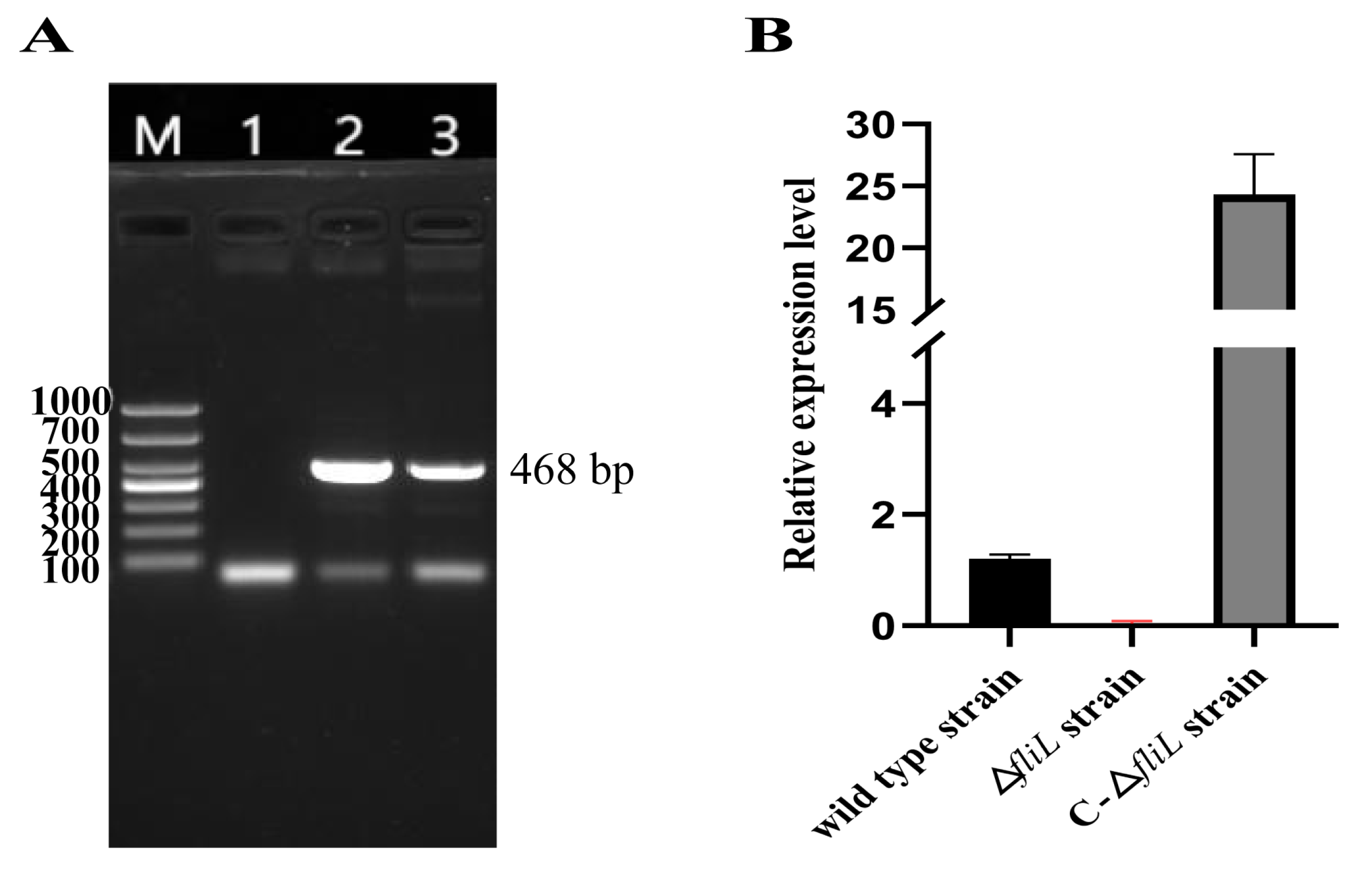
3.1. Construction of fliL Gene Deletion and Replacement Strain of P. plecoglossicida

The results of the construction process of ΔfliL and C-ΔfliL strains are shown in Supplementary Figures S1 and S2. As shown in Figure 1A, the ΔfliL strain did not exhibit the fragment of the fliL gene (468 bp), while both the NZBD9 strain and C-ΔfliL strain exhibited the fragment. Through further verification (mRNA level), as shown in Figure 1B, compared with the wild-type strain, the ΔfliL strain was almost unexpressed, while the C-ΔfliL strain showed an upregulated expression. These results demonstrate that the fliL gene deletion strain and complement strain of P. plecoglossicida was successfully constructed and can be used in subsequent experiments.

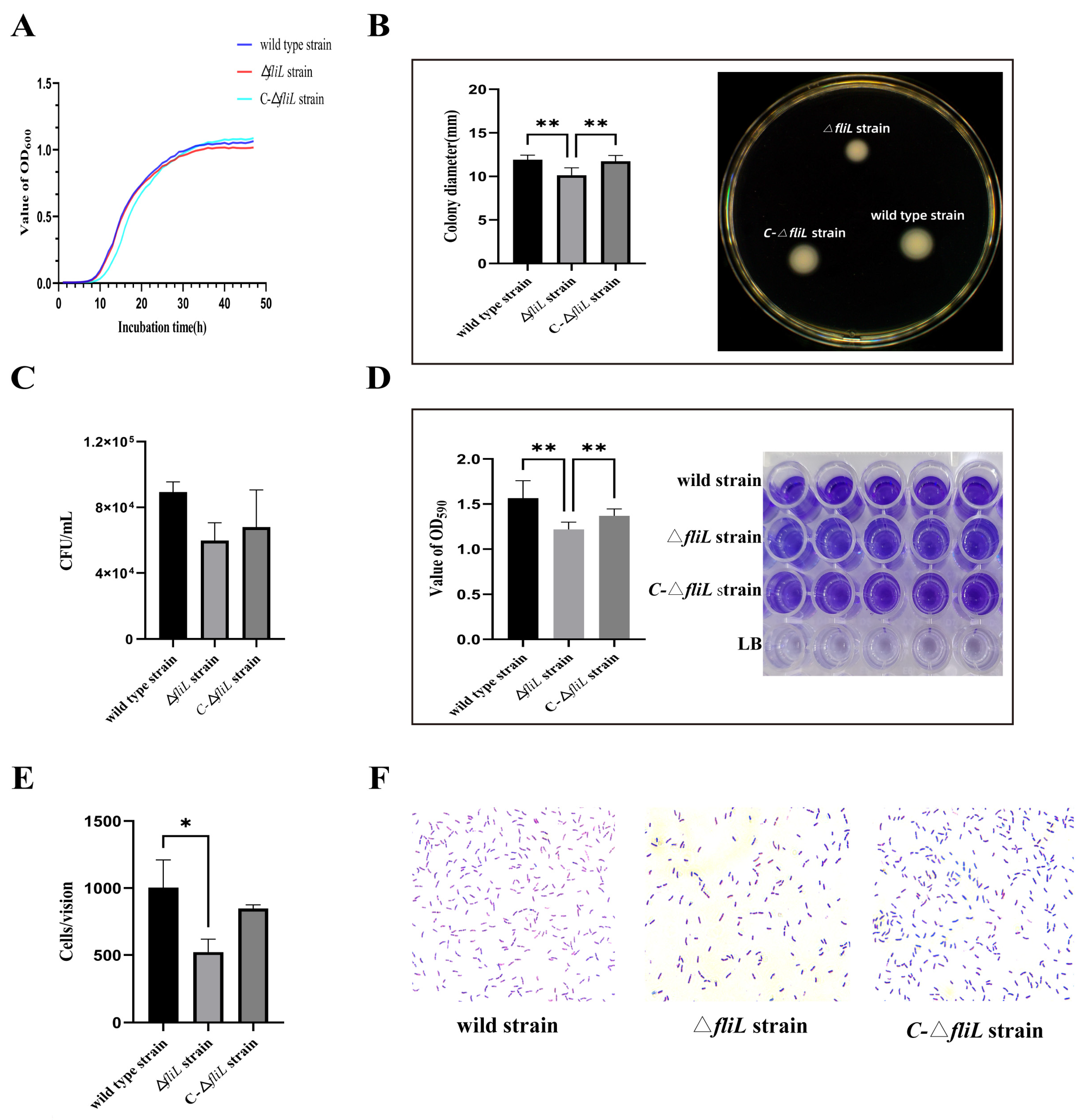
3.2. Effect of the fliL Gene on the Phenotype of P. plecoglossicida

The results of the growth curve (Figure 2A) show that there was no significant difference between the growth of the wild-type strain, ΔfliL, and C-ΔfliL strain. The motility of the three strains was determined on LB soft agar. Figure 2B shows that the colony diameter of the ΔfliL strain (10.14 ± 0.85 mm, p < 0.01) was significantly smaller than that of the wild strain (11.92 ± 0.52 mm), and the motility of the C-ΔfliL strain (11.73 ± 0.66 mm) was restored to the state of the wild type. The comparison of biofilm formation indicated that the biofilm color of the ΔfliL strain was the lightest among the three strains, and the OD590 value of the ΔfliL strain (1.22 ± 0.08, p < 0.01) was significantly lower than that of the wild strain (1.57 ± 0.19) and the C-ΔfliL strain (1.37 ± 0.08) (Figure 2D). Similarly, the adhesion of the ΔfliL strain decreased. The number of ΔfliL strain bacteria attached to the mucus-coated slide was significantly less than that of the wild strain (p < 0.05) under the same visual field (Figure 2E,F). As shown in Figure 2C, the chemotactic ability of the ΔfliL strain was weaker than that of the wild strain, but there was no significant difference.

3.3. Effect of the fliL Gene on the Virulence of P. plecoglossicida

Mortality (within 7 days) of artificial infection of pearl gentian grouper with P. plecoglossicida is presented in Table 1. No fish in the PBS group died during animal testing. Kohl’s [29] modified method was used to calculate the LD50 of the wild-type strain, ΔfliL strain, and C-ΔfliL strain, with values of 5.0 × 10³ CFU/fish (2.1 × 10³–1.2 × 10⁴, 95% confidence interval), 6.3 × 10⁴ CFU/fish (1.9 × 10⁴–2.1 × 10⁵, 95% confidence interval), and 1.3 × 10³ CFU/fish (4.8 × 10²–3.7 × 10³, 95% confidence interval), respectively. The result showed that the virulence of the ΔfliL strain was twelve-fold lower than that of the wild-type strain, while that of the C-ΔfliL strain was four-fold higher.

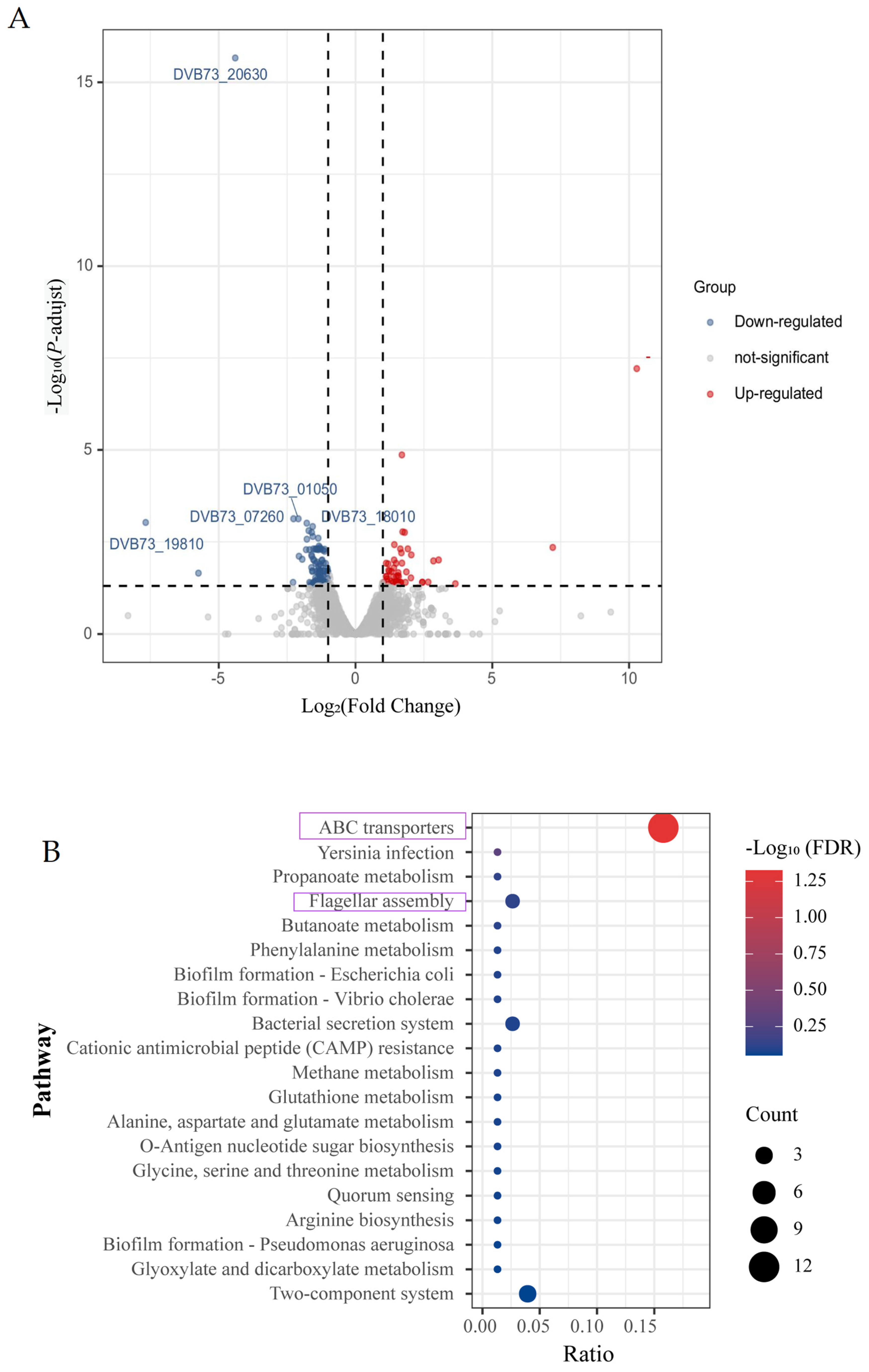
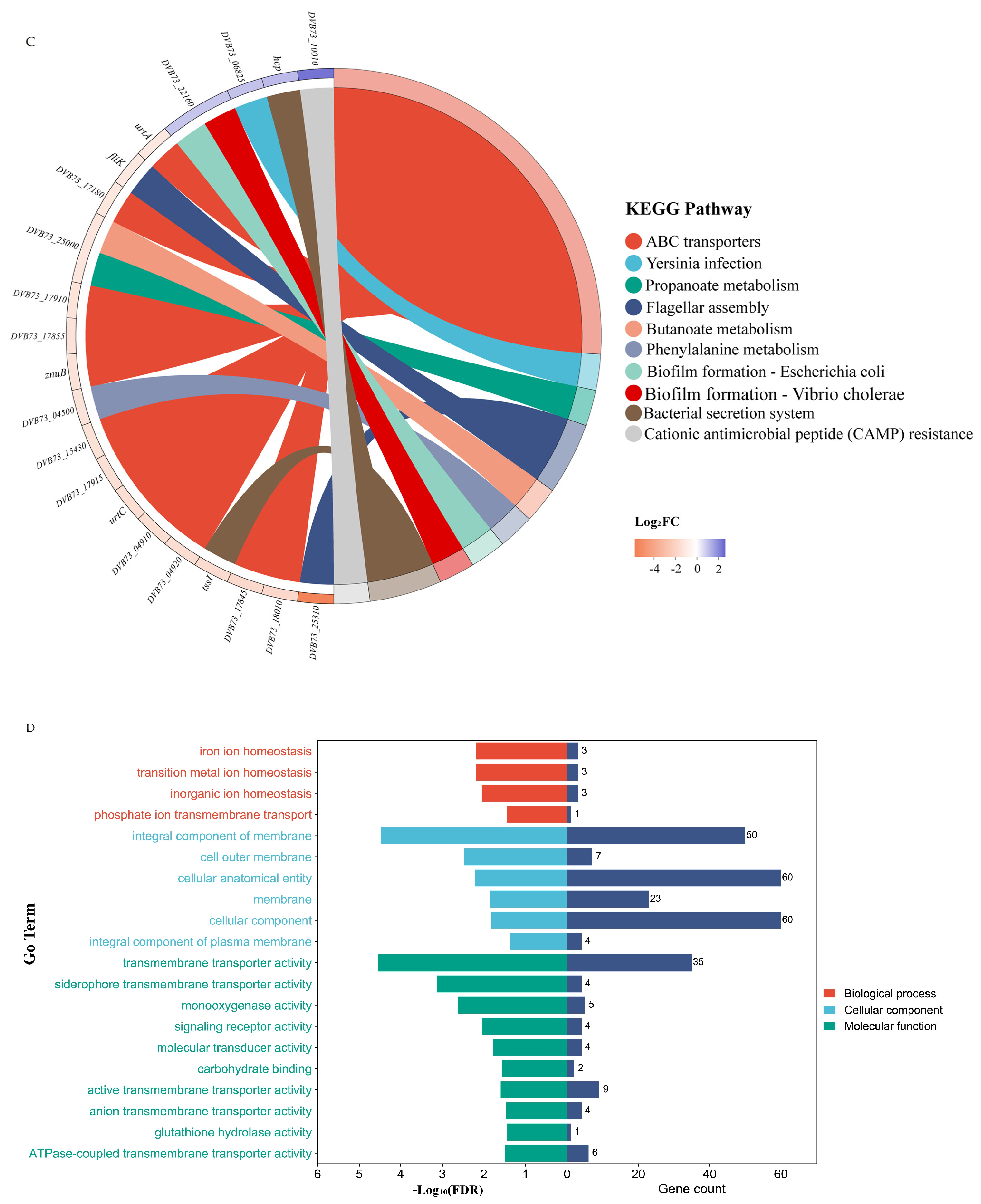
3.4. Effect of Knockout of fliL Gene on Transcriptome of Wild Strain of P. plecoglossicida

Judging from the quality of quality-controlled sequence data, the base distribution of each sample is relatively concentrated, with N% approaching 0%. The Q20 (percentage of bases with mass value ≥20) and Q30 (percentage of bases with mass value ≥30) of each sample were greater than 98.5% and 95.5%. The sequencing error rate was less than 0.1%, indicating the high quality of transcriptome sequencing results. The ratio of clean reads (mapped reads) to the reference genome was more than 98%, which indicated that the selected reference genome assembly could meet the needs of information analysis.
The analysis found that there were 126 DEGs between the transcriptome of the ΔfliL strain and wild-type strain, including 114 downregulated genes and 12 upregulated genes (Figure 3A). The first 50 differentially expressed genes were selected for thermographic analysis (Supplementary Figure S3). Five upregulated and downregulated genes were selected for qRT-PCR detection. The trend of gene expression in qRT-PCR results was consistent with sequencing results (Figure 4), which indicated the reliability of transcriptome data.
The differential genes were analyzed by GO enrichment and KEGG pathway to further explore the effect of fliL gene deletion on the transcriptome of P. pleclossicida. The GO enrichment analysis with GOATOOLS software showed that there are three types of GO enrichment functions: molecular function (MF), cellular component (CC), and biological process (BP). A total of 44 GO pathways were enriched, 42 of which were significantly enriched. The 20 GO pathways with the highest degree of enrichment are shown in Figure 3D, among which the top 5 are the intrinsic component of membrane, an integral component of cytoplasm, transporter activity, transmembrane transporter activity, and siderophore uptake transmembrane transporter activity. It is suggested that the deletion of the fliL gene may affect the membrane and transmembrane transport activity of P. plecoglossicida. KEGG Pathway enrichment analysis showed that 35 differential genes (DEGs) were enriched into 39 pathways in the KEGG database. Among them, 29 pathways downregulate differential gene enrichment, including the ABC transporter, flagellar assembly, biofilm formation, bacterial secretion system, and two-component system, etc. The top 20 enrichment pathways are shown in Figure 3B, and the genes corresponding to the top 10 KEGG pathways and their expression levels are shown in Figure 3C. It was noteworthy that the ABC transporter receptor pathway was the most significant pathway of enrichment. As one of the largest protein superfamilies, ABC transporters transport a variety of substrates across the cellular membranes of bacteria by ATP binding and hydrolysis (Figure 5A). The ABC transporter receptor pathway was analyzed, and its upregulation and downregulation are shown in Figure 5B. As shown in the Figure 5, the alkanesulfonate-binding protein (SsuA), ABC transporter permease (SsuC), and SsuB transporter are all downregulated. AfuB transporters in the iron uptake system and ZnuA and ZnuB transporters in the zinc uptake system were downregulated. In addition, the phosphate transport system permease protein PstA, methionine uptake transporter MetNI, and the urea permease UrtA, UrtC were also downregulated. The results showed that the transport and absorption ability of the ΔfliL strain to the outside world were weakened.

4. Discussion

As an important gene modification technology, gene knockout technology is widely used in the fields of animals and plants [30,31,32] and microorganisms [33,34,35]. It is one of the most powerful tools for studying the function of biological genes. The main methods include homologous recombination, the ZFN technique, TALEN technique, and CRISPR/Cas9 system mediation [36,37,38]. Homologous recombination has played a role in the study of a variety of bacteria. Examples of strains were Bifidobacterium longum [39], Helicobacter pylori [40], Mycobacterium avium subespecie paratuberculosis, (MAP) [41]. In this study, the fliL gene of P. percoglossicida was knocked out by homologous recombination of suicide plasmids, and the ΔfliL strain was constructed successfully, which laid the foundation for this study.
Most bacteria-infected hosts undergo four processes: contact, adhesion, entry, and host colonization [42], which are closely related to flagellar movement, biofilm formation, adhesion ability, and other virulence factors [43,44]. Flagellar movement is crucial to the survival and virulence of many pathogenic bacteria [17] and is one of the determinants of pathogenicity. The flagellar structure consists of three parts: a rotary motor, a universal joint, and a helical propeller [45], which consists of more than 50 genes with multiple operons that regulate flagella synthesis and function [46]. For example, the fliK gene controls the length of flagellar hooks and is crucial for the motility of Bacillus thuringiensis [47]. In the study of P. percoglossicida, the silence of genes such as fliG, flgK, and flgC that encode flagellin all lead to a significant decline in the movement, adhesion, and biofilm formation of P. percoglossicida [10,14,15]. The survival rate of infected Epinephelus coioides increased by 55%, 55%, and 80%, respectively. These results suggest that flagella genes play a key role in the pathogenicity of bacteria. In this study, we used a homologous recombination technique to knock out the flagellum-related gene fliL. The biological characterization showed that the motility, adhesion, and biofilm-forming ability of ΔfliL strain were obviously decreased, but it has no effect on the growth of P. percoglossicida. By supplementing the fliL gene, the C-ΔfliL strain has regained its motility, and its biofilm-forming ability has also been improved. These results suggest that the fliL gene may be involved in the regulation of flagella assembly, thus weakening the virulence of the P. percoglossicida. The virulence of ΔfliL strain was reduced 12-fold compared to the wild-type strain by testing the median lethal dose (LD50) of the infected pearl gentian grouper. Moreover, it was found that the virulence of C-ΔfliL strain was significantly greater than ΔfliL strain.
Bacterial virulence is regulated by a variety of factors besides flagella. Studies have shown that the bacterial outer membrane proteins OmpF and OmpA contribute to swimming motility and biofilm formation in Citrobacter werkmanii [48,49]. Ferredoxin receptor FusA is involved in iron transport, and knocking out the fusA gene significantly reduced the biofilm formation and adhesion ability of P. percoglossicida. The coregulation of flagella and other virulence factors on bacterial virulence is a complex mechanism that needs further study.
Transcriptomics is the study of the function of RNA transcripts. Transcriptome analysis can help us to study gene transcription and transcriptional regulation in cells at the overall level, which is the basis and starting point of gene function and structure research [50,51]. The transcriptome of prokaryotes adopts the chain-specific library building strategy [52,53]. The rapid development of this technology has promoted the research on the pathogenesis of pathogenic bacteria [54], and played an important role in the study of the drug resistance mechanism [55,56] and pathogenicity of pathogenic bacteria [57,58]. The ΔfliL strain in this study was different from the wild-type strain in the transcriptome level to some extent. A total of 126 genes with significant expression differences were screened in this transcriptome analysis, including 114 DEGs downregulated and 12 DEGs upregulated. The gene fliK, which is involved in flagella assembly, was significantly downregulated. Through enrichment analysis, the differential genes enriched into the GO pathway were mostly genes related to the membrane and transporter activity of bacteria, including those involved in the ferritic uptake of transporters. However, the absorption of manganese, iron, zinc, and other metal ions by bacteria is one of the key factors for their virulence [59,60,61]. There were 35 DEGs enriched ABC transporters, flagellar assembly, and the bacterial two-component system in the KEGG Pathway.
The ATP-binding cassette transporter family (ABC) transporter is a ubiquitous molecular pump [62] that can be classified as either an importer (type I and type II) or an exporter. Whereas type I and type II importers are found only in bacteria, whose role is to absorb essential nutrients, exporters transport molecules out of cells or organelles and exist in all organisms [63]. ABC exporters include type I secretory systems associated with the secretion of toxins, S-layer protein, siderophore, hydrolase, or antimicrobial peptide, which have roles in adhesion and colonization of the host. There are also glycoconjugates and polysaccharide biosynthetic pathways involved in membrane biosynthesis and immune escape. Importers are associated with nutrient acquisition (e.g., metal ions, amino acids, vitamins, and oligopeptides) and osmotic protection processes, all of which contribute significantly to bacterial pathogenicity [64]. In the transcriptome sequencing results of this study, the most significant pathway of KEGG enrichment was the ABC transport pathway, in which the expression of transporters for iron, zinc, and phosphate was significantly downregulated. These results suggest that the deletion of the fliL gene weakens the transport system of P. percoglossicida and affects its absorption of nutrients. This may be one of the reasons for the decreased virulence of the ΔfliL strain.

5. Conclusions

The fliL gene was related to the regulation of biofilm formation, migration, and adhesion ability of P. percoglossicida. After the knockout of the fliL gene, the virulence of the strain was reduced 12-fold (LD50 test), and the transcriptome of P. percoglossicida was significantly affected. The fliK gene related to flagellar assembly and several genes related to transport were significantly downregulated, and the pathway enriched by differentially expressed genes was the ABC transporter. It suggests that the fliL gene might be influencing the P. percoglossicida’s system of material transportation and flagellum assembly to reduce its virulence.

Supplementary Materials

The following supporting information can be downloaded at: https://www.mdpi.com/article/10.3390/fishes8080397/s1, Table S1: The primers used to construct strains. Table S2: Primer sequence for transcriptome validation. Figure S1: Electrophoretic diagram of the construction of the knockout strain. Figure S2: Electrophoretic diagram of the construction of the complement strain. Figure S3: Clustering analysis of differentially expressed genes.

Author Contributions

L.S.: performed experiments and data analysis, and completed the first draft. J.Z., L.Z. and Q.L.: provided useful help in the process of experiment and data analysis. L.H. and Y.Q.: provided guidance and supervised the process of developing the manuscript. Q.Y.: supervised, revised and edited the manuscript, and submitted the manuscript. All authors have read and agreed to the published version of the manuscript.

Funding

This work was supported by the Natural Science Foundation of Fujian Province under contract No. 2021J01828 and 2022J02044, National Natural Science Foundation of China under contract No. 31972836, Science and Technology Plan Project of Fuzhou City under contract No. 2022-P-014, Science and Technology Plan Project of Fujian Province under contract No. 2022L3059.

Institutional Review Board Statement

All animal protocols were approved by the Animal Ethics Committee of Jimei University (Acceptance No JMULAC201159).

Data Availability Statement

The original contributions presented in the study are included in the article/Supplementary Materials. Further inquiries can be directed to the corresponding authors.

Conflicts of Interest

The authors declare that the research was conducted in the absence of any commercial or financial relationships that could be construed as a potential conflict of interest.

References

  1. Mzula, A.; Wambura, P.N.; Mdegela, R.H.; Shirima, G.M. Present Status of Aquaculture and the Challenge of Bacterial Diseases in Freshwater Farmed Fish in Tanzania; A Call for Sustainable Strategies. Aquac. Fish. 2021, 6, 247–253. [Google Scholar] [CrossRef]
  2. Rodger, H.D. Fish Disease Causing Economic Impact in Global Aquaculture. In Fish Vaccines; Adams, A., Ed.; Birkhäuser Advances in Infectious Diseases; Springer: Basel, Switzerland, 2016; pp. 1–34. ISBN 978-3-0348-0980-1. [Google Scholar]
  3. Wu, X.Y.; Xiong, J.B.; Fei, C.J.; Dai, T.; Zhu, T.F.; Zhao, Z.Y.; Pan, J.; Nie, L.; Chen, J. Prior exposure to ciprofloxacin disrupts intestinal homeostasis and predisposes ayu (Plecoglossus altivelis) to subsequent Pseudomonas plecoglossicida-induced infection. Zool. Res. 2022, 43, 648–665. [Google Scholar] [CrossRef]
  4. Cai, H.Y.; Ma, Y.; Qin, Y.X.; Zhao, L.M.; Yan, Q.P.; Huang, L.X. Vvrr2: A New Vibrio NcRNA Involved in Dynamic Synthesis of Multiple Biofilm Matrix Exopolusaccharides, Biofilm Structuring and Virulence. Aquaculture 2023, 563, 738925. [Google Scholar] [CrossRef]
  5. Du, Z.Y.; Zhang, M.M.; Qin, Y.X.; Zhao, L.M.; Huang, L.X.; Xu, X.J.; Yan, Q.P. The Role and Mechanisms of the Two-Component System EnvZ/OmpR on the Intracellular Survival of Aeromonas hydrophila. J. Fish Dis. 2022, 45, 1609–1621. [Google Scholar] [CrossRef] [PubMed]
  6. Park, S.C.; Shimamura, I.; Fukunaga, M.; Mori, K.I.; Nakai, T. Isolation of Bacteriophages Specific to a Fish Pathogen, Pseudomonas plecoglossicida, as a Candidate for Disease Control. Appl. Environ. Microbiol. 2000, 66, 1416–1422. [Google Scholar] [CrossRef] [Green Version]
  7. Xu, K.P.; Wang, Y.S.; Yang, W.X.H.; Cai, H.Y.; Zhang, Y.Y.; Huang, L.X. Strategies for Prevention and Control of Vibriosis in Asian Fish Culture. Vaccines 2022, 11, 98. [Google Scholar] [CrossRef]
  8. Nishimori, E.; Kita-Tsukamoto, K.; Wakabayashi, H. Pseudomonas plecoglossicida Sp. Nov., the Causative Agent of Bacterial Haemorrhagic Ascites of Ayu, Plecoglossus altivelis. Int. J. Syst. Evol. Microbiol. 2000, 50 Pt 1, 83–89. [Google Scholar] [CrossRef] [Green Version]
  9. Akayli, T.; Çanak, Ö.; Başaran, B. A New Pseudomonas Species Observed in Cultured Young Rainbow Trout (Oncorhynchus mykis Walbaum, 1792): Pseudomonas plecoglossicida. Biyol. Bilim. Araştırma Derg. 2011, 4, 107–111. [Google Scholar]
  10. Yuan, B.; Zhao, L.M.; Zhuang, Z.X.; Wang, X.R.; Fu, Q.; Huang, H.B.; Huang, L.X.; Qin, Y.X.; Yan, Q.P. Transcriptomic and Metabolomic Insights into the Role of the flgK Gene in the Pathogenicity of Pseudomonas plecoglossicida to Orange-Spotted Grouper (Epinephelus coioides). Zool. Res. 2022, 43, 952–965. [Google Scholar] [CrossRef]
  11. Zhang, J.T.; Zhou, S.M.; An, S.W.; Chen, L.; Wang, G.L. Visceral Granulomas in Farmed Large Yellow Croaker, Larimichthys crocea (Richardson), Caused by a Bacterial Pathogen, Pseudomonas plecoglossicida. J. Fish Dis. 2014, 37, 113–121. [Google Scholar] [CrossRef]
  12. Xin, G.; Zhao, L.M.; Zhuang, Z.X.; Wang, X.; Fu, Q.; Huang, H.B.; Huang, L.X.; Qin, Y.X.; Zhang, J.N.; Zhang, J.L.; et al. Function of the rpoD Gene in Pseudomonas plecoglossicida Pathogenicity and Epinephelus coioides Immune Response. Fish Shellfish Immunol. 2022, 127, 427–436. [Google Scholar] [CrossRef] [PubMed]
  13. Tang, Y.; Jiao, J.P.; Zhao, L.M.; Zhuang, Z.X.; Wang, X.R.; Fu, Q.; Huang, H.B.; Huang, L.X.; Qin, Y.X.; Zhang, J.N.; et al. The Contribution of exbB Gene to Pathogenicity of Pseudomonas plecoglossicida and Its Interactions with Epinephelus coioides. Fish Shellfish Immunol. 2022, 120, 610–619. [Google Scholar] [CrossRef] [PubMed]
  14. Jiao, J.P.; Zhao, L.M.; Huang, L.X.; Qin, Y.X.; Su, Y.Q.; Zheng, W.Q.; Zhang, J.N.; Yan, Q.P. The Contributions of fliG Gene to the Pathogenicity of Pseudomonas plecoglossicida and Pathogen-Host Interactions with Epinephelus coioides. Fish Shellfish Immunol. 2021, 119, 238–248. [Google Scholar] [CrossRef] [PubMed]
  15. Yang, D.; Zhao, L.M.; Li, Q.; Huang, L.X.; Qin, Y.X.; Wang, P.; Zhu, C.Z.; Yan, Q.P. flgC Gene Is Involved in the Virulence Regulation of Pseudomonas plecoglossicida and Affects the Immune Response of Epinephelus coioides. Fish Shellfish Immunol. 2023, 132, 108512. [Google Scholar] [CrossRef] [PubMed]
  16. Zhu, S.W.; Kumar, A.; Kojima, S.; Homma, M. FliL Associates with the Stator to Support Torque Generation of the Sodium-Driven Polar Flagellar Motor of Vibrio. Mol. Microbiol. 2015, 98, 101–110. [Google Scholar] [CrossRef]
  17. Tachiyama, S.; Chan, K.L.; Liu, X.L.; Hathroubi, S.; Peterson, B.; Khan, M.F.; Ottemann, K.M.; Liu, J.; Roujeinikova, A. The Flagellar Motor Protein FliL Forms a Scaffold of Circumferentially Positioned Rings Required for Stator Activation. Proc. Natl. Acad. Sci. USA 2022, 119, e2118401119. [Google Scholar] [CrossRef]
  18. Lin, T.S.; Zhu, S.W.; Kojima, S.; Homma, M.; Lo, C.J. FliL Association with Flagellar Stator in the Sodium-Driven Vibrio Motor Characterized by the Fluorescent Microscopy. Sci. Rep. 2018, 8, 11172. [Google Scholar] [CrossRef] [Green Version]
  19. Lee, Y.Y.; Patellis, J.; Belas, R. Activity of Proteus Mirabilis FliL Is Viscosity Dependent and Requires Extragenic DNA. J. Bacteriol. 2013, 195, 823–832. [Google Scholar] [CrossRef] [Green Version]
  20. Mengucci, F.; Dardis, C.; Mongiardini, E.J.; Althabegoiti, M.J.; Partridge, J.D.; Kojima, S.; Homma, M.; Quelas, J.I.; Lodeiro, A.R. Characterization of FliL Proteins in Bradyrhizobium diazoefficiens: Lateral FliL Supports Swimming Motility, and Subpolar FliL Modulates the Lateral Flagellar System. J. Bacteriol. 2020, 202, e00708-19. [Google Scholar] [CrossRef]
  21. Partridge, J.D.; Nieto, V.; Harshey, R.M. A New Player at the Flagellar Motor: FliL Controls Both Motor Output and Bias. mBio 2015, 6, e02367. [Google Scholar] [CrossRef] [Green Version]
  22. Takekawa, N.; Isumi, M.; Terashima, H.; Zhu, S.W.; Nishino, Y.; Sakuma, M.; Kojima, S.; Homma, M.; Imada, K. Structure of Vibrio FliL, a New Stomatin-like Protein That Assists the Bacterial Flagellar Motor Function. mBio 2019, 10, e00292-19. [Google Scholar] [CrossRef] [PubMed] [Green Version]
  23. Josenhans, C.; Suerbaum, S. The Role of Motility as a Virulence Factor in Bacteria. Int. J. Med. Microbiol. 2002, 291, 605–614. [Google Scholar] [CrossRef] [PubMed]
  24. Suaste-Olmos, F.; Domenzain, C.; Mireles-Rodríguez, J.C.; Poggio, S.; Osorio, A.; Dreyfus, G.; Camarena, L. The Flagellar Protein FliL Is Essential for Swimming in Rhodobacter sphaeroides. J. Bacteriol. 2010, 192, 6230–6239. [Google Scholar] [CrossRef] [PubMed] [Green Version]
  25. Ye, H.D.; Xu, Z.J.; Tao, Z.; Li, W.Y.; Li, Y.; Yang, A.; Wang, W.; Yin, X.L.; Yan, X.J. Efficacy and Safety of Pseudomonas plecoglossicida Mutant ΔtssD-1 as a Live Attenuated Vaccine for the Large Yellow Croaker (Larimichthys crocea). Aquaculture 2021, 531, 735976. [Google Scholar] [CrossRef]
  26. Howery, K.E.; Rather, P.N. Allelic Exchange Mutagenesis in Proteus mirabilis. Methods Mol. Biol. 2019, 2021, 77–84. [Google Scholar] [CrossRef] [PubMed]
  27. Vijayan, S.P.; Rekha, P.D.; Dinesh, U.; Arun, A.B. Determination of Acute Lethal Dose 50 (LD50) of Uranyl Nitrate in Male Swiss Albino Mice. Res. J. Pharm. Technol. 2018, 11, 1086. [Google Scholar] [CrossRef]
  28. Melo Ferreira, R.; Sabo, A.R.; Winfree, S.; Collins, K.S.; Janosevic, D.; Gulbronson, C.J.; Cheng, Y.-H.; Casbon, L.; Barwinska, D.; Ferkowicz, M.J.; et al. Integration of Spatial and Single-Cell Transcriptomics Localizes Epithelial Cell-Immune Cross-Talk in Kidney Injury. JCI Insight 2021, 6, e147703. [Google Scholar] [CrossRef]
  29. Rong, Y.; Tang, L.; Yin, H.F.; Sun, D.Y. A Modified Coriolis Method Was Used to Calculate the Number of BALB/cA-Nu Mice Treated with 1,2. LD50 of Dichloropropane. Chin. J. Occup. Dis. Labor Hyg. 2019, 37, 53–55. [Google Scholar]
  30. Dong, Y.; Bai, H.T.; Dong, F.; Zhang, X.B.; Ema, H. Gene Knockout in Highly Purified Mouse Hematopoietic Stem Cells by CRISPR/Cas9 Technology. J. Immunol. Methods 2021, 495, 113070. [Google Scholar] [CrossRef]
  31. Sasidharan, S.; Saudagar, P. Knockout of Tyrosine Aminotransferase Gene by Homologous Recombination Arrests Growth and Disrupts Redox Homeostasis in Leishmania Parasite. Parasitol. Res. 2022, 121, 3229–3241. [Google Scholar] [CrossRef]
  32. Zhang, Y.X.; Zhang, Y.; Qi, Y.P. Plant Gene Knockout and Knockdown by CRISPR-Cpf1 (Cas12a) Systems. Methods Mol. Biol. 2019, 1917, 245–256. [Google Scholar] [CrossRef] [PubMed]
  33. Choi, K.R.; Cho, J.S.; Cho, I.J.; Park, D.; Lee, S.Y. Markerless Gene Knockout and Integration to Express Heterologous Biosynthetic Gene Clusters in Pseudomonas putida. Metab. Eng. 2018, 47, 463–474. [Google Scholar] [CrossRef] [PubMed] [Green Version]
  34. Han, X.; Tu, Y.Q.; Wu, H.; Zhang, L.J.; Zhao, S.N. Gene Knockout Revealed the Role of Gene FeoA in Cell Growth and Division of Lactobacillus delbrueckii Subsp. Bulgaricus. Arch. Microbiol. 2021, 203, 3541–3549. [Google Scholar] [CrossRef]
  35. Zhu, X.W.; Wu, Y.K.; Lv, X.Q.; Liu, Y.F.; Du, G.C.; Li, J.H.; Liu, L. Combining CRISPR-Cpf1 and Recombineering Facilitates Fast and Efficient Genome Editing in Escherichia coli. ACS Synth. Biol. 2022, 11, 1897–1907. [Google Scholar] [CrossRef]
  36. Carroll, D. Genome Engineering with Zinc-Finger Nucleases. Genetics 2011, 188, 773–782. [Google Scholar] [CrossRef] [PubMed] [Green Version]
  37. Dalvie, N.C.; Lorgeree, T.; Biedermann, A.M.; Love, K.R.; Love, J.C. Simplified Gene Knockout by CRISPR-Cas9-Induced Homologous Recombination. ACS Synth. Biol. 2022, 11, 497–501. [Google Scholar] [CrossRef]
  38. Kim, S.E.; Kim, J.W.; Kim, Y.J.; Kwon, D.N.; Kim, J.H.; Kang, M.J. Generation of Fibroblasts Lacking the Sal-like 1 Gene by Using Transcription Activator-like Effector Nuclease-Mediated Homologous Recombination. Asian-Australas. J. Anim. Sci. 2016, 29, 564–570. [Google Scholar] [CrossRef]
  39. Sakaguchi, K.; He, J.L.; Tani, S.; Kano, Y.; Suzuki, T. A Targeted Gene Knockout Method Using a Newly Constructed Temperature-Sensitive Plasmid Mediated Homologous Recombination in Bifidobacterium longum. Appl. Microbiol. Biotechnol. 2012, 95, 499–509. [Google Scholar] [CrossRef]
  40. Langford, M.L.; Zabaleta, J.; Ochoa, A.C.; Testerman, T.L.; McGee, D.J. In Vitro and in Vivo Complementation of the Helicobacter Pylori Arginase Mutant Using an Intergenic Chromosomal Site. Helicobacter 2006, 11, 477–493. [Google Scholar] [CrossRef] [Green Version]
  41. Alonso, M.N.; Malaga, W.; Mc Neil, M.; Jackson, M.; Romano, M.I.; Guilhot, C.; Santangelo, M.P. Efficient Method for Targeted Gene Disruption by Homologous Recombination in Mycobacterium avium Subspecie paratuberculosis. Res. Microbiol. 2020, 171, 203–210. [Google Scholar] [CrossRef]
  42. Chaussé, A.M.; Roche, S.M.; Moroldo, M.; Hennequet-Antier, C.; Holbert, S.; Kempf, F.; Barilleau, E.; Trotereau, J.; Velge, P. Epithelial Cell Invasion by Salmonella typhimurium Induces Modulation of Genes Controlled by Aryl Hydrocarbon Receptor Signaling and Involved in Extracellular Matrix Biogenesis. Virulence 2023, 14, 2158663. [Google Scholar] [CrossRef]
  43. Prada, C.F.; Casadiego, M.A.; Freire, C.C. Evolution of helicobacter Spp: Variability of Virulence Factors and Their Relationship to Pathogenicity. PeerJ 2022, 10, e13120. [Google Scholar] [CrossRef] [PubMed]
  44. Sano, G.; Takada, Y.; Goto, S.; Maruyama, K.; Shindo, Y.; Oka, K.; Matsui, H.; Matsuo, K. Flagella Facilitate Escape of salmonella from Oncotic Macrophages. J. Bacteriol. 2007, 189, 8224–8232. [Google Scholar] [CrossRef] [PubMed] [Green Version]
  45. Tan, J.X.; Zhang, X.; Wang, X.F.; Xu, C.H.; Chang, S.; Wu, H.J.; Wang, T.; Liang, H.H.; Gao, H.C.; Zhou, Y.; et al. Structural Basis of Assembly and Torque Transmission of the Bacterial Flagellar Motor. Cell 2021, 184, 2665–2679.e19. [Google Scholar] [CrossRef] [PubMed]
  46. Chilcott, G.S.; Hughes, K.T. Coupling of Flagellar Gene Expression to Flagellar Assembly in Salmonella enterica Serovar Typhimurium and Escherichia coli. Microbiol. Mol. Biol. Rev. 2000, 64, 694–708. [Google Scholar] [CrossRef] [Green Version]
  47. Attieh, Z.; Mouawad, C.; Rejasse, A.; Jehanno, I.; Perchat, S.; Hegna, I.K.; Økstad, O.A.; Kallassy Awad, M.; Sanchis-Borja, V.; El Chamy, L. The fliK Gene Is Required for the Resistance of Bacillus thuringiensis to Antimicrobial Peptides and Virulence in Drosophila melanogaster. Front. Microbiol. 2020, 11, 611220. [Google Scholar] [CrossRef]
  48. Zhou, G.; Wang, Y.S.; Peng, H.; Liu, H.Z.; Feng, J.; Li, S.J.; Sun, T.L.; Li, C.L.; Shi, Q.S.; Xie, X.B. Outer Membrane Protein of OmpF Contributes to Swimming Motility, Biofilm Formation, Osmotic Response as Well as the Transcription of Maltose Metabolic Genes in Citrobacter werkmanii. World J. Microbiol. Biotechnol. 2023, 39, 15. [Google Scholar] [CrossRef]
  49. Zhou, G.; Wang, Y.S.; Peng, H.; Li, S.J.; Sun, T.L.; Shen, P.F.; Xie, X.B.; Shi, Q.S. Roles of OmpA of Citrobacter werkmanii in Bacterial Growth, Biocide Resistance, Biofilm Formation and Swimming Motility. Appl. Microbiol. Biotechnol. 2021, 105, 2841–2854. [Google Scholar] [CrossRef]
  50. Dong, Z.C.; Chen, Y. Transcriptomics: Advances and Approaches. Sci. China Life Sci. 2013, 56, 960–967. [Google Scholar] [CrossRef] [Green Version]
  51. Wirka, R.C.; Pjanic, M.; Quertermous, T. Advances in Transcriptomics: Investigating Cardiovascular Disease at Unprecedented Resolution. Circ. Res. 2018, 122, 1200–1220. [Google Scholar] [CrossRef]
  52. Creecy, J.P.; Conway, T. Quantitative Bacterial Transcriptomics with RNA-Seq. Curr. Opin. Microbiol. 2015, 23, 133–140. [Google Scholar] [CrossRef] [Green Version]
  53. Siezen, R.J.; Wilson, G.; Todt, T. Prokaryotic Whole-Transcriptome Analysis: Deep Sequencing and Tiling Arrays. Microb. Biotechnol. 2010, 3, 125–130. [Google Scholar] [CrossRef] [PubMed] [Green Version]
  54. James, K.; Cockell, S.J.; Zenkin, N. Deep Sequencing Approaches for the Analysis of Prokaryotic Transcriptional Boundaries and Dynamics. Methods 2017, 120, 76–84. [Google Scholar] [CrossRef] [PubMed]
  55. Ahn, A.C.; Cavalca, L.; Colombo, M.; Schuurmans, J.M.; Sorokin, D.Y.; Muyzer, G. Transcriptomic Analysis of Two Thioalkalivibrio Species Under Arsenite Stress Revealed a Potential Candidate Gene for an Alternative Arsenite Oxidation Pathway. Front. Microbiol. 2019, 10, 1514. [Google Scholar] [CrossRef] [Green Version]
  56. Roe, C.; Williamson, C.H.D.; Vazquez, A.J.; Kyger, K.; Valentine, M.; Bowers, J.R.; Phillips, P.D.; Harrison, V.; Driebe, E.; Engelthaler, D.M.; et al. Bacterial Genome Wide Association Studies (BGWAS) and Transcriptomics Identifies Cryptic Antimicrobial Resistance Mechanisms in Acinetobacter baumannii. Front. Public Health 2020, 8, 451. [Google Scholar] [CrossRef]
  57. Lebreton, A.; Cossart, P. RNA- and Protein-Mediated Control of Listeria monocytogenes Virulence Gene Expression. RNA Biol. 2017, 14, 460–470. [Google Scholar] [CrossRef] [Green Version]
  58. Zhou, J.W.; Zhao, H.Q.; Yang, H.; He, C.Y.; Shu, W.; Cui, Z.L.; Liu, Q.Z. Insights into the Impact of Small RNA SprC on the Metabolism and Virulence of Staphylococcus aureus. Front. Cell. Infect. Microbiol. 2022, 12, 746746. [Google Scholar] [CrossRef]
  59. Papp-Wallace, K.M.; Maguire, M.E. Manganese Transport and the Role of Manganese in Virulence. Annu. Rev. Microbiol. 2006, 60, 187–209. [Google Scholar] [CrossRef] [PubMed] [Green Version]
  60. Wandersman, C.; Delepelaire, P. Bacterial Iron Sources: From Siderophores to Hemophores. Annu. Rev. Microbiol. 2004, 58, 611–647. [Google Scholar] [CrossRef]
  61. Zheng, C.K.; Qiu, J.; Zhai, Y.M.; Wei, M.; Zhou, X.H.; Jiao, X.N. ZrgA Contributes to Zinc Acquisition in Vibrio Parahaemolyticus. Virulence 2023, 14, 2156196. [Google Scholar] [CrossRef]
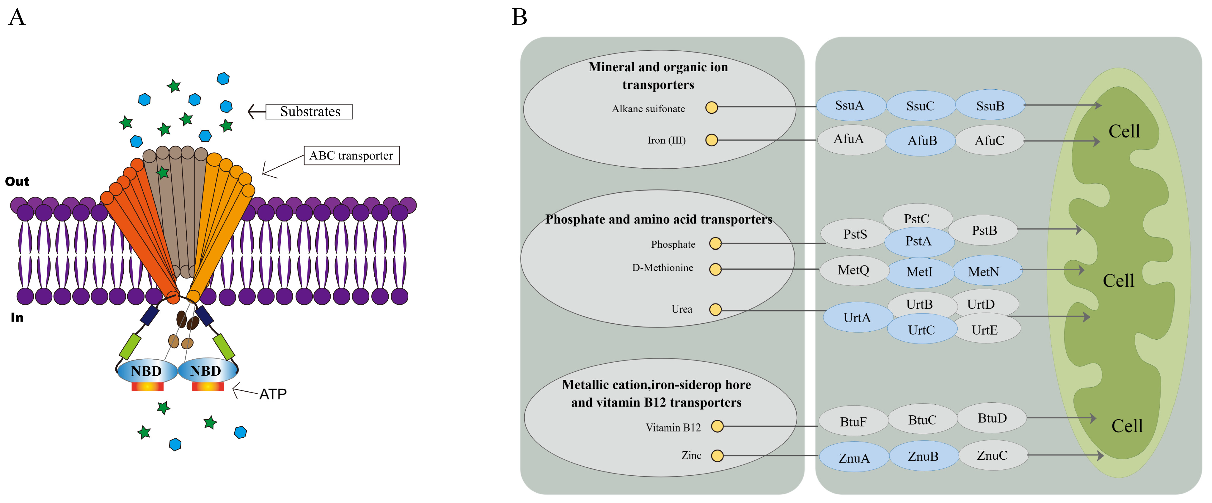
  62. Kieuvongngam, V.; Chen, J. Structures of the Peptidase-Containing ABC Transporter PCAT1 under Equilibrium and Nonequilibrium Conditions. Proc. Natl. Acad. Sci. USA 2022, 119, e2120534119. [Google Scholar] [CrossRef] [PubMed]
  63. Beis, K. Structural Basis for the Mechanism of ABC Transporters. Biochem. Soc. Trans. 2015, 43, 889–893. [Google Scholar] [CrossRef] [PubMed] [Green Version]
  64. Lewis, V.G.; Ween, M.P.; McDevitt, C.A. The Role of ATP-Binding Cassette Transporters in Bacterial Pathogenicity. Protoplasma 2012, 249, 919–942. [Google Scholar] [CrossRef] [PubMed]
Figure 1. Verification of ΔfliL and C-ΔfliL strain of P. plecoglossicida. (A) Detection of fliL DNA fragment (P5/P6 primers), M: DL1000 DNA Marker; 1: DNA of ΔfliL strain as a template; 2: DNA of C-ΔfliL strain as a template; 3: DNA of NZBD9 strain as a template; (B) fliL gene mRNA levels in three strains of P. plecoglossicida. Data were represented by mean + SD (n = 3).
Fishes 08 00397 g001
Figure 2. Comparison of the phenotype between different strains of P. plecoglossicida. (A) Growth curve; (B) Motility, left: measurement of colony diameter, right: photograph of colony morphology; (C) Chemotaxis; (D) Biofilm formation capacity, left: measurement of OD590 value, right: comparison of biofilm colors; (E) The average number of adherent bacteria; (F) Adherence of bacterial cells under the microscope (×1000). Data were mean ± SD of different assays with different biological replicates, * p < 0.05, ** p < 0.01.
Fishes 08 00397 g002
Figure 3. Comparative transcriptomic analysis between wild-type strain and ΔfliL strain of P. plecoglossicida. (A) Volcano map of transcriptome gene expression; (B) KEGG enrichment analysis of differentially expressed genes (DEGs); (C) KEGG functional enrichment chord diagram; (D) GO enrichment analysis of DEGs.
Fishes 08 00397 g003aFishes 08 00397 g003b
Figure 4. qRT-PCR validation of transcriptome. The vertical coordinate shows the name of the gene, and the horizontal coordinate shows the multiple of expression difference.
Fishes 08 00397 g004
Figure 5. ABC transporter transport diagram. (A) ABC receptor transport substance simulation; (B) ABC transport path diagram: blue means downward adjustment, gray means no significant difference.
Fishes 08 00397 g005
Table 1. Results of the artificial infection of different strains of P. plecoglossicida to pearl gentian grouper.
Dose (CFU/Fish)The Number of Test Animals Wild Type StrainΔfliL StrainC-ΔfliL Strain
The Number of Dead Mortality/%The Number of Dead Mortality/%The Number of Dead Mortality/%
5 × 10610101001010010100
5 × 105101010077010100
5 × 10410990440990
5 × 10310660330880
5 × 102100000330
Disclaimer/Publisher’s Note: The statements, opinions and data contained in all publications are solely those of the individual author(s) and contributor(s) and not of MDPI and/or the editor(s). MDPI and/or the editor(s) disclaim responsibility for any injury to people or property resulting from any ideas, methods, instructions or products referred to in the content.

