Article

Quantifying the Neural and Behavioral Correlates of Repeated Social Competition in the Fighting Fish Betta splendens

Department of Psychology, Emory University, Atlanta, GA 30322, USA
*
Author to whom correspondence should be addressed.
Fishes 2023, 8(8), 384; https://doi.org/10.3390/fishes8080384
Submission received: 8 June 2023 / Revised: 19 July 2023 / Accepted: 21 July 2023 / Published: 25 July 2023
(This article belongs to the Special Issue Causes and Consequences of Cognitive Variation in Fishes)

Abstract

:
The fighting fish Betta splendens, long studied for its aggressive territorial competitions, has the potential to be a tractable and relevant model for studying the intersection of cognitive ecology and social neuroscience. Yet, few studies have comprehensively assessed Betta behavior across both social and nonsocial contexts. Furthermore, the present study is the first to quantify the expression of phosphorylated ribosomal protein S6 (PS6), a proxy for neural response, in the Betta telencephalon. Here, we assessed male Betta behavior across a suite of tasks and found that response to a mirror, but not neophilia (a novel object) nor anxiety (scototaxis), predicted behavior in a social competition. To then explore the cognitive aspects of social competition, we exposed Betta to either a familiar or novel opponent and compared their competitive behavior as well as their neural responses in the teleost homologs of the hippocampus, basolateral amygdala, and lateral septum. We did not detect any differences between familiar-exposed and novel-exposed individuals, but by implementing the first use of a habituation–dishabituation competition design in a study of Betta, we were able to observe remarkable consistency in competitive outcomes across repeated exposures. Taken together, the present study lays the groundwork for expanding the use of Betta to explore integrative and multidimensional questions of social cognition.
Key Contribution: This multi-assay study is the first to implement an opponent habituation–dishabituation paradigm in Betta splendens and the first to characterize PS6 expression in the Betta splendens telencephalon, expanding our understanding of male territorial social competition.

Graphical Abstract

1. Introduction

Social encounters are complex, multimodal, and variable across time and space. The ability to successfully navigate a social encounter such as a competitive territorial interaction is highly consequential to an individual’s current condition and their future fitness. Displaying appropriate social behavior in these scenarios involves cognitive elements including memory, discrimination, transitive inference, and cognitive flexibility [1]. When characterizing an animal’s decision to fight or flee, ecologists have long considered the morphological conditions that “tip the scales” towards one action or the other, such as size [2,3], markings [4], testosterone [5], weaponry [6], and age [7]. But more recently, there has been the emergence of a cognitive approach to our understanding of these interactions [8]. This cognitive approach, described by Real, is where animal decisions are not simply objective weightings of external stimuli but rather the product of a series of processes (perception, encoding, storage, and representation) [9]. Put another way, we can ask: do behavioral and cognitive characteristics outside the social realm predict performance in a competitive social interaction?
Research has indeed demonstrated this broader pattern. There’s an extensive body of work on cross-context consistent individual differences in behavior, often referred to in the literature as “behavioral syndromes” or “animal personalities” [10,11]. For example, in black-capped chickadees, exploration style correlates to learning speed during a highly ecologically relevant task of acoustic discrimination [12]. In guppies, individuals from lower predation populations were “hastier” in a spatial memory task [13]. And in sailfin mollies, less anxious individuals performed better in a discrimination learning task but worse in a spatial reversal task [14]. Importantly, there is mounting evidence that these differences can indeed influence the outcomes of social encounters, especially with regard to position in dominance hierarchies [15,16,17,18], but see [19].
One particularly useful approach to exploring the relationship between behavioral tendencies and social outcomes is by utilizing an animal that has a highly stereotyped, easily inducible, and energetically expensive form of social competition. The Siamese fighting fish Betta splendens has been underutilized in this area of research despite its suitability for these experiments. They exhibit intense and metabolically costly [20] male–male territorial combat interactions [21,22] that over time result in consistent dyadic dominance relationships [23]. Researchers have developed assays to explore the robust behavioral traits of Betta, in particular the mirror assay, which has been employed in ethological studies of Betta for nearly a century [24].
Perceiving social stimuli, contextualizing the information through the recall of prior experience, and integrating the information with cues of internal state to display context-appropriate behavior is obviously a computationally complex process, requiring communication across multiple brain regions. A fantastic foundation of research has characterized [25,26] and re-evaluated [27,28] an integrated network of brain regions that coordinate social behavior known as the Social Behavior Network. Research has also demonstrated the connections between regions of this Social Behavior Network and regions involved in attention, decision-making, motivation, and reward processing, together recognized as the Social Decision-Making Network [29]. These neural mechanisms are incredibly conserved across a diverse set of taxa [26,29]. There are a growing number of studies on the neural mechanisms of cognition and social behavior in other fish species [30,31,32,33]. But, to our knowledge, the current study is the first to investigate brain regions within the Social Decision-Making Network in Betta splendens. In fact, only recently has a brain atlas of the Betta splendens telencephalon (the forebrain, the area where the SDMN regions are located) been published [34]. The tools to conduct this work are certainly available in Betta: researchers have characterized the electrophysical responses of the Betta optic tectum, a region used for visual processing in teleosts when observing an opponent [35], and a more recent transcriptomic analysis [36] on whole-brain Betta tissue uncovered neural signals (upregulated immediate early genes) produced when Bettas are synchronizing their competition behavior with their opponent.
To emphasize the potential of Betta as an emerging model at the intersection of cognitive ecology and social neuroscience, here we (1) assess how male Betta splendens behavior across three tasks (novel object exposure, scototaxis task, and mirror exposure) predicts performance in a competitive social interaction. Then, to explore the cognitive aspects of social competition, we (2) characterize differences in behavior between two social contexts: a familiar social opponent and a novel social opponent. Finally, to uncover the relevant neural mechanisms involved in the two social competitive contexts (novel vs. familiar opponent), we (3) characterize the neural response (phosphorylated ribosomal protein S6 expression) in three brain regions of the Social Decision-Making Network.
Previous literature has conducted an open field task [37,38], a scototaxis task [39], and a mirror task [23,24,40,41,42,43] in Betta splendens. Researchers have also uncovered a behavioral syndrome in Betta by correlating behavior across multiple assessments of boldness and aggression [44]. To expand on this work, we first correlate behavior across tasks. To then understand these behaviors in an ecologically relevant context, measure if these behaviors predict success when assessing a live opponent during a social competition. In our opponent assessment task, we ask if Bettas exhibit distinct responses to a familiar competitor versus a novel competitor. Previous work in Bettas has found that they respond similarly toward neighbors and strangers [45], though the previous study did not employ a habituation–dishabituation paradigm. The habituation–dishabituation paradigm consists of repeated exposures to one stimulus (i.e., the same opponent) to induce familiarization, followed by exposure to a novel stimulus (i.e., a new opponent). This paradigm is a common tool for assessing spontaneous individual recognition [46], as a change in behavior to the new stimulus is an indication of memory and discrimination. While there have been tests of repeated competitions (each with a new opponent) that show consistency in dominant/subordinate dynamics [23], this has not been paired with a novel exposure to assess opponent-specific changes in behavior.
To further our mechanistic understanding of Betta social interactions, here we pair our behavioral assessment with a cellular approach. We compare Bettas exposed to familiar vs. novel opponents and assess neural responses in regions of the brain associated with both social behavior and memory to identify potential regions implicated in opponent recognition. We assessed neural responses in the putative teleost homologs of the hippocampus (Dlv), basolateral amygdala (Dm), and lateral septum (Vv). The dorsolateral telencephalon (Dl), particularly the ventral division (Dlv), is the putative teleost homolog to the mammalian hippocampus and has been shown to influence spatial memory in fish [47,48,49]. The dorsomedial telencephalon (Dm) is considered the putative teleost homolog to the mammalian basolateral amygdala and is a region demonstrated to affect emotional learning in fish [47,48,49]. The ventral part of the ventral telencephalon (Vv) is the teleost’s putative homolog to the mammalian lateral septum, a region implicated in reproductive behavior, and is considered a hub region for the integration of social stimuli in both mammals and fish as it is a member of the Mesolimbic (Dopaminergic) Reward System, receives projections from the hippocampus, and has bidirectional projections with the hypothalamus and preoptic area [47]. We hypothesized that these regions would show differential neural responses to a novel vs. familiar opponent as they are implicated in memory and reproduction.
Taken together, the work conducted here explores behavioral and neural relationships in a species that has evolved a social decision-making phenotype (at the behavioral and mechanistic levels) uniquely shaped by both artificial and natural selective forces. Correlating behavioral traits and neural mechanisms to metrics of success in social competition can further our understanding of the adaptive value and ecological tradeoffs that may shape the evolution of social behavior and cognition.

2. Materials and Methods

2.1. Animal Husbandry

Twenty-four adult male Betta Splendens varying in color were purchased from a local pet store in October 2022 (“Traditional” Male Betta from Pet Supermarket in Atlanta, GA, USA, originally distributed by SunPet LTD, Atlanta, GA, USA as “Betta Male Large 10000145”). Standard length of subjects averaged 3.77 cm (range: 3.14–4.33 cm). Subjects were individually housed in 1 L clear aquaria with lids and a small (~3 cm diameter) plastic aquarium plant for enrichment. Tanks were visually isolated from each other via an opaque barrier made of laminated construction paper. Subjects were fed daily with Aqueon Color Enhancing Betta Food and housed on a 12:12 light–dark cycle. The room housing the tanks was set to 80 °F. The ambient room temperature was the primary source of heat for a 50-gallon water reservoir filled with reverse osmosis water for use in housing and experimental tanks, and a supplemental aquarium heater was placed in the reservoir as needed. Water was fully changed in each housing tank twice a week. Subjects were acclimated to lab conditions for one week prior to experimentation, and it was observed that a majority of individuals constructed bubble nests after a few days of acclimation, reflecting suitable housing conditions. All subjects were housed according to Emory University Institutional Animal Care and Use Committee (IACUC) regulations (PROTO202200088).

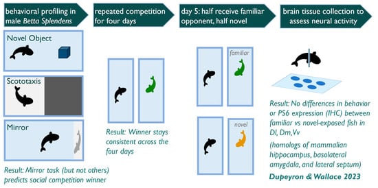
2.2. Experimental Design

The general experimental timeline is as follows: on day one, subjects underwent a novel object interaction assay immediately followed by a mirror assay. On day four, subjects underwent a scototaxis assay. On days seven through ten, subjects were exposed once per day to a competitor (the same competitor each day). Lastly, on day eleven, half the subjects were again exposed to the familiar competitor, and half the subjects were exposed to a novel competitor. Following this final social competition, subjects were euthanized for subsequent brain tissue analysis.
The first assay conducted was a novel object-interaction assay. The novel object assay (as well as the mirror assay and scototaxis assay) was conducted in a clear plastic 33.5 cm × 20 cm × 21 cm experimental tank filled to a depth of 7.5 cm with a camera positioned overhead and laminated paper placed adjacent to the tank to create a visual barrier between subjects assayed simultaneously. In the novel object-interaction assay, the subject was first removed from their home tank and placed in a white, 6-centimeter-diameter plexiglass cylinder located at one end of the experimental tank for a three-minute habituation. During this habituation period, the experimenter inserted a blue plastic rectangle (4 cm × 2 cm × 2 cm) that was attached to a clear acrylic dowel rod for ease of placement and removal (modeled after the novel object used in Lucon-Xiccato and Dadda 2016) [50] at the end of the tank opposite the habituation cylinder. After habituation, the cylinder was removed, and the subject was allowed to swim and interact freely with the object for ten minutes.
The mirror assay immediately followed the novel object interaction assay. At the end of the novel object interaction assay, the subject was gently netted and placed back into a habituation cylinder for a three-minute habituation. During this habituation period, the experimenter removed the novel object and placed a 15 cm × 10 cm mirror along the short end of the tank. The mirror was made of flexible plastic and thus was able to be inserted flush with the tank wall. After three minutes, the habituation cylinder was removed, and the subject was allowed to swim freely and interact with the mirror for ten minutes. After ten minutes, the assay concluded, and subjects were returned to their home tanks for two days of rest. Note that for all assays, the experimental tank was completely emptied of water between subjects before reuse so that no odor cues were available.
Individuals next underwent a scototaxis assay. The experimental tank for the scototaxis assay was the same as described previously, except the outside of the tank was covered on all four sides and bottom with laminated paper, one-half white and one-half black. Subjects were habituated for three minutes in a white cylinder placed in the middle of the tank. After three minutes, the cylinder was removed, and subjects were allowed to swim freely in the scototaxis tank for ten minutes. Following the assay, individuals were returned to their home tanks for two days of rest.
Subjects then underwent a repeated social competition paradigm over five consecutive days. Once a day for four consecutive days, individuals were exposed to an opponent for ten minutes. This competition was conducted in the individual’s home tank by removing the visual barrier between the subject and its neighbor, allowing for visual but not olfactory or tactile contact. This was to ensure the animals could not sustain injuries from their opponent. The plastic aquarium plant in the tank was also removed to allow full visibility of the tank for the overhead camera recording. On the fifth and final day, half of the subjects (n = 12) were again exposed to the same (familiar) opponent, and half of the subjects (n = 12) were exposed to a novel opponent. This fifth social competition was a 15-minute exposure. Immediately following this fifth competition, individuals were isolated for 45 min, then immersed in an ice bath for anesthetization and rapidly decapitated for brain tissue analysis, as described below.

2.3. Behavioral Scoring

Video images were taken from overhead cameras (Sony Handycam, HDR CX-405) throughout the experiment. Subject location data was recorded throughout the assay using the event-logging software CowLog (version 3.0.2). To assess location data, the applications “PictureInPicture” (Mac) or “OnTopReplica” (Windows) were used to overlay a transparent grid onto the video (see Supplementary Figure S1). For the mirror assay and the social competitions, additional social behaviors were quantified in Cowlog: ramming, surface breathing, opercular display, tail-beating, lateral swimming, and non-social (no behavior exhibited). Please see Table 1 for additional details on the quantification of social behavior. While multiple observers scored the videos (KW and SD), only one scorer scored each assay for consistency.

2.4. Tissue Processing

To assess a proxy for neural responses to a social competition with either a familiar or novel opponent, phosphorylated ribosomal protein S6 (PS6) was quantified in sectioned tissue using immunohistochemical (IHC) labeling. Labeling PS6 identifies ribosomal proteins that have been phosphorylated in roughly the previous hour, thus corresponding to an increase in translation [51,52]. Previous studies in other fish species have immunohistochemically labeled for PS6 to assess neural responses [31,32,53,54]. Following euthanasia, the whole head tissue was stored in 4% paraformaldehyde for four hours. Tissue was then rinsed in phosphate buffered saline (PBS) for 30 s, and the brain was extracted from the skull for storage in 30% sucrose overnight. Tissue was then embedded in TissueTek OCT (optimal cutting temperature) compound until sectioning. Brain tissue was sectioned on a cryostat (Leica CM-1860) at 20 µm in −23 °C into three series and thaw-mounted directly on microscope slides (TruBond 380 White 20 mm slides). Slides were stored at −80 °C. The immunohistochemical labeling protocol is as follows: After thawing, the slides were first rinsed 5 times for 5 min each in 1000 µL of 1X tris buffered saline (TBS) on a rocker set to low speed. Unless stated otherwise, solutions were applied as 1000 µL per slide. Note that to prevent the solutions from spilling off the slides, a barrier was drawn around the edge of the slides using an ImmEdge hydrophobic pen. The barrier was reapplied as needed throughout the IHC protocol. Additionally, slides were placed directly on a hotplate set to 40 °C for approximately 10 min, which is necessary to assist with tissue adhesion to the slide throughout the protocol. Slides were rinsed for 5 min in 1000 µL of 4% paraformaldehyde once on a shaker. Subsequently, slides were washed twice for 5 min each in 1X TBS on the shaker. Following this, block (0.3% Triton and 10% Normal Donkey Serum (NDS) in 1X TBS) was applied to each slide, and slides were transferred into a clear humid chamber (Tupperware) at room temperature and stored for 1 h. Afterwards, primary antibody (1:500 Cell Signaling 22115 235/236 Rabbit anti-PS6 antibody in diluent) was added to the slides, and slides were stored in an opaque humid chamber (an IBI Scientific immunohistochemical staining tray with water in the reservoir under the slides) at 4 °C for 24 h. This antibody has been used as a label in other studies of neural responses in fish [53]. Following primary incubation, slides were washed twice for 30 min each in 1X TBS on a shaker. Next, secondary antibodies (0.03% Donkey anti-Rabbit 594 in diluent) were applied to the slides, and slides were transferred into an opaque humid chamber at room temperature and stored for 2 h. Lastly, slides were washed in 1X TBS for 20 min once on a shaker, and ProLong with 4′,6-diamidino-2-phenylindole (DAPI, a nuclear stain [55]) was added immediately prior to coverslipping (VWR Micro Cover Glass). Coverslipped slides were set out to dry at room temperature in the dark, and once dried (typically overnight), slides were sealed with clear nail polish and stored in the dark at room temperature until imaging.

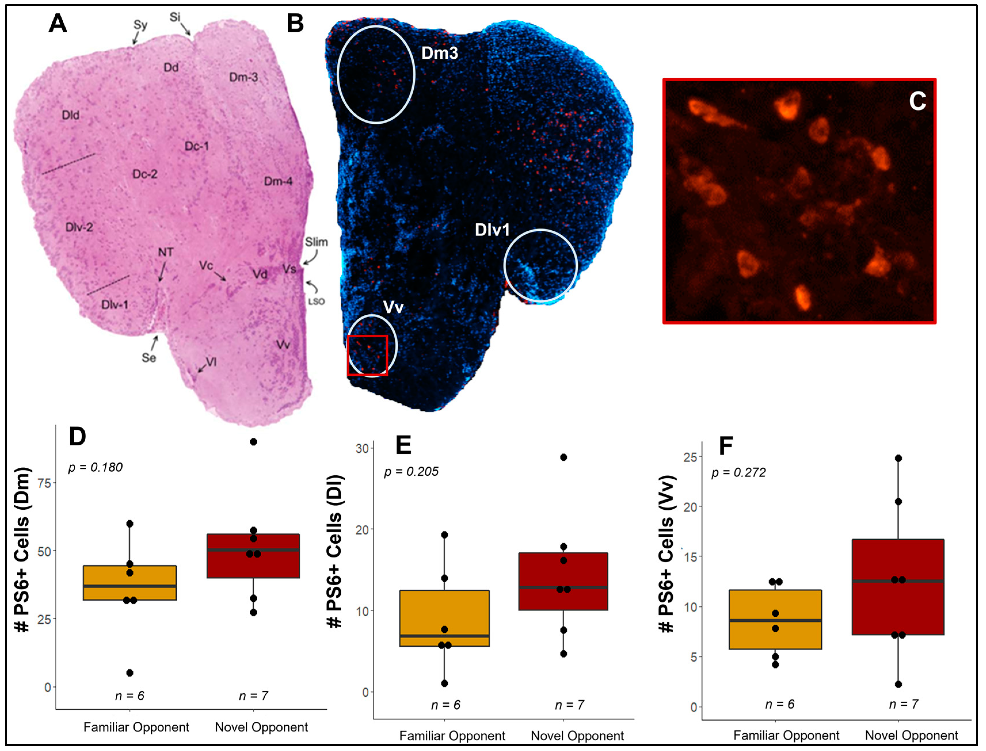
2.5. Imaging and Cell Quantification

Tissue was imaged using a Zeiss Axio Image Microscope with Apotome.2 set to 10× magnification. To select appropriate sections, we referred to a telencephalic atlas published by Magalhães Horn and Rasia-Filho (2018) [34]. Tissue was imaged between −180 µm and 0 µm, with at least one hemisphere completely visible in the image and six consecutive sections imaged when possible. Due to variability in tissue availability after extraction from the skull as well as tissue degradation during sectioning, 13 of 24 individuals (6 familiar treatments, 7 novel treatments) were successfully imaged and quantified. Composites were compiled from Z-stack images taken in both DAPI and PS6 channels and saved as TIFF files. Red channel images (corresponding to PS6 labeling) were imported into FIJI (ImageJ version 1.53q) to adjust minimum and maximum contrast values in order to subtract background fluorescence as needed, and the image was saved as a PNG (2758 × 2214 pixels). An ROI (region of interest) was aligned per region to morphological landmarks using the DAPI channel, and the area outside the ROI was erased. The brain region location was judged based on the telencephalon atlas (Magalhães Horn and Rasia-Filho 2018). Note that for consistency, each region’s ROI was saved as an object; thus, for a given brain region, the surface area of tissue that was processed for cell counting was identical across subjects. Each ROI was an oval shape (ROI width x height in pixels: Dlv1: 374 × 349, Dm3:443 × 535, Vv: 253 × 312). These standardized ROIs were sized to only capture a central “punch” of the brain region of interest and not capture any tissue in adjacent regions. This allowed us to specifically compare the number of PS6+ labelled cells in this area rather than simply capture variability in region size. PS6+ labeled cells within each region were counted by one scorer (KW) using the ImageJ “CellCounter” plugin. The number of PS6+ labelled cells reported is an average across sections. Note that if both the left and right hemispheres were quantifiable, they were considered two separate sections.

2.6. Statistical Analysis

Data were analyzed in R (version 1.1.453) [56]. The R package “cowlogdata” (version 0.1.2) [57] was used to compile individual behavioral video logs into a summary spreadsheet that included durations of time spent in each zone, time of initiation of each behavioral event, and number of events recorded for each behavior and/or number of entries into a given area. To analyze categorical data, we conducted a Chi-squared test. To analyze continuous data that was compared across categories (e.g., winner vs. loser), we first conducted a Shapiro–Wilk normality test using the R function, and then either a Wilcoxon Rank Sum test (nonparametric paired data), Wilcoxon Signed-Rank Test (nonparametric unpaired data), or t-test (parametric data), a Chi-squared test (categorical data), an ANOVA (parametric data with three or more categories), or the Kruskal–Wallis test (nonparametric data with three or more categories) was used as appropriate. To analyze two continuous variables, we used linear regressions using the “lm” function in R. All data was Bonferroni corrected for the number of behaviors analyzed within the given assay or the number of comparisons in the cross-assay analysis. To visualize behavior over time, we modified the function “clseries” in the R package “cowlogdata” to include 50 time bins of 10 seconds each. Lastly, effect sizes were either calculated using a direct formula or by using the R package “rstatix.” Data analysis R code and primary data can be found on github.com/kellyjwallace/Dupeyron_Wallace_Betta_2023 (accessed on 7 September 2022).

3. Results

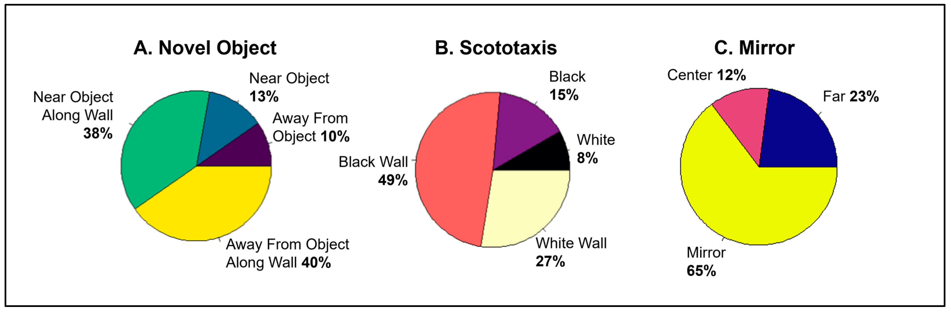
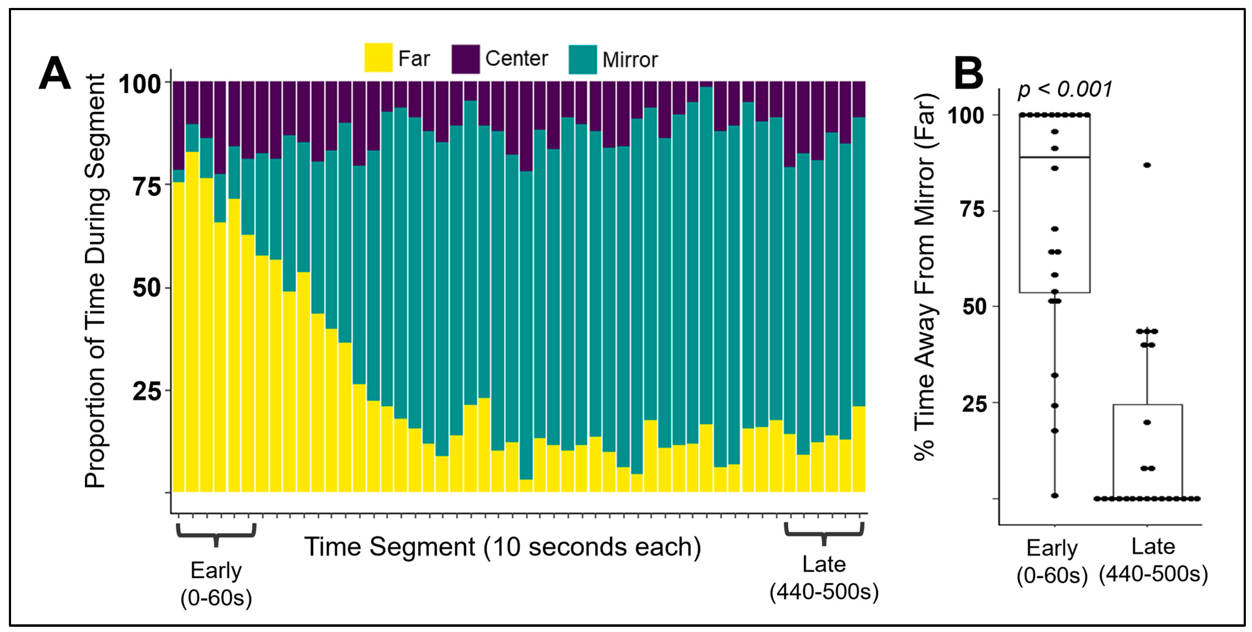
3.1. Is Individual Behavior Predictable and Consistent across Contexts?

We first quantified where individuals spent their time during the three behavior tests separately (Figure 1). In the novel object assay, individuals spent their time evenly between the half of the tank near the object (51%) and away from the object (50%) (Figure 1A, SD (sum near object) ± 8.6%). In the scototaxis assay, individuals showed a significant preference for the black half of the tank (64%) over the white half (35%) (Wilcoxon Signed Rank Test, p = 0.008, r = 0.531, V = 241) (Figure 1B, SD (sum white) ± 25.8%). In the mirror assay, individuals showed a significant preference for the third of the tank closest to the mirror (65%) than the other two-thirds of the tank (t-test, p < 0.001, Cohen’s D = 1.363, t = 6.67) (Figure 1C, SD (far zone) ± 16.1%, SD (center zone) ± 10.2%, SD (mirror zone) ± 23.3%).
To understand how individuals behaved during the assays in greater detail, we visualized behavior over time. While we did not identify obvious changes over time in the scototaxis or novel object assays (Supplementary Figure S2A,B), we observed a noticeable change over the minutes of the mirror assay, where individuals initially avoided the mirror but then began spending most of their time near it (Figure 2A). To quantify this observation, we compared the proportion of time spent in the third of the tank furthest away from the mirror in the first minute of the assay (seconds 0–60) to the latest minute included in the time series visualization (seconds 440–500) and found that individuals significantly decreased their avoidance of the mirror over time (Wilcoxon Signed Rank Test, p < 0.001, V = 294, r = 0.764, Figure 2B).
To then identify if the behaviors across the three assays correlated to each other (consistent individual variation), we selected one behavior from each assay to compare to the others using linear regression: time spent near the novel object, time spent in the black half of the scototaxis tank, and time spent near the mirror. No behaviors correlated at the individual level across assays when assessed via linear regression (Supplementary Table S1). Additionally, to verify if non-behavioral factors predicted behavior in the four assays, we correlated standard length (an indirect proxy for age) and color. Size did not correlate with behavior in any of the three assays when assessed via linear regression (Supplementary Table S1). Color morphs, quantified by the experimenters, were not evenly distributed: most fish were either “blue-red-purple” (n = 11) or “red” (n = 10), with one fish each being categorized as “turquoise”, “blue”, and “white”. Because of this, we statistically compared only the two color morphs with large enough sample sizes. Behavior in the three assays did not differ between the two color morphs when assessed via a t-test or Wilcoxon test, as appropriate (Supplementary Figure S3).

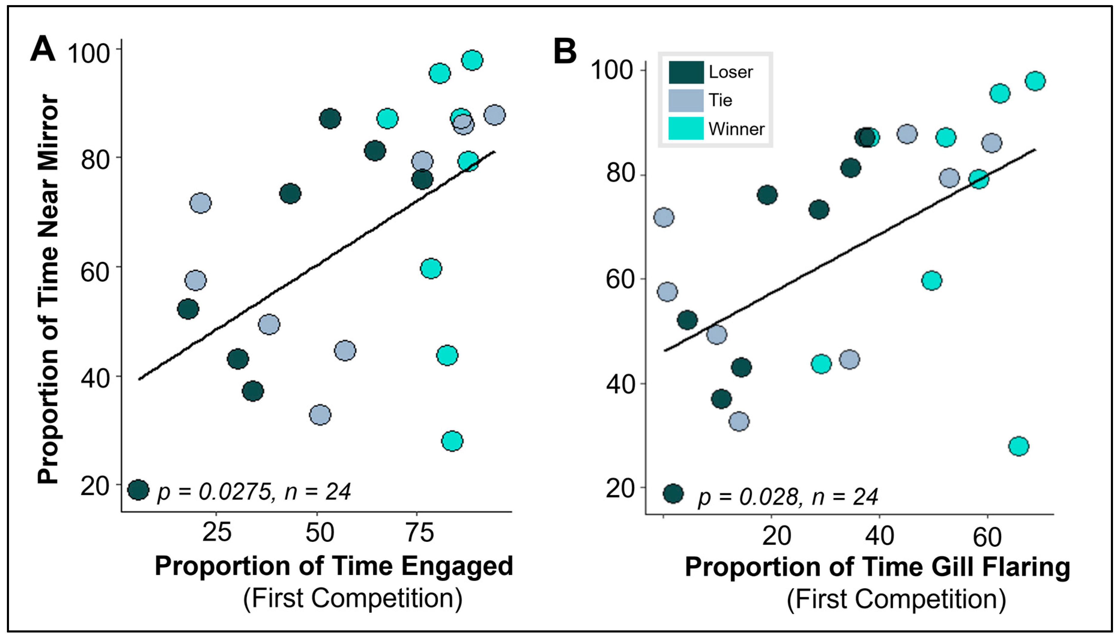
3.2. Does Behavior in Other Contexts Predict Social Competition?

To categorize a “winner” and a “loser” in the social competition, we identified which individual in the dyad had at least 20% more gill-flaring time than their opponent. Gill-flaring has been used as a metric of competition outcome in other studies [21,22,58]. While we refer to individuals as “winners” and “losers” in this experiment, we acknowledge that this criterion can only presume a winner as the opponents are not allowed to interact directly. Winners spent significantly more time active than losers (Wilcoxon Rank Sum Test comparing winners and losers (no ties), p = 0.002 following Bonferroni correction, W = 63, Z = 0.814, Figure 3A), but did not differ in the other aggressive behavior measures: tail beating, ramming/biting, lateral swimming, or surface breathing (Supplementary Figure S4). To determine if non-behavioral factors (size/age, color) predicted the outcome of the first social competition, we quantified the size of the individual relative to its opponent, such that a positive value indicates the individual was the larger fish in the dyad and a negative value indicates the individual was the smaller fish in the dyad. We found that when assessed via a Chi-squared test, the winner was more likely than chance to be the larger opponent (p = 0.011, X2 = 9.00, φ = 1.837, as seen in Figure 3B), but winners and losers did not differ in color (Supplementary Figure S5A).
To then identify if behaviors in the three assays predicted the outcome of the social competition, we compared winners and losers in the three behavior assays. Winners and losers did not differ in any of the three behaviors assessed prior to the competition: time spent near the novel object, time spent in the black half of the scototaxis tank, and time spent near the mirror (Supplementary Figure S5B–D). Finding no effect on outcome (winning or losing), we correlated behaviors across all individuals. We found that behavior in the mirror assay correlated to behavior in the social competition at the individual level: when conducting a simple linear regression, time spent near the mirror (the independent variable) significantly correlated with activity in the social competition (p = 0.0275 following Bonferroni correction, df = 22, r2 = 0.269, Figure 4A) and with gill flaring (p = 0.028 following Bonferroni correction, df = 22, r2 = 0.268, Figure 4B).

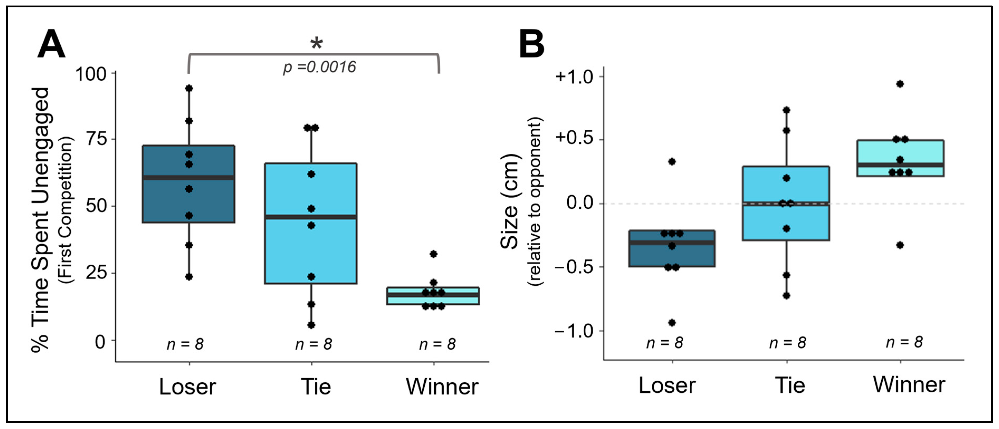
3.3. Does Competitive Behavior Remain Consistent over Repeated Exposures?

Upon assessing the first social competition and its predictors, we next determined if repeated exposures to an opponent influenced behavior (days one through four). To do so, we first examined if the winner during the first encounter was more likely than chance to remain the winner over the subsequent three encounters with the same opponent. We found that outcomes were incredibly consistent across the four social competitions—in fact, no individual who lost the first competition would go on to win any of the three subsequent competitions. Correspondingly, those who won their first competition would not lose in the following three. While in some competitions, opponents that previously won/lost would then tie, they never “flipped” the winner and loser in the first four competitions. When quantifying this via the Chi-squared test (i.e., assessing if the winner of the first competition is more likely than chance to win the second/third/fourth competition), the result is significant for each of the four days (comparing days 1 and 2: p = 0.001, X2 = 10.286, φ = 2.100; comparing days 1 and 3: p = 0.038, X2 = 4.500, φ = 0.919; and comparing days 1 and 4: p = 0.011, X2 = 6.400, φ = 1.306). Note that this analysis excludes ties. On Day One, 4/12 dyadic competitions resulted in ties; on Day Two: 4/12; on Day Three: 6/12; on Day Four: 5/12; on Day Five: 7/12. The number of ties did not significantly increase over subsequent competitions when assessed via a Chi-squared test comparing the days with the largest difference in ties (Day 1 vs. Day 5 and Day 2 vs. Day 5, both of which compared four versus seven ties). Additionally, we visualized individual behavior in each social competition over time intervals of ten seconds to observe if changes over the course of the assay differed across the four competitions (for example, noticeable habituation or increases in aggression after the first minute(s) of the task). In doing so, we did not detect any evident visual patterns (Supplementary Figure S2C–F).

3.4. Do Competitive Behavior and Neural Responses Differ between Individuals Exposed to A Novel vs. Familiar Opponent?

Interestingly, while outcomes were highly consistent during the first four competitions, the winner of the fourth social competition did not significantly predict the winner of the fifth competition for either the familiar treatment (p = 1.000, X2 = 0) or the novel treatment (p = 1.000, X2 = 0). See Supplementary Table S2 for the outcome of each competition.
We compared the behavior of individuals facing a novel opponent on Day 5 of the social competition to that of those facing a familiar opponent. No competitive behaviors significantly differed between familiar and novel opponent-exposed individuals (Supplementary Table S3). To assess if neural response differed between familiar and novel opponent-exposed, we quantified PS6+ labeled cells in the putative teleost homologs to the hippocampus (Dlv), basolateral amygdala (Dm), and lateral septum (Vv) (Figure 5A–C). No significant differences were observed in the number of PS6-labeled cells in the three regions when assessed via t-tests (Dm: p = 0.180, t = −1.438, Cohen’s D = −0.795, Dl: p = 0.205, t = −1.348, Cohen’s D = −0.745, Vv: p = 0.272, t = −1.173, Cohen’s D = −0.636) (Figure 5D–F).
Lastly, given that we had observed behavioral predictors that correlated to competitive behavior at the individual level, we similarly asked if PS6 expression correlated to individual behavior during the fifth social competition using linear regression analysis. We found that no behavior correlated to PS6 expression in any brain region (Supplementary Table S3).

4. Discussion

Examining the non-social behavioral tendencies that predict success in social competitions is of vital importance to biologists as it contextualizes these traits within the constraints of natural and sexual selection. If these tendencies indeed provide benefits to the individual during competitive interactions, then the expectation is that other compensatory tradeoffs must be regulating their presence or variation in the population. This balancing act provides the foundation for individual variation, behavioral flexibility, and/or the development of fixed alternative tactics over time. A simple example may be that a bolder individual who is more likely to win in an aggressive territorial contest may also be more likely to be preyed upon by a predator [59].
Betta splendens has long been a model for understanding the physiological mediators of aggression due to their ease of use in laboratory studies as well as their quantifiable behavioral repertoires [22]. Now, with the advancement of new techniques [36,60] and the emerging integration of molecular perspectives with ethological frameworks [9], their utility as a model is reaching new areas of investigation. Here, we expanded this work by conducting a multi-assay behavioral suite designed to further uncover the behavioral correlates of competitive behavior.
In our study, Betta males did not prefer or avoid the novel object. Previous studies conducting a novel object interaction assay have shown chemogenic effects on object preference [44,61], but have not reported if object preference or avoidance in control animals was significantly different from chance. In the scototaxis task, we found a significant preference for the black side. This differs from a previous study that found no preference [39], though the study was only conducted in females. Further exploration of sex differences and correlations with other anxiety measures is a useful next step for this species.
In the social competition, winners were unsurprisingly larger in size, which is why many studies in Betta size-match opponents [41]. When we correlated behavior in the social competition to those measured in separate assays, we found that behavior towards a mirror predicted behavior when exposed to a live competitor, as has been previously shown [42,43]. Yet neophilia, in the form of exposure to a novel object, and anxiety, in the form of exposure to a black-white (scototaxis) tank, did not predict behavior during the social competitions. When then determining if these assays could predict the winner of the social competition, curiously, neither these two tasks nor engagement with the mirror predicted the winner. This lack of correlation differed from our predictions, as we had expected correlations across the tasks based on similar observations in Betta and in other species. In Betta splendens, boldness (measured across three assays) correlates positively with gill flaring [44]. In the guppy Brachyraphis episcopi, low predation populations approach a mirror and a novel object more than high predation populations [62], and in the juvenile cichlid Astatotilapia burtoni, dominance behavior and exploration in an open field task load together in a principal component analysis [63]. Instead of identifying these relationships, our results more closely resemble those seen in zebrafish, with no correlation between boldness (assessed via depth in a trapezoidal tank) and aggression towards a mirror [64]. It is certainly possible that the details of each experimental design are what drive this variation in results. If so, adapting and standardizing assays for boldness, exploration, and stress specifically for Betta (as has been accomplished with aggressive competition and the mirror task) is an area of importance in future work. Alternatively, as results in Brachyraphis episcopi [62] and in the three-spine stickleback Gasterosteus aculeatus [65] suggest, population differences may drive a large proportion of behavioral variation. Thus, we recommend prioritizing comparisons across strains of Betta.
After this initial correlative search, we compared Bettas exposed to a familiar and novel opponent using a habituation–dishabituation paradigm. Over repeated exposure to the same opponent, we found consistency in the competitive outcomes, reflecting previous literature on stable dominance interactions in this species [23]. This consistency was abolished on Day 5, as expected for the novel treatment, but curiously, it was also abolished for the familiar treatment. We do not believe this is a methodological artifact, as the social competition procedure was identical to the prior days and all days were consecutive with no temporal gap. It is possible that, in using a repeated measures barrier design, we have identified a particularly relevant timepoint of habituation or of the establishment of dominance that is not seen when individuals freely interact. For example, in when paradise fish are exposed to the same competitor consecutively for three days, they showed habituation toward the opponent on the third day (though, unlike our experiment, this was distinct to a familiar competitor, as repeated exposure to a novel competitor each day did not elicit this habituation) [66].
On the final exposure, with some individuals receiving a familiar opponent and some individuals receiving a novel opponent, we did not find differences between these two treatments in behavior, as has been previously observed [45]. We note that with a larger sample size in the future, we could statistically compare four groups in both behavior and PS6 expression to better capture the variation in social outcomes: familiar-exposed winners, familiar-exposed losers, novel-exposed winners, and novel-exposed losers. Similarly, the lack of differences in behavior may reflect the design limitations of our social competition test. Because of the barrier between individuals, not only were animals unable to process olfactory information [67], but additionally, they did not receive feedback from direct interaction (such as sustaining an injury or successfully causing the opponent to flee). With this said, in our experiment, Betta consistently exhibited dramatic fighting behavior upon initiation of the social competition and during the mirror task, suggesting that visual information is sufficient to induce ecologically relevant behavior. Previous work on Betta courtship has used computer-generated videos of conspecifics to disentangle relevant features [68], and this approach can be adapted to further study the results described here.
In order to facilitate the further expansion of Betta splendens as a model for integrated studies of behavior, cognition, and neuroscience [69], we included a more detailed analysis of our behavioral data. To showcase the depth of data analysis that can be conducted on behavioral data, we incorporated an analysis of behavior and location over time within each assay, similar to an analysis conducted by Ramos et al. (2021) [41]. This yielded an interesting and significant insight worth additional exploration: behavior towards a mirror, but not towards live opponents or towards a novel object, changes over the minutes of the task.
Neural analyses can provide valuable insight into the underlying factors driving individual social behavior. Importantly, in the present study, we have now conducted the first characterization of PS6 expression in response to social stimuli in the Betta telencephalon. In doing so, we saw expression in regions of the SDMN as well as noticeable variation across individuals, providing a foundation for subsequent cellular work in this species. Yet, we did not discover differences between familiar and novel opponents in the brain, as hypothesized. Why might PS6 labelling not differ between familiar and novel-exposed individuals or correlate to competitive behaviors at the individual level? It is certainly possible that Bettas do not recognize previously encountered opponents or only exhibit recognition-related behavior under specific circumstances, such as with a female audience present or an opponent bubble nest already built [70]. Additionally, even if they do exhibit recognition, they may not alter their behavior due to a generalized “aggression ceiling effect.” Alternatively, it is possible that these brain regions may indeed differentially process novel vs. familiar social stimuli and that we were unable to detect this via assessment of PS6 labeling. Future studies could examine more brain regions of relevance beyond the three quantified here. We highly recommend future studies also explore Betta neural responses using other markers as well as assess nonapeptide expression [71] in the preoptic area and nucleus accumbens, as these regions are more directly implicated in aggression in fish. From an ecological perspective, we should also compare neural differences between winners and losers under more naturalistic group conditions. Historical work has shown that groups of Betta can form stable communities under certain circumstances. In mixed-sex communities, aggression wanes after hierarchy establishment after roughly ten days [72], but in male-only communities, aggression does not wane over ten weeks [73], suggesting that reproductive context is a highly relevant dimension for further analysis.

5. Conclusions

The Siamese fighting fish Betta splendens has been long used as a model for understanding the behavioral and physiological predictors of success in territorial competitions. Here we expanded our understanding of social competition in this species by assessing behavior in alternative contexts and by assessing the neurobiological mechanisms involved in opponent recognition. Employing novel assays [74] and neural analyses [75] has yielded important insights into the social world of other species of fish. While we did not identify opponent-specific recognition within the scope of our experiment, we employed the first use of a habituation–dishabituation competition design and the first characterization of neural response in the telencephalon in Betta.
In future work, we advocate for greater incorporation of cognitive testing into studies on Betta social behavior. Previous cognitive tasks have found that Betta can complete spatial memory tasks [76], radial arm mazes [77], and a serial reversal discrimination task [78]. More generally, current efforts have been overturning the assumption that fish do not display complex cognitive abilities, which has unfortunately limited their inclusion in cognitive and neuroscientific fields historically [30,79]. Establishing Betta as a tractable and relevant model for studying the intersection of cognitive ecology and social neuroscience [74] will yield insights into not only this charismatic species but into our conceptual understanding of social cognition.

Supplementary Materials

The following supporting information can be downloaded at: https://www.mdpi.com/article/10.3390/fishes8080384/s1, Table S1: Results of linear regressions correlating individual behavior across the three assays and subject size; Table S2: A visualization of the outcomes of the five social competitions. Each row is an individual. Note that the first twelve individuals were in the familiar treatment, and thus their pairing stayed the same during the fifth competition, whereas the novel treatment individuals encountered new opponents during the fifth competition; Table S3: The leftmost column shows the results of a t-test or Wilcoxon test (determined by a Shapiro-Wilk normality test) comparing familiar and novel treatment individuals in their behavior during the last (Day 5) social competition. The three columns to the right show the results of a linear regression analysis correlating the neural response of the stated brain region with behavior during the last social competition; Figure S1: Visualizations of the locations that fish were quantified in during the three behavior assays: (A) novel object, (B) scototaxis, and (C) mirror. In addition to location, during the mirror assay fish were quantified for social behaviors; Figure S2: Visualizations of average time spent in the various locations of the scototaxis assay (A), novel object assay (B), and the first four social competitions (C–F), averaged across all individuals. Note the visualization includes time 0 to time 500 (seconds). Figure S3: Color morph does not predict behavior in the three assays. Note the p-values reported are the results of t-test or Wilcoxon test comparing the BRP and RED color morphs. Note that the y-axes are all % of time, with 0 = 0% and 1 = 100%. Figure S4: Assessed during the first social competition, winners did not significantly differ from losers in time spent tail beating, ramming/biting, lateral swimming, or surface breathing. Note the p values shown are t-tests or Wilcoxon tests comparing winners to losers (not including ties) and are not Bonferroni corrected. Note that the y-axes are all % of time, with 0 = 0% and 1 = 100%. Figure S5: Assessed during the first social competition, winners did not significantly differ from losers in color (A). Note the color comparison here was simply the “blue-red-purple” morph compared to “red” morph as the other color morphs only had one individual per category. Furthermore, winners and losers did not differ in behaviors from the other three assays: time spent near the novel object (B), time spent in the black half of the scototaxis assay (C), or time spent near the mirror (D).

Author Contributions

Conceptualization, K.J.W.; Data curation, S.D.; Formal analysis, K.J.W.; Methodology, S.D. and K.J.W.; Writing—original draft, S.D.; Writing—review & editing, K.J.W. All authors have read and agreed to the published version of the manuscript.

Funding

This work was supported by the National Science Foundation (National Science Foundation PRFB no. 2011001 to K.J.W.).

Institutional Review Board Statement

All procedures were approved by the Institutional Animal Care and Use Committee of Emory University (Protocol 202200088). The IACUC oversees compliance with the Guide for the Care and Use of Laboratory Animals, Eighth Edition, published by the National Research Council of the National Academies [80]. Emory University is an AAALAC-fully accredited institution (Association for Assessment and Accreditation of Laboratory Animal Care International).

Data Availability Statement

R code and raw data are accessible via github.com/kellyjwallace/Dupeyron_Wallace_Betta_2023 (accessed 24 July 2023).

Acknowledgments

We thank Richmond Thompson and Aubrey Kelly for their guidance, feedback, and assistance during data collection, analysis, and writing.

Conflicts of Interest

The authors declare no conflict of interest.

References

  1. Shettleworth, S.J. Cognition, Evolution, and Behavior; Oxford University Press: Oxford, UK, 2009. [Google Scholar]
  2. Haley, M.P.; Deutsch, C.J.; Le Boeuf, B.J. Size, dominance and copulatory success in male northern elephant seals, Mirounga angustirostris. Anim. Behav. 1994, 48, 1249–1260. [Google Scholar] [CrossRef]
  3. Funghi, C.; Leitão, A.V.; Ferreira, A.C.; Mota, P.G.; Cardoso, G.C. Social Dominance in a Gregarious Bird is Related to Body Size but not to Standard Personality Assays. Ethology 2014, 121, 84–93. [Google Scholar] [CrossRef]
  4. Tibbetts, E.A.; Dale, J. A socially enforced signal of quality in a paper wasp. Nature 2004, 432, 218–222. [Google Scholar] [CrossRef]
  5. Wingfield, J.C.; Hegner, R.E.; Dufty, A.M., Jr.; Ball, G.F. The “Challenge Hypothesis”: Theoretical Implications for Patterns of Testosterone Secretion, Mating Systems, and Breeding Strategies. Am. Nat. 1990, 136, 829–846. [Google Scholar] [CrossRef]
  6. Formica, V.; Donald, H.; Marti, H.; Irgebay, Z.; Brodie, E. Social network position experiences more variable selection than weaponry in wild subpopulations of forked fungus beetles. J. Anim. Ecol. 2020, 90, 168–182. [Google Scholar] [CrossRef] [PubMed]
  7. Favre, M.; Martin, J.G.; Festa-Bianchet, M. Determinants and life-history consequences of social dominance in bighorn ewes. Anim. Behav. 2008, 76, 1373–1380. [Google Scholar] [CrossRef]
  8. Real, L.A. Toward a cognitive ecology. Trends Ecol. Evol. 1993, 8, 413–417. [Google Scholar] [CrossRef]
  9. Wallace, K.J.; Hofmann, H.A. Decision-making in a social world: Integrating cognitive ecology and social neuroscience. Curr. Opin. Neurobiol. 2021, 68, 152–158. [Google Scholar] [CrossRef]
  10. Réale, D.; Reader, S.M.; Sol, D.; McDougall, P.T.; Dingemanse, N.J. Integrating animal temperament within ecology and evolution. Biol. Rev. 2007, 82, 291–318. [Google Scholar] [CrossRef]
  11. Sih, A.; Del Giudice, M. Linking behavioural syndromes and cognition: A behavioural ecology perspective. Philos. Trans. R. Soc. B Biol. Sci. 2012, 367, 2762–2772. [Google Scholar] [CrossRef]
  12. Guillette, L.M.; Reddon, A.R.; Hurd, P.L.; Sturdy, C.B. Exploration of a novel space is associated with individual differences in learning speed in black-capped chickadees, Poecile atricapillus. Behav. Process. 2009, 82, 265–270. [Google Scholar] [CrossRef] [PubMed]
  13. Burns, J.G.; Rodd, F.H. Hastiness, brain size and predation regime affect the performance of wild guppies in a spatial memory task. Anim. Behav. 2008, 76, 911–922. [Google Scholar] [CrossRef]
  14. Gibelli, J.; Aubin-Horth, N.; Dubois, F. Individual differences in anxiety are related to differences in learning performance and cognitive style. Anim. Behav. 2019, 157, 121–128. [Google Scholar] [CrossRef]
  15. Colléter, M.; Brown, C. Personality traits predict hierarchy rank in male rainbowfish social groups. Anim. Behav. 2011, 81, 1231–1237. [Google Scholar] [CrossRef]
  16. David, M.; Auclair, Y.; Cézilly, F. Personality predicts social dominance in female zebra finches, Taeniopygia guttata, in a feeding context. Anim. Behav. 2011, 81, 219–224. [Google Scholar] [CrossRef]
  17. Favati, A.; Leimar, O.; Løvlie, H. Personality Predicts Social Dominance in Male Domestic Fowl. PLoS ONE 2014, 9, e103535. [Google Scholar] [CrossRef]
  18. Wallace, K.J.; Choudhary, K.D.; Kutty, L.A.; Le, D.H.; Lee, M.T.; Wu, K.; Hofmann, H.A. Social ascent changes cognition, behaviour and physiology in a highly social cichlid fish. Philos. Trans. R. Soc. B Biol. Sci. 2022, 377, 20200448. [Google Scholar] [CrossRef]
  19. Devost, I.; Jones, T.B.; Cauchoix, M.; Montreuil-Spencer, C.; Morand-Ferron, J. Personality does not predict social dominance in wild groups of black-capped chickadees. Anim. Behav. 2016, 122, 67–76. [Google Scholar] [CrossRef]
  20. Castro, N.; Ros, A.F.; Becker, K.; Oliveira, R.F. Metabolic costs of aggressive behaviour in the Siamese fighting fish, Betta splendens. Aggress. Behav. 2006, 32, 474–480. [Google Scholar] [CrossRef]
  21. Evans, C. Display vigour and subsequent fight performance in the siamese fighting fish, Betta splendens. Behav. Process. 1985, 11, 113–121. [Google Scholar] [CrossRef]
  22. Portugal, S.J. Siamese fighting fish. Curr. Biol. 2023, 33, R341–R343. [Google Scholar] [CrossRef]
  23. Wallen, K.; Wojciechowski-Metzlar, C.I. Social conditioning and dominance in male Betta splendens. Behav. Process. 1985, 11, 181–188. [Google Scholar] [CrossRef] [PubMed]
  24. Lissmann, H.W. Die Umwelt des Kampffisches (Betta splendens Regan). J. Comp. Physiol. A 1932, 18, 65–111. [Google Scholar] [CrossRef]
  25. Newman, S.W. The Medial Extended Amygdala in Male Reproductive Behavior A Node in the Mammalian Social Behavior Network. Ann. N. Y. Acad. Sci. 1999, 877, 242–257. [Google Scholar] [CrossRef] [PubMed]
  26. Goodson, J.L. The vertebrate social behavior network: Evolutionary themes and variations. Horm. Behav. 2005, 48, 11–22. [Google Scholar] [CrossRef]
  27. Kelly, A.M. A consideration of brain networks modulating social behavior. Horm. Behav. 2022, 141, 105138. [Google Scholar] [CrossRef]
  28. Wallace, K.J.; Chun, E.K.; Manns, J.R.; Ophir, A.G.; Kelly, A.M. A test of the social behavior network reveals differential patterns of neural responses to social novelty in bonded, but not non-bonded, male prairie voles. Horm. Behav. 2023, 152, 105362. [Google Scholar] [CrossRef]
  29. O’Connell, L.A.; Hofmann, H.A. Evolution of a Vertebrate Social Decision-Making Network. Science 2012, 336, 1154–1157. [Google Scholar] [CrossRef]
  30. Calvo, R.; Schluessel, V. Neural substrates involved in the cognitive information processing in teleost fish. Anim. Cogn. 2021, 24, 923–946. [Google Scholar] [CrossRef]
  31. Fan, R.; Reader, S.M.; Sakata, J.T. Alarm cues and alarmed conspecifics: Neural activity during social learning from different cues in Trinidadian guppies. Proc. R. Soc. B Boil. Sci. 2022, 289, 20220829. [Google Scholar] [CrossRef]
  32. Baran, N.M.; Streelman, J.T. Ecotype differences in aggression, neural activity and behaviorally relevant gene expression in cichlid fish. Genes Brain Behav. 2020, 19, e12657. [Google Scholar] [CrossRef] [PubMed]
  33. Rodriguez-Santiago, M.; Jordan, A.; Hofmann, H.A. Neural activity patterns differ between learning contexts in a social fish. Proc. R. Soc. B Boil. Sci. 2022, 289, 20220135. [Google Scholar] [CrossRef] [PubMed]
  34. Horn, C.M.; Rasia-Filho, A.A. The Cytoarchitecture of the Telencephalon of Betta splendens Regan 1910 (Perciformes: Anabantoidei) with a Stereological Approach to the Supracommissural and Postcommissural Nuclei. Anat. Rec. 2017, 301, 88–110. [Google Scholar] [CrossRef]
  35. McDonald, C.G.; Paul, D.H.; Hawryshyn, C.W. Visual Sensation of an Ethological Stimulus, the Agonistic Display of Betta splendens, Revealed using Multi-Unit Recordings from Optic Tectum. Environ. Biol. Fishes 2004, 70, 285–291. [Google Scholar] [CrossRef]
  36. Vu, T.-D.; Iwasaki, Y.; Shigenobu, S.; Maruko, A.; Oshima, K.; Iioka, E.; Huang, C.-L.; Abe, T.; Tamaki, S.; Lin, Y.-W.; et al. Behavioral and brain- transcriptomic synchronization between the two opponents of a fighting pair of the fish Betta splendens. PLoS Genet. 2020, 16, e1008831. [Google Scholar] [CrossRef] [PubMed]
  37. Carvalho, P.D.P.; Mayer, P.C.M.; Walsh-Monteiro, A.; Gouveia, A. Effects of menthol on aggressive behavior in Siamese fighting fish (Betta splendens). Psychol. Neurosci. 2015, 8, 290–296. [Google Scholar] [CrossRef]
  38. Forsatkar, M.N.; Nematollahi, M.A.; Biro, P.A.; Beckmann, C. Individual boldness traits influenced by temperature in male Siamese fighting fish. Physiol. Behav. 2016, 165, 267–272. [Google Scholar] [CrossRef]
  39. Tudor, M.S.; Lopez-Anido, R.N.; Yocius, C.A.; Conlin, S.M.; Hamlin, H.J. Ecologically relevant arsenic exposure alters female mate preference and anxiety-like behavior in Betta splendens. Heliyon 2019, 5, e02646. [Google Scholar] [CrossRef]
  40. Bronstein, P.M. Onset of combat in male Betta splendens. J. Comp. Psychol. 1983, 97, 135–139. [Google Scholar] [CrossRef]
  41. Ramos, A.; Alex, D.; Cardoso, S.D.; Gonçalves, D. Androgens and corticosteroids increase in response to mirror images and interacting conspecifics in males of the Siamese fighting fish Betta splendens. Horm. Behav. 2021, 132, 104991. [Google Scholar] [CrossRef]
  42. Baenninger, R. Fighting by Betta splendens: Effects on aggressive displaying by conspecifics. Psychon. Sci. 1968, 10, 185–186. [Google Scholar] [CrossRef]
  43. Meliska, J.A.; Meliska, C.J.; Hoyenga, K.T.; Hoyenga, K.B.; Ward, E.F. Approach tendency and threat display as related to social status of Siamese fighting fish, Betta splendens. Anim. Learn. Behav. 1975, 3, 135–139. [Google Scholar] [CrossRef]
  44. Hebert, O.L.; Lavin, L.E.; Marks, J.M.; Dzieweczynski, T.L. The effects of 17α-ethinyloestradiol on boldness and its relationship to decision making in male Siamese fighting fish. Anim. Behav. 2014, 87, 203–212. [Google Scholar] [CrossRef]
  45. Alyan, S. Male Betta splendens are equally aggressive toward neighbors and strangers. J. Ichthyol. 2010, 50, 1066–1069. [Google Scholar] [CrossRef]
  46. Gheusi, G.; Bluthé, R.-M.; Goodall, G.; Dantzer, R. Social and individual recognition in rodents: Methodological aspects and neurobiological bases. Behav. Process. 1994, 33, 59–87. [Google Scholar] [CrossRef]
  47. O′Connell, L.A.; Hofmann, H.A. The Vertebrate mesolimbic reward system and social behavior network: A comparative synthesis. J. Comp. Neurol. 2011, 519, 3599–3639. [Google Scholar] [CrossRef]
  48. Northcutt, R.G. Connections of the lateral and medial divisions of the goldfish telencephalic pallium. J. Comp. Neurol. 2006, 494, 903–943. [Google Scholar] [CrossRef]
  49. Portavella, M.; Vargas, J.; Torres, B.; Salas, C. The effects of telencephalic pallial lesions on spatial, temporal, and emotional learning in goldfish. Brain Res. Bull. 2002, 57, 397–399. [Google Scholar] [CrossRef]
  50. Lucon-Xiccato, T.; Dadda, M. Guppies Show Behavioural but Not Cognitive Sex Differences in a Novel Object Recognition Test. PLoS ONE 2016, 11, e0156589. [Google Scholar] [CrossRef] [PubMed]
  51. Butler, J.M.; Whitlow, S.M.; Roberts, D.A.; Maruska, K.P. Neural and behavioural correlates of repeated social defeat. Sci. Rep. 2018, 8, 6818. [Google Scholar] [CrossRef] [PubMed]
  52. Magnuson, B.; Ekim, B.; Fingar, D.C. Regulation and function of ribosomal protein S6 kinase (S6K) within mTOR signalling networks. Biochem. J. 2012, 441, 1–21. [Google Scholar] [CrossRef]
  53. Calvo, R.; Hofmann, M.H.; Schluessel, V. Brain areas activated during visual learning in the cichlid fish Pseudotropheus zebra. Brain. Struct. Funct. 2023, 228, 859–873. [Google Scholar] [CrossRef] [PubMed]
  54. Fischer, E.K.; Westrick, S.E.; Hartsough, L.; Hoke, K.L. Differences in neural activity, but not behavior, across social contexts in guppies, Poecilia reticulata. Behav. Ecol. Sociobiol. 2018, 72, 131. [Google Scholar] [CrossRef]
  55. Tarnowski, B.I.; Spinale, F.G.; Nicholson, J.H. DAPI as a Useful Stain for Nuclear Quantitation. Biotech. Histochem. 1991, 66, 296–302. [Google Scholar] [CrossRef]
  56. R Core Team. R: A Language and Environment for Statistical Computing; R Foundation for Statistical Computing: Vienna, Austria, 2021; Available online: https://www.R-project.org (accessed on 7 September 2022).
  57. Wallace, K.J. Cowlogdata: An R Package to Analyze and Visualize Observations Generated by the Event Logging Software CowLog. 2020. Available online: github.com/kellyjwallace/cowlogdata (accessed on 7 September 2022).
  58. Simpson, M. The Display of the Siamese Fighting Fish, Betta splendens. Anim. Behav. Monogr. 1968, 1, 1–73. [Google Scholar] [CrossRef]
  59. Balaban-Feld, J.; Mitchell, W.A.; Kotler, B.P.; Vijayan, S.; Elem, L.T.T.; Rosenzweig, M.L.; Abramsky, Z. Individual willingness to leave a safe refuge and the trade-off between food and safety: A test with social fish. Proc. R. Soc. B Boil. Sci. 2019, 286, 20190826. [Google Scholar] [CrossRef] [PubMed]
  60. Kwon, Y.M.; Vranken, N.; Hoge, C.; Lichak, M.R.; Norovich, A.L.; Francis, K.X.; Camacho-Garcia, J.; Bista, I.; Wood, J.; McCarthy, S.; et al. Genomic consequences of domestication of the Siamese fighting fish. Sci. Adv. 2022, 8, eabm4950. [Google Scholar] [CrossRef] [PubMed]
  61. Forsatkar, M.N.; Nematollahi, M.A.; Brown, C. The toxicological effect of Ruta graveolens extract in Siamese fighting fish: A behavioral and histopathological approach. Ecotoxicology 2016, 25, 824–834. [Google Scholar] [CrossRef]
  62. Archard, G.A.; Braithwaite, V.A. Variation in aggressive behaviour in the poeciliid fish Brachyrhaphis episcopi: Population and sex differences. Behav. Process. 2011, 86, 52–57. [Google Scholar] [CrossRef]
  63. Solomon-Lane, T.K.; Hofmann, H.A. Early-life social environment alters juvenile behavior and neuroendocrine function in a highly social cichlid fish. Horm. Behav. 2019, 115, 104552. [Google Scholar] [CrossRef]
  64. Way, G.P.; Kiesel, A.L.; Ruhl, N.; Snekser, J.L.; McRobert, S.P. Sex differences in a shoaling-boldness behavioral syndrome, but no link with aggression. Behav. Process. 2015, 113, 7–12. [Google Scholar] [CrossRef]
  65. Lacasse, J.; Aubin-Horth, N. A test of the coupling of predator defense morphology and behavior variation in two threespine stickleback populations. Curr. Zool. 2012, 58, 53–65. [Google Scholar] [CrossRef]
  66. Miklósi, A.; Haller, J.; Csányi, V. The Influence of Opponent-Related and Outcome-Related Memory on Repeated Aggressive Encounters in the Paradise Fish (Macropodus opercularis). Biol. Bull. 1995, 188, 83–88. [Google Scholar] [CrossRef]
  67. Colyer, S.W.; Jenkins, C. Pheromonal Control of Aggressive Display in Siamese Fighting Fish (Betta splendens). Percept. Mot. Ski. 1976, 42, 47–54. [Google Scholar] [CrossRef] [PubMed]
  68. Clotfelter, E.D.; Curren, L.J.; Murphy, C.E. Mate Choice and Spawning Success in the Fighting Fish Betta splendens: The Importance of Body Size, Display Behavior and Nest Size. Ethology 2006, 112, 1170–1178. [Google Scholar] [CrossRef]
  69. Lichak, M.R.; Barber, J.R.; Kwon, Y.M.; Francis, K.X.; Bendesky, A. Care and Use of Siamese Fighting Fish (Betta Splendens) for Research. Comp. Med. 2022, 72, 169–180. [Google Scholar] [CrossRef]
  70. Dzieweczynski, T.L.; Earley, R.L.; Green, T.M.; Rowland, W.J. Audience effect is context dependent in Siamese fighting fish, Betta splendens. Behav. Ecol. 2005, 16, 1025–1030. [Google Scholar] [CrossRef]
  71. Godwin, J.; Thompson, R. Nonapeptides and Social Behavior in Fishes. Horm. Behav. 2012, 61, 230–238. [Google Scholar] [CrossRef]
  72. Goldstein, S.R. Observations on the establishment of a stable community of adult male and female siamese fighting fish (Betta splendens). Anim. Behav. 1975, 23, 179–185. [Google Scholar] [CrossRef]
  73. Cain, N.W.; Baenninger, R. Social organization and the maintenance of aggressive behavior in community-housed male Siamese fighting fish (Betta splendens). Anim. Learn. Behav. 1980, 8, 171–176. [Google Scholar] [CrossRef]
  74. Wallace, K.J.; Hofmann, H.A. Equal performance but distinct behaviors: Sex differences in a novel object recognition task and spatial maze in a highly social cichlid fish. Anim. Cogn. 2021, 24, 1057–1073. [Google Scholar] [CrossRef]
  75. Foran, C.M.; Bass, A.H. Preoptic AVT Immunoreactive Neurons of a Teleost Fish with Alternative Reproductive Tactics. Gen. Comp. Endocrinol. 1998, 111, 271–282. [Google Scholar] [CrossRef]
  76. Luchiari, A.C. How Betta splendens finds its way. Behav. Process. 2016, 124, 47–51. [Google Scholar] [CrossRef] [PubMed]
  77. Roitblat, H.L.; Tham, W.; Golub, L. Performance of Betta splendens in a radial arm maze. Anim. Learn. Behav. 1982, 10, 108–114. [Google Scholar] [CrossRef]
  78. Rnic, A. Food and Mirror Image as Reinforcers in Discrimination Reversal Learning with Siamese Fighting Fish (Betta splendens). Ph.D. Thesis, Lakehead University, Thunder Bay, ON, Canada, 2017. [Google Scholar]
  79. Patton, B.W.; Braithwaite, V.A. Changing tides: Ecological and historical perspectives on fish cognition. WIREs Cogn. Sci. 2015, 6, 159–176. [Google Scholar] [CrossRef] [PubMed]
  80. National Research Council. Guide for the Care and Use of Laboratory Animals, 8th ed.; The National Academies Press: Washington, DC, USA, 2011. [Google Scholar] [CrossRef]
Figure 1. Proportions of time spent in various zones during the three behavior assays: novel object assay (A), scototaxis assay (B), and mirror assay (C).
Fishes 08 00384 g001
Figure 2. Visualizations of average time spent in the three areas of the mirror assay (near mirror, central, and away from mirror) in 10 s bins, averaged across all individuals (A). When comparing seconds 0–60 of the assay to seconds 440–500, subjects significantly decreased their avoidance of the mirror (B).
Fishes 08 00384 g002
Figure 3. (A) Assessed during the first social competition, winners (defined as those who had at least 20% more time spent gill flaring relative to their opponent) were also significantly more active. (B) Subjects who won were also more likely than chance to be the larger of the two opponents. Asterisks indicate significance at α = 0.05.
Fishes 08 00384 g003
Figure 4. Time spent near the mirror correlated at an individual level to time spent engaged (A) and gill flaring (B) during the first social competition.
Fishes 08 00384 g004
Figure 5. A hematoxylin–eosin-stained Betta splendens telencephalic section (−130 µm) showing regions and subdivisions, from Magalhães Horn and Rasia-Filho 2018 [34] (A). A telencephalic section (brightness and color adjusted) from the present experiment stained for DAPI (blue) and PS6 (red), with approximate region of interest designations circled (B). A magnified visualization of the Vv shows PS6+ labelled cells in red (C). Note that the location of (C) is highlighted in a red box in (B). Neural responses did not differ between familiar and novel-exposed individuals in the teleost homologs of the basolateral amygdala (D), hippocampus (E), and lateral septum (F).
Fishes 08 00384 g005
Table 1. Ethogram of social behaviors quantified in the mirror assay and social competitions.
BehaviorDescription
ramming/bitingFish making rapid, targeted contact with the mirror or opponent-facing barrier using their mouth.
surface breathingFish swimming up to the surface of the water to inhale oxygen. Often, but not always, an emitted bubble can be seen during this behavior.
gill flaringFish flaring their operculum. To be tallied during the social exposure assay, this behavior must occur in the opponent-facing half of the chamber.
tail beatingFish bending body continually to making the motion of an “S”. This is a conspicuous display that occurs more slowly than simply bending the body to swim forward. To be tallied during the social exposure assay, this behavior had to occur in the opponent-facing half of the chamber.
lateral swimmingFish swimming with body in close proximity to (roughly one fish-width) and in parallel to the mirror or opponent-facing barrier.
unengaged (N/A)Fish that are not engaging in any of the above behaviors, or in the case of the social exposure assay are in the half of the tank far away from the opponent.
Disclaimer/Publisher’s Note: The statements, opinions and data contained in all publications are solely those of the individual author(s) and contributor(s) and not of MDPI and/or the editor(s). MDPI and/or the editor(s) disclaim responsibility for any injury to people or property resulting from any ideas, methods, instructions or products referred to in the content.

