Article

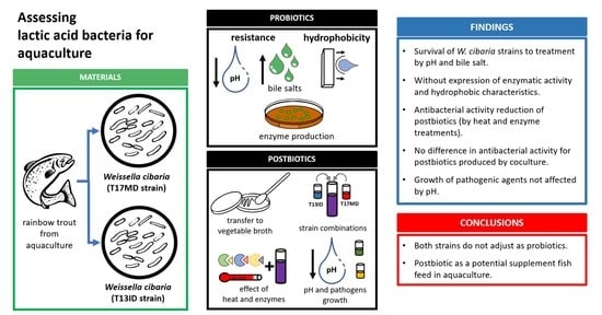
Profiling a New Postbiotic Product for Its Application in Fish Aquaculture

by
Mario Quintanilla-Pineda
1,2,*,
Jesús Díaz
2,
Ana Gutiérrez-Falcon
2,
Francisco C. Ibañez
1 and
Florencio Marzo
1,*
1
Laboratorio de Fisiología y Nutrición Animal, Universidad Pública de Navarra, 31006 Pamplona, Spain
2
Productos Específicos de Nutrición y Técnicas Alimentarias SL, 31191 Pamplona, Spain
*
Authors to whom correspondence should be addressed.
Fishes 2023, 8(6), 304; https://doi.org/10.3390/fishes8060304
Submission received: 9 May 2023 / Revised: 30 May 2023 / Accepted: 5 June 2023 / Published: 6 June 2023
(This article belongs to the Special Issue Feed Additives in Aquaculture)

Abstract

:
Weissella cibaria is a lactic acid bacteria that has been receiving great interest in the last decade; however, its research into aquaculture remains insufficient. In this context, the resistance to pH 3–5, bile salts (10–20%), hydrophobicity, and carbohydrate metabolisms of two W. cibaria strains isolated from rainbow trout were evaluated. At the same time, the postbiotic products of both W. cibaria were produced under different conditions, and their stability to heat (95–130 °C) and proteolytic enzymes were determined. The low pH sensitivity of two pathogens (Aeromonas salmonicida subsp. salmonicida and Yersinia ruckeri) was also recorded. Both strains of W. cibaria survived pH and bile salt treatments. Neither expressed enzymatic activity or hydrophobic characteristics with the executed methods. Treatment with heat, enzymes, and transfer to vegetal broth significantly reduced (p < 0.05) the postbiotic product’s antibacterial activity. On the other hand, no significant difference (p < 0.05) was reported in antibacterial characteristics when postbiotics were produced by coculturing both strains. Low pH does not significantly influence pathogen growth (p < 0.05). Our findings suggest that the studied strains do not adjust as probiotics, but the postbiotic product obtained from them could become a promising tool as a supplement food for fish aquaculture applications.
Key Contribution: The postbiotic products obtained from Weissella cibaria show the potential to become useful tools in the aquaculture industry.

Graphical Abstract

1. Introduction

Weissella is a genus closely linked to other acid lactic bacteria [1]. They have been isolated from diverse sources such as plants, sediments, fermented foods, and animals, including rainbow trout [2]. This genus has been studied as an applicable probiotic option since it has shown the ability to produce metabolites that can promote the growth of other probiotic bacteria [3], improve the fermentation process in food production [4], inhibit undesired bacteria during the fermentation process [5], and have antibacterial properties against pathogens in in vivo and in vitro studies [6,7,8].
At present, probiotics are known as live microorganisms that, when administered in adequate amounts, confer a health benefit on the host [9]. In that respect, microorganisms should have certain characteristics to be called “probiotics”, among them the ability to survive low pH and bile salt conditions, adhere to intestine walls, and produce enzymes. Some Weissella strains have exhibited these desirable characteristics [10], such as the production of dextransucrase with a nutraceutical effect on Bifidobacterium [1], β-D-glucosidase relevant to the dietary and sensory attributes of fermented food products, or pectin lyases, useful for the wine and juice industries for maceration and clarification of the products [2]. Although they have not yet been recognized as GRAS (generally recognized as safe) [11], the genus has awakened great interest in the scientific community for its ability to produce great amounts of non-digestible oligosaccharides and extracellular polysaccharides and the applications of these metabolic products in the food, feed, clinical, and cosmetics industries [12].
On the other hand, postbiotics were defined as preparations of inanimate microorganisms and/or their components that confer a health benefit on the host. In order to fit within the term “postbiotic”, this preparation must come from a well-defined microorganism, its inactivation process must be described, and its benefit to the host should be proven [13]. In this scenario, a postbiotic product containing two strains of W. cibaria isolated from rainbow trout has been previously studied in vitro for its applications as a biological tool against the causative agents of furunculosis and red mouth disease in aquaculture [8], demonstrating that a postbiotic preparation made from these strains might be a suitable option in the industry.
Interest in postbiotics has emerged over the past few years due to their technological advantages over probiotic products. Some of these benefits include fewer concerns about bacterial viability during storage and the need for less investment in cold chain technologies. Moreover, the accuracy of labeling postbiotics is considerably higher since the number of viable microorganisms at the time of production and inactivation will not vary through storage time or conditions. Moreover, the intellectual property of the product is safer since the microorganism cannot be isolated from the final product [13].
In this context, two W. cibaria strains with proven antibacterial activity as live cells and postbiotics [8] will be studied to evaluate the hypothesis of whether the postbiotic products obtained from them can be developed into a novel product suitable for the aquaculture industry. Therefore, this work aimed to assess bacterial survival under different experimental conditions and to determine their adhesion and enzymatic potential using the indicators caseinate, carboxymethylcellulose, and tributyrin. Consequently, the postbiotic products obtained from the two isolates were produced under different experimental conditions to determine the influence of heat, treatment with the enzymes proteinase K, pepsin, and α-chymotrypsin, alterations in product performance when produced in cost-effective, standardized plant-based broth medium, and coculture on the previously reported characteristics, and to establish their potential applications in the fish aquaculture industry.

2. Materials and Methods

2.1. Strains

Two strains previously isolated from rainbow trout [8] were selected to be characterized under different experimental conditions. Both strains were identified by 16S RNA sequencing as Weissella cibaria, codified as T17MD isolated from skin mucus registered in the Spanish Collection of Type Cultures (CECT) by the number 30731, and T13ID isolated from gut mucus with the CECT number 30732. The strains were cultured on Man Rogosa Sharp agar and broth (MRS-A and MRS-B; Laboratorios Conda S.A., Madrid, Spain) at 30 ± 0.2 °C with agitation at 150 rpm in aerobic conditions; the time of culture was dependent on the performed assay (24 h for bile salts, pH sensitivity, hydrophobicity test, and enzyme production, and 72 h for postbiotic production).

2.2. Pathogens

The pathogens A. salmonicida subsp. salmonicida and Y. ruckeri were used as indicators of antibacterial activity; both strains were isolated from rainbow trout disease outbreaks in Leitza (Spain) in 2019 and Brescia (Italy) in 2020, respectively. They were incubated in aerobiosis at 30 ± 0.2 °C; A. salmonicida subsp. salmonicida was cultured in Müller Hilton agar and broth (Laboratorios Conda S.A., Madrid, Spain); and Y. ruckeri was cultured in Trypticase Soy Agar and broth (Laboratorios Conda S.A., Madrid, Spain).

2.3. Resistance to Bile Salts and pH Values

Fresh bile fluid from rainbow trout was used by sacrificing four fully grown and healthy fish with an overdose of 200 mg/L of tricaine methanesulfonate solution (MSS 222, Pharmaq Ltd., Fordingbridge, UK) according to the recommendations of Directive 2010/63/EU of the European Parliament. The bile fluid was extracted aseptically using a sterile syringe. The evaluation of the resistance to bile salts and acidic conditions was carried out according to the method described by Medina et al. [14], for which 24 h cultures of T17MD and T13ID strains (1 × 10⁹ CFU/mL) were used by centrifuging 1 mL of each culture at 2400× g for 10 min. The supernatants were discarded, and the pellets were resuspended in 1 mL of phosphate-buffered saline solution (PBS, Fisher BioReagents, Geel, Belgium) adjusted to 10 and 20% concentrations of bile fluid or with PBS adjusted to 3, 4, and 5 pH values using HCl 10% (Honeywell FlukaTM, Vienna, Austria) and NaOH 10% (Honeywell FlukaTM, Vienna, Austria). As a control, standard PBS (pH 6.68) was used. The tubes were then incubated in microaerophilic conditions at 30 ± 0.2 °C for 90 min, in triplicate. After incubation, ten-fold serial dilutions of each tube were made using PBS, to then be spread over MRS-A for colony-forming unit counting and incubated at 30 ± 0.2 °C for 24 h to calculate the survival rate. The data were expressed as log CFU/mL.

2.4. Hydrophobicity Test

Overnight cultures of the strains T17MD and T13ID (1 × 10⁹ CFU/mL) were used to determine their hydrophobic characteristics. The Xu et al. method was executed [15] by centrifuging 3 mL of each culture at 2400× g for 10 min; the supernatants were discarded, and the pellets were resuspended in 3 mL of PBS. Immediately after suspension, the absorbance was measured (A0min) at 600 nm using a spectrophotometer model 4251/50 (Auxilab S.L., Pamplona, Spain); then, 1 mL of Xylene (SumanLab Sdad., Pamplona, Spain) was added to each bacterial suspension; afterward, they were vortexed for 1 min, to be incubated for 5 min at room temperature; after the incubation time, the aqueous phase was carefully recovered and its absorbance was measured (A5min). The experiment was carried out in triplicate and expressed as a percentage of hydrophobicity using the formula: H% = (1 − Amin5/Amin0) × 100.

2.5. Enzyme Production

To determine proteolytic activity, caseinate was used as an indicator protein, for which Calcium Caseinate Agar was used (Laboratorios Conda S.A., Madrid, Spain) [14]. For cellulolytic activity, the Jiménez Paillié [16] method was executed by using carboxymethylcellulose as an indicator in a culture medium composed of carboxymethylcellulose (La Mezcla Perfecta SCPP, Madrid, Spain) 10 g/L, as a carbon source along with NH4Cl (Merck KGaA, Darmstadt, Germany) 1 g/L, (NH4)2SO4 (Merck KGaA, Darmstadt, Germany); 1 g/L, KH2PO4 (Labbox Labware S.L., Barcelona, Spain); 0.1 g/L, CaCl2. 2 H2O (Sigma-Aldrich, St. Louis, MI, USA) 0.4 g/L, MgSO4·7H2O (Laboratoriumdiscounter, Zevenhuizen, The Netherlands) 0.1 g/L, and European bacteriological agar (Laboratorios Conda S.A., Madrid, Spain) 15 g/L. For lipolytic activity, tributyrin was used as an indicator, for which Tributyrin Agar (Scharlab, S.L., Barcelona, Spain) was used. All culture media were inoculated with three drops of 5 µL and spread with 100 µL of overnight cultures (1 × 10⁹ CFU/mL) of the strains T17MD and T13ID. The plates were then incubated for 24 h at 30 ± 0.2 °C. After incubation, the formation of a clear halo around the site of inoculation was recorded as positive enzymatic activity. For cellulolytic activity, the halos were revealed by covering the incubated plate with 3 mL of Congo Red 1% (Labbox Labware S.L., Barcelona, Spain) for 15 min at room temperature. Afterward, the excess colorant was decanted, and the plate was then covered with 2 mL of NaCl 0.1 M (Honeywell International Inc., Offenbach, Germany) for 15 min at room temperature. Afterward, the excess was discarded, and plates were stored at 4 °C for 24 h prior to reading. Other metabolic characteristics were evaluated using the API 50CH test (CBH, Biomerieux, Lyon, France), according to manufacturer instructions.

2.6. Postbiotic Production

Postbiotics were produced as previously described [8] by incubating the strains T17MD and T13ID in MRS-B for 72 h at 30 ± 0.2 °C in constant agitation at 150 rpm. Afterward, the culture was inactivated by heat at 80 °C for 1 h. Complete inactivation was confirmed by spreading 100 µL of the product into MRS-A and incubating for 48 h at 30 ± 0.2 °C. The absence of growth ensures the complete inactivation of the product.

2.7. Transfer to New Culture Media

The strains T17MD and T13ID were transferred to a new culture medium provided by PENTABIOL S.L. (https://www.pentabiol.es/) (accessed on 9 May 2023), whose composition does not include animal protein in its formula (vegetal broth). Subsequently, samples at 24, 48, and 72 h of culture were taken, microdiluted using PBS, and inoculated on standard MRS-A for colony-forming unit counting. pH was also measured using a PHS-3BW microprocessor pH/MV/Temperature meter (Bante Instrument, Qingdao, China). After 72 h of incubation, the bacterial cultures were used to produce postbiotics as described above. A coculture challenge was executed as previously described [8] against the pathogens Y. ruckeri and A. salmonicida subsp. salmonicida to determine if the previously reported antibacterial activity remains [8].

2.8. Strain Combinations

Postbiotics from the strains T17MD and T13ID have been produced in monoculture. To evaluate changes in bacterial behavior and determine if antibacterial activity remains when produced in coculture, both strains were simultaneously incubated in the same broth. To do so, standard MRS-B (standard broth) and vegetal broth were used. 500 mL of each broth were inoculated with 0.5 mL of overnight monocultures of the strains T17MD and T13ID (1 × 10⁹ CFU/mL), to then be incubated at 30 °C for 72 h in constant agitation at 150 rpm. Aliquots were taken at 24, 48, and 72 h for colony-forming unit counting and pH measurements as previously described. After 72 h of incubation, the bacterial cultures were used to produce postbiotics as described above, and a coculture challenge was executed against Y. ruckeri and A. salmonicida subsp. salmonicida to determine antibacterial activity.

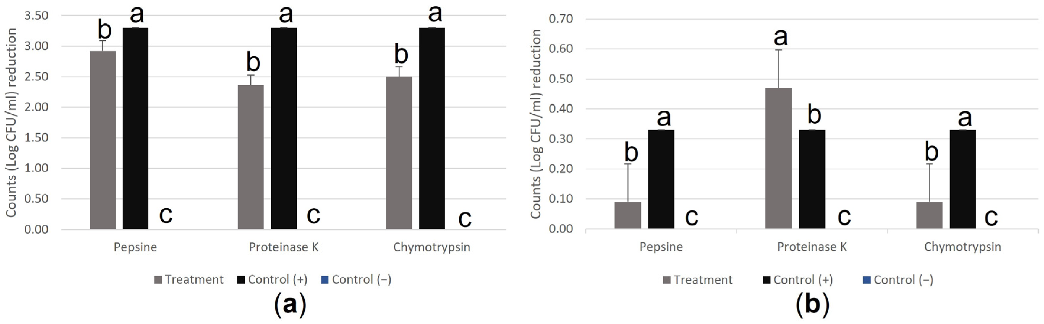
2.9. Effect of Heat and Enzymes on Antibacterial Activity

Postbiotics produced in coculture using standard MRS-B after 72 h of culture were used to determine the stability of the product under the following experimental conditions:
For thermal stability, the product was exposed to similar temperatures as the extrusion process for fish feed production, for which the product was heated at 95, 105, and 130 °C for 12 and 30 s, respectively. As a control, the postbiotic product without heat treatment was used.
The influence of the proteolytic enzymes pepsin (Sigma-Aldrich, Darmstadt, Germany), proteinase K (Fisher Bioreagents, Geel, Belgium), and α-chymotrypsin (MP Biomedicals LLC., Illkirch, France) were tested on the product. The methods described by Touraki et al. [17] were followed with a few modifications. Briefly, 1 mL of the product was treated with 2.5 mg/mL of each enzyme and incubated at 30 °C for 2 h. The reaction was stopped by inactivating the enzyme at 100 °C for 5 min using a MINI Dry Bath (Hangzhou Miu Instruments Co., Ltd., Hangzhou, China). Standard MRS-B was treated and incubated with the enzymes as a control.
Following this, the antibacterial activity of all samples was tested by coculture challenge against the pathogens Y. ruckeri and A. salmonicida subsp. salmonicida. As previously described [8].

2.10. Influence of pH on Pathogen Growth

To determine if the exposure to the acidic pH of the postbiotic treatment has an influence on the expressed reductions of counts of the pathogens Y. ruckeri and A. salmonicida subsp. salmonicida, the pH of sterile PBS was adjusted using HCl 10% and NaOH 10% to pH 3, 4, and 5. Afterward, the challenge against both pathogens was performed in triplicate by exposing them to the acidic treatments for 24 h using the coculture technique as previously described [8]. Standard PBS (pH 6.68) was used as a control.

2.11. Statistical Analysis

The results are expressed as mean ± SD. One-way analysis of variance (ANOVA) was used for analysis, followed by the least significant difference test (LSD) in cases where the values presented significant differences. The statistically significant difference was set at 0.05. The statistical analyses were performed using InfoStat version 2017 (InfoStat Group, Cordoba, Argentina).

3. Results

3.1. Resistance to Bile Salts, pH, and Hydrophobicity Test

Results are presented in Table 1, particularly that a significant reduction in the counts (p < 0.05) of the strain T13ID was observed when exposed to bile salts at 20% when compared to the control. No significant difference (p < 0.05) was observed in both strains growth when exposed to 10% bile salts for 90 min. On the other hand, a significant bacterial viability reduction (p < 0.05) of the T17MD strain was observed when exposed to pH 4. Furthermore, the strain T13ID showed a significant increase in bacterial viability (p < 0.05) when exposed to pH 3 and 4 for 90 min. No hydrophobic characteristics were expressed; values below 3% hydrophobicity were recorded for both strains.

3.2. Enzyme Production and Biochemical Analysis

Both strains tested negative for the indicators casein, tributyrin, and carboxymethylcellulose for proteolytic, lipolytic, or cellulolytic activity, respectively. Regarding carbohydrates fermentation, both strains tested positive for L-arabinose, D-xylose, D-galactose, D-glucose, D-fructose, D-mannose, N-acetylglucosamine, amygdalin, esculin ferric citrate, salicin, D-cellobiose, gentiobiose, and potassium gluconate. No difference in carbohydrate fermentation between strains was reported.

3.3. Transfer to the New Culture Median and Coculture of Strains

The changes in bacterial behavior when incubated in monoculture and coculture in two different broth mediums are presented in Table 2, where no significant difference in counts (p < 0.05) between strains is reported at 24 h of incubation when they are individually grown in the same culture medium. At 48 h of incubation, cultures on standard broth showed significantly higher counts (p < 0.05) compared to those cultured on vegetal broth. All cultures showed complete loss of viability at 72 h of incubation, except for the strain T17MD, which had significantly higher counts (p < 0.05) when incubated in monoculture on vegetal broth. Acidification of the culture medium is significantly higher (p < 0.05) when strains are incubated on vegetal broth at the three reading points.
The results of the coculture challenge are presented in Table 3. Strains that were incubated in vegetal broth in monoculture and coculture had significantly less antibacterial activity (p < 0.05) against A. salmonicida subsp. salmonicida when compared to those cultured in standard broth. Significantly higher antibacterial activity (p < 0.05) was observed on strain T13ID incubated in standard broth, reaching 4.64 ± 0.26 log CFU/mL. On the other hand, the most effective challenge against Y. ruckeri was the one made using the coculture of the two strains in standard broth, reaching significantly higher antibacterial activity (p < 0.05) when compared to the rest of the challenges.

3.4. Sensitivity of the Postbiotic Product

Changes in antibacterial activity when the postbiotic product is treated with heat are presented in Table 4. Particularly, the challenges against A. salmonicida subsp. salmonicida showed a significant reduction of antibacterial activity (p < 0.05) when compared to the positive control when the product was treated at 95 and 130 °C for 12 and 30 s, respectively. On Y. ruckeri challenges, differences were reported when the product was treated at 130 °C for 30 s, significantly reducing the antibacterial activity (p < 0.05) of the product compared to the positive control of inhibition. On the other hand, the antibacterial activity against this pathogen significantly increases (p < 0.05) with respect to the positive control of inhibition when the product is treated at 95 °C for 12 s. All employed treatments are significantly different (p < 0.05) from the negative control.
The influence of three proteolytic enzymes on the antibacterial activity of the postbiotic product against Y. ruckeri and A. salmonicida subsp. salmonicida is presented in Figure 1. A significant reduction in the antibacterial activity (p < 0.05) of the product against A. salmonicida subsp. salmonicida on the three employed enzymes is expressed. Nevertheless, the product’s antibacterial activity remains when compared to the negative control. On Y. ruckeri challenges, the exposure of the product to the proteolytic enzymes pepsin and chymotrypsin significantly reduced its antibacterial activity (p < 0.05). On the other hand, the fractions remaining after treatment of the postbiotic with the enzyme proteinase K show significantly higher antibacterial activity (p < 0.05) when compared to the positive control of inhibition. The product maintains its antibacterial properties against Y. ruckeri after the employed treatments since its activity remains significantly higher (p < 0.05) than the negative control.

3.5. Influence of Acidic pH on Pathogen Growth

Results obtained after recreating the coculture challenge using PBS at different pH values as a treatment are presented in Table 5. Where no significant changes in pathogen growth (p < 0.05) are reported when treated with pH 3, 4, or 5.

4. Discussion

Postbiotics have been considered an alternative in fish aquaculture for many applications thanks to their immunostimulant, antimicrobial, and growth promoter effects [18]. These biological tools have shown to have an influence over the expression of genes correlated with growth and nonspecific immunity in in vivo studies, as well as a positive effect on growth performance after diet supplementation [19] and for improving disease resistance in giant freshwater prawns [20].
In this context, the Weissella genus has been widely studied for its antibacterial activity against several fish pathogens, such as Streptococcus agalactiae, Photobacterium piscicida, Aeromonas hydrophila, Vibrio anguillarum, Edwardsiella tarda, A. salmonicida subsp. salmonicida, and Y. ruckeri [6,8,21], while also playing an important role from a technological perspective [12].
In this study, two strains of W. cibaria isolated from rainbow trout [8] were studied to better understand their potential as probiotics or to produce an applicable postbiotic product in fish aquaculture. In that respect, the strains T17MD and T13ID were tested under different experimental conditions. First, one of the parameters considered to evaluate potential probiotic bacteria was their ability to survive and adhere to the intestinal tract of the fish [22], mimicking their passage through the gastrointestinal tract of the fish. The results obtained demonstrated that the two strains can survive under these conditions (Table 1), and even though a significant reduction in bacterial counts (p < 0.05) of the strain T17MD was expressed when exposed to pH 4, bacterial counts were still high. A significant reduction in counts (p < 0.05) of the strain T13ID when exposed to 20% bile salt concentration was also reported, but bacterial counts were still higher than 9 log CFU/mL, the same as the rest of the tested scenarios. Most of the studies focused on potential probiotic strain characterizations have found similar survival rates when treating strains with pH ranges of 2–6 and bile salts up to 10% [14,17,22,23]. In contrast to our research, other scientists have found that some W. cibaria and W. confusa strains are highly sensitive to pH and bile salt exposure, leading to almost 100% mortality of cultures after treatment with pH 2 and 3 and bile salts at a 0.3% concentration for 2 h [10].
When probiotic selection is made, the production of enzymes is a desirable characteristic to have [14], since its production can lead to efficient utilization of ingested nutrients in the host [10]. However, in this research, the two studied strains showed no enzymatic activity with the executed methods for the indicators casein, tributyrin, and carboxymethylcellulose for proteolytic, cellulolytic, and lipolytic activity, respectively.
Fermentation of carbohydrates was also studied, and no difference between strains was reported, which is useful information to better comprehend substrate utilization during postbiotic production since antibacterial metabolic products can be mediated by substrate utilization during the culture phase of the microorganisms [24]. Hydrophobicity properties and adhesion ability are strongly correlated since biofilm formation is dependent on the cell’s surface hydrophobicity as an initial attachment process for bacteria to hydrophobic surfaces. These evaluations are necessary to estimate the ability of potential probiotic bacteria to adhere to intestinal mucus. This characteristic allows them to adhere and potentially colonize intestinal walls, which is a helpful characteristic when preventing pathogen attachment [14,25,26]. Nevertheless, in this study, neither strain showed hydrophobic characteristics with the employed solvent.
Transfer of strains to vegetal broth had a significant effect (p < 0.05) on bacterial behavior since changes in acidification and counts were recorded when compared to the cultures made in standard broth medium (Table 2). A significant reduction (p < 0.05) in antibacterial effect was also reported when strains were incubated in the vegetal substrate (Table 3). These variations in behavior might be correlated with the fact that the metabolic production during bacterial growth is mediated by the composition of the medium, which satisfies the elemental requirements of the cells during incubation. A study by Jain and Pundir [24] supports this hypothesis, where the antimicrobial metabolite production of a strain was optimized by incubating it in different culture mediums. Meaning that the vegetal broth formula employed in this study might not be adequate to optimize the antimicrobial metabolite production of the W. cibaria strains, and new substrates must be considered to better express the desirable characteristics of the product.
On the other hand, the synergetic interaction that can occur during the production and administration of mixtures of probiotic-derived products is still unclear. These interactions might result in an additive effect on the product or mutual inhibition by the released components during incubation [27]. In our study, the mixture of the two studied strains exhibited no significant influence (p < 0.05) over antibacterial activity (Table 3). Although the antibacterial effect was not enhanced through the coculture of both strains, the lack of mutual inhibition may present significant technological benefits during industrial-scale production since simultaneous production of both strains could potentially reduce the required time and equipment by half.
When designing a postbiotic intended for a food supplement, it is essential to consider the temperature it might be exposed to during the addition to the feed since the procedure might interact with its characteristics [13]. In fact, the metabolic products responsible for the reported antibacterial activity might be sensitive to high-temperature exposure [28]. In this study, the postbiotic product suffered a significant reduction in antibacterial activity (p < 0.05) against A. salmonicida subsp. salmonicida when the product was treated at 95 and 130 °C (Table 4), meaning that some of the metabolites responsible for antibacterial activity have sensitivity to heat and their properties might decrease during the extrusion process. Nevertheless, it is important to note that the effectiveness of the product remains significantly high (p < 0.05) when compared to the negative control, meaning that some metabolites involved in the antimicrobial activity are still functional after heat treatment. This evidence is of great relevance for the delivery design since it might confer the advantage of adding the postbiotic during the formulation of fish feed before the extrusion process. The antibacterial activity of the postbiotic was significantly reduced (p < 0.05) when exposed to the proteolytic enzymes pepsin, chymotrypsin, and proteinase K (Figure 1), which agrees with the hypothesis that the protein-like molecules significantly contribute to the antimicrobial activity of metabolic products produced during bacterial growth [29].
Other studies have demonstrated that the influence of pH changes on the growth of pathogens must be considered when evaluating the antimicrobial activity of metabolic products of bacteria [18], since the culture media employed for the challenge can suffer severe modifications due to alterations of the pH induced by the postbiotic treatment. In that respect, A. salmonicida subsp. salmonicida and Y. ruckeri were treated with an acidic pH (Table 5) to evaluate if it had any influence on the bacterial growth reductions previously reported [8]. No significant change in the growth of pathogens (p < 0.05) was observed during the trials, meaning that the changes in pH on the culture media have no influence over the reported antibacterial activity. Studies like ours have demonstrated that the antibacterial activity of postbiotic treatments is independent of the acidic pH [30], meaning that the molecules present in the product are responsible for the reported antibacterial activity.

5. Conclusions

This study has proven that certain desirable probiotic characteristics are not present in the studied W. cibaria strains; this evidence leads to the conclusion that they are not well-suited candidates for the development of a probiotic product. Consequently, the evaluation of the postbiotics obtained from these strains under different experimental conditions has proven that although some functionality might be lost during the process, the final product remains effective against the targeted pathogens, demonstrating its potential to become a valuable biological tool as a supplementary food in aquaculture. In addition, this research has provided insight into the role of protein-like molecules and substrate source influences on antibacterial activity. Altogether, these findings contribute to our understanding of postbiotics, their advantages, and their applications as biological tools for the control and prevention of bacteria-provoked disease in aquaculture.

Author Contributions

Conceptualization, M.Q.-P.; methodology, M.Q.-P.; validation, M.Q.-P.; formal analysis, M.Q.-P.; investigation, M.Q.-P.; resources, J.D.; writing—original draft preparation, M.Q.-P.; writing—review and editing, F.M., F.C.I. and A.G.-F.; visualization, M.Q.-P.; supervision, A.G.-F., J.D., F.C.I. and F.M.; project administration, J.D. All authors have read and agreed to the published version of the manuscript.

Funding

PENTA S.L. has been funded by the Navarre Government through the aid for hiring industrial Ph.D. students (CE: 0011-1408-2020-000023) to fulfill the Ph.D. agrifood program of the Universidad Pública de Navarra.

Institutional Review Board Statement

Not applicable.

Informed Consent Statement

Not applicable.

Data Availability Statement

Data is contained within the article.

Acknowledgments

The authors wish to extend their gratitude to PENTA S.L., whose trust and support in our research have made all this possible. The authors would also like to extend their gratitude to Eric Greenwald for selflessly providing guidance and advice during the English editing process of the manuscript.

Conflicts of Interest

Author Mario Quintanilla was employed through the aid for hiring industrial Ph.D. students: CE: 0011-1408- 5492020-000023 granted by the Navarre Government by the company Productos Especificos de Nutrición y Técnicas Alimentarias S.L. (https://www.pentabiol.es/) (accessed on 9 May 2023), authors Jesús Díaz was employed by the company Productos Específicos de Nutrición y Técnicas Alimentarias S.L., and author Ana Gutiérrez-Falcon was employed as a consultant and supervisor by Productos Específicos de Nutrición y Técnicas Alimentarias S.L. The remaining authors declare that the research was conducted in the absence of any commercial or financial relationships that could be construed as a potential conflict of interest. The company or this cooperation did not affect the authenticity and objectivity of the experimental results of this work.

References

  1. Teixeira, C.G.; Fusieger, A.; Milião, G.L.; Martins, E.; Drider, D.; Nero, L.A.; de Carvalho, A.F. Weissella: An Emerging Bacterium with Promising Health Benefits. Probiotics Antimicrob. 2021, 13, 915–925. [Google Scholar] [CrossRef] [PubMed]
  2. Fanelli, F.; Montemurro, M.; Chieffi, D.; Cho, G.S.; Franz CM, A.P.; Dell’Aquila, A.; Rizzello, C.G.; Fusco, V. Novel Insights into the Phylogeny and Biotechnological Potential of Weissella Species. Front. Microbiol. 2022, 13, 914036. [Google Scholar] [CrossRef] [PubMed]
  3. Tingirikari, J.M.R.; Kothari, D.; Shukla, R.; Goyal, A. Structural and biocompatibility properties of dextran from Weissella cibaria JAG8 as food additive. Int. J. Food Sci. Nutr. 2014, 65, 686–691. [Google Scholar] [CrossRef] [PubMed]
  4. Galle, S.; Schwab, C.; Arendt, E.; Gänzle, M. Exopolysaccharide-forming Weissella strains as starter cultures for sorghum and wheat sourdoughs. J. Agric. Food Chem. 2010, 58, 5834–5841. [Google Scholar] [CrossRef] [PubMed]
  5. Srionnual, S.; Yanagida, F.; Lin, L.H.; Hsiao, K.N.; Chen, Y.S. Weissellicin 110, a newly discovered bacteriocin from Weissella cibaria 110, isolated from plaa-som, a fermented fish product from Thailand. Appl. Environ. Microbiol. 2007, 73, 2247–2250. [Google Scholar] [CrossRef] [PubMed] [Green Version]
  6. Cai, Y.; Benno, Y.; Nakase, T.; Oh, T.-K. Specific probiotic characterization of Weissella hellenica DS-12 isolated from flounder intestine. J. Gen. Appl. Microbiol. 1998, 44, 311–316. [Google Scholar] [CrossRef] [PubMed] [Green Version]
  7. Zhu, X.-K.; Yang, B.-T.; Hao, Z.-P.; Li, H.-Z.; Cong, W.; Kang, Y.-H. Dietary supplementation with Weissella cibaria C-10 and Bacillus amyloliquefaciens T-5 enhance immunity against Aeromonas veronii infection in crucian carp (Carassiu auratus). Microb. Pathog. 2022, 167, 105559. [Google Scholar] [CrossRef]
  8. Quintanilla-Pineda, M.; Garrote Achou, C.; Díaz, J.; Gutiérrez-Falcon, A.; Bravo, M.; Herrera-Muñoz, J.I.; Peña-Navarro, N.; Alvarado, C.; Ibañez, F.C.; Marzo, F. In Vitro Evaluation of Postbiotics Produced from Bacterial Isolates Obtained from Rainbow Trout and Nile Tilapia against the Pathogens Yersinia ruckeri and Aeromonas salmonicida subsp. salmonicida. Foods 2023, 12, 861. [Google Scholar] [CrossRef]
  9. Food and Agricultural Organization of the United Nations and World Health Organization. Evaluation of Health and Nutritional Properties of Probiotics in Food including Powder Milk with Live Lactic Acid Bacteria; Food and Agricultural Organization of the United Nations and World Health Organization: Córdoba, Argentina, 2001; Available online: https://www.fao.org/3/a0512e/a0512e.pdf (accessed on 5 May 2023).
  10. Lee, K.W.; Park, J.Y.; Jeong, H.R.; Heo, H.J.; Han, N.S.; Kim, J.H. Probiotic properties of Weissella strains isolated from human faeces. Anaerobe 2012, 18, 96–102. [Google Scholar] [CrossRef]
  11. Fessard, A.; Remize, F. Why are Weissella spp. not used as commercial starter cultures for food fermentation? Fermentation 2017, 3, 38. [Google Scholar] [CrossRef] [Green Version]
  12. Abriouel, H.; Lerma, L.L.; Muñoz, M.D.C.C.; Montoro, B.P.; Kabisch, J.; Pichner, R.; Cho, G.S.; Neve, H.; Fusco, V.; Franz, C.M.A.P.; et al. The controversial nature of the Weissella genus: Technological and functional aspects versus whole genome analysis-based pathogenic potential for their application in food and health. Front. Microbiol. 2015, 6, 1197. [Google Scholar] [CrossRef] [Green Version]
  13. Salminen, S.; Collado, M.C.; Endo, A.; Hill, C.; Lebeer, S.; Quigley, E.M.M.; Sanders, M.E.; Shamir, R.; Swann, J.R.; Szajewska, H.; et al. The International Scientific Association of Probiotics and Prebiotics (ISAPP) consensus statement on the definition and scope of postbiotics. Nat. Rev. Gastroenterol. Hepatol. 2021, 18, 649–667. [Google Scholar] [CrossRef]
  14. Medina, M.; Sotil, G.; Flores, V.; Fernández, C.; Sandoval, N. In vitro assessment of some probiotic properties and inhibitory activity against Yersinia ruckeri of bacteria isolated from rainbow trout Oncorhynchus mykiss (Walbaum). Aquac. Rep. 2020, 18, 100447. [Google Scholar] [CrossRef]
  15. Xu, H.; Jeong, H.S.; Lee, H.Y.; Ahn, J. Assessment of cell surface properties and adhesion potential of selected probiotic strains. Lett. Appl. Microbiol. 2009, 49, 434–442. [Google Scholar] [CrossRef]
  16. Jiménez Paillié, E.M. Determinación de la Actividad Celulolítica, Ligninolítica y Amilolítica de Actinobacterias Aisladas de Suelo Rizosférico de Trébol Blanco (Trifolium repens). Undergraduate Thesis, Pontificia Universidad Javeriana, Bogotá D.C., Colombia, 2012. [Google Scholar]
  17. Touraki, M.; Frydas, I.; Karamanlidou, G.; Mamara, A. Partial purification and characterization of a bacteriocin produced by Bacillus subtilis NCIMB 3610 that exhibits antimicrobial activity against fish pathogens. J. Biol. Res. 2012, 18, 310–319. [Google Scholar]
  18. Ang, C.Y.; Sano, M.; Dan, S.; Leelakriangsak, M.; Lal, T.M. Postbiotics Applications as Infectious Disease Control Agent in Aquaculture. Biocontrol Sci. 2020, 25, 1–7. [Google Scholar] [CrossRef] [Green Version]
  19. Wu, X.; Teame, T.; Hao, Q.; Ding, Q.; Liu, H.; Ran, C.; Yang, Y.; Zhang, Y.; Zhou, Z.; Duan, M.; et al. Use of a paraprobiotic and postbiotic feed supplement (HWFTM) improves the growth performance, composition and function of gut microbiota in hybrid sturgeon (Acipenser baerii x Acipenser schrenckii). Fish Shellfish Immunol. 2020, 104, 36–45. [Google Scholar] [CrossRef]
  20. Dash, G.; Raman, R.P.; Pani Prasad, K.; Makesh, M.; Pradeep, M.A.; Sen, S. Evaluation of paraprobiotic applicability of Lactobacillus plantarum in improving the immune response and disease protection in giant freshwater prawn, Macrobrachium rosenbergii (de Man, 1879). Fish Shellfish Immunol. 2015, 43, 167–174. [Google Scholar] [CrossRef]
  21. Mortezaei, F.; Royan, M.; Allaf Noveirian, H.; Babakhani, A.; Alaie Kordghashlaghi, H.; Balcázar, J.L. In vitro assessment of potential probiotic characteristics of indigenous Lactococcus lactis and Weissella oryzae isolates from rainbow trout (Oncorhynchus mykiss Walbaum). J. Appl. Microbiol. 2020, 129, 1004–1019. [Google Scholar] [CrossRef]
  22. Fečkaninová, A.; Koščová, J.; Mudroňová, D.; Schusterová, P.; Cingeľová Maruščáková, I.; Popelka, P. Characterization of two novel lactic acid bacteria isolated from the intestine of rainbow trout (Oncorhynchus mykiss, Walbaum) in Slovakia. Aquaculture 2019, 506, 294–301. [Google Scholar] [CrossRef]
  23. Sica, M.G.; Brugnoni, L.I.; Marucci, P.L.; Cubitto, M.A. Characterization of probiotic properties of lactic acid bacteria isolated from an estuarine environment for application in rainbow trout (Oncorhynchus mykiss, Walbaum) farming. Antonie van Leeuwenhoek 2012, 101, 869–879. [Google Scholar] [CrossRef] [PubMed]
  24. Jain, P.; Pundir, R.K. Effect of fermentation medium, pH and temperature variations on antibacterial soil fungal metabolite production. J. Agric. Technol. 2011, 7, 247–269. [Google Scholar]
  25. Del Re, B.; Sgorbati, B.; Miglioli, M.; Palenzona, D. Adhesion, autoaggregation and hydrophobicity of 13 strains of Bifidobacterium longum. Lett. Appl. Microbiol. 2000, 31, 434–442. [Google Scholar] [CrossRef] [PubMed]
  26. Rühs, P.A.; Böcker, L.; Inglis, R.F.; Fischer, P. Studying bacterial hydrophobicity and biofilm formation at liquid-liquid interfaces through interfacial rheology and pendant drop tensiometry. Colloids Surf. B 2014, 117, 174–184. [Google Scholar] [CrossRef]
  27. Chapman, C.M.C.; Gibson, G.R.; Rowland, I. Health benefits of probiotics: Are mixtures more effective than single strains? Eur. J. Nutr. 2011, 50, 1–17. [Google Scholar] [CrossRef]
  28. Bravo Santillana, M. Caracterización de Bacterias Ácido-Lácticas con Propiedades Antimicrobianas e Inmunomoduladoras y su Investigación Aplicada en Sanidad. Ph.D. Thesis, Universidad de Extremadura, Extremadura, Spain, 2021. [Google Scholar]
  29. Mirkovic, N.; Radulovic, Z.; Uzelac, G.; Lozo, J.; Obradovic, D.; Topisirovic, L.; Kojic, M. Isolation and Characterisation of Bacteriocin and Aggregation-Promoting Factor Production in Lactococcus lactis ssp. lactis BGBM50 Strain. Food Technol. Biotechnol. 2015, 53, 237–242. [Google Scholar] [CrossRef]
  30. Bravo, M.; Combes, T.; Martinez, F.O.; Cerrato, R.; Rey, J.; Garcia-Jimenez, W.; Fernandez-Llario, P.; Risco, D.; Gutierrez-Merino, J. Lactobacilli Isolated from Wild Boar (Sus scrofa) Antagonize Mycobacterium bovis Bacille Calmette-Guerin (BCG) in a Species-Dependent Manner. Front. Microbiol. 2019, 10, 1663. [Google Scholar] [CrossRef] [Green Version]
Figure 1. Changes in antibacterial activity in the co-culture challenge of the postbiotic product after enzyme treatment: (a) changes in antibacterial activity against A. salmonicida subsp. salmonicida; (b) changes in antibacterial activity against Y. ruckeri. Bars represent the means of count (log CFU/mL) reduction (n = 3). Bars with different superscripts are significantly different (p < 0.05) from each other. Error bars express SE.
Fishes 08 00304 g001
Table 1. Bacterial resistance to rainbow trout bile salts and acidic pH.
Counts (Log CFU/mL)
TreatmentT13IDT17MD
Bile 10%8.97 ± 0.04 b9.24 ± 0.05 a
Bile 20%8.70 ± 0.00 c9.21 ± 0.00 a
pH 39.30 ± 0.00 a9.22 ± 0.02 a
pH 49.39 ± 0.08 a9.16 ± 0.04 b
pH 59.05 ± 0.02 b9.19 ± 0.04 a
Control9.06 ± 0.02 b9.30 ± 0.02 a
Results are expressed in means ± SD (n = 3). Means in the same column with different superscripts are significantly different (p < 0.05).
Table 2. Changes in counts (expressed as log CFU/mL) and pH of cocultures and monocultures of two strains of W. cibaria at three points after 72 h of incubation in two different culture mediums.
Strain24 h48 h72 hStandardVegetal
CountspHCountspHCountspHpHpH
T17MD
Vegetal broth9.18 ± 0.07 b4.12 ± 0.01 a8.60 ± 0.04 d4.03 ± 0.01 a3.83 ± 0.10 a4.02 ± 0.01 a6.40 ± 0.006.90 ± 0.00
Standard broth9.42 ± 0.02 a4.44 ± 0.01 b9.24 ± 0.02 a4.43 ± 0.00 c0.00 ± 0.00 b4.49 ± 0.00 b6.40 ± 0.006.90 ± 0.00
T13ID
Vegetal broth9.06 ± 0.07 b4.12 ± 0.02 a8.53 ± 0.02 e4.02 ± 0.01 a0.00 ± 0.00 b4.02 ± 0.00 a6.40 ± 0.006.90 ± 0.00
Standard broth9.32 ± 0.01 a4.42 ± 0.01 b9.07 ± 0.02 b4.42 ± 0.00 c0.00 ± 0.00 Bb4.47 ± 0.06 b6.40 ± 0.006.90 ± 0.00
T17MD + T13ID
Vegetal broth9.30 ± 0.03 a4.10 ± 0.02 a6.23 ± 0.05 f4.09 ± 0.00 b0.00 ± 0.00 b4.02 ± 0.02 a6.40 ± 0.006.90 ± 0.00
Standard broth9.14 ± 0.08 b4.48 ± 0.00 c8.86 ± 0.03 c4.48 ± 0.01 d0.00 ± 0.00 b4.48 ± 0.01 b6.40 ± 0.006.90 ± 0.00
Results are expressed as means ± SD (n = 3). Means in the same column with different superscripts are significantly different (p < 0.05). All pH readings showed a significant difference (p < 0.05) from the control.
Table 3. Reduction in counts (expressed as log CFU/mL) of A. salmonicida subsp. salmonicida and Y. ruckeri in coculture challenge against postbiotics produced from two strains in monoculture and coculture, produced in two different culture mediums after 72 h of incubation prior to inactivation.
StrainsA. salmonicida subsp. salmonicidaY. ruckeri
T17MD
Vegetal broth1.47 ± 0.21 c0.24 ± 0.07 b
Standard broth4.24 ± 0.05 b0.30 ± 0.12 b
T13ID
Vegetal broth1.21 ± 0.03 c0.20 ± 0.02 b
Standard broth4.64 ± 0.26 a0.26 ± 0.05 b
T17MD + T13ID
Vegetal broth1.40 ± 0.10 c0.22 ± 0.07 b
Standard broth3.46 ± 0.10 b0.40 ± 0.04 a
Control0.00 ± 0.00 d0.00 ± 0.00 c
Results are expressed as means ± SD (n = 3). Means in the same column with different superscripts are significantly different (p < 0.05).
Table 4. Changes in antibacterial activity (expressed as log CFU/mL) of postbiotic products treated with heat.
TreatmentA. Salmonicida subsp. salmonicidaY. ruckeri
95 °C/12 s3.30 ± 0.05 b0.57 ± 0.05 a
95 °C/30 s3.28 ± 0.04 b0.31 ± 0.08 bcd
105 °C/12 s3.55 ± 0.16 ab0.27 ± 0.01 cd
105 °C/30 s3.49 ± 0.07 ab0.34 ± 0.03 bc
130 °C/12 s2.53 ± 0.17 c0.39 ± 0.04 b
130 °C/30 s2.29 ± 0.10 c0.23 ± 0.05 d
Control (+)3.70 ± 0.26 a0.34 ± 0.06 bc
Control (−)0.00 ± 0.00 d0.00 ± 0.00 e
Results are expressed in means ± SD (n = 3). Means in the same column with different superscripts are significantly different (p < 0.05).
Table 5. Changes in pathogen growth (expressed as log CFU/mL) after 24 h of acidic treatment.
pHY. ruckeriA. salmonicida subsp. salmonicida
38.92 ± 0.058.77 ± 0.09
48.88 ± 0.018.68 ± 0.15
58.90 ± 0.078.65 ± 0.07
Control8.92 ± 0.068.82 ± 0.02
Results are expressed as means ± SD (n = 3).
Disclaimer/Publisher’s Note: The statements, opinions and data contained in all publications are solely those of the individual author(s) and contributor(s) and not of MDPI and/or the editor(s). MDPI and/or the editor(s) disclaim responsibility for any injury to people or property resulting from any ideas, methods, instructions or products referred to in the content.

