Article

Effects of γ-Irradiated Date (Phoenix dactylifera) Fruit on Growth, Immunological and Antioxidant Parameters of Goldfish (Carassius auratus)

by
Marzieh Heidarieh
1,
Amin Gholamhosseini
2,
Najmeh Sheikhzadeh
3,* and
Maria Angeles Esteban
4
1
Nuclear Science and Technology Research Institute, Tehran 19866-12354, Iran
2
Division of Aquatic Animal Health, Department of Clinical Sciences, School of Veterinary Medicine, Shiraz University, Shiraz 11731-71441, Iran
3
Department of Food Hygiene and Aquatic Animals, Faculty of Veterinary Medicine, University of Tabriz, Tabriz 51666-14766, Iran
4
Department of Cell Biology & Histology, Faculty of Biology, University of Murcia, 30100 Murcia, Spain
*
Author to whom correspondence should be addressed.
Fishes 2023, 8(5), 251; https://doi.org/10.3390/fishes8050251
Submission received: 10 April 2023 / Revised: 4 May 2023 / Accepted: 6 May 2023 / Published: 9 May 2023
(This article belongs to the Special Issue Effects of Feed Additives on Fish Health and Fillet Quality)

Abstract

:
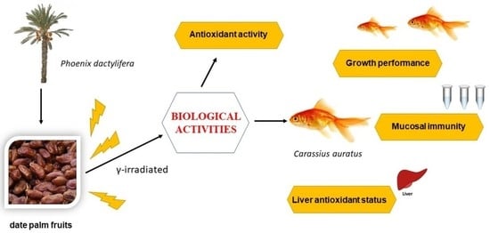
The aim of this study was to evaluate the potential effects of γ-irradiated date palm fruit (Phoenix dactylifera), which is rich in phenolic acids and has important and varied biological bioactivities, on growth performance, mucosal immunity and liver antioxidant status in goldfish (Carassius auratus). The antioxidant activity, alongside the phenolic and flavonoid contents, were also determined in irradiated palm fruit extracts (IPFE). The results showed that IPFE5 had a higher antioxidant activity as well as heightened phenolic and flavonoid contents compared to IPFE10 and IPFE0. Meanwhile, goldfish specimens were randomly divided into three groups with three replicates in each. Subsequently, skin immunity and antioxidant activity in the livers of the goldfish were studied. The growth rates of fish fed with IPFE5 and IPFE10 were significantly higher (p < 0.05) compared to the CTR group. In addition, fish fed with the IPFE5 diet demonstrated increased mucosal immunity compared to the CTR group (p < 0.05). Lipid peroxidation levels as well as antioxidant enzyme activities were also higher in all the IPFE-fed groups compared to the CTR group (p < 0.05). These data showed that 40-day dietary administration of γ-irradiated date extract, especially IPFE5, improved growth performance, mucosal immunity, and liver antioxidant capacity in goldfish. The suitability of administrating this additive in the diet of farmed fish is discussed.
Key Contribution: The current study showed that γ-irradiated palm fruit extracts (IPFE) could enhance the growth performance, skin mucosal immunity, and liver antioxidant capacity of goldfish.

Graphical Abstract

1. Introduction

Globally, ornamental pet fish represent an important component of the pet market, and the United States, Europe and Japan are among the pioneers of this industry [1]. Transport and handling of fish, poor water quality, high stocking density and contaminated feed can cause stress, resulting in infectious diseases in these fish [2]. Among these bacterial diseases, one Gram-negative species which is frequently observed in ornamental fish is Aeromonas hydrophilla [2]. Typical clinical signs of the infection caused by this bacterium include anorexia, dark coloration, septicemia and skin lesions ranging from superficial to deep [3]. Although effective administration and prophylaxis are the prime factors in safeguarding ornamental fish against bacterial diseases [4], a recent approach to mitigate the response caused by typical stressors is the use of dietary additives [5], which are important for improving fish growth, health and welfare [6,7,8]. Among the different feed additives, natural feed additives, including plants, are preferable as they are biocompatible, biodegradable, inexpensive, available and environmentally safe [9].
Date palm (Phoenix dactylifera) is a key commodity worldwide, and it plays an important economic role in the producing regions. Fresh dates are a rich source of polyphenols, including flavonoids (luteolin, quercetin, anthocyanidins) and phenolic acids (e.g., ferulic acid, vannillic acid, syringic acid, gallic acid, caffeic acid, coumaric acid, sinapic acid) in both free and bound forms [10]. Despite the importance of this plant to humans, date palms have only recently begun attracting attention in both human and animal studies, with the understanding that they can be used as functional foods and nutraceuticals [11,12]. To date, some studies have demonstrated the beneficial effects of dates on growth, immunity and antioxidant parameters in fish species [13,14,15,16]. Meanwhile, the biological responses of γ-ray irradiated plants have been widely discussed. The available studies show that γ-ray irradiation can enhance the physiological and biochemical effects of the bioactive products present in plants [17,18]. More specifically, γ-ray irradiation of dates has also been found to be a good substitute for fumigation and chemical use, preventing aflatoxin formation and improving sensory quality [10,19]. Interestingly, despite all these available data, as far as we are aware, there are insufficient data on the effects of γ-irradiated dates on aquatic organisms. Therefore, this study was conducted to evaluate the effect of γ-irradiated date fruits on the growth rate, skin mucosal immunity and antioxidant levels in the livers of goldfish.

2. Materials and Methods

2.1. P. dactylifera Extract and Diet Preparation

For the preparation of the aqueous extract, palm fruits of the Barhi variety were used according to the previous study [13]. The seedless nuts were rinsed with distilled water and chopped into small pieces. Distilled water (500 mL) was then added to the pieces, which were then incubated for 2 h at 55 °C. The final mixture was ground using Moulinex machine (Moulinex AR11083, Paris, France) and centrifuged (3500× g) for 15 min. A 0.5% yield was achieved. The final collected supernatant, referred to as the irradiated palm fruit extract (IPFE), was divided into three parts. One part was not irradiated (positive control, IPFE0), the second part was irradiated at 5 kGy (IPFE5) and the third part was irradiated at 10 kGy (IPFE10) using a cobalt-60 γ irradiator (a Gamma cell-220 irradiator, Nordion, Canada) at a dose of 1.02 Gy s−1.The extracts were stored at 4 °C in solar darkness. A negative control group (CTR) without any palm extract was also used.
Four different diets were prepared by mixing the feed ingredients described in a previous study [20], to which 200 mL kg−1 of the corresponding extract (CTR without any extract, IPFE0, IPFE5 and IPFE10) was added [20]. Then, all of the ingredients were mixed with a mixer (Isfahan Jahan Kar, Isfahan, Iran) before being pelleted using a meat grinder equipped with a 2-mm perforated disc. Each dose of IPFE was replaced by an equivalent amount of cellulose (Table 1). The experimental diets were air-dried and stored in plastic bags at 4 °C until use.

2.2. Characterization of IPFE

The in vitro antioxidant capacities of the extracts (IPFE0, IPFE5 and IPFE10) were determined spectrophotometrically using the ABTS (2,2′-azino-bis (3-ethylbenzothiazoline-6-sulfonic acid) method [22]. The radical scavenging activities of these extracts were also determined against stable 2,2- diphenyl-2-picrylhydrazyl hydrate (DPPH) [23]. The total concentrations of phenolic compounds present in IPFE were determined spectrophotometrically in accordance with the Folin-Ciocalteu method [24], with gallic acid (GAE) being used as a calibration standard. The concentrations of the flavonoid content of IPFE were also determined spectrophotometrically according to the aluminum chloride colorimetric method [25], with quercetin serving as the calibration standard.

2.3. Fish Rearing and Experimental Design

This research was implemented in the fish farm of Shiraz University (Shiraz, Iran). A total of 180 goldfish (5.60 ± 0.11 g) purchased from a private fish farm in Sepidan (Fars, Iran) were randomly distributed into 12 glass tanks of 40 L (n = 15). Three tanks (triplicates) were assigned to each experimental group. Throughout the study, the temperature and pH values were stabilized between 24.5 and 25 °C and between 7 and 7.5 mg L−1, respectively. The fish were adapted to the experimental setting for 10 days and fed the control diet three times per day (10:00, 13:00, 17:00) until satiation. Then, the fish belonging to the various groups were fed their respective diets (CTR, IPFE0, IPFE5 or IPFE10) three times a day (10:00, 13:00 and 17:00 h) until satiety for 40 days.

2.4. Fish Growth and Sampling

At the inception (0 day) and at the end (40 days) of the feeding trial, 24 h-starved fish were anesthetized via bathing in clove oil extract (50 μL L−1), and the length and weight of each fish were measured. The feed intake, specific growth rate (SGR) and feed conversion ratio (FCR) were also determined:
SGR = (Ln final weight − Ln initial weight)/feeding days
FCR = dry feed intake/(Final weight − initial weight)
After the biometric study, mucus specimens were collected from the euthanized fish. To this end, fish mucus was gathered by delicately scraping the fish surfaces with a plastic spatula. Skin mucus samples from five specimens were pool sampled to acquire enough samples for all the assays. The fish mucus was homogenized using tris-buffered saline (pH = 8.0) and centrifuged (4000× g, 30 min) at 4 °C. The supernatants were lyophilized and stored at −20 °C for the determination of immune parameters. The livers of five fish from each tank were homogenized with tris-buffered saline (pH = 7.4), centrifuged (8000× g, 25 min, 4 °C) and stored at 20 °C for the investigation of antioxidant activities.

2.5. Skin Mucus Immunity

Micrococcus lysodeikticus suspensions (75 mg mL−1) (Sigma-Aldrich, Burlington, VT, USA) in 0.1 M phosphate citrate buffer (75 μL) were mixed with 25 μL of mucus samples to determine their lysozyme activity. The turbidity of the mixture was calculated at 450 nm for 10 min using a microplate reader (Hiperion, Neuss, Germany). A decrease in the total absorbance at 0.001 per minute was recorded as one U of lysozyme activity per mg of fish mucus sample [26]. Aliquots of 1 mL of alkaline buffer solution (Sigma-Aldrich, Burlington, VT, USA) were mixed with 20 μL of mucus samples to determine the alkaline phosphatase activity. After incubation (37 °C, 5 min), 1 mL of 0.05 N NaOH solution was added to the mixture. The absorbance was recorded at 410 nm with a spectrophotometer (UNICO, Shanghai, China) [26].
To determine the mucus protease activity, 200 μL of mucus sample was mixed with the same volume of 100 mM ammonium bicarbonate buffer containing 0.7% azocasein, pH 7.8 for 19 h on a shaker at 30 °C. The reaction was terminated by adding 4.6% trichloroacetic acid followed by centrifugation (10,000 rpm, 10 min). The supernatant was mixed with 0.5 M NaOH, and the absorbance was recorded at 405 nm [26]. To determine the total Ig (Immunoglobulin) level, the protein content was calculated prior to and after mucus precipitation with polyethylene glycol [27]. To determine the in vitro bactericidal activity, Aeromonas hydrophila (ATCC 7966) grown in tryptic soy broth for 24 h at 25 °C was adjusted to 109 CFU mL−1. Afterwards, 25 μL of phosphate buffer saline (PBS) was added to the second to eighth wells of a plate. Then, 50 μL of skin mucus samples were added to the first wells and serially diluted from the second to eighth wells. Aliquots of 25 μL of the bacterium suspension were added to each well, and the samples were incubated overnight at room temperature. The last well with clear bacterial inhibition was recorded as the minimal inhibitory concentration (MIC) for each experimental condition or group [26].

2.6. Liver Antioxidant Parameters

The malondialdehyde (MDA) level in the fish liver samples was determined using the thiobarbituric acid test [28]. Fish liver samples (5 g) were homogenized in 15 mL of deionized water and 7.2% butylated hydroxytoluene. Afterwards, 2 mL of 15 mM 2-thiobarbituric acid and 15% trichloroacetic acid were added to 1 mL of the treated samples. After incubation for 10 min in boiling water, the mixture was centrifuged (2500× g, 15 min) and the absorbance was recorded at 532 nm [28]. Commercial kits were used to calculate the functions of some antioxidant enzymes, specifically superoxide dismutase (Ransod, Randox/SD 125) and glutathione peroxidase (Ransel, Randox/RS 505). Catalase activity was assessed using a hydrogen peroxidase assay upon the formation of its stable complex with ammonium molybdate. Briefly, 200 µL of the supernatant was incubated in a working solution consisting of 1000 µL of hydrogen peroxide and 500 µL of phosphate buffer (pH: 7.4) at 25 °C for 60 s. Then, 1000 µL of 32.4 mmol L−1 ammonium molybdate was added to the reaction solution, and the concentration of the yellow complex of molybdate and hydrogen peroxide was measured at a 405 nm wavelength using a spectrophotometer (UNICO, Shanghai, China) [29]. Glutathione S-transferase was assayed following the method proposed by Habig et al. (1974) using 1-chloro-2,4-dinitrobenzene (CDNB) as the substrate [30]. A total of 20 µL of the supernatant was incubated in a working solution (1 mL) containing 1 mM CDNB, 1 mM reduced glutathione and 0.1 M phosphate buffer, pH 6.5 at 25 °C for 3 min. Afterwards, the absorbance changes were recorded at 340 nm for 3 min using a spectrophotometer (UNICO, Shanghai, China) [30]. One unit of activity was defined as the amount of enzyme catalyzing the formation of 1 nmol of product/min under the specific assay conditions.

2.7. Statistical Analysis

The experimental results were statistically analyzed using SPSS package version 22. The homogeneity of variance and normality were checked using Levene and Shapiro–Wilk tests, respectively. All groups were compared using one-way ANOVA and Tukey posthoc tests. Differences were regarded statistically meaningful when the p value was below 0.05.

3. Results

3.1. Analysis of IPFE

The antioxidant activity determined by the DPPH method was significantly (p < 0.05) highest in IPFE5 (0.71 µ/mL), followed by IPFE10 (0.59 µ/mL) and IPFE0 (0.59 µ/mL). Furthermore, the ABTS method also indicated that the significantly (p < 0.05) highest antioxidant capacity (4.76 µ/mL) was present in IPFE5, followed by IPFE10 (3.96 µ/mL), and the lowest was in IPFE0 (3.02 µ/mL). In the present study, the significantly (p < 0.05) highest total phenolic compounds were observed in IPFE5 (7.62 mg GAE g−1 of extract), followed by IPFE10 (6.81 mg), and IPFE0 (6.13 mg) had the lowest values according to the data obtained using the Folin-Ciocalteu colorimetric method. On the other hand, according to the aluminum chloride colorimetric method, a significantly (p < 0.05) higher level of total flavonoids was recorded in IPFE5 (4.80 mg quercetin g−1 of extract) and IPFE10 (3.90 mg) extracts rather than the IPFE0 (3.61 mg) extract (Table 2).

3.2. Fish Growth

In terms of growth performance, the group fed the CTR diet had significantly (p < 0.05) enhanced growth indices, including final weight, final length, SGR and FCR compared to the other groups. In contrast, all of the aforementioned indices significantly (p < 0.05) improved in the IPFE5 group, followed by the IPFE10 group. However, in the IPFE0 group, the final weight and SGR had improved compared to the CTR group (p < 0.05) (Table 3).

3.3. Mucus Immune Parameters

In the group of fish fed the CTR diet, some immunological parameters, specifically lysozyme, alkaline phosphatase, protease and Ig showed significantly (p < 0.05) lower levels compared to the values obtained for fish belonging to other groups. However, all the fish fed IPFE (IPFE5, IPFE10 or IPFE0) had significantly (p < 0.05) higher levels than those found in fish fed with the CTR diet. In addition, the bactericidal activity present in the mucus of the fish fed with the IPFE5 diet was significantly (p < 0.05) more than that observed in the mucus of fish from the CTR group (Table 4).

3.4. Liver Antioxidant Parameters

The level of lipid peroxidation products was significantly (p < 0.05) higher in the CTR group compared to the values obtained for the rest of the fish in the present experiment. The analysis of antioxidant enzyme activities also showed that the values of superoxide dismutase, catalase and glutathione S-transferase were significantly (p < 0.05) higher in the fish fed the IPFE5 or IPFE10 diets compared to those found in the fish from the CTR or IPFE0 groups. The levels of glutathione peroxide were highest in the fish fed with the IPFE5 diet, followed by those in fish fed with the IPFE10 diet, which were significantly higher compared to the values in the CTR and IPFE0 groups (p < 0.05). Finally, the fish fed the IPFE0 diet also had significantly (p < 0.05) higher levels of all enzyme activities than those found in the fish fed the CTR diet (Table 5).

4. Discussion

Date fruit has several potential benefits owing to the presence of carotenoids, phenolic acids, flavonoids, tocopherols and phytosterols, which has led many researchers to consider this product as a convenient source of natural antioxidants and functional food ingredients [31]. In addition, some studies have demonstrated the benefits of dates when used as a feed additive for different fish species [13,14,15]. Moreover, the efficacy of γ-irradiation in improving the quality and shelf life of vegetables and fruits has been investigated [10,17,19,32]. To the best of our knowledge, there is no information on the possible use of γ-irradiated date fruits in aquatic animals. Taking all these considerations into account, in this work, we studied the effects exerted on growth, mucosal immunity and liver antioxidant systems in goldfish fed with γ-irradiated palm fruit. In this study, an analysis of IPFE was performed prior to its use as a dietary additive for the goldfish.
The antioxidant activity of the used date fruit extract was studied prior to being used as a feed additive for goldfish. The results of these assays showed that aqueous extraction, coupled with γ-irradiation at 5 kGy, could increase the antioxidant activity in date fruit compared to other samples of the same non-irradiated dates. Our results are in agreement with previous studies demonstrating that date extract preparation can increase antioxidant activity, as substantiated by various methods such as ABTS, FRAP and DPPH assays [13,33,34]. In parallel with increased antioxidant activity in the date extract following the γ-irradiation, some authors also reported elevated antioxidant activities in irradiated dates and other fruits [19,35]. It has been assumed that the increased antioxidant activity of fruits after being irradiated might be due to the degradation of polymeric phenolic compounds into smaller units during the course of γ-irradiation [36]. Phenolic compounds are the chief elements in advancing the antioxidant activities of date fruits, which contain flavonoids, ferulic and sinapic acid, p-coumaric and procyanidins [37]. In the present study, similar to what was observed for antioxidant activity, phenolic compounds and total flavonoid levels were highest in the IPFE5 samples. These results indicate that the use of optimal γ-irradiation is of great importance for the correct preparation of food additives for any species, including fish species.
The present study demonstrated that growth performance was improved in fish fed with γ-irradiated date extracts. These results align with previous findings on the improvement of irradiated dietary ingredients in fish species. For example, γ-irradiation of feed with 20% soybean meal improved protein digestibility and increased growth in fighting fish (Betta splendens) [38]. Rainbow trout (Oncorhynchus mykiss) also showed a higher uptake of γ-irradiated Ergosan in the intestine compared to crude Ergosan, which resulted in improved growth performance [39]. More recently, it was revealed that the use of γ-irradiated propolis extract at 10 and 30 kGy in the diet of common carp (Cyprinus carpio) boosted growth rates [40]. In the current study, the use of γ-irradiated dates as a natural feed additive could act as an attractant, improving feed intake and eventually increasing the fish weight and growth performance. Interestingly, no studies have been carried out to investigate the impacts of γ-irradiated food additives on fish gut flora, but a former study showed that γ-irradiated rice bran could induce positive changes in the gut microflora of broiler chicken [41]. It was also found that the effects of γ-processing on the inhibition of microbes in dates [19] could indirectly affect the gut bacterial community of goldfish. Although the mechanism underlying the enhancement of fish growth after IPFE administration is unclear, γ-irradiation may change the structure of date polyphenols into smaller units, as discussed above. It has also been demonstrated that humans receiving high levels of glucose, fructose and sucrose in the form of date fruits exhibited a higher relative abundance of Bifidobacteria, as well as a reduction in Bacteroides [42]. It is now a well-established fact that a balanced gut microbiota plays a vital role in metabolism, pathogen resistance and immune enhancement [43]. Therefore, increasing polyphenol content after date irradiation might enhance gut microbiota, leading to improved growth performance in goldfish. However, further studies are needed to corroborate this hypothesis.
The mucosal surfaces of fish, including the skin, gills and gut, are in constant contact with various unfavorable abiotic and biotic agents [44]. The outcomes of the current study demonstrated that feeding with γ-irradiated date extract elevated the immune parameters, specifically lysozyme, alkaline phosphatase, protease, total Ig as well as antibacterial activity in goldfish skin. There have been few works carried out to study the effects of irradiated feed additives in fish mucosal immunity. For example, γ-irradiated alginic acid boosted skin mucosal immunity and resistance to crowding stress in rainbow trout [39,45]. Feeding with date fruit extract also increased mucosal immune parameters (lysozyme, protease and alkaline phosphatase activity) in common carp fry skin [16]. The effects of polyphenols on the fish immune system have been reviewed previously [46]; however, the mechanism of different polyphenols’ actions has not been described completely. It is widely acknowledged that polyphenols predominantly exert their activity in the gut, where they modulate the microbiome and confer immunoprotective effects on host immunologic and metabolic markers [47,48]. The current study also showed that polyphenol sources from irradiated dates magnified bactericidal activity against Aeromonas hydrophilla in goldfish skin mucus. It has been previously demonstrated that polyphenols have defensive properties against pathogens, not only by regulating the host immune system, but also by counteracting the pathogen itself [49]. In this study, fish fed with IPFE5 had higher skin mucosal immunity than those fed with IPFE10. It was previously assumed that the modulatory effects of polyphenols on cellular and humoral components of the immune system were dependent on their individual structures, doses and duration of use of these compounds [49].
Previous studies also demonstrated that date extracts have potent antioxidant activities [13,15,50]. Moreover, γ-irradiation could increase the antioxidant activity of date extracts. The present study demonstrated that the aqueous extract of γ-irradiated dates enhanced antioxidant enzyme activities such as superoxide dismutase, catalase, glutathione S-transferase and glutathione peroxidase activities in goldfish livers. In fact, γ-irradiation can degrade antioxidant ingredients or break down some components into antioxidant components. Hence, the total composition and antioxidant content could be affected, thus changing the antioxidant properties [51]. In addition, it was previously proposed that γ-irradiation is able to disintegrate the chemical bonds of polyphenols, thus releasing soluble, low-molecular-weight phenols and reinforcing the antioxidant activity of these compounds [52]. The mechanism of action of various polyphenols in antioxidant activity is regarded as the direct scavenging of free radicals by hydrogen atom transfer or single electron transfer mechanisms and the transition metal chelation mechanism [53,54].
In the present study, γ-irradiation at 5 kGy enhanced the antioxidant activity, phenolic compounds and total flavonoids of the date extracts. The best responses were achieved in goldfish fed with date extract irradiated at 5 kGy, whereas γ-irradiation at 10 kGy did not show the same potential for inducing antioxidant activity in the date extract or physiological responses in goldfish. It was previously demonstrated that food exposed to high doses of γ-irradiation could induce some adverse biological effects such as DNA damage, oxidative stress, apoptosis and genetic and epigenetic changes in different animals [55]. Meanwhile, because of the altered structure, vitamins and other nutrients from irradiated food are not absorbed properly, leading to their deficiencies [55]. Therefore, utilizing an optimum dose of γ-irradiation in the feed additives is of great significance.

5. Conclusions

The current results demonstrate that γ-irradiated date fruit, especially at 5 kGy, is a key factor in enhancing the growth performance, skin mucosal immunity, and liver antioxidant capacity of goldfish. These results suggest that this feed additive has the potential to be considered in farmed fish due to its beneficial effects on growth, immune system and antioxidant status.

Author Contributions

M.H.: designing the study, experimental work, funding acquisition and manuscript writing. A.G.: experimental work and data analysis. N.S.: designing the study, experimental work, funding acquisition and manuscript writing. M.A.E.: advising during the experiment, editing the manuscript. All authors have read and agreed to the published version of the manuscript.

Funding

This study was supported by the Research affairs of Nuclear Science and Technology Research Institute, Tehran, Iran and the University of Tabriz.

Institutional Review Board Statement

All methods were carried out in accordance with ARRIVE guidelines for the animal experiments. The study was carried out in accordance with the guidelines and regulations approved by the Animal Experimentation Committee of the Tabriz University (Approval Code: FVM.REC.1396.937; Approval Date: 2 October 2022), Tabriz, Iran. Consent from the fish farm owner was obtained prior to using the goldfish in the present study.

Data Availability Statement

All data that support the findings are presented in the manuscript.

Acknowledgments

The authors are grateful to the Research affairs of Nuclear Science and Technology Research Institute and the University of Tabriz for their financial support. This research forms part of the ThinkInAzul programme supported by MCIN with funding from European Union Next Generation EU (PRTR-C17. I1) and by the Comunidad Autónoma de la Región de Murcia-Fundación Séneca (Spain).

Conflicts of Interest

The authors declare no conflict of interest.

References

  1. Noga, E.J. Fish Disease: Diagnosis and Treatment, 2nd ed.; Willey-Blackwell: Hoboken, NJ, USA, 2010; 536p. [Google Scholar]
  2. Cardoso, P.H.M.; Moreno, A.M.; Moreno, L.Z.; Oliveira, C.H.; Baroni, F.A.; Maganha, S.R.L. Infectious diseases in aquarium ornamental pet fish: Prevention and control measures. Braz. J. Vet. Res. Anim. Sci. 2019, 56, e151697. [Google Scholar] [CrossRef]
  3. Roberts, H.E.; Palmeiro, B.; Weber, E.S. Bacterial and parasitic diseases of pet fish. Vet. Clin. N. Am. Exot. Anim. Pract. 2009, 12, 609–638. [Google Scholar] [CrossRef] [PubMed]
  4. Soni, M.; Oureshi, Q.A.; Mishra, M.; Nishad, C.S.; Chhaba, B.; Das, S.A. Common Aeromonas Infections in Ornamental Fishes: A review. Biol. Forum. 2021, 13, 433–439. [Google Scholar]
  5. Herrera, M.; Mancera, J.M.; Costas, B. The Use of Dietary Additives in Fish Stress Mitigation: Comparative Endocrine and Physiological Responses. Front. Endocrinol. 2019, 10, 447. [Google Scholar] [CrossRef]
  6. Hasan, M.T.; Jang, W.J.; Lee, J.M.; Lee, B.J.; Hur, S.W.; Lim, S.G. Effects of Immunostimulants, Prebiotics, Probiotics, Synbiotics, and Potentially Immunoreactive Feed Additives on Olive Flounder (Paralichthys olivaceus): A Review. Rev. Fish. Sci. Aquac. 2019, 27, 417–437. [Google Scholar] [CrossRef]
  7. Mohan, K.; Ravichandran, S.; Muralisankar, T.; Uthayakumar, V.; Chandirasekar, R.; Seedevi, P.; Rajan, D.K. Potential uses of fungal polysaccharides as immunostimulants in fish and shrimp aquaculture: A review. Aquaculture 2019, 500, 250–263. [Google Scholar] [CrossRef]
  8. Vijayaram, S.; Sun, Y.Z.; Zuorro, A.; Ghafarifarsani, H.; Doan, H.V.; Hoseinifar, S.H. Bioactive immunostimulants as health-promoting feed additives in aquaculture: A review. Fish Shellfish. Immunol. 2022, 130, 294–308. [Google Scholar] [CrossRef]
  9. Hai, N.V. The use of medicinal plants as immunostimulants in aquaculture: A review. Aquaculture 2015, 446, 88–96. [Google Scholar]
  10. Ashraf, Z.; Hamidi-Esfahani, Z. Date and Date Processing: A Review. Food Rev. Int. 2011, 27, 101–133. [Google Scholar] [CrossRef]
  11. Hussain, M.I.; Farooq, M.; Syed, Q.A. Nutritional and biological characteristics of the date palm fruit (Phoenix dactylifera L.)—A review. Food Biosci. 2020, 34, 100509. [Google Scholar] [CrossRef]
  12. Younas, A.; Naqvi, S.A.; Khan, M.R.; Shabbir, M.A.; Jatoi, M.A.; Anwar, F. Functional food and nutra-pharmaceutical perspectives of date (Phoenix dactylifera L.) fruit. J. Food Biochem. 2020, 44, e13332. [Google Scholar] [CrossRef] [PubMed]
  13. Esteban, M.A.; Cordero, H.; Martínez-Tomé, M.; Jiménez-Monreal, A.M.; Bakhrouf, A.; Mahdhi, A. Effect of dietary supplementation of probiotics and palm fruits extracts on the antioxidant enzyme gene expression in the mucosae of gilthead seabream (Sparus aurata L.). Fish Shellfish. Immunol. 2014, 39, 532–540. [Google Scholar] [CrossRef] [PubMed]
  14. Guardiola, F.A.; Porcino, C.; Cerezuela, R.; Cuesta, A.; Faggio, C.; Esteban, M.A. Impact of date palm fruits extracts and probiotic enriched diet on antioxidant status, innate immune response and immune-related gene expression of European seabass (Dicentrarchus labrax). Fish Shellfish. Immunol. 2016, 52, 298–308. [Google Scholar] [CrossRef] [PubMed]
  15. Hoseinifar, S.H.; Dadar, M.; Khalili, M.; Cerezuela, R.; Esteban, M.Á. Effect of dietary supplementation of palm fruit extracts on the transcriptomes of growth, antioxidant enzyme and immune-related genes in common carp (Cyprinus carpio) fingerlings. Aquac. Res. 2017, 48, 3684–3692. [Google Scholar] [CrossRef]
  16. Hoseinifar, S.H.; Khalili, M.; Rufchaei, R.; Raeisi, M.; Attar, M.; Cordero, H. Effects of date palm fruit extracts on skin mucosal immunity, immune related genes expression and growth performance of common carp (Cyprinus carpio) fry. Fish Shellfish Immunol. 2015, 47, 706–711. [Google Scholar] [CrossRef]
  17. Ahuja, S.; Kumar, M.; Kumar, P.; Gupta, V.K.; Singhal, R.K.; Yadavet, A. Metabolic and biochemical changes caused by gamma irradiation in plants. J. Radioanal. Nucl. Chem. 2014, 300, 199–212. [Google Scholar] [CrossRef]
  18. Rezanejad, R.; Ojagh, S.M.; Heidarieh, M.; Raeisi, M.; Rafiee, G.; Alishahi, A.R. Characterization of Gamma-Irradiated Rosmarinus officinalis L. (Rosemary). Turk. J. Pharm. Sci. 2019, 16, 43–47. [Google Scholar] [CrossRef]
  19. Zarbakhsh, S.; Rastegar, S. Influence of postharvest gamma irradiation on the antioxidant system, microbial and shelf life quality of three cultivars of date fruits (Phoenix dactylifera L.). Sci. Hortic. 2019, 247, 275–286. [Google Scholar] [CrossRef]
  20. Sheikhzadeh, N.; Ahmadifar, E.; Dawood, M.A.O.; Soltani, M. Dietary sodium propionate enhanced the growth performance, immune-related genes expression, and resistance against Ichthyophthirius multifiliis in goldfish (Carassius auratus). Aquaculture 2021, 540, 736720. [Google Scholar] [CrossRef]
  21. Mousavi, S.; Sheikhzadeh, N.; Tayefi-Nasrabadi, H.; Alizadeh-Salteh, S.; Khani Oushani, A.; Firouzamandi, M.; Mardani, K. Administration of grape (Vitis vinifera) seed extract to rainbow trout (Oncorhynchus mykiss) modulates growth performance, some biochemical parameters, and antioxidant-relevant gene expression. Fish Physiol. Biochem. 2020, 46, 777–786. [Google Scholar] [CrossRef]
  22. Re, R.; Pellegrini, N.; Proteggente, A.; Pannala, A.; Yang, M.; Rice-Evans, C. Antioxidant activity applying an improved ABTS radical cation decolorization assay. Free Radic. Biol. Med. 1999, 26, 1231–1237. [Google Scholar] [CrossRef] [PubMed]
  23. Yen, G.C.; Chen, H.Y. Antioxidant Activity of Various Tea Extracts in Relation to Their Antimutagenicity. J. Agric. Food Chem. 1995, 43, 27–32. [Google Scholar] [CrossRef]
  24. Singleton, V.L.; Orthofer, R.; Lamuela-Raventós, R.M. Analysis of total phenols and other oxidation substrates and antioxidants by means of folin-ciocalteu reagent. Meth Enzymol. 1999, 299, 152–178. [Google Scholar]
  25. Chang, C.; Yang, M.; Wen, H.; Chern, J. Estimation of total flavonoid content in propolis by two complementary Colorimetric methods. J. Food Drug Anal. 2002, 10, 178–182. [Google Scholar]
  26. Sheikhzadeh, N.; Karimi Pashaki, A.; Nofouzi, K.; Heidarieh, M.; Tayefi-Nasrabadi, H. Effects of dietary Ergosan on cutaneous mucosal immune response in rainbow trout (Oncorhynchus mykiss). Fish Shellfish. Immunol. 2012, 32, 407–410. [Google Scholar] [CrossRef] [PubMed]
  27. Siwicki, A.K.; Anderson, D.P.; Rumsey, G.L. Dietary intake of immunostimulants by rainbow trout affects non-specific immunity and protection against furunculosis. Vet. Immunol. Immunop. 1994, 41, 125–139. [Google Scholar] [CrossRef]
  28. Ahn, D.U.; Olson, D.G.; Jo, C.; Chen, X.; Wu, C.; Lee, J.I. Effect of muscle type, packaging, and irradiation on lipid oxidation, volatile production and color in raw pork patties. Meat Sci. 1998, 49, 27–39. [Google Scholar] [CrossRef]
  29. Góth, L.A. Simple method for determination of serum catalase and revision of reference range. Clin. Chim. Acta 1991, 196, 143–152. [Google Scholar] [CrossRef]
  30. Habig, W.H.; Pabst, M.J.; Jacoby, W.B. Glutathione S-transferases, the first enzymatic step in mercapturic acid formation. J. Biol. Chem. 1974, 249, 321–336. [Google Scholar]
  31. Maqsood, S.; Adiamo, O.; Ahmad, M.; Mudgil, P. Bioactive compounds from date fruit and seed as potential nutraceutical and functional food ingredients. Food Chem. 2020, 308, 125522. [Google Scholar] [CrossRef]
  32. Heidarieh, M.; Nabipour, A.; Shahbazi, S.; Shawrang, P.; Zhang, B. Effect of gamma irradiation processing on total phenol and antioxidant capacities of the Iranian extract of propolis. Radiochim. Acta 2021, 109, 635–641. [Google Scholar] [CrossRef]
  33. Saafi, E.B.; Arem, A.E.; Issaoui, M.; Hammami, M.; Achour, L. Phenolic content and antioxidant activity of four date palm (Phoenix dactylifera L.) fruit varieties grown in Tunisia. Int. J. Food Sci. Technol. 2009, 44, 2314–2319. [Google Scholar] [CrossRef]
  34. Hasan, N.; Amon, Z.; Nor, A.I.; Monkhtarrudin, N.; Esa, N.; Azlan, A. Nutritional composition and in vitro evaluation of the antioxidant properties of various dates extracts (Phoenix dactylifera L.) from Libya. Asian J. Clin. Nutr. 2010, 2, 208–214. [Google Scholar] [CrossRef]
  35. Cabo Verde, S.; Trigo, M.J.; Sousa, M.B.; Ferreira, A.; Ramos, C.; Nunes, I. Effects of gamma radiation on raspberries: Safety and quality issues. J. Toxicol. Environ. Health 2013, 76, 291–303. [Google Scholar] [CrossRef] [PubMed]
  36. Harrison, K.; Were, L.M. Effect of gamma irradiation on total phenolic content yield and antioxidant capacity of Almond skin extracts. Food Chem. 2007, 102, 932–937. [Google Scholar] [CrossRef]
  37. Guo, C.; Yang, J.; Wei, J.; Li, Y.; Xu, J.; Jiang, Y. Antioxidant activities of peel, pulp and seed fractions of common fruits as determined by FRAP assay. Nutr. Res. 2003, 23, 1719–1726. [Google Scholar] [CrossRef]
  38. Thongprajukaew, K.; Kovitvadhi, U.; Kovitvadhi, S.; Somsueb, P.; Rungruangsak-Torrissen, K. Effects of different modified diets on growth, digestive enzyme activities and muscle compositions in juvenile Siamese fighting fish (Betta splendens Regan, 1910). Aquaculture 2011, 322, 1–9. [Google Scholar] [CrossRef]
  39. Sheikhzadeh, N.; Chehrara, F.; Heidarieh, M.; Katayoon, N.; Baradaran, B. Effects of irradiated Ergosan on the growth performance and mucus biological components of rainbow trout Oncorhynchus mykiss. Chin. J. Ocean. Limnol. 2016, 34, 13–18. [Google Scholar] [CrossRef]
  40. Heidarieh, M.; Banaee, M.; Heidarieh, H.; Gholamhosseini, A. Effects of gamma-irradiated ethanolic extract of Iranian propolis on growth performance, immunological and haematological parameters in juvenile common carp (Cyprinus carpio L.). Int. J. Aquat. Biol. 2022, 9, 393–402. [Google Scholar]
  41. Khosravi, M.; Dastar, B.; Aalami, M.; Shawrang, P.; Ashayerizade, O. Comparison of gamma–Irradiation and enzyme supplementation to eliminate antinutritional factors in rice bran in broiler chicken diets. Livest. Sci. 2016, 191, 51–56. [Google Scholar] [CrossRef]
  42. Eid, N.; Enani, S.; Walton, G.; Corona, G.; Costabile, A.; Gibson, G. The impact of date palm fruits and their component polyphenols, on gut microbial ecology, bacterial metabolites and colon cancer cell Proliferation. J. Nutr. Sci. 2014, 3, e46. [Google Scholar] [CrossRef] [PubMed]
  43. Han, H.; Yi, B.; Zhong, R.; Wang, M.; Zhang, S.; Ma, J. From gut microbiota to host appetite: Gut microbiota-derived metabolites as key regulators. Microbiome 2021, 9, 162. [Google Scholar] [CrossRef] [PubMed]
  44. Gomez, G.; Pei, J.; Mwangi, W.; Adams, L.G.; Rice-Ficht, A.; Ficht, T.A. Immunogenic and Invasive Properties of Brucella melitensis 16M Outer Membrane Protein Vaccine Candidates Identified via a Reverse Vaccinology Approach. PLoS ONE 2013, 8, e59751. [Google Scholar] [CrossRef] [PubMed]
  45. Nematallahi, M.A.; Izadi, M.; Ziaie, F.; Mirvaghefi, A.; Heidarieh, M.; Mahmoodi, S. Comparison between of effect of Ergosan particles and nano-particles as produced by using Gamma rays and ultrasonic waves on the growth and stress parameters indices of rainbow trout (Oncorhynchus mykiss). Aquac. Res. 2020, 7, 123–131. [Google Scholar]
  46. Ahmadifar, E.; Yousefi, M.; Karimi, M.; Fadaei Raieni, R.; Dadar, M.; Sevdan Yilmaz, S. Benefits of Dietary Polyphenols and Polyphenol-Rich Additives to Aquatic Animal Health: An Overview. Rev. Fish Sci. Aquac. 2020, 29, 478–511. [Google Scholar] [CrossRef]
  47. Yahfoufi, N.; Alsadi, N.; Jambi, M.; Matar, C. The Immunomodulatory and Anti-Inflammatory Role of Polyphenols. Nutrients 2018, 10, 1618. [Google Scholar] [CrossRef]
  48. Lin, S.; Wang, Z.; Lam, K.L.; Zeng, S.; Tan, B.K.; Hu, J. Role of intestinal microecology in the regulation of energy metabolism by dietary polyphenols and their metabolites. Food Nutr. Res. 2019, 63, 1518. [Google Scholar] [CrossRef]
  49. Neyestani, T.R. Polyphenols and Immunity. In Wild-Type Food in Health Promotion and Disease Prevention; Watson, R.R., DeMeester, F., Eds.; Humana Press Inc.: Totowa, NJ, USA, 2008; pp. 413–434. [Google Scholar]
  50. Mohammadi, M.; Soltani, M.; Siahpoosh, A.; Shamsaei Mehrjan, M. Effects of date palm (Phoenix dactylifera) seed extract on heavy metals concentrations in carp (Cyprinus carpio). Pol. J. Environ. Stud. 2016, 25, 1117–1123. [Google Scholar] [CrossRef]
  51. Huang, S.J.; Mau, J.L. Antioxidant properties of methanolic extracts from Antrodia camphorata with various doses of γ-irradiation. Food Chem. 2007, 105, 1702–1710. [Google Scholar] [CrossRef]
  52. Adamo, M.; Capitani, D.; Mannina, L.; Cristinzio, M.; Ragni, P.; Tata, A. Decontamination treatment by ionizing radiation. Radiat. Phys. Chem. 2004, 71, 167–170. [Google Scholar] [CrossRef]
  53. Olszowy, M. What is responsible for antioxidant properties of polyphenolic compounds from plants? Plant Physiol. Biochem. 2019, 144, 135–143. [Google Scholar] [CrossRef] [PubMed]
  54. Bhuyan, U.; Handique, J.G. Chapter 6-Plant polyphenols as potent antioxidants: Highlighting the mechanism of antioxidant activity and synthesis/development of some polyphenol conjugates. In Studies in Natural Products Chemistry; Atta-Ur Rahman, Ed.; Elsevier: Amsterdam, The Netherlands, 2022; Volume 75, pp. 243–266. [Google Scholar]
  55. Harrell, C.R.; Djonov, V.; Fellabaum, C.; Volarevic, V. Risks of Using Sterilization by Gamma Radiation: The Other Side of the Coin. Int. J. Med. Sci. 2018, 15, 274–279. [Google Scholar] [CrossRef] [PubMed]
Table 1. Feed composition used for the control (CTR) group.
Components(%)
Kilkafish meal a18
Soybean meal b35
Wheat flour26
Cottonseed meal15
Cellulose1
Vitamin mixture c2.5
Mineral mixture d2.5
Chemical composition (% dry matter)
Dry matter87.80
Crude protein33.14
Crude lipid6.18
Ash5.57
Gross energy (kcal kg−1)3948.91
a Crude protein: 60.6%. b Crude protein: 44.2%. c, d Mixture described previously [21].
Table 2. Analysis of IPFE.
ExtractDPPH
(µ/mL)
ABTS
(µ/mL)
Total Phenol
(mg GAE/g Extract)
Total Flavonoid
(mg Quercetin/g Extract)
IPFE00.53 ± 0.01 c3.02 ± 0.15 c6.13 ± 0.32 c3.61 ± 0.22 b
IPFE50.71 ± 0.01 a4.76 ± 0.29 a7.62 ± 0.19 a4.80 ± 0.34 a
IPFE100.59 ± 0.01 b3.96 ± 0.34 b6.81 ± 0.08 b3.90 ± 0.42 a
Data in each column superscripted by different letters are significantly different (p < 0.05) following the Tukey posthoc test. Data are shown as means ± SE. n = 3 for each group. IPFE: irradiated palm fruit extract; DPPH: 2,2-diphenyl-2-picrylhydrazyl hydrate; ABTS: 2,2′-azino-bis (3-ethylbenzothiazoline-6-sulfonic acid); GAE: gallic acid.
Table 3. Growth parameters in goldfish fed with IPFE for 40 days.
Treatment Groups Growth Indices
Initial Weight (g)Initial Length (cm)Final Weight (g)Final Length (cm)Dry Feed Intake (g)SGRFCR
CTR5.53 ± 0.124.83 ± 0.109.21 ± 0.38 c6.69 ± 0.36 c10.12 ± 0.05 c1.25 ± 0.02 c2.77 ± 0.09 c
IPFE05.50 ± 0.114.77 ± 0.1210.18 ± 0.25 b7.06 ± 0.11 c12.08 ± 0.08 b1.53 ± 0.07 b2.58 ± 0.15 bc
IPFE55.58 ± 0.114.85 ± 0.0911.45 ± 0.51 a7.93 ± 0.20 a13.35 ± 0.08 a1.79 ± 0.22 a2.29 ± 0.06 a
IPFE105.52 ± 0.104.80 ± 0.1311.08 ± 0.09 a7.51 ± 0.09 b13.67 ± 0.09 a1.71 ± 0.06 b2.46 ± 0.09 b
Data in each column superscripted by different letters are significantly different (p < 0.05) following the Tukey posthoc test. Data are shown as means ± SE. For feed intake, SGR and FCR, n = 3 for each group. For final weight and length, n = 45 for each group. IPFE: irradiated palm fruit extract; CTR: control group; SGR: specific growth rate; FCR: feed conversion ratio.
Table 4. Skin immunological parameters in goldfish fed with IPFE for 40 days.
Treatment Groups Mucus Immunological Parameters
Lysozyme Activity (U mg−1)Alkaline Phosphatase (U mg−1)Protease (U mg−1)Ig Level (mg ml−1)MIC
CTR36.13 ± 0.46 d23.19 ± 0.16 c35.08 ± 2.11 d8.01 ± 1.62 b0.67 ± 0.12 b
IPFE060.27 ± 1.01 c88.27 ± 1.21 b58.11 ± 3.07 c12.86 ± 2.98 a2.33 ± 0.48 ab
IPFE578.97 ± 1.89 a96.27 ± 1.55 a69.42 ± 1.88 a13.98 ± 1.31 a6.67 ± 0.33 a
IPFE1070.15 ± 1.58 b87.29 ± 0.98 b63.61 ± 2.09 b12.65 ± 1.02 a5.33 ± 0.33 ab
Data in each column superscripted by different letters are significantly different (p < 0.05) following the Tukey posthoc test. Data are shown as means ± SE. n = 3 for each group. IPFE: irradiated palm fruit extract; CTR: control group; MIC: minimal inhibitory concentration.
Table 5. Liver antioxidant indices in goldfish fed with IPFE for 40 days.
Treatment Groups Liver Antioxidant Parameters
Lipid Peroxidation Product (µmol mg−1)Superoxide Dismutase (U mg−1)Catalase (U mg−1)Glutathione S-Transferase (U mg−1)Glutathione Peroxidase
(U mg−1)
CTR30.36 ± 1.28 b6.25 ± 0.15 c9.25 ± 0.93 c9.02 ± 0.21 c10.09 ± 0.26 d
IPFE015.75 ± 0.33 a7.18 ± 0.26 b10.55 ± 1.02 b9.83 ± 0.15 b14.88 ± 0.48 c
IPFE516.11 ± 1.07 a9.04 ± 0.82 a12.81 ± 1.03 a11.63 ± 0.19 a20.06 ± 0.31 a
IPFE1015.87 ± 1.08 a8.93 ± 1.03 a12.51 ± 1.19 a11.41 ± 0.32 a17.43 ± 0.38 b
Data in each column superscripted by different letters are significantly different (p < 0.05) following the Tukey posthoc test. Data are shown as means ± SE. n = 15 for each group. IPFE: irradiated palm fruit extract; CTR: control group.
Disclaimer/Publisher’s Note: The statements, opinions and data contained in all publications are solely those of the individual author(s) and contributor(s) and not of MDPI and/or the editor(s). MDPI and/or the editor(s) disclaim responsibility for any injury to people or property resulting from any ideas, methods, instructions or products referred to in the content.