Article

Isolation and Identification of Staphylococcus saprophyticus from Diseased Hybrid Sturgeon

1
College of Life Sciences, Wuchang University of Technology, Wuhan 430200, China
2
Yangtze River Fisheries Research Institute, Chinese Academy of Fishery Sciences, Wuhan 430223, China
3
Wuhan Dynamic Life Science Co., Ltd., Wuhan 430223, China
4
Zhaoqing Dahuanong Biology Medicine Co., Ltd., Zhaoqing 526238, China
*
Author to whom correspondence should be addressed.
These authors contributed equally to this work.
Fishes 2023, 8(5), 250; https://doi.org/10.3390/fishes8050250
Submission received: 4 March 2023 / Revised: 2 May 2023 / Accepted: 6 May 2023 / Published: 9 May 2023
(This article belongs to the Special Issue Disease Control in Fish and Shrimp Aquaculture)

Abstract

:
Hybrid sturgeon is an important economic fish species in China. In 2021, a bacterium was isolated from the liver and kidneys of freshwater-farmed hybrid sturgeon in Yichang City, Hubei Province, causing a disease with high mortality and surface bleeding. Through morphological observation, 16S rDNA gene sequence analysis, pathogenicity, an antimicrobial sensitivity test, as well as serum physiological and biochemical analysis, it was identified as Staphylococcus saprophyticus and named E702. The 16S rDNA gene sequence of E702 is highly homologous to S. saprophyticus in GenBank. Phylogenetic analysis showed that E702 and S. saprophyticus clustered into one clade. The 50% lethal dose of E702 was 2.14 × 10⁵ CFU/g. The percentages of monocytes and eosinophils were markedly increased in the diseased sturgeon’s blood, whereas the percentages of platelets and lymphocytes were decreased. The activity levels of aspartate aminotransferase, alanine aminotransferase and alkaline phosphatase in the diseased fish were significantly increased. The diseased fish suffered obvious damage to many tissues and organs, especially the liver and kidney, showing swelling, hyperemic and inflammatory cell infiltration. E702 was sensitive to antibiotics such as neomycin, cefazolin, norfloxacin, carbenicillin, gentamicin and ciprofloxacin. The study not only proved that S. saprophyticus was responsible for a great deal of hybrid sturgeon deaths, but also shed light on its potential risks in hybrid sturgeon farming. The research results provided the theoretical basis for the diagnosis as well as prevention of this disease.
Key Contribution: A bacterial strain was isolated from artificially bred diseased hybrid sturgeon, an important commercial fish. The strain (E702) was identified as Staphylococcus saprophyticus by 16S rDNA sequencing and biochemical analysis. A hispathological analysis of the hybrid sturgeon infected with S. saprophyticus was carried out. An antimicrobial sensitivity test showed that the E702 was sensitive to various antibiotics. S. saprophyticus was the cause of lesions in multiple organs for the breeding hybrid sturgeon.

Graphical Abstract

1. Introduction

Acipenseriformes is the only extant order of cartilaginous animals, represented by sturgeon and white sturgeon. It is the “living fossil” of the origin and evolution of fish and even vertebrates, so it is of great importance in science and research [1]. In addition, the sturgeon also has extremely high economic value. Caviar, which has been likened to “black soft gold”, is usually made from lightly salted sturgeon roe (http://language.chinadaily.com.cn/2011-11/01/content_14013902.htm, (accessed on 1 November 2011)) [2]. Traditional caviar comes from wild sturgeon [3]. However, due to the influence of human factors such as dams being built, environmental pollution and ecological destruction, wild sturgeons are gradually diminishing in number and are even endangered [4]. The Convention on International Trade in Endangered Species of Wild Fauna and Flora (CITES) listed the all the world’s existing sturgeons in Appendix II of the CITES in 1998 and implemented a wild sturgeon fishing and caviar export quota system. Owing to the decline in resources as well as in production year by year, the supply of caviar falls short [5,6]. It is an inevitable trend to produce caviar from farmed sturgeon instead of wild sturgeon. The hybrid sturgeon (Acipenser baerii ♀ × Acipenser schrenckii ♂) obtained from the hybridization between Acipenser baerii and Acipenser schrenckii is one of the main commercial breeding species in China, with significant economic benefits [7]. However, infectious diseases including bacterial [8], fungal [9] and viral [10] diseases have always limited the scale and quality of hybrid sturgeon farming.
In 2021, a mass of hybrid sturgeons died in Yichang, a city in Hubei province, China. This study aimed to investigate the etiology of this disease. The reason for the disease was clarified through a series of experiments such as morphological observation, biochemical identification, 16S rDNA gene sequence analysis and physiological and biochemical analysis of the serum. Finally, a strain was isolated from the liver and kidney of the diseased hybrid sturgeons. Further drug susceptibility testing was performed to determine the highly sensitive drugs for the treatment of the disease, so as to achieve better therapeutic effects and reduce the generation of drug-resistant strains. This study provided a theoretical basis for the follow-up pathogenesis research of this pathogenic bacteria and offered guidance for the prevention and treatment of diseases caused by this pathogen.

2. Materials and Methods
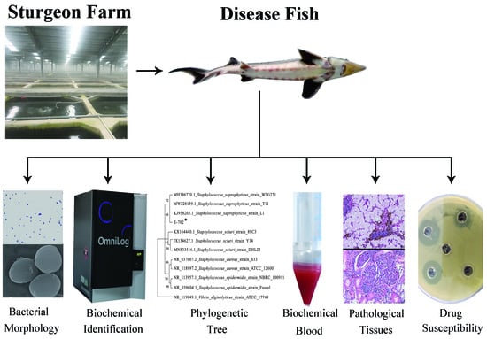
2.1. Fish

In 2021, a large number of breeding hybrid sturgeons died in a breeding base in Yichang, China, and diseased fish were collected for further experiments. The healthy hybrid sturgeons used for pathogenicity tests came from Yichang, and the fish length was (15 ± 2) cm. Healthy hybrid sturgeons (about 500 fish) were reared in 25 aquariums (0.8 m × 0.6 m × 0.6 m) at a temperature of 20 ± 1 °C and fed twice a day. Fish with similar growth and weight were selected for subsequent experiments. All experimental procedures were conducted according to the guidelines of the appropriate Animal Experimental Ethical Inspection of Laboratory Animal Centre, Yangtze River Fisheries Research Institute, Chinese Academy of Fishery Sciences (ID: YFI2021-zhouyong-15).

2.2. Pathogen Isolation

The surface of the sick fish was sterilized with 75% ethanol and placed in a biological safety cabinet for dissection. Liver and kidney tissues were homogenized and spread on brain heart infusion (BHI) agar plates [11], and the plates were incubated at 28 °C for 36 h. Subsequently, individual colonies were selected by inoculation loops and streaked on new BHI agar plates for further isolation and purification. The purified strain E702 was inoculated into 800 μL of liquid BHI medium and cultured at 28 °C at 200 rpm, until the OD600 of the bacterial liquid was about 0.5. An amount of 100 µL of bacterial solution was used for Gram staining, and 600 µL of the bacterial solution was mixed with 300 µL of 50% glycerol solution and stored at −80 °C.

2.3. Morphological Observation

Bacteria were collected by centrifugation at 5000 rpm for 2 min and suspended in phosphate-buffered saline (PBS). The suspension was then spread onto glass slides to dry, followed by Gram staining, as described by Xiao [11]. In addition, the strain was observed with scanning electron microscopy and recorded.

2.4. 16S rDNA Sequence Analysis

The nucleic acid of strain E702 was extracted using the Bacterial Genomic DNA Kit (Axygen, Somerville, MA, USA), and the 16S rDNA gene of strain E702 was amplified using universal primers (F: 5’-AGAGTTTGATCATGGCTCAG-3’ and R: 5’-TACGGTTACCTTGTTACGACTT-3’) [12]. The amplification program was as follows: 30 s at 96 °C, 45 s at 57 °C, and 1 min at 72 °C for 32 cycles. The amplified products were identified by 1.0% agarose gel electrophoresis (Liuyi, China). The target bands were recovered and sequenced by Tsingke Bio-technology Co., Ltd. (Wuhan, China). The sequencing results were analyzed through the NCBI website (http://blast.ncbi.nlm.nih.gov, (accessed on 1 May 2022), and a phylogenetic tree was constructed with the software MEGA 4.0.

2.5. Biochemical Identification of Bacteria

The E702 strain was grown on Biolog Universal Anaerobe (BUA) agar medium at 30 °C for 24 h. In terms of the operating requirements of the Biolog bacterial identification kit (Biolog, Hayward, CA, USA), a single colony was selected and placed in the IF-inoculum (Biolog, USA). After the Biolog bacterial identification plate was inoculated with the inoculum containing E702 at a dose of 100 μL per well, it was placed in the Biolog automatic microorganism identification system for identification, and the results were recorded.

2.6. Toxicity Identification

Strain E702 was grown in liquid BHI medium for 20 h at 200 rpm with shaking at 30 °C. Bacteria were collected after centrifugation (5000 rpm, 2 min), and the pellet was rinsed twice with aseptic PBS (Cytiva, Washington, DC, USA). Bacteria suspensions of distinct concentrations (104, 105, 106 and 107 CFU/mL) were prepared with sterile PBS after the concentration was detected by a bacterial colony counter. The healthy hybrid sturgeons were randomly separated into 5 groups (4 groups for infection test, 1 group for control test), with 30 fish in each group. The infection test group was injected with 0.3 mL bacterial suspension with the above distinct concentrations, respectively, and the control group was injected with 0.3 mL aseptic PBS. After infection, the number of deaths in each group was observed and recorded for 14 consecutive days. The pathogenic bacteria were isolated and re-identified from the diseased fish. Experiments were performed in triplicate with biological replicates. The 50% lethal dose (LD50) value of the E702 strain was calculated with the method of Reed and Muench [13].

2.7. Histopathological Observation

Pathological tissues were sampled from four parts of each diseased fish, including the gills, liver, kidneys and intestines, and these were sectioned for observation. The significantly changed ones were selected for analysis. These collected organs and tissues were fixed with 4% paraformaldehyde. Histopathological section preparation included ethanol gradient dehydration (80%, 90%, 95% and 100%), paraffin embedding, sectioning (5 µm thickness, Leica, Wetzlar, Germany), hematoxylin-eosin staining (Solarbio, Beijing, China) and glass slides sealed with neutral resin [14]. Pathological changes in different tissues were observed by light microscopic examination of histopathological sections (Mount Olympus, Tokyo, Japan) [15].

2.8. Analysis of Blood Parameters

Blood was collected from the tail vein of the control (n = 6) and diseased hybrid sturgeon (n = 6) via sterile disposable syringes and used for smear preparation. By staining with Wright-Giemsa stain (Baso, Zhuhai, China), the cells on each blood smear were observed, classified and counted under the oil lens [16].
Blood was collected in a 2 mL centrifuge tube and left at 4 °C for 4 h before centrifugation at 5000 rpm for 15 min. The supernatant was analyzed with a biochemical analyzer (HITACHI, Hitachi City, Japan), and the data were analyzed with Excel software (Microsoft Office software 2013).

2.9. Antimicrobial Sensitivity Testing

The Kirby–Bauer disc diffusion method was adopted to perform antimicrobial sensitivity testing [17]. A suspension of strain E702 was spread evenly on the surface of Mueller–Hinton (MH) agar medium, with 100 μL of liquid bacterial suspension added to each plate, and 4 antimicrobial sensitivity test paper discs were then placed equally on the surface of the medium. Subsequently, the diameter of the inhibition zone was measured after 24 h of incubation at 28 °C. Vancomycin, furazolidone, streptomycin, chloramphenicol, gentamicin, neomycin, cefazolin, norfloxacin, carbenicillin and ciprofloxacin (Hangwei, Hefei, China) were selected for the antimicrobial sensitivity test.

2.10. Statistical Analyses

Statistical analyses were carried out using the SPSS software (version 13, SPSS, Chicago, IL, USA). Differences between means of treatments were performed with Duncan’s test, with means considered significantly different at p < 0.05.

3. Results

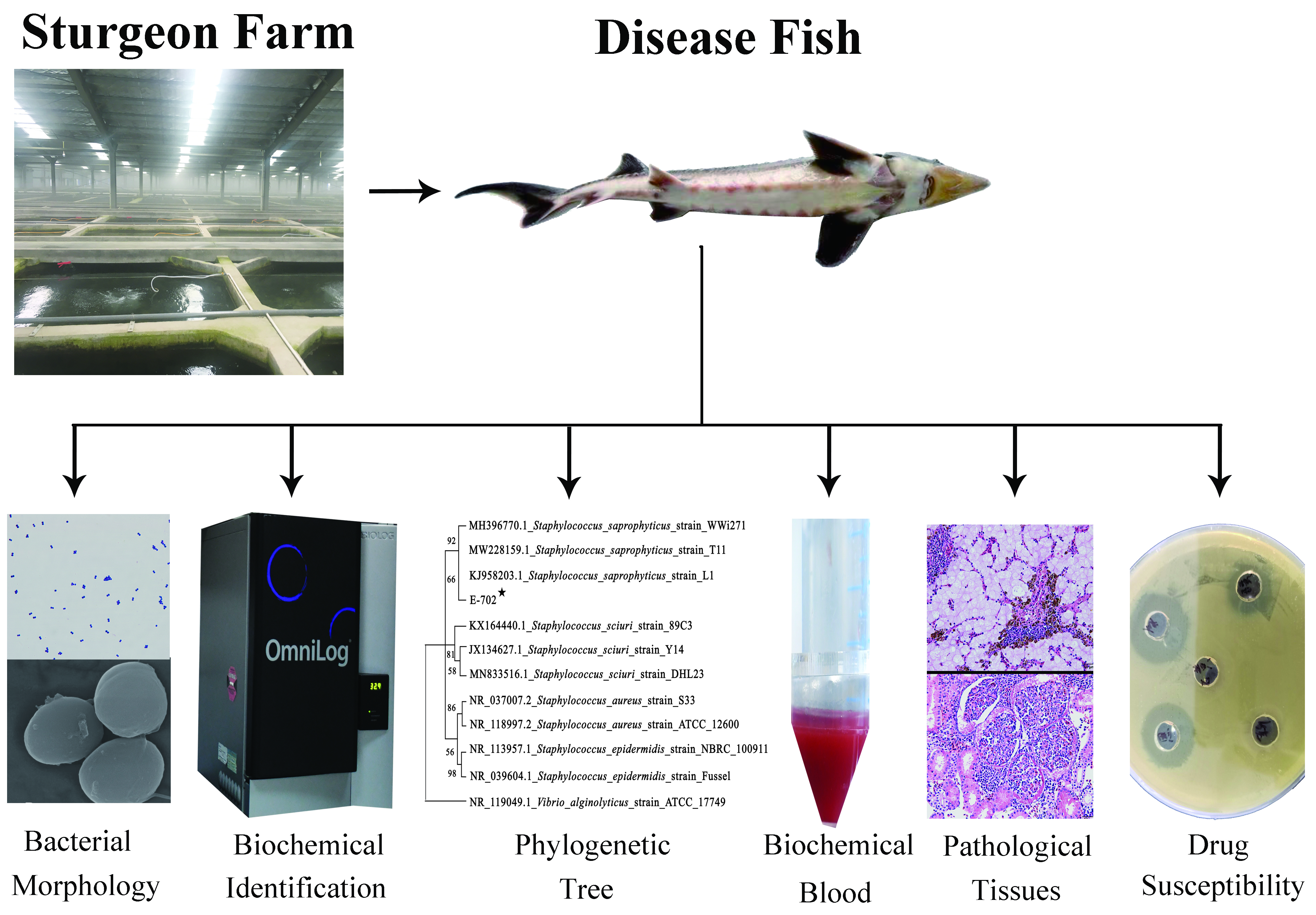
3.1. Symptoms of Diseased Hybrid Sturgeons

The diseased fish were about 60 cm long (Figure 1A), and their mobility was reduced. The blood spots on both sides of the abdomen were evenly and symmetrically distributed (Figure 1A), and the cloacas were red or even purple (Figure 1A,B). The swim bladder, liver, kidneys, intestines and stomach all had varying degrees of hyperemia and swelling (Figure 1B). No parasites were found on the body surface, gills or in vivo.

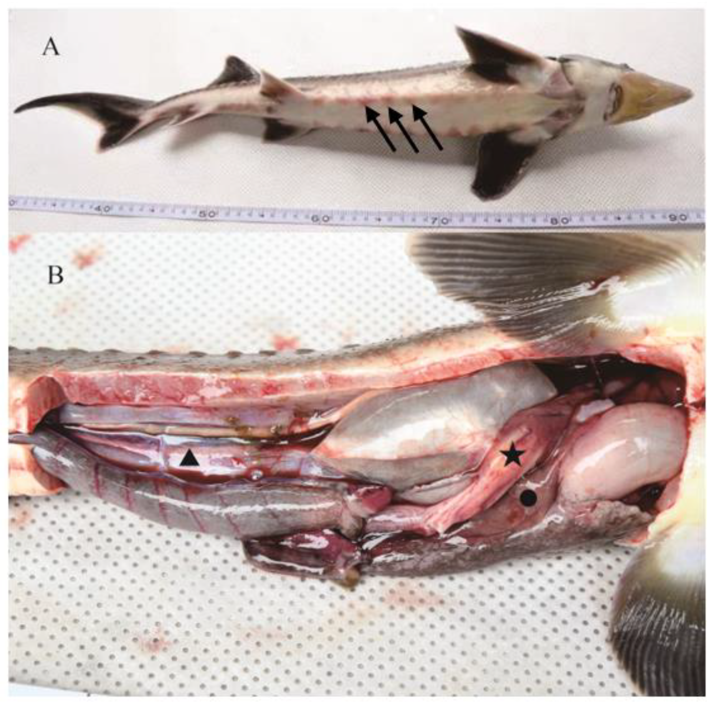
3.2. Bacterial Morphological Analysis

The bacteria isolated from diseased fish were shown as purple cocci after Gram staining (Figure 2A), indicating that they were Gram-positive bacteria. The bacteria were further observed with a scanning electron microscope, and it was found that the diameter of the bacteria was about 700 nm, the surface was not smooth, and there were flagella (Figure 2B).

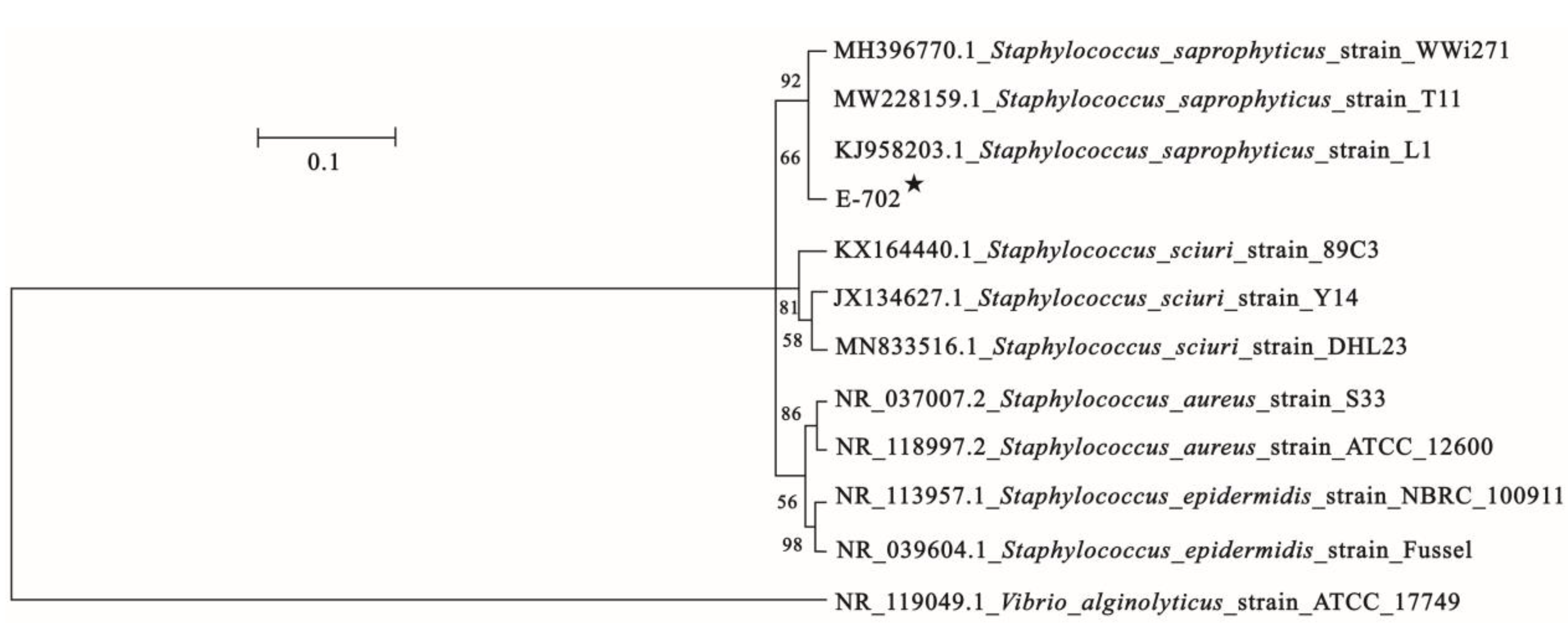
3.3. 16S rDNA Identification

The 16S rDNA of this strain was amplified and sequenced, and the full-length (1443 bp) fragment was subjected to NCBI BLAST analysis. The results revealed that this fragment had the greatest similarity to Staphylococcus saprophyticus and was clustered in the same clade with S. saprophyticus (GenBank accession numbers: MH396770.1, KJ958203.1 and MW228159.1) in the phylogenetic tree (Figure 3). These results indicated that this strain belongs to S. saprophyticus.

3.4. Biochemical Identification of Bacteria

To identify strain E702, biochemical analysis was performed using the Biolog microbial identification system, and the results were then matched to the database, identifying the strain as S. saprophyticus (Table 1).

3.5. Pathogenicity

The healthy hybrid sturgeon died after being injected with different concentrations of the E702 strain. According to the experimental results (Table 2), the LD50 of E702 was 2.14 × 10⁵ CFU/g. S. saprophyticus was isolated from artificially infected hybrid sturgeons again.

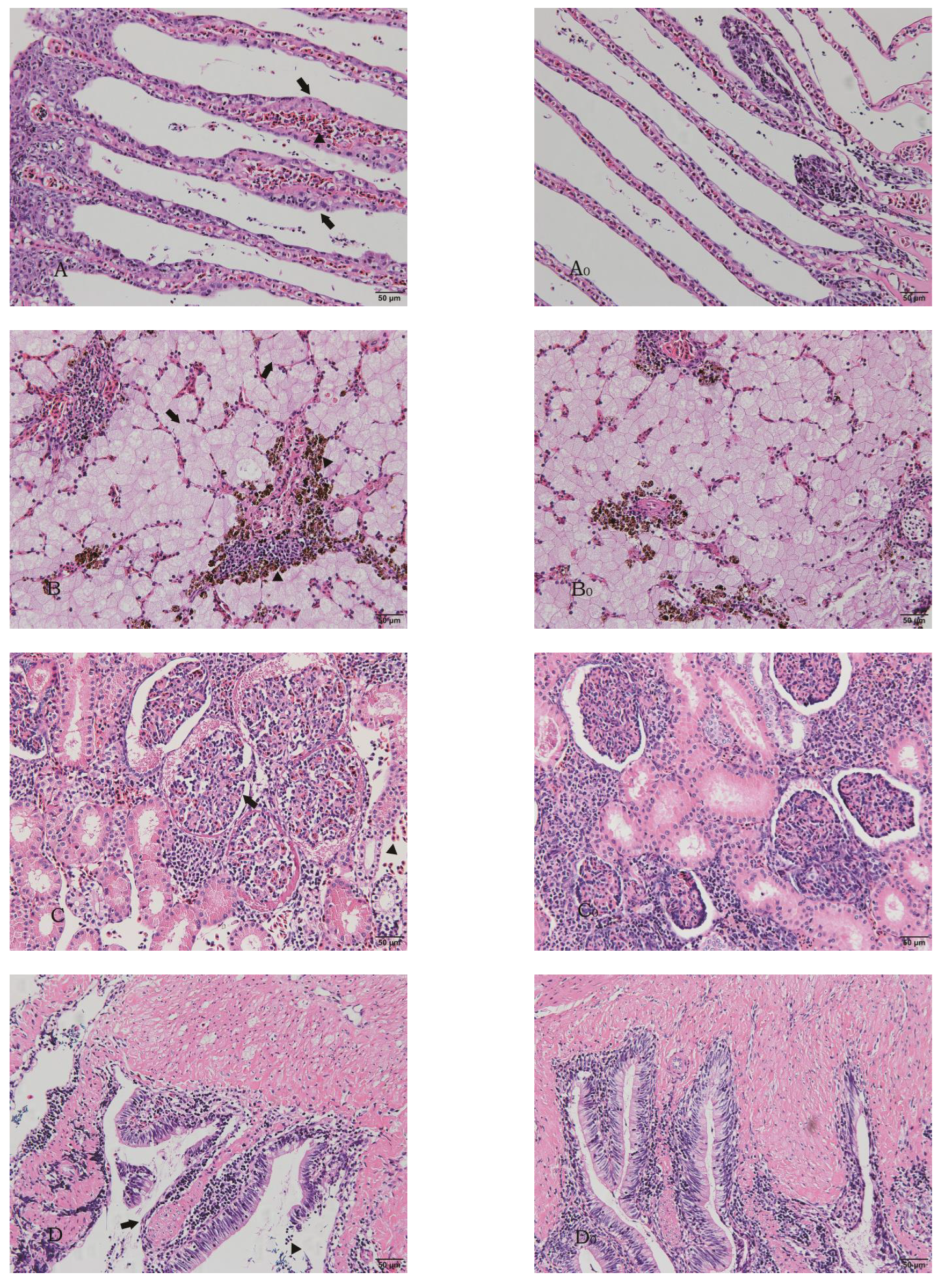
3.6. Pathological Analysis

Organs of diseased hybrid sturgeon from farms were collected for histopathology observation. After isolating the pathogenic bacteria, they were verified using the Koch rule. The experimental group was used to detect its pathogenicity and artificially infected fish with this bacterium. The gill structure of the experimental group showed partial fusion, accompanied by infiltration of the inflammatory cells (Figure 4A). There was a large amount of inflammatory cell infiltration in the liver of diseased fish (Figure 4B). Renal cells infiltrated and merged with inflammatory cells (Figure 4C). A large number of fused cells were found in the intestinal section, some of which were accompanied by cell infiltration (Figure 4D).

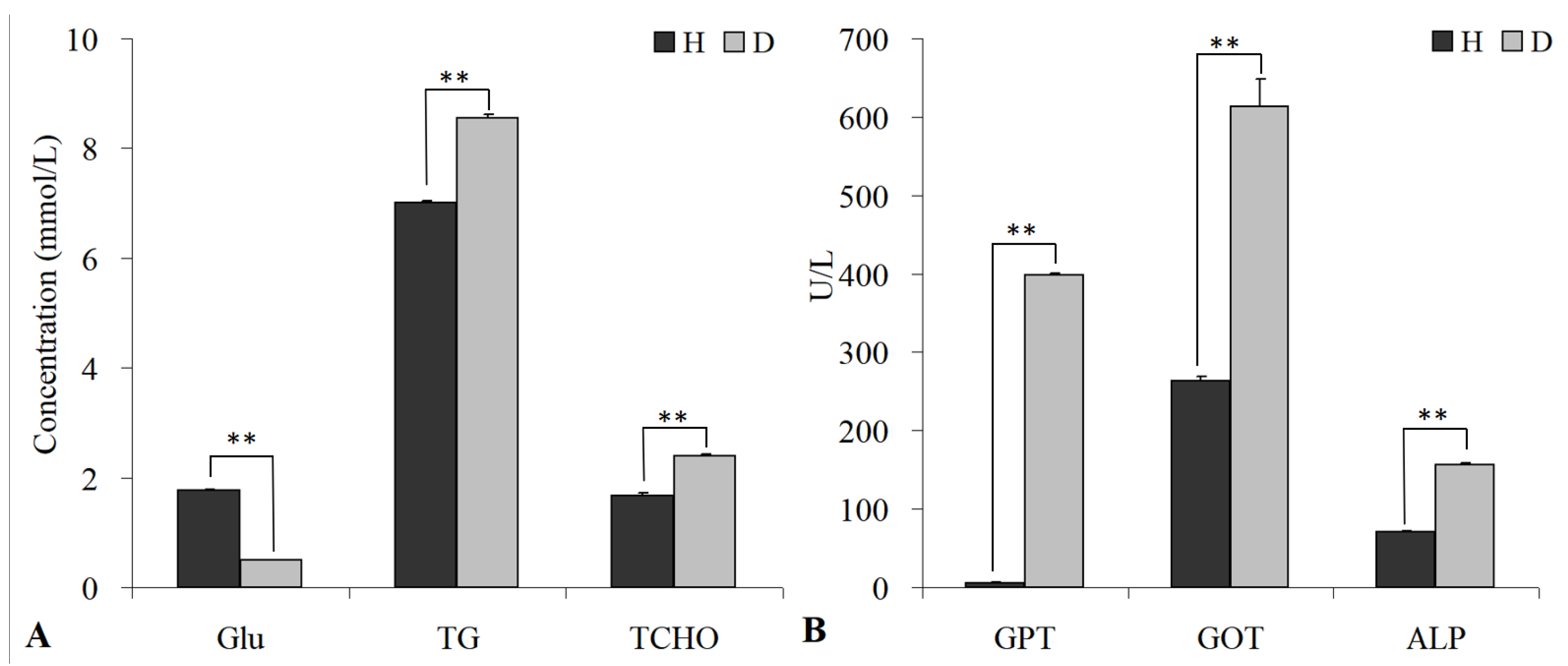
3.7. Blood Biochemical Analysis

By serum physiology and biochemical analysis, the glucose concentration of the diseased hybrid sturgeon was significantly reduced compared with the control, whereas the triglycerides (TG) and total cholesterol (TCHO) rose (Figure 5A). The glutamate-pyruvate transaminase (GPT), aspartate transaminase (GOT) and alkaline phosphatase (ALP) concentrations of the diseased hybrid sturgeons were significantly increased compared with the control (Figure 5B). The calculated p value was 0.0037.

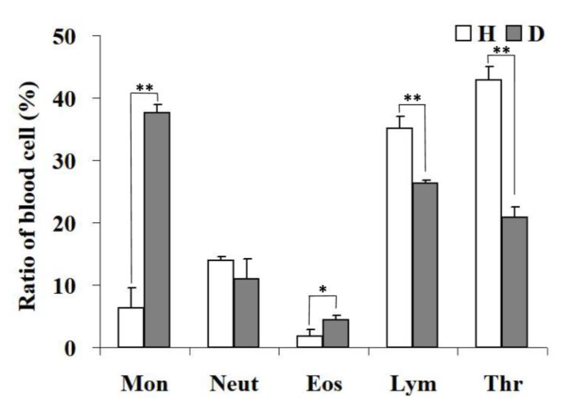
3.8. Blood Count Analysis

In terms of leukocyte numbers (Figure 6), monocytes and lymphocytes were the most abundant cells in the diseased hybrid sturgeons, and the least abundant were eosinophils. The white blood cells of the control group were mostly platelets and lymphocytes, and a lower number were eosinophils. Compared with healthy hybrids, diseased hybrids had significantly higher proportions of monocytes (p < 0.01) and eosinophils (p < 0.05) and noticeably lower proportions of lymphocytes and platelets (p < 0.01).

3.9. Drug Sensitivity

A number of drugs were used to test the antimicrobial sensitivity of the E702 strain, according to the diameter of the inhibition zone (Table 3). The results showed that the strain was highly sensitive to neomycin, cefazolin, norfloxacin, carbenicillin, gentamicin and ciprofloxacin, moderately sensitive to vancomycin, furazolidone and streptomycin and resistant to chloramphenicol.

4. Discussion

S. saprophyticus isolated in this study is a common colonizing bacterium that can cause urinary tract infection [18], acute pyelonephritis, urethritis [19] and endocarditis [20,21] in humans. It has been found in the human gastrointestinal tract, rectum, blood [19,20,22] and intestines of some animals [23]. Previous studies have shown that S. saprophyticus, as the pathogenic bacteria of most aquatic products [24], reproduces rapidly, and its metabolites have a rancid smell [25], which seriously threatens the yield and quality of aquatic products and even causes water pollution. However, fish infected with S. saprophyticus have been rarely reported, and the pathogenesis of S. saprophyticus has been poorly understood so far. In the present investigation, the pathogenic strain E702 was isolated from the diseased hybrid sturgeon, which was Gram positive, and its surface was not smooth and had flagella as observed by scanning electron microscopy. The 16S rDNA of this strain was amplified, sequenced and compared, and the results showed that this fragment had the highest similarity with S. saprophyticus, which belongs to the Staphylococcus genus. The pathogenicity test clarified that the LD50 was 2.14 × 10⁵ CFU/g, and the E702 strain had strong virulence to hybrid sturgeons. In addition, the results of reinfection experiments revealed that Staphylococcus saprophyticus was the pathogenic agent leading to the death of the hybrid sturgeons.
Lymphocytes are a crucial part of the immune system, exert a positive effect during the adaptive immune response [26] and can be used to assess the body’s immune response after pathogen invasion. In the blood of the experimental group hybrid sturgeon in this study, the proportion of lymphocytes was significantly reduced, indicating a decline in immune function. Monocytes and granulocytes eliminate invading pathogens through phagocytosis, thereby participating in the immune response [27]. The percentage of monocytes and eosinophils in the blood of S. saprophyticus-infected hybrid sturgeons was dramatically increased, accordingly promoting phagocytosis. The function of platelets is mainly to promote hemostasis and accelerate coagulation, and the abnormal quantity and quality of platelets can cause bleeding diseases [28]. In addition, platelets have the function of phagocytosing viruses, bacteria and other particles [29,30,31]. As the line of defense against bacterial infections, a low number of platelets means that the body has a heavy infection. The diseased sturgeons in this study had markedly reduced platelets, which may be the main cause of systemic hemorrhage.
Aspartate aminotransferase (GOT) and alanine aminotransferase (ALT) are mainly distributed in liver cells and are usually less abundant in serum, but when liver cells are damaged, the cell membrane permeability increases, and the release of GOT and ALT into the blood will lead to a significant increase in enzyme activity in the serum [32]. Therefore, transaminases (especially ALT) are sensitive markers of hepatocyte damage. Alkaline phosphatase (ALP) is a critical regulatory enzyme in the biological metabolism of animals and is widely distributed in various tissues of the body, mostly in the liver [33,34]. Consequently, an abnormal ALP concentration can be used for the diagnosis and identification of liver diseases. The serum levels of GOT, ALT and ALP in the diseased hybrid sturgeons in this study were remarkably increased, indicating that the liver cells were damaged, and the liver function decreased.
Histopathologically, the lesions of E702-infected sturgeon had symptoms such as inflammatory cell infiltration, cell swelling, and cell fusion, which were similar to Bester sturgeon infected with Lactococcus lactis subsp. Lactis [35]. These results suggest that bacterial infection may lead to simultaneous lesions and reactions in multiple organs.
Staphylococci are very sensitive to antibiotics [36]. However, with the widespread use and abuse of antibiotics, S. saprophyticus has shown a trend of multiple drug resistance and has become a common pathogen of animal infection in recent years [37]. The E702 strain separated from the diseased hybrid sturgeon has a rough surface, as well as flagella, and is vulnerable to neomycin, cefazolin, norfloxacin, carbenicillin and ciprofloxacin, but moderately sensitive to vancomycin, furazolidone and streptomycin. In a previous study, a strain of S. saprophyticus named JY08 was isolated from Acipenser schrenckii, which has a smooth surface, is sensitive to doxycycline and is resistant to the compound sulfamethoxazole [38]. Differences in morphology and resistance may be related to environmental differences or their different origins. As a result, the results of the drug sensitivity test of pathogenic bacteria should first be referred to the diagnosis and treatment of bacterial diseases. In addition, it is also crucial to reasonably select drugs with good antibacterial treatment effects and to avoid multiple drug resistance caused by the abuse of antibiotics.

5. Conclusions

In this study, a germ strain was isolated from diseased hybrid sturgeon and identified as S. saprophyticus. The virulence test showed that the strain was pathogenic bacteria to sturgeon, and its LD50 was 2.14 × 10⁵ CFU/g. The diseased hybrid sturgeon exhibited severe histopathological changes and marked changes in hematological parameters. This strain was sensitive to neomycin, cefazolin, norfloxacin, carbenicillin and ciprofloxacin. In sum, S. saprophyticus was the pathogen responsible for the mass mortality of hybrid sturgeon. The present research emphasizes its underlying ventures in hybrid sturgeon breeding and aquaculture.

Author Contributions

Conceptualization, Y.W., Y.F. and Y.Z.; Methodology, Y.F., T.L. and Y.C.; Software, T.L.; Validation, Y.W. and Y.Z.; Investigation, M.X. and Z.X.; Data Curation, L.J.; Writing—Original Draft Preparation, Y.W. and Y.F.; Writing—Review and Editing, Y.W., Y.F. and Y.Z.; Visualization, R.G.; Supervision, Y.Z.; Project Administration, Y.W.; Funding Acquisition, Y.Z. All authors have read and agreed to the published version of the manuscript.

Funding

This work was supported by grants from the Central Public Interest Scientific Institution Basal Research Fund, CAFS (2020TD44) and the National Freshwater Aquatic Germplasm Resource Center (FGRC18537).

Institutional Review Board Statement

All experimental procedures were conducted according to the guidelines of the appropriate Animal Experimental Ethical Inspection of Laboratory Animal Centre, Yangtze River Fisheries Research Institute, Chinese Academy of Fishery Sciences (ID Number: YFI2021-zhouyong-15).

Data Availability Statement

The data presented in this study are available on request from the corresponding author.

Acknowledgments

We would like to thank Wuhan Donglikang Life Science Co., Ltd. for providing therapeutic products for the diseased sturgeons involved in this study.

Conflicts of Interest

The authors declare no conflict of interest.

References

  1. Boschung, H.T.; Bailey, R.M. Acipenseriformes: AccessScience; McGraw Hill: New York, NY, USA, 2019. [Google Scholar] [CrossRef]
  2. Caviar: Symbol of Luxury and Excess. 2011. Available online: http://language.chinadaily.com.cn/2011-11/01/content_14013902.htm (accessed on 1 November 2011).
  3. Bledsoe, G.; Bledsoe, C.; Rasco, B. Caviars and fish roe products. Crit. Rev. Food Sci. 2003, 43, 317–356. [Google Scholar] [CrossRef] [PubMed]
  4. Bronzi, P.; Chebanov, M.; Michaels, J.; Wei, Q.; Rosenthal, H.; Gessner, J. Sturgeon meat and caviar production: Global update 2017. J. Appl. Ichthyol. 2019, 3, 257–266. [Google Scholar] [CrossRef]
  5. Ferreira, B.; Bertoncini, T. The IUCN Red List of Threatened Species. 2018. Available online: http://dx.doi.org/10.2305/IUCN.UK.2018-2.RLTS.T39303A46914065.en (accessed on 14 September 2018).
  6. Jarić, I.; Bronzi, P.; Cvijanović, G.; Lenhardt, M.; Gessner, J. Paddlefish (Polyodon spathula) in Europe: An aquaculture species and a potential invader. J. Appl. Ichthyol. 2019, 35, 267–274. [Google Scholar] [CrossRef]
  7. Chen, X.; Li, C.; Yang, C.; Zhang, S.; Wu, J. Status and prospects of techniques in the sturgeon aquaculture industry in China. Freshw. Fish. 2017, 47, 108–112. [Google Scholar] [CrossRef]
  8. Pierezan, F.; Shahin, K.; Heckman, T.; Ang, J.; Soto, E. Outbreaks of severe myositis in cultured white sturgeon (Acipenser transmontanus L.) associated with Streptococcus iniae. J. Fish Dis. 2020, 43, 485–490. [Google Scholar] [CrossRef]
  9. Soto, E.; Fast, M.; Purcell, S.; Coleman, D.; Yazdi, Z.; Kenelty, K.; Yun, S.; Camus, A. Expression of immune markers of white sturgeon (Acipenser transmontanus) during Veronaea botryosa infection at different temperatures. Comp. Biochem. Physiol. Part D Genom. Proteom. 2022, 41, 100950. [Google Scholar] [CrossRef]
  10. Mugetti, D.; Pastorino, P.; Menconi, V.; Pedron, C.; Prearo, M. The old and the new on viral diseases in sturgeon. Pathogens 2020, 9, 146. [Google Scholar] [CrossRef]
  11. Xiao, Z.; Xue, M.; Wu, X.; Zeng, L.; Zhu, Y.; Jiang, N.; Fan, Y.; Zhou, Y. Isolation and identification of Staphylococcus warneri from diseased Coreius guichenoti. Aquacult. Rep. 2022, 22, 100988. [Google Scholar] [CrossRef]
  12. Lane, D.J. 16S/23S rRNA sequencing. In Nucleic Acid Techniques in Bacterial Systematics; John Wiley & Sons Ltd.: New York, NY, USA, 1991; pp. 115–175. [Google Scholar] [CrossRef]
  13. Reed, L.; Muench, H. A simple method of estimating fifty percent endpoints. Am. J. Epidemiol. 1938, 27, 493–497. [Google Scholar] [CrossRef]
  14. Jiang, N.; Xu, J.; Ma, J.; Fan, Y.; Zhou, Y.; Liu, W.; Zeng, L. Histopathology and ultrastructural pathology of cyprinid herpesvirus II (CyHV-2) infection in gibel carp, Carassius auratus gibelio. Wuhan Univ. J. Nat. Sci. 2015, 20, 413–420. [Google Scholar] [CrossRef]
  15. Xu, Y.; Li, Y.; Xue, M.; Xiao, Z.; Fan, Y.; Zeng, L.; Zhou, Y. Effects of dietary Enterococcus faecalis YFI-G720 on the growth, immunity, serum biochemical, intestinal morphology, intestinal microbiota, and disease resistance of crucian carp (Carassius auratus). Fishes 2022, 7, 18. [Google Scholar] [CrossRef]
  16. Liu, W.; Xu, J.; Ma, J.; LaPatra, S.E.; Meng, Y.; Fan, Y.; Zhou, Y.; Yang, X.; Zeng, L. Immunological responses and protection in Chinese giant salamander Andrias davidianus immunized with inactivated iridovirus. Vet. Microbiol. 2014, 174, 382–390. [Google Scholar] [CrossRef] [PubMed]
  17. Barry, A.L.; Coyle, M.B.; Thornsberry, C.; Gerlach, E.H.; Hawkinson, R.W. Methods of measuring zones of inhibition with the Bauer-Kirby disk susceptibility test. J. Clin. Microbiol. 1979, 10, 885–889. [Google Scholar] [CrossRef] [PubMed]
  18. Lawal, O.U.; Barata, M.; Fraqueza, M.J.; Worning, P.; Bartels, M.D.; Goncalves, L.; Paixão, P.; Goncalves, E.; Toscano, C.; Empel, J.; et al. Staphylococcus saprophyticus from clinical and environmental origins have distinct biofilm composition. Front. Microbiol. 2021, 7, 663768. [Google Scholar] [CrossRef]
  19. Hovelius, B.; Colleen, S.; Mardh, P.A. Urinary tract Infections in men caused by Staphylococcus saprophyticus. Scand. J. Infect. Dis. 1984, 16, 37–41. [Google Scholar] [CrossRef]
  20. Choi, S.H.; Woo, J.H.; Jeong, J.Y.; Kim, N.J.; Kim, M.N.; Yang, S.K.; Ryu, J. Clinical significance of Staphylococcus saprophyticus identified on blood culture in a tertiary care hospital. Diagn. Microbiol. Infect. Dis. 2006, 56, 337–339. [Google Scholar] [CrossRef]
  21. Garduño, E.; Quez, I.M.; Beteta, A.; Said, I.; Blanco, J.; Pineda, T.S. Staphylococcus saprophyticus causing native valve endocarditis. Scand. J. Infect. Dis. 2005, 37, 690–704. [Google Scholar] [CrossRef]
  22. Rupp, M.E.; Soper, D.E.; Archer, G.L. Colonization of the female genital tract with Staphylococcus saprophyticus. J. Clin. Microbiol. 1992, 30, 2975–2979. [Google Scholar] [CrossRef]
  23. Hedman, P.; Ringertz, O.; Lindström, M.; Olsson, K. The origin of Staphylococcus saprophyticus from cattle and pigs. Scand. J. Infect. Dis. 1993, 25, 57–60. [Google Scholar] [CrossRef]
  24. Fu, L.; Wang, C.; Liu, N.; Ma, A.; Wang, Y. Quorum sensing system-regulated genes affect the spoilage potential of Shewanella baltica. Food Res. Int. 2018, 107, 1–9. [Google Scholar] [CrossRef]
  25. Odeyemi, O.A.; Burke, C.M.; Bolch, C.; Stanley, R. Seafood spoilage microbiota and associated volatile organic compounds at different storage temperatures and packaging conditions. Int. J. Food Microbiol. 2018, 280, 87–99. [Google Scholar] [CrossRef] [PubMed]
  26. Scapigliati, G. Functional aspects of fish lymphocytes. Dev. Comp. Immunol. 2013, 41, 200–208. [Google Scholar] [CrossRef] [PubMed]
  27. Döring, M.; Stanchi, K.M.C.; Erbacher, A.; Haufe, S.; Schwarze, C.P.; Handgretinger, R.; Hofbeck, M.; Kerst, G. Phagocytic activity of monocytes, their subpopulations and granulocytes during post-transplant adverse events after hematopoietic stem cell transplantation. Immunobiology 2015, 220, 605–613. [Google Scholar] [CrossRef] [PubMed]
  28. Yao, D.; Chai, Y.; Wang, Y.; Guo, K.; Hang, J. Microscopic structures of peripheral blood cells in chinese sturgeon (Acipenser sinensis). J. Yangtze Univ. (Nat. Sci. Ed.) 2015, 12, 22–26. [Google Scholar] [CrossRef]
  29. Kullaya, V.I.; Mast, Q.D.; Ven, A.; Elmoussaoui, H.; Kibiki, G.; Simonetti, E.; Jonge, M.; Ferwerda, G. Platelets modulate innate immune response against human respiratory syncytial virus in vitro. Viral Immunol. 2017, 30, 576–581. [Google Scholar] [CrossRef]
  30. Nording, H.M.; Seizer, P.; Langer, H.F. Platelets in inflammation and atherogenesis. Front. Immunol. 2015, 6, 98. [Google Scholar] [CrossRef]
  31. Rossaint, J.; Kühne, K.; Skupski, J.; Aken, H.V.; Looney, M.R.; Hidalgo, A.; Zarbock, A. Directed transport of neutrophil-derived extracellular vesicles enables platelet-mediated innate immune response. Nat. Commun. 2016, 7, 13464. [Google Scholar] [CrossRef]
  32. Chuang, C.; Tsai, C.; Lin, E.; Huang, C.; Lin, Y.; Lan, C.; Huang, C. Heat-killed Lactobacillus salivarius and Lactobacillus johnsonii reduce liver injury induced by alcohol in vitro and in vivo. Molecules 2016, 21, 1456. [Google Scholar] [CrossRef]
  33. Whyte, M.P. Physiological role of alkaline phosphatase explored in hypophosphatasia. Ann. N. Y. Acad. Sci. 2010, 1192, 190–200. [Google Scholar] [CrossRef]
  34. Orimo, H. The mechanism of mineralization and the role of alkaline phosphatase in health and disease. J. Nippon Med. Sch. 2010, 77, 4–12. [Google Scholar] [CrossRef]
  35. Chen, M.; Hung, S.; Shyu, C.; Lin, C.; Liu, P.; Chang, C.; Shia, W.; Cheng, C.; Lin, S.; Tu, C. Lactococcus lactis subsp. lactis infection in Bester sturgeon, a cultured hybrid of Huso huso × Acipenser ruthenus, in Taiwan. Res. Vet. Sci. 2012, 93, 581–588. [Google Scholar] [CrossRef] [PubMed]
  36. Waisbren, B.A. The Sensitivity of Staphylococci to Antibiotics: A Five-Year Study of Sensitivity, Usage, and Cross Resistance in a General Hospital. AMA Arch. Intern. Med. 1958, 101, 397. [Google Scholar] [CrossRef] [PubMed]
  37. De, S.; Souza, D.; Leif, S.; Paiva, P.R.; Fernandes, R.R.; Helena, C.E.; Soares, P.M.; Dos, S.; Natacha, M.; Nunes, B. Staphylococcus saprophyticus recovered from humans, food, and recreational waters in Rio de Janeiro, Brazil. Int. J. Microbiol. 2017, 2017, 4287547. [Google Scholar] [CrossRef]
  38. Zhou, H. Isolation and identification of Staphylococcus saprophyticus from Acipenser schrenckii and its genomics analysis. J. Fish. Res. 2021, 43, 461–471. [Google Scholar] [CrossRef]
Figure 1. Clinical symptoms of E702-infected hybrid sturgeon. (A): The cloacas is red, and the abdomen is hyperemic (arrows). (B): Ascites (triangle), liver hemorrhage (dot), intestinal hyperemia (asterisk).
Fishes 08 00250 g001
Figure 2. Gram staining and scanning electron microscopy of bacterium E702. (A): Gram staining (bar scale: 10 µm); (B): scanning electron microscopy (bar scale: 500 nm).
Fishes 08 00250 g002
Figure 3. The values at the node indicate the percentage of trees in which this grouping occurred after bootstrapping the data (1000 replicates). The scale bar indicates the number of substitutions per site. The asterisk is bacterium E-702.
Fishes 08 00250 g003
Figure 4. Histopathological observation of diseased hybrid sturgeon (scale: 50 µm). (A,A0) Gills: swollen gills, enlarged nuclei, hyperplasia of epithelial cells (arrows) and infiltration of inflammatory cells (triangles) in the diseased sturgeon. (B,B0) Liver: hepatocyte enlargement (arrow) with a large infiltration of inflammatory cells (triangles). (C,C0) Kidney: glomerular lesions (arrows) with numerous inflammatory cells (triangles). (D,D0) Intestine: Intestinal villi with structural damage, partial shedding of epidermal cells (arrows), with numerous inflammatory cells (triangles).
Fishes 08 00250 g004
Figure 5. Blood biochemical analysis of hybrid sturgeon.(A) glucose (Glu), albumin (TG) and total cholesterol (TCHO). (B) Glutamate-pyruvate transaminase (GPT), aspartate transaminase (GOT), alkaline phosphatase (ALP); H, healthy hybrid sturgeon; D, diseased hybrid sturgeon; ** p < 0.01.
Fishes 08 00250 g005
Figure 6. Blood cell count of hybrid sturgeon (Mon, monocytes; Neut, neutrophils; Eos, eosinophils; Lym, lymphocytes; Thr, thrombocytes; H, healthy hybrid sturgeon; D, diseased hybrid sturgeon; * p < 0.05; ** p < 0.01).
Fishes 08 00250 g006
Table 1. Biochemical identification results of strain E702.
ReagentResult *ReagentResult
A1 Negative ControlNE1 GelatinB
A2 DextrinPE2 Glycyl-L-ProlineB
A3 D-MaltosePE3 L-AlanineB
A4 D-TrehalosePE4 L-ArginineP
A5 D-CellobioseBE5 L-Aspartic AcidB
A6 GentiobiosePE6 L-Glutamic AcidP
A7 SucrosePE7 L-HistidineP
A8 D-TuranosePE8 L-Pyroglutamic AcidB
A9 StachyoseNE9 L-SerineP
A10 Positive ControlPE10 LincomycinN
A11 PH6PE11 Guanidine HClB
A12 PH5NE12 Niaproof 4N
B1 D-RaffinoseNF1 PectinP
B2 α-D-LactosePF2 D-Galacturonic AcidB
B3 D-MelibioseBF3 L-Galactonic Acid LactoneB
B4 β-Methyl-D-GlucosidePF4 D-Gluconic AcidP
B5 D-SalicinBF5 D-Glucuronic AcidP
B6 N-Acetyl-D-GlucosaminePF6 GlucuronamideP
B7 N-Acetyl-β-D-MannosaminePF7 Mucic AcidN
B8 N-Acetyl-D-GalactosamineNF8 Quinic AcidN
B9 N-Acetyl Neuraminic AcidBF9 D-Saccharic AcidN
B10 1% NaClPF10 VancomycinN
B11 4% NaClPF11 Tetrazolium VioletP
B12 8% NaClPF12 Tetrazolium BlueP
C1 α-D-GlucosePG1 P-Hydroxy-Phenylacetic AcidN
C2 D-MannosePG2 Methyl PyruvateP
C3 D-FructosePG3 D-Lactic Acid Methyl EsterP
C4 D-GalactosePG4 L-Lactic AcidP
C5 3-Methyl GlucoseBG5 Citric AcidP
C6 D-GlucoseBG6 6α-Keto-Glutaric AcidB
C7 L-FucoseBG7 D-Malic AcidN
C8 L-RhamnoseBG8 L-Malic AcidP
C9 InosineBG9 Bromo-Succinic AcidP
C10 1%Sodium LactateBG10 Nalidixic AcidP
C11 Fusidic AcidNG11 Lithium ChlorideP
C12 D-SerinePG12 Potassium TelluriteP
D1 D-SorbitolPH1 Tween 40P
D2 D-MannitolPH2 γ-Amino-Butyric AcidP
D3 D-ArabitolBH3 α-Hydroxy-Butyric AcidP
D4 Myo-lnositolNH4 β-Hydroxy-D, L-Butyric AcidB
D5 GlycerolPH5 α-Keto-Butyric AcidN
D6 D-Glucose-6-PO4BH6 Acetoacetic AcidP
D7 D-Fructose-6-PO4BH7 Propionic AcidN
D8 D-Aspartic AcidNH8 Acetic AcidP
D9 D-SerineNH9 Formic AcidP
D10 TroleandomycinNH10 AztreonamP
D11 Rifamycin SVNH11 Sodium ButyrateP
D12 MinocyclineNH12 Sodium BromateN
* Abbreviations: P, positive; N, negative; B, borderline.
Table 2. Mortality of S. saprophyticus infected with strain E702 at different concentrations.
GroupBacteria Concentration/(CFU·mL−1)Injection Dose/mLCumulative Death NumberMortality
Test group6.7 × 1070.32893.33%
6.7 × 1060.32170.00%
6.7 × 1050.32066.67%
6.7 × 1040.31033.33%
ControlPBS0.300%
Table 3. Detection of drug sensitivity of the E702 strain.
Medicine NameInhibition Zone (mm)SensitivityMedicine NameInhibition Zone (mm)Sensitivity
Vancomycin15INeomycin21S
Furazolidone17ICefazolin34S
Streptomycin18INorfloxacin21S
Chloramphenicol9RCarbenicillin22S
Gentamicin16SCiprofloxacin23S
Abbreviations: S, susceptible; I, intermediate; R, resistant.
Disclaimer/Publisher’s Note: The statements, opinions and data contained in all publications are solely those of the individual author(s) and contributor(s) and not of MDPI and/or the editor(s). MDPI and/or the editor(s) disclaim responsibility for any injury to people or property resulting from any ideas, methods, instructions or products referred to in the content.

