Article

Cinnamaldehyde Decreases the Pathogenesis of Aeromonas hydrophila by Inhibiting Quorum Sensing and Biofilm Formation

1
Yangtze River Fisheries Research Institute, Chinese Academy of Fishery Sciences, Wuhan 430223, China
2
College of Fisheries and Life Science, Shanghai Ocean University, Shanghai 201306, China
*
Authors to whom correspondence should be addressed.
Fishes 2023, 8(3), 122; https://doi.org/10.3390/fishes8030122
Submission received: 8 February 2023 / Revised: 18 February 2023 / Accepted: 20 February 2023 / Published: 21 February 2023
(This article belongs to the Special Issue Interactions between Fish and Pathogens in Aquaculture)

Abstract

:
Antibiotics were the main fishery drugs for treating Aeromonas hydrophila (A. hydrophila) infection, which would generate selective pressure and result in the appearance of antibiotic-resistant bacteria. The bacterial quorum sensing (QS) system provides a new alternative strategy against A. hydrophila infection. QS inhibitors can reduce bacterial virulence behaviors by disrupting QS, which has no effect on bacterial growth. Therefore, we studied the effect of cinnamaldehyde from a natural plant extract on the QS of A. hydrophila aiming to reduce its pathogenicity. The efficacy of cinnamaldehyde against A. hydrophila was evaluated from various aspects, including the effects on aerolysin, lipase, protease, swarming motility, biofilm formation, acyl-homoserine lactones (AHLs), and QS-related genes. Moreover, the therapeutic effect of cinnamaldehyde in vitro and in vivo was studied. The results showed that cinnamaldehyde could decrease the virulence phenotypes of A. hydrophila regulated by QS. Moreover, the transcriptions of related genes (aerA, ahyR, and ahyI) were downregulated following the addition of cinnamaldehyde. The in vitro and in vivo therapeutic assays show that cinnamaldehyde could reduce the aerolysin-mediated A549 cell injury and increase the survival rate of crucian carp infected with A. hydrophila. These results indicate that cinnamaldehyde would be a candidate QS inhibitor against A. hydrophila infection.

1. Introduction

Aquatic products have become favorite foods for their rich, high-quality protein and the improvement of life quality, which accelerates the development of the aquaculture industry. Nevertheless, the fast growth of aquaculture brings a number of problems, particularly infectious diseases caused by bacteria. Antibiotics as the main drugs for fish health management and disease treatment were used in all stages of fish growth [1]. However, the overuse of antibiotics provided selective pressures to bacterial strains and resulted in the emergence of drug-resistant strains, which then decreased the drug efficacy or even resulted in treatment failure [2]. The invention of an anti-virulence strategy targeting bacterial quorum sensing (QS) provided a novel approach facing the incidence of antibiotic-resistant bacterial infections. The QS systems rely on signaling molecules secreted by bacteria that vary with bacterial density, which initiate the expression of related genes and further regulate their virulence behaviors and biofilm formation [3]. Therefore, the alternative drugs targeting QS can attenuate the pathogenicity and drug resistance of bacteria.
A. hydrophila is the main pathogen that threatens fish health and causes septicemia in fish [4]. A. hydrophila can secrete a variety of virulence factors, such as aerolysin, lipase, protease, and hemolysin, which determine the pathogenicity of the bacterium [5]. Moreover, A. hydrophila has a strong biofilm-forming ability, which can resist the effects of many adverse conditions by attaching to surfaces, thereby promoting the drug resistance and pathogenicity of bacteria [6]. Studies have found that the virulence of A. hydrophila could be regulated by the QS [7]. Therefore, disrupting QS is an alternative approach to combating A. hydrophila infection.
Cinnamaldehyde (Figure 1A) is the main content of Cinnamomum cassia Presl, which exhibited a number of biological activities, such as antioxidant, antibacterial, antitumor, anti-inflammatory, and antidiabetic effects [8]. Studies have demonstrated that cinnamaldehyde performed against the growth of A. hydrophila by disrupting cell membranes and interfering with protein metabolism [9]. However, the function of cinnamaldehyde against the virulence of A. hydrophila has not been reported. Our study found that cinnamaldehyde could influence the pathogenicity of A. hydrophila by interfering with the QS.

2. Materials and Methods

2.1. Microorganisms and Reagents

A. hydrophila XS-91-4-1 and Chromobacterium violaceum CV026 were stored in our lab. Cinnamaldehyde (purity ≥ 95%) was purchased from Shanghai Aladdin Biochemical Technology Co., Ltd. Enrofloxacin was a commercial product purchased from the National Institutes for Food and Drug Control (Beijing, China). Cinnamaldehyde and enrofloxacin were dissolved in DMSO to obtain stock solutions of 40,960 μg/mL for in vitro assays and dissolved in 10% Tween 80 to obtain cinnamaldehyde emulsion for in vivo study.

2.2. Determination of Minimum Inhibitory Concentrations (MICs)

The MICs were determined according to a previously published protocol [10]. Briefly, cinnamaldehyde was diluted from 512 μg/mL to 2 μg/mL with MHB medium by 2-fold dilution in a 96-well plate, while enrofloxacin was diluted from 32 μg/mL to 0.125 μg/mL. Then, the bacterial cells at a density of 5 × 10⁵ CFU/mL were added to each well of the plate and cultured at 28 °C overnight. The wells without visible bacterial growth were defined as MICs.

2.3. Growth Curves Assay

Bacterial suspension cultured in LB broth was equally divided into 5 flasks when the optical density (OD) at 600 nm reached 0.3. Then, cinnamaldehyde was added into each flask to cause the final concentrations of cinnamaldehyde in the mixtures to reach 0, 1, 2, 4, 8, and 16 μg/mL, respectively. The mixtures were incubated at 28 °C for 5 h, and samples were taken every 30 min to read the OD600nm values.

2.4. Hemolysis

The hemolysis was measured using bacterial supernatants, as previously described [11]. Briefly, bacterial cultures at OD600nm of 0.3 were added to indicated concentrations of cinnamaldehyde (0, 1, 2, 4, 8, 16 μg/mL) and then co-cultured to OD600nm of 1.5. The bacterial suspensions were then centrifuged and then the supernatants were activated with the addition of trypsin, 100 μL of activated supernatant, 25 μL of sheep erythrocytes, and 875 μL of hemolysis buffer to obtain a 1 mL reaction system. The systems were then incubated at 37 °C for 20 min. The hemolytic activity of each drug group was read at OD543nm with a spectrophotometer.

2.5. Immuno-Blot

The concentrations of total proteins of bacterial supernatants described above were determined using the bicinchoninic acid method [12]. Then, the supernatants were sampled with Laemmli buffer and boiled for 10 min, then loaded for protein electrophoresis. A semi-dry transfer cell was used to transfer the proteins to the PVDF membrane. Then, the PVDF membrane was blocked with milk for 2 h, followed by incubation with the primary anti-aerolysin antibody and HRP-conjugated secondary goat anti-rabbit antiserum for 1 h, respectively. The levels of aerolysin in the supernatants were detected by an ECL detection kit.

2.6. Lipase Assay

Lipase assay was determined using the method reported by Ramanathan et al. [13]. In brief, the substrate mixture was composed of 0.3% (w/v) p-nitrophenyl palmitate in isopropanol and 50 mM Na2HPO4 buffer in an appropriate ratio (1:9). A total of 100 µL bacterial supernatants after different treatments were added to 900 µL of substrate mixture and incubated for 1 h. Then, the reaction was terminated by the addition of 1 M sodium carbonate buffer at the same volume of the system. After centrifugation, the values of OD410nm were determined in each reaction system.

2.7. Protease Activity Assay

According to a previous study, azocasein was used as the substrate [14]. Briefly, the supernatants of A. hydrophila co-cultured with cinnamaldehyde at concentrations ranging from 1 to 16 μg/mL were incubated with 20 mg/mL azocasein for 1 h, and 100 g/L trichloroacetic acids were added to the mixture to precipitate the protein. Then, an equal volume of NaOH (1 mol/L) was added, and the values were read at the OD of 440 nm.

2.8. Swarming Motility Assay

Sterilized swarming agar consists of 1% glucose, 0.5% agar, 0.5% peptone, and 0.2% yeast extract [15]. A total of 15 mL of swarming agar with different concentrations of cinnamaldehyde (1, 2, 4, 8, and 16 μg/mL) were added to the plates, and then 2 μL of bacterial suspension (OD600nm = 1.0) was added in the middle of the agar. Swarming agar plus DMSO was set as the control group. All plates were incubated for 24 h at 28 °C and the diameters of swarming were determined.

2.9. Biofilm Formation

Cinnamaldehyde was 2-fold diluted at volumes of 100 μL in a 96-well plate, and then bacterial cultures at volumes of 100 μL were added to each well after a dilution of 1:10. After incubation at 28 °C for 24 h, the cultures in 96-well plates were washed twice with PBS to remove unattached cells. The plate was air-dried and 0.5% crystal violet was added to stain bacterial cells. After washing, 95% ethanol was added into each well to release crystal violet in bacterial cells, and the absorption values were further determined at OD570nm.
For microscopic analysis, cinnamaldehyde and bacterial cultures were added to the 24-well plate containing glass slides and then incubated for 24 h. Bacterial cells attached in glass slides were stained with crystal violet after washing and images were pictured under a microscope.

2.10. qPCR Assay

Bacterial cells collected in hemolysis assay were used for qPCR assay. Briefly, a commercial RNA isolation kit was used to obtain the total RNA of the samples, then DNA remaining in the total RNA was removed. After reverse transcription, qPCR reactions were carried out to determine the expression levels of target genes. Primer pairs used in the study were listed in Table 1. Consequently, 16s rRNA was defined as the internal standard. Ct values were obtained and the expression levels were calculated by 2−ΔΔCt.

2.11. AHLs Production Assay

The effect of cinnamaldehyde on the AHLs production of A. hydrophila was determined by the previous method [16]. Briefly, A. hydrophila and C. violaceum CV026 cultured overnight were streaked in parallel at equal intervals on agar plates plus indicated concentrations of cinnamaldehyde at 28 °C for 24 h. To quantify violacein production, A. hydrophila was co-cultured with cinnamaldehyde for 24 h, then bacterial supernatants were collected and sterilized. An overnight C. violaceum CV026 culture was sub-inoculated into the sterilized supernatants and further cultured for 24 h. Then, bacterial cells of C. violaceum CV026 were collected by centrifugation and DMSO was added to release violacein pigment. The production of violacein was determined by a microplate reader at 585 nm.

2.12. Cell Viability Assays

A549 cells cultured in DMEM plus 10% fetal bovine serum and 5% CO2 were used to determine the protective effect of cinnamaldehyde against aerolysin-induced cell injury. Cells were seeded into a 96-well plate after being digested by trypsin. Sterile bacterial supernatants after treatment with cinnamaldehyde were co-cultured with the cells for 1.5 h. Then, the cells were acquired for live/dead cell staining, and cell-free supernatants for LDH release assays, respectively. The percent of LDH release was calculated by determining the values of OD490nm. Cell injury was evaluated by photographing cells treated with live/dead regents.

2.13. Animal Study

Animal studies were performed under the guidance of the Animal Welfare and Research Ethics Committee at the Yangtze River Fisheries Research Institute (Permission No. YFI-2022DJ-011, 10 June 2022). All the experimental protocols were approved and supervised by the animal care committee. A total of 60 crucian carps weighing 200 ± 10 g were maintained in 100 L glass tanks for 7 days and then were separated into 3 groups. For the experimental conditions, the dissolved oxygen in culture water was more than 5.0 mg/L, the test water temperature was 28 ± 2 °C, and the pH was 7.5~8.0. A. hydrophila XS-91-4-1 was cultured to the mid-log phase and diluted to a concentration of 1.5 × 10⁸ CFU/mL with sterile PBS. The fish infection model was established with the injection of the bacterial suspension at a volume of 100 μL intraperitoneally, and 100 μL of the sterile PBS for the negative control group. The cinnamaldehyde treatment group was given 25 mg/kg cinnamaldehyde by a gavage needle, while the positive and negative control group were administered with 10% Tween 80. The course was maintained for 3 days at 12-h intervals. The mortality in each group was recorded every day for 8 days. Three independent tests were carried out in this experiment.

2.14. Statistical Analysis

Statistical analysis was performed using GraphPad Prism 8.0 software. Data on survival rate were analyzed by Kaplan–Meier estimates and log-rank test, while other data were firstly analyzed by one sample K-S and explore tests, and then by Student’s t-test to determine the statistical significance. p < 0.05 indicates statistical significance.

3. Results

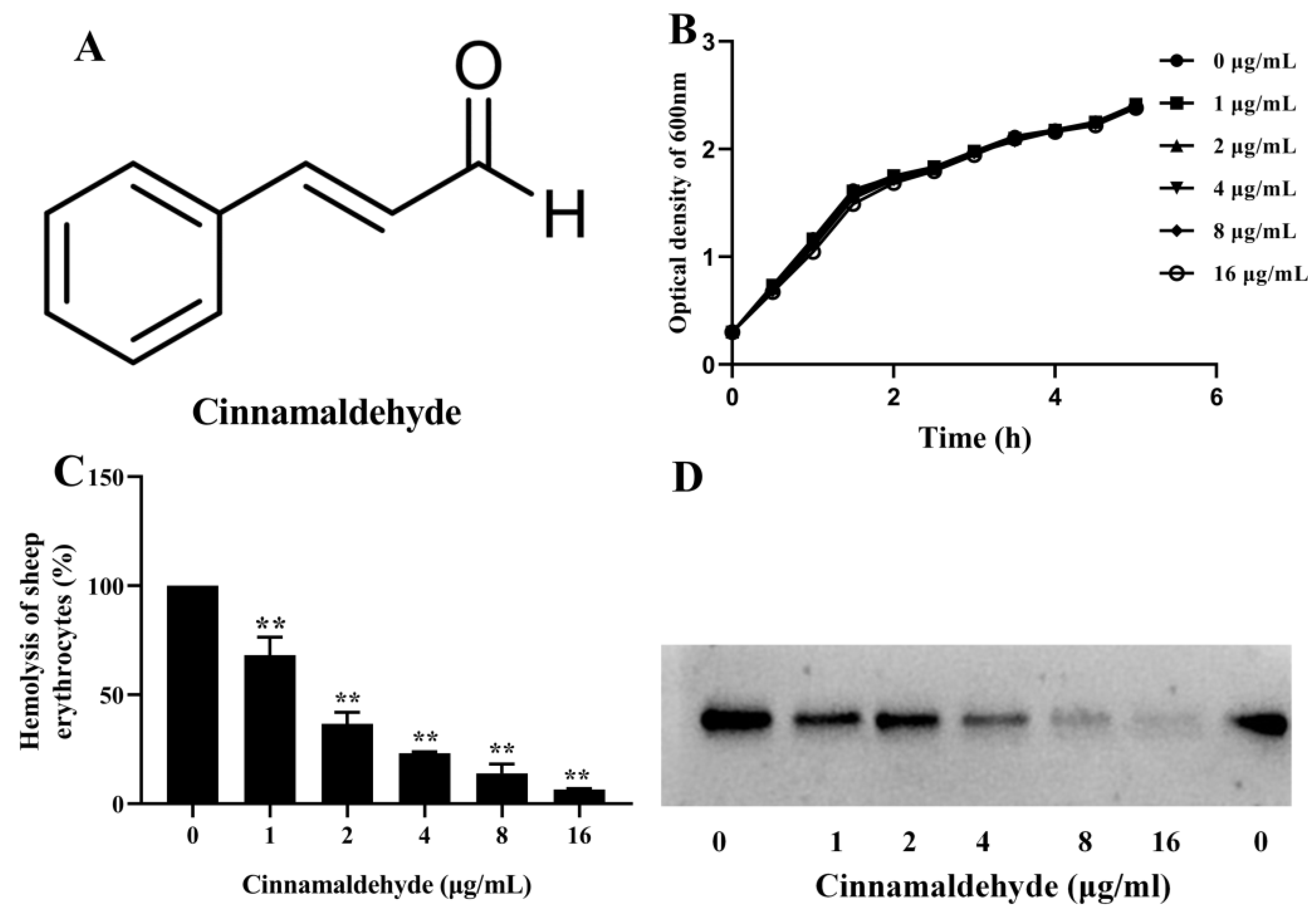
3.1. Effect of Cinnamaldehyde on A. hydrophila Growth

The MICs were 128 μg/mL and 4 μg/mL for cinnamaldehyde and enrofloxacin, respectively. The results indicated that cinnamaldehyde had little anti-A. hydrophila activity. Moreover, the growth curves assay was performed to evaluate the influence of cinnamaldehyde on bacterial growth in 5 h. As shown in Figure 1B, cinnamaldehyde had no role in bacterial growth when the concentrations reached 1 to 16 μg/mL. Furthermore, the addition of DMSO in the drug-free group showed that DMSO could not inhibit bacterial growth. Taken together, the results demonstrated that A. hydrophila co-cultured with cinnamaldehyde under our experimental conditions could not provide selective pressure to the bacterium.

3.2. Cinnamaldehyde Inhibited the Hemolysis of A. hydrophila

The hemolysis results showed that cinnamaldehyde could reduce the hemolytic activity of bacterial supernatants obtained from A. hydrophila plus cinnamaldehyde dose-dependently (Figure 1C). The hemolytic activity declined to 68.14 ± 8.25, 36.64 ± 5.31, 23.21 ± 0.60, 13.94 ± 4.30, and 6.50 ± 0.42% when co-cultured with cinnamaldehyde at concentrations of 1, 2, 4, 8, and 16 μg/mL compared with the cinnamaldehyde-free group. Hemolysis was remarkably inhibited by the addition of cinnamaldehyde at concentrations higher than 1 μg/mL. Moreover, the immune-blot assay was conducted to determine the relationship between aerolysin production and hemolysis. As expected, aerolysin production decreased with increasing drug concentrations (Figure 1D). Therefore, cinnamaldehyde could decrease the hemolysis of A. hydrophila supernatants by reducing the production of aerolysin.

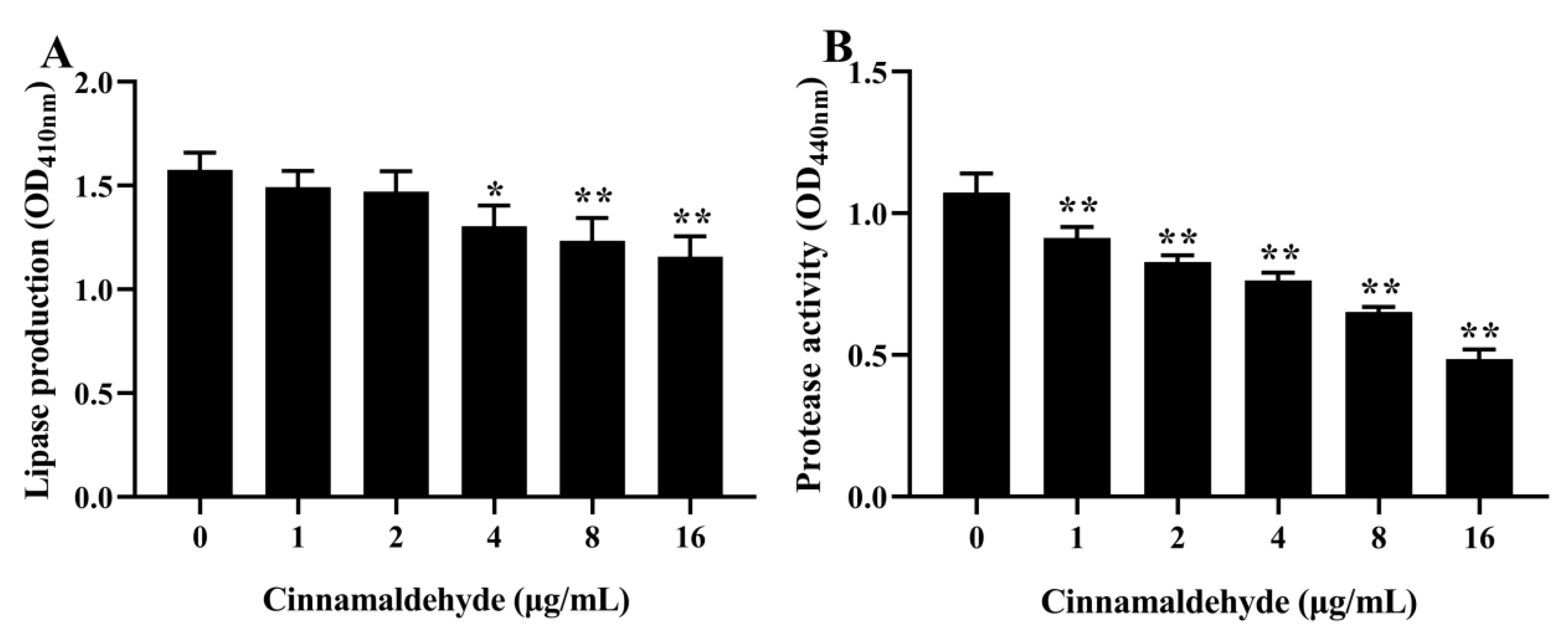
3.3. Inhibitory Effect on Lipase Production

Cinnamaldehyde could dose-dependently reduce lipase production of A. hydrophila when the concentrations of cinnamaldehyde range from 4 to 16 μg/mL (Figure 2A). Compared with the cinnamaldehyde-free group, lipase production reduced to 73.49 ± 5.91% when co-cultured with 16 μg/mL cinnamaldehyde.

3.4. Inhibitory Effect on Protease Activity

As shown in Figure 2B, cinnamaldehyde could dose-dependently inhibit the protease activity of A. hydrophila. Compared with the drug-free group, the protease activity decreased to 85.24 ± 5.59, 7.22 ± 2.47, 71.14 ± 2.19, 60.87 ± 2.16, and 45.28 ± 1.50%, plus cinnamaldehyde at concentrations ranging from 1 to 16 μg/mL.

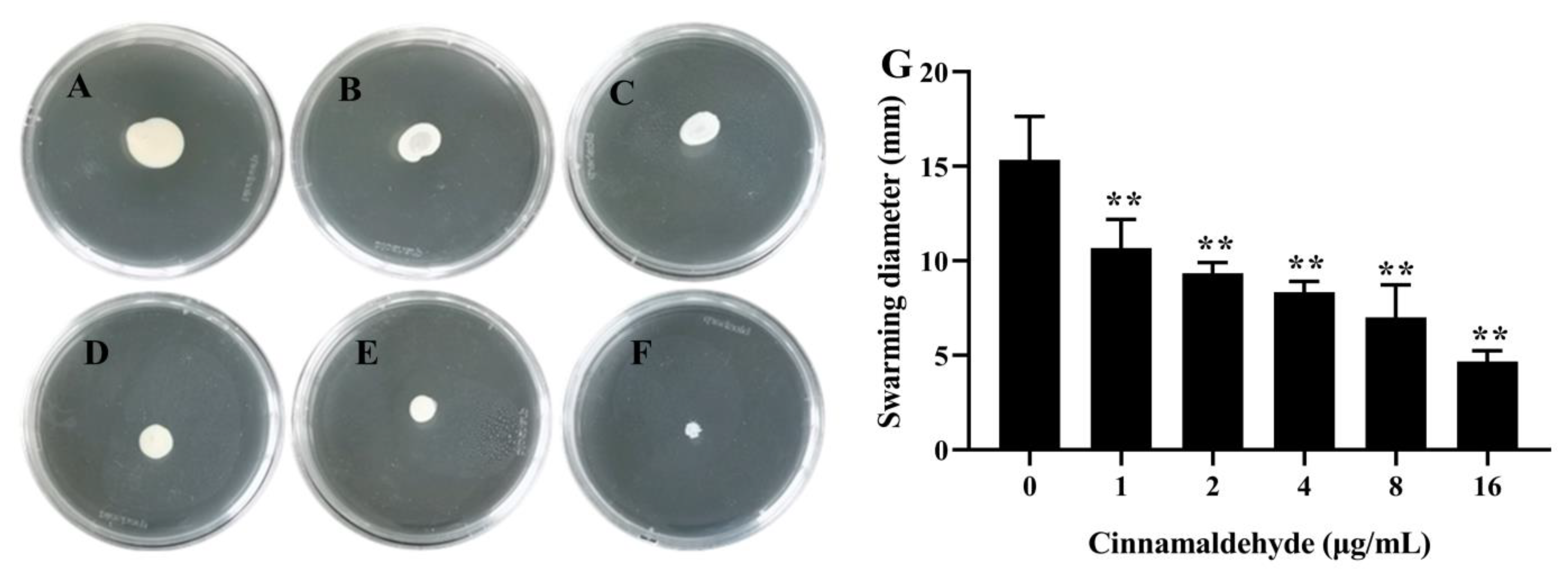
3.5. Inhibitory Effect of Cinnamaldehyde on Swarming Motility

As shown in Figure 3A, the swarming zone of A. hydrophila was visible on drug-free agar, while the swarming range became narrow following the addition of cinnamaldehyde at indicated concentrations (Figure 3B–F). Moreover, the swarming diameter was determined. As shown in Figure 3G, the swarming diameter decreased to 10.67 ± 1.25, 9.33 ± 0.47, 8.33 ± 0.47, 7.00 ± 1.41, and 4.67 ± 0.47 mm, and 15.33 ± 1.89 mm for the drug-free group. The swarming motility was significantly reduced when co-cultured with cinnamaldehyde at concentrations ranging from 1 to 16 μg/mL.

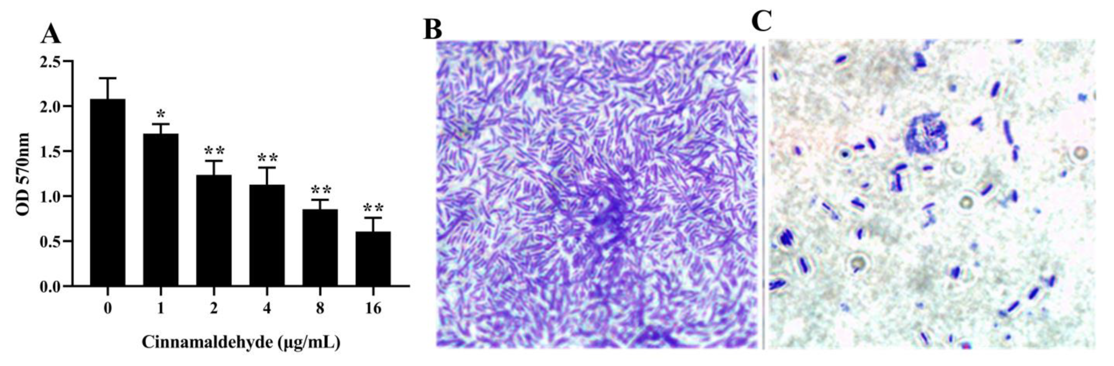
3.6. Inhibition of Biofilm Formation

As shown in Figure 4A, cinnamaldehyde could reduce A. hydrophila biofilm formation in a dose-dependent manner. The inhibition rates of biofilm formation ranged from 17.54 ± 11.62% to 70.26 ± 8.22%, as the drug concentration was increased from 1 to 16 μg/mL. Moreover, the biofilm on glass slides was observed by microscopy. As shown in Figure 4B, a large number of bacterial cells stained purple could be observed on a drug-free slide, while a few cells were presented in the 16 μg/mL drug group (Figure 4C).

3.7. Cinnamaldehyde Reduced the Transcription of Related Genes

The above results showed that cinnamaldehyde could reduce the production of virulence factors and biofilm formation, which are regulated by the QS system of A. hydrophila. Therefore, we determined the effect of cinnamaldehyde on aerolysin coding genes (aerA) and QS-related genes (ahyR, ahyI). As shown in Figure 5, the transcription of aerA gene was reduced to 5.58-fold under the 16 μg/mL drug compared with the drug-free group, and 13.48-fold and 8.35-fold for ahyI and ahyR, respectively. Moreover, statistical significance was observed in the lowest tested concentration of 1 μg/mL.

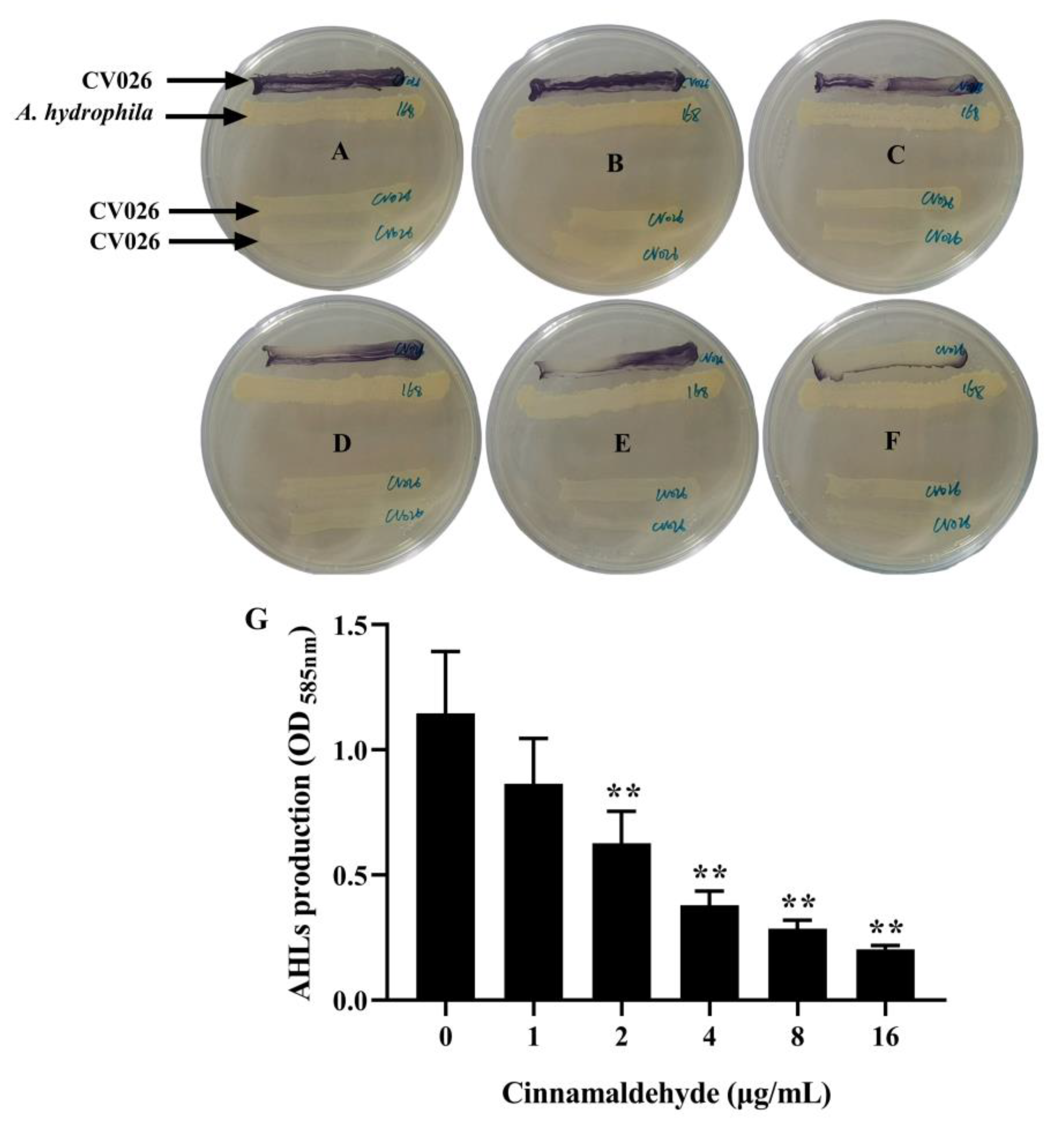
3.8. Cinnamaldehyde Reduced AHLs Production

qPCR results showed that cinnamaldehyde could downregulate the transcript of related genes, while AHLs are the main signal molecules in the QS system regulating the expression of related genes of A. hydrophila. The results indicated that cinnamaldehyde might inhibit AHLs production. Thus, bioreporter C. violaceum CV026 was used to investigate the production of AHLs of A. hydrophila after a co-incubation with indicated concentrations of cinnamaldehyde. As shown in Figure 6A, C. violaceum CV026 produced a visible purple color in the drug-free agar, while a little purple color was produced on 16 μg/mL of the drug agar (Figure 6F). Moreover, the violacein production of C. violaceum CV026 was quantitatively determined by releasing it in DMSO. As shown in Figure 6G, cinnamaldehyde could dose-dependently reduce violacein production in C. violaceum CV026, and statistical significance was observed in the 2 μg/mL drug group.

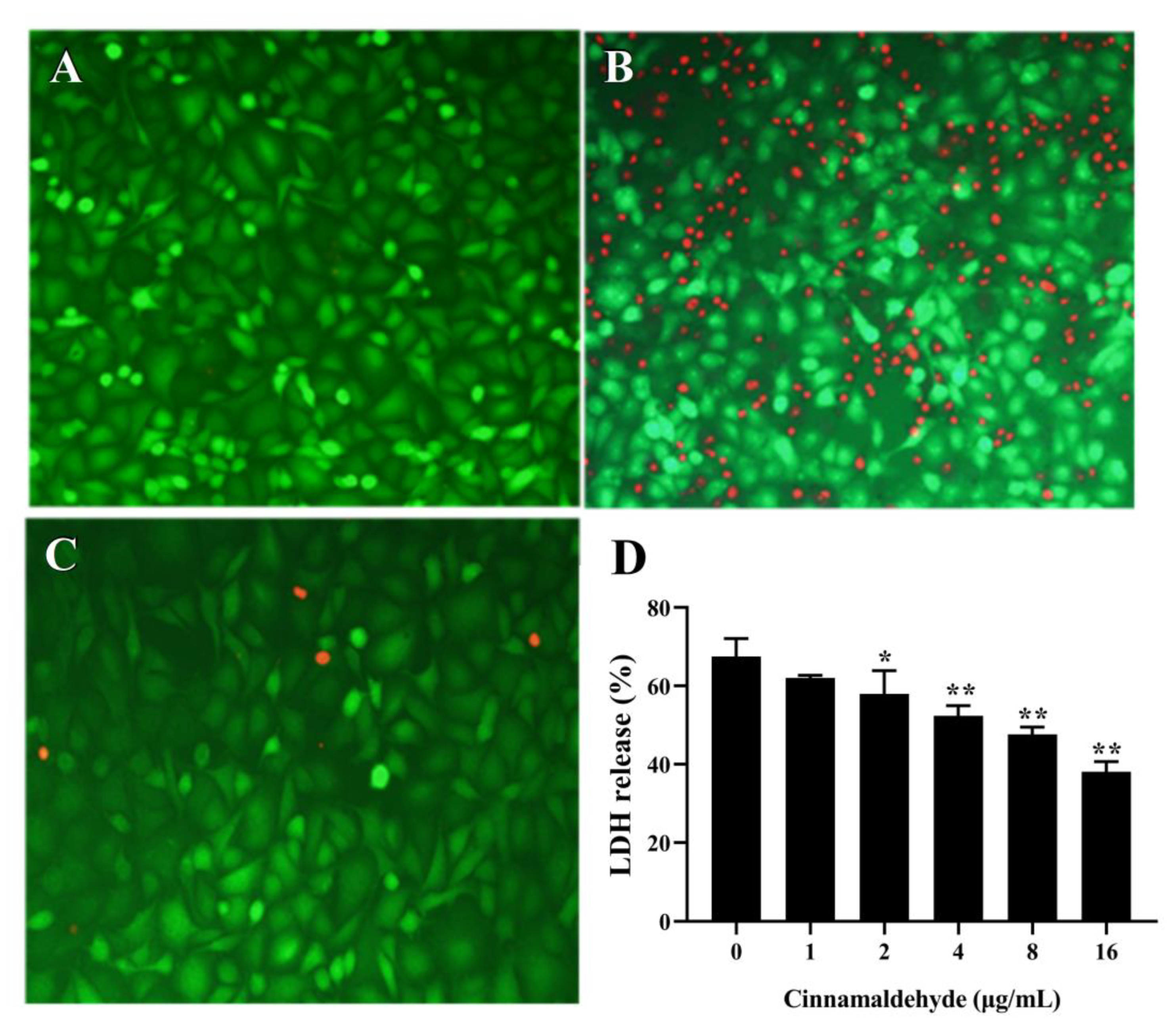
3.9. Cell Viability Results

As shown in Figure 7A, untreated cells were stained green indicating live cells, while cells after treatment with a drug-free supernatant indicating dead cells were stained red (Figure 7B). The ratio of dead cells in the visual field was obviously decreased compared with cells in the cinnamaldehyde-free group (Figure 7C). Moreover, LDH release results showed that LDH release reduced with increasing concentrations of cinnamaldehyde (Figure 7D). When co-cultured with 16 μg/mL drug-treated supernatant, LDH release decreased to 38.12 ± 2.12%, and to 67.50 ± 3.77% for the drug-free group.

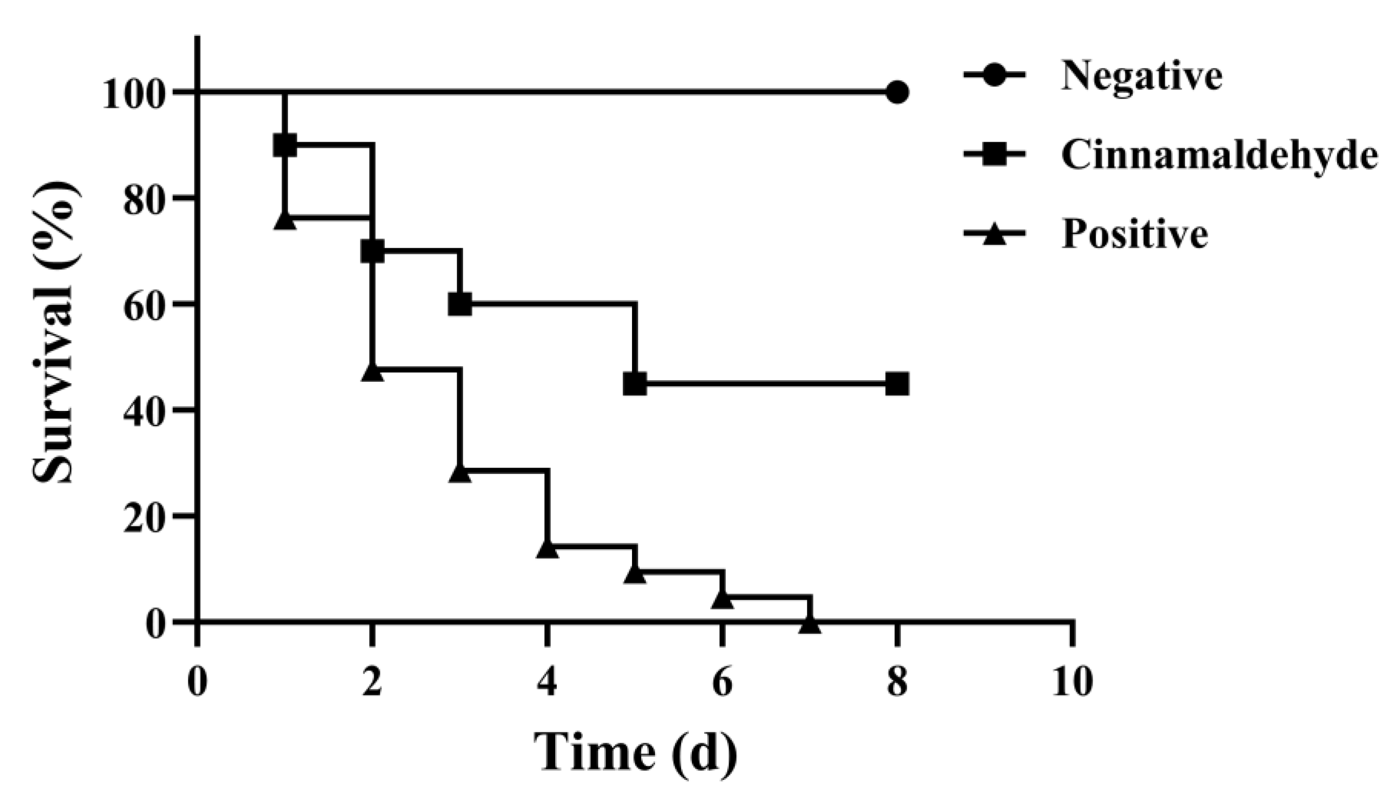
3.10. Protective Effect of Cinnamaldehyde on Crucian Carp Infected by A. hydrophila

The results above showed that cinnamaldehyde could significantly suppress the product of virulence factors and biofilm formation and provided protection to A549 cells against cell injury. The findings above revealed that cinnamaldehyde might have therapeutic effects on a fish model challenged with A. hydrophila, Therefore, the infection model of crucian carp was established. In the positive control group, swelling showed around the fins and ascites in the abdominal cavity. As shown in Figure 8, deaths occurred on the first day in the cinnamaldehyde-treated group and the positive control group. The mortality of fish in the positive control group was 100% in 7 days (Figure 8), and 45% of the cinnamaldehyde-treated group. The results showed statistical significance by log-rank test analysis. All fish were alive in the negative control group during the course.

4. Discussion

A. hydrophila threatens the healthy development of freshwater aquaculture [17]. Antibiotics are the primary measures for dealing with bacterial diseases in aquaculture, but the occurrence of resistance restricted the practice of antibiotics [18]. Therefore, new therapeutic drugs are needed for increasing bacterial diseases in aquaculture. Studies have demonstrated that herbal medicines have benefits in controlling bacterial infections [19]. A number of studies reported that cinnamaldehyde inhibited the growth of both Gram-positive and Gram-negative bacteria [20,21]. Sapna Rani et al. studied the in vitro antimicrobial activity of cinnamaldehyde using the agar disk diffusion method and showed that cinnamaldehyde could inhibit Streptococcus agalactiae at a concentration of 0.66 μg/mL [22]. Xing et al. found that natural cinnamaldehyde could be used as an alternative for fungicide in the field with a MIC of 50 mL/L for Fusarium verticillioides [23]. Cinnamaldehyde has been reported to inhibit the growth of bacteria from aquatic sources. Yin et al. found that the MIC of cinnamaldehyde was effective against the growth of A. hydrophila CW at 256 μg/mL by disrupting the cell membrane and impacting protein metabolism [24]. Barbara Rossi tested the inhibitory effect of cinnamaldehyde against two Vibrio spp.; the results showed that the MICs of cinnamaldehyde were effective against Vibrio harveyi and Vibrio anguillarum at 1.88 mM and 3.75 mM, respectively [25]. In our study, the MIC of cinnamaldehyde against A. hydrophila XS-91-4-1 was 128 μg/mL, which was about two times lower than that reported by Yin et al., and may be due to differences in strains [24].
A variety of naturally extracted compounds have anti-virulence effects on A. hydrophila. Jing et al. found that 8 μg/mL of genistein could significantly inhibit the hemolytic activity and biofilm formation of A. hydrophila [26]. Sun et al. showed that 25 μg/mL of esculetin could significantly reduce the biofilm formation of A. hydrophila, while in our study, 1 μg/mL of cinnamaldehyde could significantly reduce the biofilm formation of A. hydrophila [27]. Moreover, cinnamaldehyde has been reported as an inhibitor targeting bacterial virulence and this results in the reduction of the pathogenicity of a number of bacterial pathogens. Ferro et al. reported that cinnamaldehyde could diminish the hemolytic activity and adhere to the latex of Staphylococcus aureus (S. aureus) at sub-inhibitory concentrations of 0.125 mg/mL, which protected Galleria mellonella larvae from S. aureus infection [28]. Studies found that cinnamaldehyde could significantly inhibit the biofilms of Porphyromonas gingivalis and Streptococcus mutans at sub-MIC concentrations [29,30]. Li et al. demonstrated that cinnamaldehyde could interact with the LuxR-type protein of Pseudomonas fluorescens and result in the reduction of virulence factors [31]. Mary et al. found that uropathogenic Escherichia coli treated with 750 μM cinnamaldehyde could significantly downregulate the expression of the related virulence genes at subinhibitory concentrations, which could decrease the attachment and invasion of bacteria to urinary tract epithelial cells [32]. Moreover, cinnamaldehyde had the same inhibitory effect on the production of virulence factors and the expression of the virulence gene (aerA) of A. hydrophila in our study.
Gilles Brackman determined the ability of cinnamaldehyde to inhibit AI-2-based QS system in Vibrio harveyi; the results showed that cinnamaldehyde and its derivatives could interfere with AI-2 QS at concentrations without anti-bacterial growth and lead to a neutralizing virulence in Artemia shrimp [33]. However, the effect of cinnamaldehyde against the A. hydrophila QS system was not reported. Dong et al. found that A. hydrophila co-incubated with thymol could significantly downregulate QS-related genes ahyI and ahyR, and provide protection to channel catfish challenged with A. hydrophila [34]. The same methods were used by us and showed that cinnamaldehyde could significantly downregulate the QS-related genes ahyI and ahyR at the concentrations in our study. Moreover, QS mediated by AHLs regulates the pathogenicity of A. hydrophila strains [35]. C. violaceum CV026 is a bioreporter that can produce purple violacein in response to AHLs [36]. In our present study, cinnamaldehyde could reduce AHLs production of A. hydrophila by using the C. violaceum CV026. The results indicated that cinnamaldehyde could inhibit QS by inhibiting the expression of QS-related genes and the production of AHLs.
In aquaculture, the cinnamon essential oil has been shown to have antibacterial, anesthetic, growth-promoting, and antioxidant effects, indicating a promising use in aquaculture [37]. Moreover, Abdelhamed et al. found that feeding commercial feed mixed with 15 and 20 mg/kg of cinnamaldehyde could increase the survival rate of catfish infected with Edwardsiella ictaluri [38]. Faikoh et al. examined zebrafish infected with Streptococcus agalactiae and A. hydrophila. The zebrafish were immersed in water with 75 μL/L of liposome-encapsulated cinnamaldehyde, which significantly increased the survival rate of the zebrafish (31.1 ± 10.18% and 35.6 ± 3.85%) compared with the control group [39]. Our study showed a higher survival rate of 45% to crucian carp infected with A. hydrophila compared with the control group, which may be due to the method of administration and the species of fish. Taken together, our findings provide a candidate alternative drug for A. hydrophila infection.

5. Conclusions

Cinnamaldehyde could reduce the production of aerolysin, lipase, protease, and AHLs, and inhibit swarming motility and biofilm formation by interfering with the QS of A. hydrophila at sub-inhibitory concentrations. qPCR analysis indicated that cinnamaldehyde could downregulate the transcription of aerolysin coding genes (aerA) and QS-related genes (ahyR, ahyI). Moreover, significant therapeutic effects were shown in vitro and in vivo therapeutic assays. Taken together, cinnamaldehyde is a potential QS inhibitor against A. hydrophila infection.

Author Contributions

J.D. and X.A. designed the experiments. J.D., S.L. and S.Z. carried out the experiments, collected the data, and wrote this manuscript. Y.L., Y.Y. and N.X. checked and revised the manuscript. Q.Y., S.L. and S.Z. contributed data analysis. All authors have read and agreed to the published version of the manuscript.

Funding

This work is supported by the National Key R&D Program of China (No. 2019YFD0900104) and the China Agriculture Research System of MOF and MARA (CARS-46).

Institutional Review Board Statement

Animal studies were performed under the guidance of the Animal Welfare and Research Ethics Committee at the Yangtze River Fisheries Research Institute (Permission No. YFI-2022DJ-011, 10 June 2022).

Data Availability Statement

The data that support the findings of this study are available from the corresponding author upon request.

Conflicts of Interest

The authors declare no conflict of interest.

References

  1. Patwary, Z.P.; Ali, M.M.; Faruk, M.A.R. Evaluation of the status of use of chemicals and antibiotics in freshwater aquaculture activities with special emphasis to fish health management. J. Bangladesh Agric. Univ. 1970, 6, 381–390. [Google Scholar] [CrossRef] [Green Version]
  2. Smith, P. Antibiotics in aquaculture: Reducing their use and maintaining their efficacy. In Infectious Disease in Aquaculture; Woodhead Publishing: Cambridge, UK, 2012; pp. 161–189. [Google Scholar] [CrossRef]
  3. Saleh, A.A. Bacterial quorum sensing and biofilm formation. Bangladesh J. Med. Microbiol. 2014, 8, 1. [Google Scholar] [CrossRef]
  4. Zmyslowska, I.; Korzekwa, K.; Szarek, J. Aeromonas Hydrophila in Fish Aquaculture. J. Comp. Pathol. 2009, 141, 313. [Google Scholar] [CrossRef]
  5. Radosavljevi, V.; Irkovi, M.; Ljubojevi, D.; Novakov, N.; Milievi, V. Detection of Aerolysin (aerA) Gene in Aeromonas hydrophila Strains Isolated from Diseased Carp. Arch. Vet. Med. 2014, 8, 1. [Google Scholar] [CrossRef]
  6. Mamun, M.; Nasren, S.; Abhiman, P.B.; Rathore, S.S.; Shankar, K.M. Investigation of production, formation and characterization of biofilm cells of Aeromonas hydrophila for oral vaccination of fish. J. Exp. Zool. India 2019, 22, 1115–1123. [Google Scholar]
  7. Chu, W. Role of the quorum-sensing system in biofilm formation and virulence of Aeromonas hydrophila. Afr. J. Microbiol. Res. 2011, 5, 5819–5825. [Google Scholar] [CrossRef] [Green Version]
  8. Haripriya, D.; Nadhiya, K.; Vijayalakshmi, K. Antioxidant Potential of Cinnamaldehyde: An In vitro study. Int. J. Pharm. Res. Bio-Sci. 2013, 2, 270–278. [Google Scholar]
  9. Sagar, S.; Rani, S.; Sanusree, P.S.; Sru, K.V. Exploration of Targeting Mechanisms of Cinnamon Essential Oil against Water Borne Multiple Drug Resistant Isolate Aeromonas hydrophila C4. Res. Sq. 2021, 1–19. [Google Scholar] [CrossRef]
  10. Dong, J.; Ding, H.; Liu, Y.; Yang, Q.; Xu, N.; Yang, Y.; Ai, X. Magnolol protects channel catfish from Aeromonas hydrophila infection via inhibiting the expression of aerolysin. Vet. Microbiol. 2017, 211, 119–123. [Google Scholar] [CrossRef]
  11. Dong, J.; Zhang, L.; Liu, Y.; Xu, N.; Ai, X. Luteolin decreases the pathogenicity of Aeromonas hydrophila via inhibiting the activity of aerolysin. Virulence 2021, 12, 165–176. [Google Scholar] [CrossRef]
  12. Smith, P.K.; Krohn, R.I.; Hermanson, G.T.; Mallia, A.K.; Gartner, F.H.; Provenzano, M.D.; Fujimoto, E.K.; Goeke, N.M.; Olson, B.J.; Klenk, D.C. Measurement of protein using bicinchoninic acid. Anal. Biochem. 1985, 150, 76–85. [Google Scholar] [CrossRef] [PubMed]
  13. Ramanathan, S.; Kannan, R.D.; Sivasubramanian, S.; Arunachalam, K.; Lin, X. Anti-quorum Sensing and Protective Efficacies of Naringin Against Aeromonas hydrophila Infection in Danio rerio. Front. Microbiol. 2020, 11, 600622. [Google Scholar] [CrossRef]
  14. Qing, L.; Tengteng, X.; Jiahui, Z.; Huijuan, Z.; Zunying, L. Inhibition of Aeromonas hydrophila quorum sensing by extract from Zanthoxylum bungeanum. Chin. J. Bioprocess Eng. 2020, 18, 263–268. [Google Scholar]
  15. Patel, B.; Kumari, S.; Banerjee, R.; Samanta, M.; Das, S. Disruption of the quorum sensing regulated pathogenic traits of the biofilm-forming fish pathogen Aeromonas hydrophila by tannic acid, a potent quorum quencher. Biofouling 2017, 33, 580–590. [Google Scholar] [CrossRef]
  16. Jahid, I.K.; Han, N.; Ha, S.D. Inactivation kinetics of cold oxygen plasma depend on incubation conditions of Aeromonas hydrophila biofilm on lettuce. Food Res. Int. 2014, 55, 181–189. [Google Scholar] [CrossRef]
  17. Vignesh, S.; Krishnaveni, G.; Devaa, J.C.W.; Muthukumar, S.; Uthandakalaipandian, R. Experimental challenge of the freshwater fish pathogen Aeromonas hydrophila Ah17 and its effect on snakehead murrel Channa striata. Aquac. Int. 2022, 30, 1221–1238. [Google Scholar] [CrossRef]
  18. Caruffo, M.; Navarrete, P. Antibiotics in aquaculture: Impacts and alternatives. APUA Newsletter. 2015, 33, 4–7. [Google Scholar]
  19. Akinyemi, A. Efficacy of Traditional Herbs as an Alternative to Antibiotics Used in Aquaculture. J. Sci. Multidiscip. Res. 2013, 5, 43–51. [Google Scholar]
  20. Alsalim, H.A.A.; Shawkat, M.S.; Ibrahem, M. Antibacterial Activity of Cinnamomum Zeylanicum Bark Oil and Cinnamaldehyde on some Locally Isolated Pathogenic Bacteria. World J. Pharm. Res. 2017, 6, 174–185. [Google Scholar] [CrossRef]
  21. Awang, A.; Taher, M.; Susanti, D. The mode of antimicrobial action of Cinnamomum burmannii’s essential oil & cinnamaldehyde. J. Teknol. 2016, 78, 41–47. [Google Scholar] [CrossRef] [Green Version]
  22. Rani, S.; Verma, S.; Singh, H.; Ram, C. Antibacterial activity and mechanism of essential oils in combination with medium-chain fatty acids against predominant bovine mastitis pathogens. Lett. Appl. Microbiol. 2022, 74, 959–969. [Google Scholar] [CrossRef] [PubMed]
  23. Xing, F.; Hua, H.; Selvaraj, J.N.; Zhao, Y.; Zhou, L.; Liu, X.; Liu, Y. Growth inhibition and morphological alterations of Fusarium verticillioides by cinnamon oil and cinnamaldehyde. Food Control 2014, 46, 343–350. [Google Scholar] [CrossRef]
  24. Yin, L.; Chen, J.; Wang, K.; Geng, Y.; Ouyang, P. Study the antibacterial mechanism of cinnamaldehyde against drug-resistant Aeromonas hydrophila in vitro. Microb. Pathog. 2020, 145, 104208. [Google Scholar] [CrossRef] [PubMed]
  25. Barbara, R.; Angéles, E.M.; María, G.J.; Giulia, G.; Alberto, C.; Andrea, P.; Ester, G. Antimicrobial Power of Organic Acids and Nature-Identical Compounds against Two Vibrio spp.: An In Vitro Study. Microorganisms 2021, 9, 966. [Google Scholar] [CrossRef]
  26. Jing, D.; Defu, Z.; Jianrong, L.; Yongtao, L.; Shun, Z.; Yibin, Y.; Ning, X.; Qiuhong, Y.; Xiaohui, A. Genistein Inhibits the Pathogenesis of Aeromonas hydrophila by Disrupting Quorum Sensing Mediated Biofilm Formation and Aerolysin Production Front. Pharmacology 2021, 12, 753581. [Google Scholar] [CrossRef]
  27. Sun, B.; Luo, H.; Jiang, H.; Wang, Z.; Jia, A. Inhibition of Quorum Sensing and Biofilm Formation of Esculetin on Aeromonas hydrophila. Front. Microbiol. 2021, 12, 737626. [Google Scholar] [CrossRef]
  28. Ferro, T.A.; Araujo, J.M.; Dos Santos Pinto, B.L.; Dos Santos, J.S.; Souza, E.B.; da Silva, B.L.; Colares, V.L.; Novais, T.M.; Filho, C.M.; Struve, C.; et al. Cinnamaldehyde Inhibits Staphylococcus aureus Virulence Factors and Protects against Infection in a Galleria mellonella Model. Front. Microbiol. 2016, 7, 2052. [Google Scholar] [CrossRef] [Green Version]
  29. Wang, Y.; Zhang, Y.; Shi, Y.Q.; Pan, X.H.; Cao, P. Antibacterial effects of cinnamon (Cinnamomum zeylanicum) bark essential oil on Porphyromonas gingivalis. Microb. Pathog. 2018, 116, 26–32. [Google Scholar] [CrossRef]
  30. He, Z.; Huang, Z.; Jiang, W.; Zhou, W. Antimicrobial Activity of Cinnamaldehyde on Streptococcus mutans Biofilms. Front. Microbiol. 2019, 10, 2241. [Google Scholar] [CrossRef] [Green Version]
  31. Li, T.; Wang, D.; Liu, N.; Ma, Y.; Ding, T.; Mei, Y.; Li, J. Inhibition of quorum sensing-controlled virulence factors and biofilm formation in Pseudomonas fluorescens by cinnamaldehyde. Int. J. Food Microbiol. 2018, 269, 98–106. [Google Scholar] [CrossRef]
  32. Amalaradjou, M.A.; Narayanan, A.; Venkitanarayanan, K. Trans-cinnamaldehyde decreases attachment and invasion of uropathogenic Escherichia coli in urinary tract epithelial cells by modulating virulence gene expression. J. Urol. 2011, 185, 1526–1531. [Google Scholar] [CrossRef] [PubMed]
  33. Brackman, G.; Defoirdt, T.; Miyamoto, C.; Bossier, P.; Calenbergh, S.V.; Nelis, H.; Coenye, T. Cinnamaldehyde and cinnamaldehyde derivatives reduce virulence in Vibrio spp. by decreasing the DNA-binding activity of the quorum sensing response regulator LuxR. BMC Microbiol. 2008, 8, 149. [Google Scholar] [CrossRef] [PubMed] [Green Version]
  34. Dong, J.; Zhang, L.; Liu, Y.; Xu, N.; Zhou, S.; Yang, Q.; Yang, Y.; Ai, X. Thymol Protects Channel Catfish from Aeromonas hydrophila Infection by Inhibiting Aerolysin Expression and Biofilm Formation. Microorganisms 2020, 8, 636. [Google Scholar] [CrossRef] [PubMed]
  35. Jin, L.; Zhang, X.; Shi, H.; Wang, W.; Du, W. Identification of a Novel N-Acyl Homoserine Lactone Synthase, AhyI, in Aeromonas hydrophila and Structural Basis for Its Substrate Specificity. J. Agric. Food Chem. 2020, 68, 2516–2527. [Google Scholar] [CrossRef]
  36. Nasuno, E.; Okano, C.; Limura, K.; Morohoshi, T.; Ikeda, T.; Kato, N. Quick detection of cell to cell communication in gram negative bacteria by colour change of polymer matrix entrapping reporter bacteria. Mater. Res. Innov. 2014, 18, S4.879–S874.883. [Google Scholar] [CrossRef]
  37. Guerino, B.J.; Erbice, B.A.; Carine, d.F.S.; Nunes, D.S.; Liana, d.S.F.; Lenise, d.L.S.; Felipetto, C.J.; Bernardo, B. The Use of Cinnamon Essential Oils in Aquaculture: Antibacterial, Anesthetic, Growth-Promoting, and Antioxidant Effects. Fishes 2022, 7, 133. [Google Scholar] [CrossRef]
  38. Abdelhamed, H.; Ozdemir, O.; Ibrahim, I.; Lawrence, M.; Karsi, A. Antibacterial activities of trans-cinnamaldehyde, caprylic acid, and β-resorcylic acid against catfish pathogens. Aquaculture 2019, 504, 334–344. [Google Scholar] [CrossRef]
  39. Faikoh, E.N.; Hong, Y.H.; Hu, S.Y. Liposome-encapsulated cinnamaldehyde enhances zebrafish (Danio rerio) immunity and survival when challenged with Vibrio vulnificus and Streptococcus agalactiae. Fish Shellfish Immun. 2014, 38, 15–24. [Google Scholar] [CrossRef]
Figure 1. Inhibitory effects of cinnamaldehyde on hemolytic activity induced by aerolysin. (A) Chemical structure of cinnamaldehyde; (B) growth curves of A. hydrophila co-cultured with indicated concentrations of cinnamaldehyde; (C) effect of cinnamaldehyde on the hemolytic activity of bacterial supernatants; sheep erythrocytes treated with the bacterial supernatant of the drug-free group were used as the positive control; (D) detection of aerolysin production in bacterial supernatant plus cinnamaldehyde. The hemolysis assays in Figure 1C consist of data from three independent experiments. The results shown are the mean ± SD of the three experimental data compared to the drug-free group, **, p < 0.01.
Fishes 08 00122 g001
Figure 2. Inhibitory effect of cinnamaldehyde on two extracellular enzymes of A. hydrophila. (A) Lipase production of A. hydrophila plus cinnamaldehyde; (B) the effect of cinnamaldehyde on the protease activity of A. hydrophila. The data in both assays are mean ± SD of three independent experiments. *, 0.01 < p < 0.05 and **, p < 0.01 when compared with drug-free group.
Fishes 08 00122 g002
Figure 3. Effects of cinnamaldehyde on swarming motility. (A) DMSO control group; (B) 1 μg/mL; (C) 2 μg/mL; (D) 4 μg/mL; (E) 8 μg/mL; (F) 16 μg/mL; (G) swarming diameter. All data are presented as mean ± SD in the swarming diameter of three independent experiments. **, p < 0.01 when compared to the DMSO group.
Fishes 08 00122 g003
Figure 4. Effect of cinnamaldehyde on biofilm formation of A. hydrophila. (A) Determination of the cinnamaldehyde effect on bacterial biofilms, all data are the mean ± SD of three independent experiments. *, 0.01 < p < 0.05 and **, p < 0.01 when compared with drug-free group; (B) cinnamaldehyde-free group; (C) 16 μg/mL.
Fishes 08 00122 g004
Figure 5. The effects on related genes of A. hydrophila plus cinnamaldehyde. Three independent qPCR assays were performed for aerA, ahyI, and ahyR genes, and the data are mean ± SD. *, 0.01 < p < 0.05 and **, p < 0.01 when compared to drug-free group.
Fishes 08 00122 g005
Figure 6. Cinnamaldehyde reduces AHLs production by A. hydrophila. (A) Cinnamaldehyde-free group; (B) 1 μg/mL; (C) 2 μg/mL; (D) 4 μg/mL; (E) 8 μg/mL; (F) 16 μg/mL; (G) violacein production of C. violaceum CV026 in response to AHLs at indicated cinnamaldehyde concentrations, all data are the mean ± SD of three independent experiments. **, p < 0.01 when compared to the drug-free group.
Fishes 08 00122 g006
Figure 7. The protective effect of cinnamaldehyde on aerolysin-mediated cell injury. (A) Untreated cells; (B) cells treated with drug-free supernatant; (C) cells treated with 16 μg/mL cinnamaldehyde-treated bacterial supernatant; (D) LDH release of A549 cells co-cultured with cinnamaldehyde-treated bacterial supernatants; LDH assay was performed in triplicate; data were mean value ± SD. *, 0.01 < p < 0.05 and **, p < 0.01.
Fishes 08 00122 g007
Figure 8. Cinnamaldehyde can increase the survival rate of crucian carp infected with A. hydrophila. Infected crucian carp were administered with 25 mg/kg of cinnamaldehyde or sterile PBS every 12 h for 3 days; deaths were recorded for 8 days. Treatment with cinnamaldehyde had a significant protective effect on crucian carp infected with A. hydrophila when analyzed by log-rank test (p < 0.0001).
Fishes 08 00122 g008
Table 1. Primer pairs used in qPCR assay.
PrimerSequencePCR Amplicon (bp)Accession No.
aerA-FTCTACCACCACCTCCCTGTC218NC008570.1
aerA-RGACGAAGGTGTGGTTCCAGT
ahyI-FGTCAGCTCCCACACGTCGTT202CP000462.1
ahyI-RGGGATGTGGAATCCCACCGT
ahyR-FTTTACGGGTGACCTGATTGAG206CP000462.1
ahyR-RCCTGGATGTCCAACTACATCTT
16S rRNA-FTAATACCGCATACGCCCTAC164NR074841.1
16S rRNA-RACCGTGTCTCAGTTCCAGTG
Disclaimer/Publisher’s Note: The statements, opinions and data contained in all publications are solely those of the individual author(s) and contributor(s) and not of MDPI and/or the editor(s). MDPI and/or the editor(s) disclaim responsibility for any injury to people or property resulting from any ideas, methods, instructions or products referred to in the content.

