Article

Effects of Short-Term Intermittent Fasting on Growth Performance, Fatty Acids Profile, Glycolysis and Cholesterol Synthesis Gene Expression in European Seabass Dicentrarchus labrax

by
Olga Ntantali
1,*,
Emmanouil E. Malandrakis
2,
Wout Abbink
3,
John Bastiaansen
3,
Evanthia Chatzoglou
2,
Ioannis T. Karapanagiotidis
1,
Eleni Golomazou
1 and
Panagiota Panagiotaki
1
1
Laboratory of Aquaculture, Department of Ichthyology and Aquatic Environment, University of Thessaly, Fitokou Street, 38446 Volos, Greece
2
Laboratory of Applied Hydrobiology, Department of Animal Science, Agricultural University of Athens, Iera Odos 75, 11855 Athens, Greece
3
Animal Breeding & Genomics, Wageningen Livestock Research, Wageningen University and Research, Droevendaalsesteeg 1, 6708 PB Wageningen, The Netherlands
*
Author to whom correspondence should be addressed.
Fishes 2023, 8(12), 582; https://doi.org/10.3390/fishes8120582
Submission received: 30 September 2023 / Revised: 12 November 2023 / Accepted: 22 November 2023 / Published: 29 November 2023
(This article belongs to the Section Sustainable Aquaculture)

Abstract

:
The present study was applied to evaluate the effects of alternate feeding and feed restriction on gene expression, growth, proximate composition and biochemical indices in European seabass, Dicentrarchus labrax. Fish were randomly divided into six indoor tanks with 90 fish per tank in a recirculating aquaculture system. Two feeding strategies were applied, in which the first group was fed daily to satiation and the second was intermittently fed (8 days feeding to satiation–2 days starvation) for 40 days. At the end of the experiment, outlier fish were sorted as fast growers (FG) and slow growers (SG) according to their final body weight. The differential gene expression tested was related to glycolysis (pk, ldha, hk, g3pdh, eno1 and alda), fatty acid metabolism (lpl and acc) and cholesterol synthesis (7dhcr and sqle). In addition, muscle ldha and gpi expressions were positively correlated with fish weight. The concentrations of glucose, triglycerides, cholesterol and non-esterified fatty acids (NEFA) were not affected by the dietary treatments. Glucose and NEFA differed significantly between SG and FG fed groups. Overall, the physiological responses of glucose and fatty acid metabolism in fish, as recorded by gene expression assays, were triggered by minor interventions in feeding rather than the different growth rates. Expression of specific genes and biochemical parameters could be used as potential biomarkers to improve aquaculture practices and benefit fish husbandry through selective breeding, feeding strategies and farm management. The study provides new insights on the impact of intermittent feeding of European seabass, with gene markers and their potential effects, for European seabass aquaculture.
Key Contribution: Our study complements the expanding research regarding the potential effects of feed restriction on gene expression related to the nutritional status of European seabass. Quantification of specific genes and certain biochemical parameters may identify potential biomarkers to assess dietary status and/or fish growth.

1. Introduction

In the wild, fish regularly must deal with a lack of food and, consequently, have evolved mechanisms of coping with limited food availability. Most fish species can survive periods of fasting by altering their resting metabolic rate [1]. When food is limited, fish tend to reduce their physiological activity, which is upregulated again when there is an abundance of nourishment [2]. Feed restriction is tested in aquaculture settings to reduce feed utilization without compromising production by leveraging physiological adaptations of fish [3,4,5,6,7].
Fish nutrition plays a key role in the production cycle as it constitutes the most important growth factor and is the major operating cost of the aquaculture production. Enhanced growth has been targeted by aquaculture for many years; for example, fasting and refeeding regimes, which have been used to accelerate growth [8], have been extensively reviewed [9]. Compensatory growth, during a specific period of time, is considered significantly faster than the growth rate of fish that have not experienced feed deprivation [10]. Intermittent fasting has been proposed for achieving compensatory growth in various economically important fish species such as the Atlantic salmon (Salmo salar) [11,12], rainbow trout (Oncorhynchus mykiss) [10], Nile tilapia (Oreochromis niloticus) [13], European seabass (Dicentrarchus labrax) [3,4,6], gilthead seabream (Sparus aurata) [14,15], gibel carp (Carassius auratus gibelio) and Chinese longsnout catfish (Leiocassis longirostris) [16].
The nutritional status of fish influences the muscle growth pathways. Low food intake impairs growth proportionally to deprivation intensity and duration of fasting. Growth is regulated by hormones such as growth hormone (GH) in the central nervous system, which, in turn, controls feeding behavior [17]. In addition, many other physiological and environmental factors, such as fish density, food intake, temperature, diet formulation, size or sex, moderate the growth rate. During fasting, the catabolism of lipids and the release of fatty acids are of paramount importance for fish to cope with restriction in food intake. These fatty acids are transported across the plasma membrane into different tissues for oxidation or storage. Ad libitum feeding of fish tends to increase lipid accumulation in the viscera, liver and muscle, which are mobilized during fasting [18].
The role of many interacting environmental and genetic factors can explain the physiological routes of fish growth. An analytical approach to mRNA levels provides important and useful insights into growth performance in fish given that expression of specific genes could affect fish muscle growth [19]. Expression levels of specific mRNAs can explain variation in growth rates and therefore can be used as valid biomarkers for fish selection. Although fish husbandry could have an effect on gene expression, the basal regulation of various genes is strongly correlated with productive traits [20]. For example, in rainbow trout selected for fat muscle content, lipogenic genes were both affected by refeeding and selection [21]. Furthermore, muscle recovery was correlated with upregulation of genes related to RNA processing, translation and maturation of proteins, ribosome biogenesis, cell proliferation and mitochondrial bioenergetics at an early stage, while, in the later phase, several genes regulating Golgi and reticulum dynamics and genes involved in muscle remodeling were induced [22]. In European seabass, fasting activates lipolytic genes in the adipose tissue, liver and muscle; to the contrary, lipogenesis is downregulated in the liver and adipose tissue. Phospholipid- and oxidative-metabolism-related genes are differentially expressed in the liver and the skeletal muscle of fasted European sea bass, and their regulation returns back to normal 12 days after refeeding [23]. Fasting affects growth through direct inhibition of the GH/IGF axis, by decreasing IGF-I and IGF-II mRNA levels, which are restored during refeeding [24].
European seabass, Dicentrarchus labrax, is an important Mediterranean aquaculture species and is considered of great commercial importance [25]. European seabass (Dicentrarchus labrax) and gilthead seabream (Sparus aurata), with estimated productions of, respectively, 208,000 and 200,000 tons per year, are the most important aquaculture finfish species of the Mediterranean Sea [26]. European seabass accounts for 30% of the value of all the finfish species produced in the Mediterranean [27]. The present study aims to identify biochemical markers and target genes regulated by growth and nutritional status in short-term intermittent feeding of European seabass that can be used as potential biomarkers for breeding programs.

2. Materials and Methods

European seabass (Dicentrarchus labrax) juveniles were transferred from the fish farm Ecloserie Marine de Gravelines (Gravelines, France) to the CARUS research facility of Wageningen University and Research in the Netherlands. Fish were acclimatized to laboratory conditions before the start of the trial and fed on formulated diets (Altech Coppens, Helmond, The Netherlands) to fully satisfy the known nutritional requirements of the species. After acclimatization, 540 graded fish weighing 87.5 ± 16.2 g were uniformly dispersed to six cylindrical tanks of 1000 L in a recirculating aquaculture system (RAS). More specifically, fish were randomly distributed into two groups with three replicate tanks per group. Fish were hand fed with commercial 3 mm pellets twice daily (09:00 and 16:00). Fish of the first group (fed) were fed continuously to apparent satiation for 40 days, whereas the second group was fed in a feeding–fasting scheme (fasted) of 80–20% (2 days of fasting followed by 8 days of refeeding to apparent satiation) for 40 days. Water quality parameters were monitored and controlled, including NH4+, NO2, NO3, dissolved oxygen, temperature, salinity and pH. Fish were kept under a controlled light regime (12:12 h light/dark) at 21 °C. During sampling, fish were graded in two groups according to their final weight (FG—fast growers and SG—slow growers from both fed and fasted groups) and sacrificed by phenoxyethanol overdose (1 ppt). Blood from the caudal vein was drawn with heparinized syringes and centrifuged (4000 rpm for 10 min at 4 °C), and the plasma was separated and stored at −80 °C for biochemical analysis. Tissues from the liver and the white muscle were dissected and stored in RNAlater (Thermofisher Scientific, MA, USA) at −20 °C for downstream analysis. Whole carcasses were stored at −20 °C for proximate composition analysis and fatty acid analysis.
Growth parameters were calculated according to the following formulas:
Weight gain rate (WGR, %) = 100 × (final body weight-initial body weight)/initial body weight;
Specific growth rate (SGR, % day−1) = 100 × (Ln final body weight − Ln initial body weight)/number of days;
Condition factor (CF, g/cm3) = 100 × (body weight, g)/(body length, cm3);
Hepatosomatic index (HSI, %) = 100 × (liver weight, g)/(whole body weight, g);
Viscerosomatic index (VSI, %) = 100 × (viscera weight, g)/(whole body weight, g);
Intestine somatic index (ISI, %) = 100 × (intestine weight, g)/(body weight, g);
Spleen somatic index (SI, %) = 100 × (spleen weight, g)/(whole body weight, g);
Intraperitoneal fat rate (IFR, %) = 100 × (intraperitoneal fat weight, g)/(whole body weight, g).
Whole carcasses of nine fish per group were ground prior to proximate analysis, and moisture content was analyzed by drying the samples to a constant weight at 105 °C for 24 h. Crude fat was determined by the Soxhlet method, based on the extraction of the lipids, using a Soxtherm Multistat/SX PC (Sox-416 Macro, Gerhard, Germany) (AOAC, 1996). Crude protein content was determined using Kjeldahl method by measuring total nitrogen (N × 6.25; Βehr Labor-Technik, Germany). To determine ash quantity, samples were placed in a muffle furnace (Nabertherm L9/12/C6, Lilienthal, Germany) at 600 °C for 5 h. Gross energy was assessed adiabatically utilizing an IKA oxygen bomb calorimeter (C5000, IKA Werke, Staufen, Germany).
Serum glucose, cholesterol, triglycerides, non-esterified fatty acids (NEFA) and lactate dehydrogenase (LDH) activities were measured colorimetrically according to standard commercial protocols. Glucose (Cayman Chemical Company, Ann Arbor, MI, USA) values were extracted from absorbance at 505 nm using a standard curve in the range of 0–25 mg/dL. Triglycerides (Cayman Chemical Company) were quantified at 540 nm. LDH activity (Abcam, Cambridge, UK) was measured at 450 nm in a kinetic mode at 37 °C for 30 min while protected from light. Serum cholesterol was quantified with a commercial fluorometric assay kit (BIOSIS Ltd., Athens, Greece) with absorbance wavelength at 510 nm according to the manufacturer’s instructions. NEFA were measured enzymatically (Instruchemie B.V., Delfzijl, the Netherlands) at 540 nm at 37 °C for 4.5 min. All biochemical analyses were carried out with a BMG Fluostar Omega microplate reader with different set-ups. To determine fatty acids profiles, the Folch extraction method was used, followed by esterification and separation using a capillary column in an Agilent GC 6890.
Levene and Shapiro–Wilk tests were used to assess homogeneity of variance and normality, respectively. Data were tested with nested ANOVA where growth (random factor) was nested within treatment (fixed factor).
Total RNA was extracted with Nucleospin®RNA (Macherey-Nagel, Düren, Germany) according to the manufacturer’s protocol. Isolated RNA quantity and quality were determined via spectrophotometry, using a Quawell Q5000 UV–Vis spectrophotometer and on a 1% denaturing agarose gel, respectively. For cDNA synthesis, 500 ng of RNA was reverse transcribed with PrimeScript RT Reagent Kit according to the manufacturer’s instructions, diluted 10-fold and stored at −20 °C. The qPCR analysis was carried out with RT2 SYBR Green qPCR Mastermix (Qiagen) in a Rotor-Gene Q 5plex using 1ml of cDNA per reaction and 300 nM of each primer in a final volume of 10 mL. The qPCR assay was designed for the profiling of six glycolytic genes (hk, gpi, eno1, g3pdh, alda, pk), two genes related to fatty acid metabolism (lpl and acc) and two genes involved in cholesterol biosynthesis (sqle, 7dhcr). The primers designed for the qPCR experiment are presented in Table 1. β-actin (actb) and elongation factor 1 (ef1) were used as reference genes (geometric mean). Each sample was analyzed for primer dimer, contamination or mispriming with melting curve analysis and 2.5% agarose gel electrophoresis. Relative gene expression was determined with the Pfaffl method (REST) [28], and fold change (FC) values were transformed to log2FC.

3. Results

3.1. Growth Performance and Proximate Composition

Fasted and fed fish exhibited a feeding rate of 2.5 ± 0.1% and 2.9 ± 0.1%, and the FCR was calculated at 2.98 ± 0.2 and 3.23 ± 0.3, respectively. Growth performance body indices and proximate composition of the European seabass fed with two feeding regimes and divided into weight groups (FG and SG) are presented in Table 2. The experimental feeding regime did not significantly affect final body weight of either FG or SG groups. In general, all the somatometric indices were not significantly affected by short-term fasting, except for the ISI. In addition, CF and IFR were substantially higher in the FG group than the SG group (p < 0.001), in both fasted and fed fish. Fasting did not affect proximate composition.
All fasted fish, both SG and FG, had similar (p > 0.05) proximate composition, but moisture, crude protein, crude lipid and ash contents were significantly increased in SG compared to FG fish.

3.2. Biochemical Parameters

The values of plasma glucose, triglycerides, cholesterol, non-esterified fatty acids (NEFA) and LDH activity are presented in Table 3. Significantly lower NEFA levels were found in FG compared to SG fish, and the levels of triglycerides and cholesterol were higher in fed fish (p < 0.05). On the other hand, growth did not affect their blood concentration levels.

3.3. Fatty Acids (FA) Profiles

Nutritional status did not significantly affect FA profiles. On the other hand, growth significantly affected the FA profile since most of the FAs (10 out 16) showed different carcass concentrations between growth groups. The lipids of SG contained significantly lower proportions of 18:2n-6, 18:3n-3 and Σn-6 and higher proportions of 14:0, 18:4n-3, 20:1n-9, 22:1n-11, EPA and DHA compared to the FG (Table 4).

3.4. Gene Expression

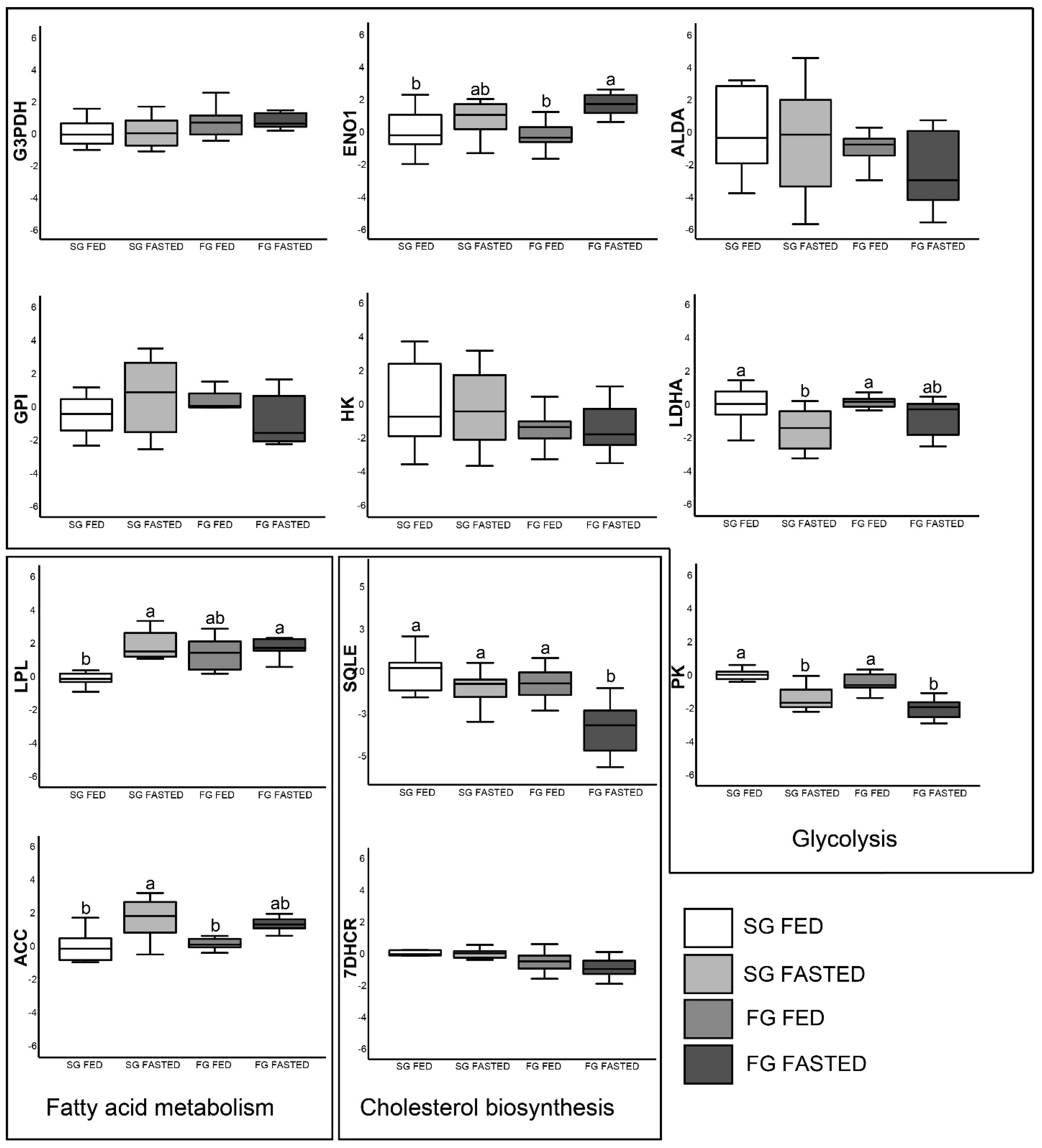
Differential gene expression (log2FC) among the different treatments and fish groups is presented in Figure 1 for muscle tissue and Figure 2 for liver. In muscle, all genes demonstrated significant differential expression among fish group treatments, while, in the liver, 5 genes (g3pdh, alda, gpi, hk, 7dhcr) out of 11 exhibited no significant difference (p > 0.05). Transcription levels of g3pdh, alda, gpi and ldha showed an increased trend in fed FG compared to fed SG, although such a trend was not observed for fasted fish groups. Furthermore, mRNA levels of g3pdh, hk, pk, lpl, acc, sqle and 7dhcr were significantly different (p < 0.05) between fish groups with different dietary statuses (fed versus fasted), regardless of the growth group (SG or FG). In the liver, only pk was significantly upregulated (p < 0.05) in fed fish compared with the fasted ones, for both growth groups. ldha, lpl and acc mRNA levels were differentially expressed between fed and fasted fish, only for the SG group.

4. Discussion

In aquaculture, feeding strategies are of critical importance as feeds and feeding represent 57–59% of the production cost [29], and both underfeeding and overfeeding can have negative consequences for the production [30,31,32]. Thus, successful farming strongly relies on feed management [3]. Many feeding methods are being practiced, such as ad libitum feeding, restricted feeding and intermittent feeding, to achieve the best feeding scheme for each species and developmental stage [33]. A key factor in view of obtaining the desired growth compensation after feed deprivation for a specific species is to manage the appropriate duration and intensity of feed deprivation.
In the present study, the growth performance (final weight and length, WGR and SGR) within the fast growers (FG) and slow growers (SG) groups was not significantly affected by the short-term food deprivation periods. Any potential loss of weight during the 2-day (out of 10 days) fasting period was compensated during the refeeding-to-satiation periods. Under the current experimental conditions, feed restriction for 2 days followed by an 8-day refeeding period seemed to be an adequate management strategy for European seabass. Both duration and intensity of feed restriction in compensatory growth responses need to be further evaluated to be incorporated in commercial production of the species.
This short-term intermittent fasting did not have a substantial effect either on condition factors or other morphometric indices. It is noteworthy that intraperitoneal fat (IFR) was significantly higher in FG than SG, independently of the dietary regime followed. This was due to intraperitoneal fat accumulation in the FG fish, which is not a desirable trait, supported by the fact that these fish (either fed or fasted) had also an increased body lipid content, although this was not significant, compared to the SG fish. Also, no significant difference was observed in any parameter tested between fasted SG and fasted FG groups concerning the whole-body proximate composition. Similar results were reported in other studies, where there was no significant difference in the proximate composition of the fish under different feeding regimes [34] or feeding ratios/feeding rates [35,36,37]. In addition, in the present study, whole-body protein, lipid, moisture, dry matter and ash were greatly reduced in fed FG groups compared to fed SG. The values of protein, moisture and ash were not found to be statistically different in terms of nutritional composition, but only lipid values were affected by the feeding strategies in barramundi (Lates calcarifer) experiencing starvation and refeeding cycles [38]. Also, other studies reported that the body lipid content of fish decreased proportionally to feed consumption [39,40]. The proximate composition of fish generally fluctuates with food quantity, with lipid deposition likely to be increased with elevated food supply [41]. It is a fact that utilization of lipid stores is the most usual response of fish subjected to feed deprivation followed by refeeding in order to satisfy energy requirements [15,42,43,44,45]. In the present study, the deprivation period was short and did not substantially affect fatty acids (FAs) contents of the fish. FAs such as 20:1n-9 and 22:1n-11 are heavily catabolized for energy in the growth of farmed fish; therefore, FG tend to have lower amounts of these FAs. The same fact stands for EPA, which can be readily β-oxidized, and DHA, which requires peroxisomal and mitochondrial β-oxidation. Through this process, these FAs serve as energy deposits in fish if necessary for homeostasis [46].
Blood biochemical indices of reared fish are determined to evaluate the physiological status of fish [47] and to investigate conditions that might lead to adaptations due to the their nutritional status [48]. The biochemical features of fish blood could be adequate to monitor the metabolic balance and health status in intensive aquaculture conditions [49]. In this study, we used the biochemical profile of D. labrax to assess the physiological status during short-term restricted feeding. Differences between fed and fasted groups were not observed, a fact that supports the hypothesis that short-term intermittent fasting (2 days out of 10) does not affect fish biochemical indices in the long term. Some biochemical parameters were significantly different between SG and FG fish, and thus were related to growth, within the fasted or fed group. During short-term starvation of seabass, the prediction and prognosis of the nutritional or physiological status, measured by biochemical blood parameters, seem to have potential as useful tools which could manage and monitor feeding practices during production of the species [48,50,51,52].
Plasma cholesterol concentration was not affected, possibly due to the short-term nature of the fasting period, in contrast to more prolonged periods of fasting where differences in plasma cholesterol do occur [3]. LDH activity in plasma was not affected either by fasting or different growth, although it has been established that LDH activity in muscle increases in larger fish [53,54,55,56,57,58]. Although plasma LDH activity was considered as equal among groups, significant differences in plasma glucose and NEFA content between FG and SG imply growth-related divergence in glucose and lipid metabolism. LDH is a key enzyme of anaerobic glycolysis that catalyzes pyruvate reduction to lactate and has been used as a dietary status biomarker even for wild fish populations [59].
A higher conversion rate of glucose into pyruvate in muscle to increase growth rate is depicted by increased expression of gpi, alda and g3pdh. In turbot, Scophthalmus maximus, a comparison between fast- and slow-growing fish revealed a set of 16 upregulated genes in muscle coding for enzymes of the glycolytic pathway [60]. Larger rainbow trout individuals possessed many transcripts with functions in glycolysis and gluconeogenesis, as well as lipid metabolism, compared to smaller individuals [61]; this upregulation has been associated with an increased muscle energy demand in fast-growing fish. Glucose homeostasis in general is critical for the fish in order to meet these demands [62]. When rainbow trout juveniles were subjected to high dietary carbohydrate first feeding, glucose metabolism glucose transport and glycolysis-related genes were upregulated, and their expression was permanently modified, demonstrating efficient programming of the glucose metabolism [63]. Therefore, gpi, alda and g3pdh muscle expression can be used as a reliable biomarker for growth estimation of fish individuals.
Pk and ldha gene expression in liver was suppressed after 40 days of intermediate fasting. Both enzymes play an essential role in glucose homeostasis, with PK being the final enzyme for ATP production and pyruvate production, while LDHA regulates interconversion of pyruvate and l-lactate. Therefore, these enzymes are strongly inter-related, and their gene expression is positively correlated. Glycolysis and gluconeogenesis are metabolic processes that degrade and synthesize glucose and are essential for fish survival. The gluconeogenic pathway, which results in the generation of glucose, uses ATP that is supplied from NEFA catabolism [64] and may be associated with increased feed intake [65]. G3PDH and LDH are significantly downregulated under prolonged hypoxia stress [66]. Significant feed deprivation has been shown to have a profound downregulating effect in genes involved in glycolysis [67]. Among others, glycolysis genes are differentially regulated between growth hormone transgenic and wild-type coho salmon during periods of long-term food shortage [68].
Cholesterol synthesis genes (7dhcr and sqle) were differentiated by dietary status. In Atlantic salmon, dietary substitution of fish oil with vegetable oils regulated cholesterol biosynthesis genes such as 7dhcr and sqle [69]. Furthermore, liver transcriptome and tissue lipid composition are driven by cholesterol synthesis upregulation in fish fed with animal by-products and vegetable-based diets [70]. In tilapia, cholesterol biosynthesis and fatty acid metabolism genes have been associated with growth [71]. Cholesterol biosynthetic gene expression was not associated with plasma cholesterol, which did not exhibit differences among groups.
Positive correlation of growth and ldha gene expression implies the promotion of accelerated cell growth and replication. A positive correlation of LDH activity and growth has been demonstrated in the past for salmon [72] and rainbow trout [73]. Furthermore, transgenic fish that perform better in terms of growth exhibit increased LDH activity [74]. Although, correlations between gene expression and weight could be more complex than those for ldh and gpi, these correlations depict glycolytic rates.
Research on mRNA quantification may identify potential biomarkers and use gene expression profiles either to assess dietary status or predict fish growth. Results of the present study showed that body weight growth of Dicentrarchus labrax was not affected by repeated intermitted fasting for 40 days. The study provides new insights on the impact of intermittent feeding of European seabass, with gene markers and their potential effects, for European seabass aquaculture. Expression of specific genes and blood biochemical parameters could be used as potential biomarkers to improve aquaculture practices and benefit fish husbandry through selective breeding, feeding strategies and farm management.

5. Conclusions

Research on mRNA quantification may identify potential biomarkers and use gene expression profiles either to assess dietary status or predict fish growth. Results of the present study showed that body weight growth of Dicentrarchus labrax was not affected by repeated intermitted fasting. The study provides new insights on the impact of intermittent feeding of European seabass, with gene markers and their potential effects, for European seabass aquaculture. Expression of specific genes and blood biochemical parameters could be used as potential biomarkers to improve aquaculture practices and benefit fish husbandry through selective breeding, feeding strategies and farm management.

Author Contributions

Conceptualization, E.E.M. and P.P.; Data curation, O.N. and E.E.M.; Funding acquisition, W.A. and P.P.; Methodology, E.E.M., W.A. and J.B.; Project administration, W.A.; Supervision, E.E.M., W.A. and P.P.; Visualization, O.N., E.E.M. and W.A.; Writing—original draft, O.N. and E.E.M.; Writing—review and editing, E.E.M., W.A., J.B., E.C., I.T.K., E.G. and P.P. All authors have read and agreed to the published version of the manuscript.

Funding

The current work was funded by the AQUAEXCEL2020 project (grant agreement no. 652831) under the TNA program GeneComp (project ID AE120002) granted to O.N. and E.E.M.

Institutional Review Board Statement

Animal manipulations for this experiment were carried out by legally permitted and competent personnel in accordance with the European Directive (2010/63/EU) and approved by the Dutch Central Committee for Animal Experimentation (CCD) under the license number AVD1040020197805 on 27 May 2019 and by the Animal Experimental Committee of Wageningen University and Research.

Informed Consent Statement

Not applicable.

Data Availability Statement

Data is contained within the article.

Acknowledgments

The authors thankfully acknowledge the staff of CARUS from Wageningen University and Research for technical support and fish husbandry. Aristeidis Tsopelakos and Antonios Tsakiris are kindly thanked for their assistance on gas chromatography and FA profiling.

Conflicts of Interest

The authors declare no conflict of interest.

References

  1. Fu, S.J.; Xie, X.J.; Cao, Z.D. Effect of Fasting on Resting Metabolic Rate and Postprandial Metabolic Response in Silurus Meridionalis. J. Fish Biol. 2005, 67, 279–285. [Google Scholar] [CrossRef]
  2. Ng, W.K.; Lu, K.S.; Hashim, R.; Ali, A. Effects of Feeding Rate on Growth, Feed Utilizationand Body Composition of a Tropical Bagrid Catfish. Aquac. Int. 2000, 8, 19–29. [Google Scholar] [CrossRef]
  3. Chatzifotis, S.; Papadaki, M.; Despoti, S.; Roufidou, C.; Antonopoulou, E. Effect of Starvation and Re-Feeding on Reproductive Indices, Body Weight, Plasma Metabolites and Oxidative Enzymes of Sea Bass (Dicentrarchus labrax). Aquaculture 2011, 316, 53–59. [Google Scholar] [CrossRef]
  4. Rubio, V.C.; Sánchez, E.; Cerdá-Reverter, J.M. Compensatory Feeding in the Sea Bass after Fasting and Physical Stress. Aquaculture 2010, 298, 332–337. [Google Scholar] [CrossRef]
  5. McKenzie, D.J.; Vergnet, A.; Chatain, B.; Vandeputte, M.; Desmarais, E.; Steffensen, J.F.; Guinand, B. Physiological Mechanisms Underlying Individual Variation in Tolerance of Food Deprivation in Juvenile European Sea Bass, Dicentrarchus labrax. J. Exp. Biol. 2014, 217, 3283–3292. [Google Scholar] [CrossRef]
  6. Adaklı, A.; Taşbozan, O. The Effects of Different Cycles of Starvation and Refeeding on Growth and Body Composition on European Sea Bass (Dicentrarchus labrax). Turkish J. Fish. Aquat. Sci. 2015, 15, 425–433. [Google Scholar] [CrossRef]
  7. Daulé, S.; Vandeputte, M.; Vergnet, A.; Guinand, B.; Grima, L.; Chatain, B. Effect of Selection for Fasting Tolerance on Feed Intake, Growth and Feed Efficiency in the European Sea Bass Dicentrarchus labrax. Aquaculture 2014, 420–421, S42–S49. [Google Scholar] [CrossRef]
  8. Hornick, J.L.; Van Eenaeme, C.; Gérard, O.; Dufrasne, I.; Istasse, L. Mechanisms of Reduced and Compensatory Growth. Domest. Anim. Endocrinol. 2000, 19, 121–132. [Google Scholar] [CrossRef]
  9. Ali, M.; Nicieza, A.; Wootton, R.J. Compensatory Growth in Fishes: A Response to Growth Depression. Fish Fish. 2003, 4, 147–190. [Google Scholar] [CrossRef]
  10. Nikki, J.; Pirhonen, J.; Jobling, M.; Karjalainen, J. Compensatory Growth in Juvenile Rainbow Trout, Oncorhynchus Mykiss (Walbaum), Held Individually. Aquaculture 2004, 235, 285–296. [Google Scholar] [CrossRef]
  11. Stefansson, S.O.; Imsland, A.K.; Handeland, S.O. Food-Deprivation, Compensatory Growth and Hydro-Mineral Balance in Atlantic Salmon (Salmo salar) Post-Smolts in Sea Water. Aquaculture 2009, 290, 243–249. [Google Scholar] [CrossRef]
  12. Nicieza, A.G.; Metcalfe, N.B. Growth Compensation in Juvenile Atlantic Salmon: Responses to Depressed Temperature and Food Availability. Ecology 1997, 78, 2385–2400. [Google Scholar] [CrossRef]
  13. Ali, T.E.S.; Martínez-Llorens, S.; Moñino, A.V.; Cerdá, M.J.; Tomás-Vidal, A. Effects of Weekly Feeding Frequency and Previous Ration Restriction on the Compensatory Growth and Body Composition of Nile Tilapia Fingerlings. Egypt. J. Aquat. Res. 2016, 42, 357–363. [Google Scholar] [CrossRef]
  14. Bavčević, L.; Klanjšček, T.; Karamarko, V.; Aničić, I.; Legović, T. Compensatory Growth in Gilthead Sea Bream (Sparus aurata) Compensates Weight, but Not Length. Aquaculture 2010, 301, 57–63. [Google Scholar] [CrossRef]
  15. Peres, H.; Santos, S.; Oliva-Teles, A. Lack of Compensatory Growth Response in Gilthead Seabream (Sparus aurata) Juveniles Following Starvation and Subsequent Refeeding. Aquaculture 2011, 318, 384–388. [Google Scholar] [CrossRef]
  16. Zhu, X.; Xie, S.; Zou, Z.; Lei, W.; Cui, Y.; Yang, Y.; Wootton, R.J. Compensatory Growth and Food Consumption in Gibel Carp, Carassius Auratus Gibelio, and Chinese Longsnout Catfish, Leiocassis longirostris, Experiencing Cycles of Feed Deprivation and Re-Feeding. Aquaculture 2004, 241, 235–247. [Google Scholar] [CrossRef]
  17. Canosa, L.F.; Bertucci, J.I. Nutrient Regulation of Somatic Growth in Teleost Fish. The Interaction between Somatic Growth, Feeding and Metabolism. Mol. Cell. Endocrinol. 2020, 518, 111029. [Google Scholar] [CrossRef]
  18. Jezierska, B.; Hazel, J.R.; Gerking, S.D. Lipid Mobilization during Starvation in the Rainbow Trout, Salmo gairdneri Richardson, with Attention to Fatty Acids. J. Fish Biol. 1982, 21, 681–692. [Google Scholar] [CrossRef]
  19. Johnston, I.A.; Bower, N.I.; Macqueen, D.J. Growth and the Regulation of Myotomal Muscle Mass in Teleost Fish. J. Exp. Biol. 2011, 214, 1617–1628. [Google Scholar] [CrossRef]
  20. Fuller, S.A.; Beck, B.H.; McEntire, M.E.; Peatman, E.; Abernathy, J.; Bahler, J. Heritability of Growth Traits and Correlation with Hepatic Gene Expression among Hybrid Striped Bass Exhibiting Extremes in Performance. Cogent Biol. 2018, 4, 1453319. [Google Scholar] [CrossRef]
  21. Skiba-Cassy, S.; Lansard, M.; Panserat, S.; Médale, F. Rainbow Trout Genetically Selected for Greater Muscle Fat Content Display Increased Activation of Liver TOR Signaling and Lipogenic Gene Expression. Am. J. Physiol. Regul. Integr. Comp. Physiol. 2009, 297, 1421–1429. [Google Scholar] [CrossRef] [PubMed]
  22. Rescan, P.Y.; Montfort, J.; Rallière, C.; Le Cam, A.; Esquerré, D.; Hugot, K. Dynamic Gene Expression in Fish Muscle during Recovery Growth Induced by a Fasting-Refeeding Schedule. BMC Genom. 2007, 8, 438. [Google Scholar] [CrossRef] [PubMed]
  23. Rimoldi, S.; Benedito-Palos, L.; Terova, G.; Pérez-Sánchez, J. Wide-Targeted Gene Expression Infers Tissue-Specific Molecular Signatures of Lipid Metabolism in Fed and Fasted Fish. Rev. Fish Biol. Fish 2016, 26, 93–108. [Google Scholar] [CrossRef]
  24. Terova, G.; Rimoldi, S.; Chini, V.; Gornati, R.; Bernardini, G.; Saroglia, M. Cloning and Expression Analysis of Insulin-like Growth Factor I and II in Liver and Muscle of Sea Bass (Dicentrarchus labrax, L.) during Long-Term Fasting and Refeeding. J. Fish Biol. 2007, 70, 219–233. [Google Scholar] [CrossRef]
  25. Bjørndal, T.; Guillen, J.; Rad, F. Are Farmed European Seabass (Dicentrarchus labrax) Prices in European Union Markets Affected by Turkish Exports of Farmed European Seabass? Aquac. Econ. Manag. 2019, 23, 341–357. [Google Scholar] [CrossRef]
  26. FEAP. European Aquaculture Production Report; IEMed: Barcelona, Spain, 2021; Volume 2020. [Google Scholar]
  27. Carvalho, N.; Guillen, J. Aquaculture in the Mediterranean; IEMed: Barcelona, Spain, 2021. [Google Scholar]
  28. Pfaffl, M.W. A New Mathematical Model for Relative Quantification in Real-Time RT-PCR. Nucleic Acids Res. 2001, 29, e45. [Google Scholar] [CrossRef] [PubMed]
  29. Hellenic Aquaculture Producers Organisation. Greek Aquaculture: Annual Report 2022; HAPO: Athens, Greece, 2023. [Google Scholar]
  30. Yılmaz, H.A.; Eroldogan, O.T. Combined Effects of Cycled Starvation and Feeding Frequency on Growth and Oxygen Consumption of Gilthead Sea Bream, Sparus aurata. J. World Aquac. Soc. 2011, 42, 522–529. [Google Scholar] [CrossRef]
  31. Mccarthy, I.D.; Carter, C.G.; Houlihan, D.F. The Effect of Feeding Hierarchy on Individual Variability in Daily Feeding of Rainbow Trout, Oncorhynchus Mykiss (Walbaum). J. Fish Biol. 1992, 41, 257–263. [Google Scholar] [CrossRef]
  32. Talbot, C.; Corneillie, S.; Korsøen, Ø. Pattern of Feed Intake in Four Species of Fish under Commercial Farming Conditions: Implications for Feeding Management. Aquac. Res. 1999, 30, 509–518. [Google Scholar] [CrossRef]
  33. Lovell, T. Fish Nutrition and Feeding Experiments. In Nutrition and Feeding of Fish; Springer: New York, NY USA, 1998; pp. 123–134. [Google Scholar]
  34. Cho, S.H.; Lee, S.M.; Park, B.H.; Ji, S.C.; Lee, J.; Bae, J.; Oh, S.Y. Compensatory Growth of Juvenile Olive Flounder, Paralichthys olivaceus L., and Changes in Proximate Composition and Body Condition Indexes during Fasting and after Refeeding in Summer Season. J. World Aquac. Soc. 2006, 37, 168–174. [Google Scholar] [CrossRef]
  35. Cho, S.H.; Lee, S.M.; Park, B.H.; Ji, S.C.; Choi, C.Y.; Lee, J.H.; Kim, Y.C.; Lee, J.H.; Oh, S.Y. Effect of Daily Feeding Ratio on Growth and Body Composition of Subadult Olive Flounder, Paralichthys olivaceus, Fed an Extruded Diet during the Summer Season. J. World Aquac. Soc. 2007, 38, 68–73. [Google Scholar] [CrossRef]
  36. Kim, Y.O.; Oh, S.Y.; Lee, W.S. Feeding Ratio Affects Growth, Body Composition, and Blood Chemistry of Mandarin Fish (Siniperca scherzeri) in Recirculating Aquaculture System. Fish. Aquat. Sci. 2021, 24, 219–227. [Google Scholar] [CrossRef]
  37. Kim, K.D.; Kang, Y.J.; Kim, K.W.; Kim, K.M. Effects of Feeding Rate on Growth and Body Composition of Juvenile Flounder, Paralichthys olivaceus. J. World Aquac. Soc. 2007, 38, 169–173. [Google Scholar] [CrossRef]
  38. Tian, X.; Qin, J.G. A Single Phase of Food Deprivation Provoked Compensatory Growth in Barramundi Lates Calcarifer. Aquaculture 2003, 224, 169–179. [Google Scholar] [CrossRef]
  39. Rueda, F.M.; Martinez, F.J.; Zamora, S.; Kentouri, M.; Divanach, P. Effect of Fasting and Refeeding on Growth and Body Composition of Red Porgy, Pagrus pagrus L. Aquac. Res. 1998, 29, 447–452. [Google Scholar] [CrossRef]
  40. Gaylord, I.G.; Gatlin, D.M. Assessment of Compensatory Growth in Channel Catfish Ictalurus punctatus R. and Associated Changes in Body Condition Indices. J. World Aquac. Soc. 2007, 31, 326–336. [Google Scholar] [CrossRef]
  41. Jobling, M. Fish Bioenergetics; Chapman & Hall: London, UK, 1994. [Google Scholar]
  42. Mattila, J.; Koskela, J.; Pirhonen, J. The Effect of the Length of Repeated Feed Deprivation between Single Meals on Compensatory Growth of Pikeperch Sander lucioperca. Aquaculture 2009, 296, 65–70. [Google Scholar] [CrossRef]
  43. Tian, X.; Fang, J.; Dong, S. Effects of Starvation and Recovery on the Growth, Metabolism and Energy Budget of Juvenile Tongue Sole (Cynoglossus semilaevis). Aquaculture 2010, 310, 122–129. [Google Scholar] [CrossRef]
  44. Urbinati, E.C.; Sarmiento, S.J.; Takahashi, L.S. Short-Term Cycles of Feed Deprivation and Refeeding Promote Full Compensatory Growth in the Amazon Fish Matrinxã (Brycon amazonicus). Aquaculture 2014, 433, 430–433. [Google Scholar] [CrossRef]
  45. Xu, H.; Li, X.; Sun, W.; Chen, J.; Gao, Q.; Shuai, K.; Leng, X. Effects of Different Feeding Rates of Extruded and Pelleted Feeds on Growth and Nutrient Retention in Channel Catfish (Ictalurus punctatus). Aquac. Int. 2017, 25, 1361–1372. [Google Scholar] [CrossRef]
  46. Tocher, D.R. Metabolism and Functions of Lipids and Fatty Acids in Teleost Fish. Rev. Fish. Sci. 2003, 11, 107–184. [Google Scholar] [CrossRef]
  47. Peruzzi, S.; Varsamos, S.; Chatain, B.; Fauvel, C.; Menu, B.; Falguière, J.C.; Sévère, A.; Flik, G. Haematological and Physiological Characteristics of Diploid and Triploid Sea Bass, Dicentrarchus labrax L. Aquaculture 2005, 244, 359–367. [Google Scholar] [CrossRef]
  48. Peres, H.; Santos, S.; Oliva-Teles, A. Blood Chemistry Profile as Indicator of Nutritional Status in European Seabass (Dicentrarchus labrax). Fish Physiol. Biochem. 2014, 40, 1339–1347. [Google Scholar] [CrossRef] [PubMed]
  49. Svetina, A.; Matašin, Ž.; Tofant, A.; Vučemilo, M.; Fijan, N. Haematology and Some Blood Chemical Parameters of Young Carp till the Age of Three Years. Acta Vet. Hung. 2002, 50, 459–467. [Google Scholar] [CrossRef] [PubMed]
  50. Islam, M.J.; Slater, M.J.; Bögner, M.; Zeytin, S.; Kunzmann, A. Extreme Ambient Temperature Effects in European Seabass, Dicentrarchus Labrax: Growth Performance and Hemato-Biochemical Parameters. Aquaculture 2020, 522, 735093. [Google Scholar] [CrossRef]
  51. Sinha, A.K.; Dasan, A.F.; Rasoloniriana, R.; Pipralia, N.; Blust, R.; De Boeck, G. Hypo-Osmotic Stress-Induced Physiological and Ion-Osmoregulatory Responses in European Sea Bass (Dicentrarchus labrax) Are Modulated Differentially by Nutritional Status. Comp. Biochem. Physiol. Part A Mol. Integr. Physiol. 2015, 181, 87–99. [Google Scholar] [CrossRef]
  52. Caruso, G.; Denaro, M.G.; Caruso, R.; Mancari, F.; Genovese, L.; Maricchiolo, G. Response to Short Term Starvation of Growth, Haematological, Biochemical and Non-Specific Immune Parameters in European Sea Bass (Dicentrarchus labrax) and Blackspot Sea Bream (Pagellus bogaraveo). Mar. Environ. Res. 2011, 72, 46–52. [Google Scholar] [CrossRef]
  53. Somero, G.N.; Childress, J.J. A Violation of the Metabolism-Size Scaling Paradigm: Activities of Glycolytic Enzymes in Muscle Increase in Larger-Size Fish. Physiol. Zool. 1980, 53, 322–337. [Google Scholar] [CrossRef]
  54. Churova, M.V.; Shulgina, N.; Kuritsyn, A.; Krupnova, M.Y.; Nemova, N.N. Muscle-Specific Gene Expression and Metabolic Enzyme Activities in Atlantic Salmon Salmo salar L. Fry Reared under Different Photoperiod Regimes. Comp. Biochem. Physiol. Part B Biochem. Mol. Biol. 2020, 239, 110330. [Google Scholar] [CrossRef]
  55. Pribyl, A.L.; Hyde, J.R.; Robertson, L.; Vetter, R. Defining an Ideal Temperature Range for the Northern Subpopulation of Pacific Sardine, Sardinops Sagax Caeruleus. Environ. Biol. Fishes 2016, 99, 275–291. [Google Scholar] [CrossRef]
  56. Mittakos, I.; Ayala, M.D.; López-Albors, O.; Grigorakis, K.; Lenas, D.; Kakali, F.; Nathanailides, C. Muscle Cellularity, Enzyme Activities, and Nucleic Acid Content in Meagre (Argyrosomus regius). Can. J. Zool. 2012, 90, 1270–1277. [Google Scholar] [CrossRef]
  57. Bar, N.; Volkoff, H. Adaptation of the Physiological, Endocrine, and Digestive System Functions to Prolonged Food Deprivation in Fish. In Comparative Physiology of Fasting, Starvation, and Food Limitation; Springer Berlin Heidelberg: Berlin/Heidelberg, Germany, 2012; pp. 69–89. [Google Scholar]
  58. Gaudreau, C.M.; Le François, N.R.; Tveiten, H.; Blier, P.U. Evaluation of the Physiological Relationship Between Oxidative Stress and Metabolic Characters: Insights from Hybridization Between Anarhichas Minor × A. Lupus. Open Fish Sci. J. 2014, 6, 107–114. [Google Scholar] [CrossRef]
  59. Sherwood, G.D.; Pazzia, I.; Moeser, A.; Hontela, A.; Rasmussen, J.B. Shifting Gears: Enzymatic Evidence for the Energetic Advantage of Switching Diet in Wild-Living Fish. Can. J. Fish. Aquat. Sci. 2002, 59, 229–241. [Google Scholar] [CrossRef]
  60. Robledo, D.; Rubiolo, J.A.; Cabaleiro, S.; Martínez, P.; Bouza, C. Differential Gene Expression and SNP Association between Fast- and Slow-Growing Turbot (Scophthalmus maximus). Sci. Rep. 2017, 7, 12105. [Google Scholar] [CrossRef] [PubMed]
  61. Danzmann, R.G.; Kocmarek, A.L.; Norman, J.D.; Rexroad, C.E.; Palti, Y. Transcriptome Profiling in Fast versus Slow-Growing Rainbow Trout across Seasonal Gradients. BMC Genom. 2016, 17, 60. [Google Scholar] [CrossRef]
  62. Polakof, S.; Panserat, S.; Soengas, J.L.; Moon, T.W. Glucose Metabolism in Fish: A Review. J. Comp. Physiol. B Biochem. Syst. Environ. Physiol. 2012, 182, 1015–1045. [Google Scholar] [CrossRef]
  63. Song, Y.; Alami-Durante, H.; Skiba-Cassy, S.; Marandel, L.; Panserat, S. Higher Glycolytic Capacities in Muscle of Carnivorous Rainbow Trout Juveniles after High Dietary Carbohydrate Stimulus at First Feeding. Nutr. Metab. 2019, 16, 77. [Google Scholar] [CrossRef] [PubMed]
  64. Petersen, M.C.; Vatner, D.F.; Shulman, G.I. Regulation of Hepatic Glucose Metabolism in Health and Disease. Nat. Rev. Endocrinol. 2017, 13, 572–587. [Google Scholar] [CrossRef]
  65. Hemre, G.-I.; Mommsen, T.P.; Krogdahl, Å. Carbohydrates in Fish Nutrition: Effects on Growth, Glucose Metabolism and Hepatic Enzymes. Aquac. Nutr. 2002, 8, 175–194. [Google Scholar] [CrossRef]
  66. Ma, J.-L.; Qiang, J.; Tao, Y.-F.; Bao, J.-W.; Zhu, H.-J.; Li, L.-G.; Xu, P. Multi-Omics Analysis Reveals the Glycolipid Metabolism Response Mechanism in the Liver of Genetically Improved Farmed Tilapia (GIFT, Oreochromis niloticus) under Hypoxia Stress. BMC Genom. 2021, 22, 105. [Google Scholar] [CrossRef]
  67. Kondo, H.; Suda, S.; Kawana, Y.; Hirono, I.; Nagasaka, R.; Kaneko, G.; Ushio, H.; Watabe, S. Effects of Feed Restriction on the Expression Profiles of the Glucose and Fatty Acid Metabolism-Related Genes in Rainbow Trout Oncorhynchus Mykiss Muscle. Fish. Sci. 2012, 78, 1205–1211. [Google Scholar] [CrossRef]
  68. Abernathy, J.; Panserat, S.; Welker, T.; Plagne-Juan, E.; Sakhrani, D.; Higgs, D.A.; Audouin, F.; Devlin, R.H.; Overturf, K. Food Shortage Causes Differential Effects on Body Composition and Tissue-Specific Gene Expression in Salmon Modified for Increased Growth Hormone Production. Mar. Biotechnol. 2015, 17, 753–767. [Google Scholar] [CrossRef] [PubMed]
  69. Leaver, M.J.; Villeneuve, L.A.N.; Obach, A.; Jensen, L.; Bron, J.E.; Tocher, D.R.; Taggart, J.B. Functional Genomics Reveals Increases in Cholesterol Biosynthetic Genes and Highly Unsaturated Fatty Acid Biosynthesis after Dietary Substitution of Fish Oil with Vegetable Oils in Atlantic Salmon (Salmo salar). BMC Genom. 2008, 9, 299. [Google Scholar] [CrossRef] [PubMed]
  70. Caballero-Solares, A.; Xue, X.; Parrish, C.C.; Foroutani, M.B.; Taylor, R.G.; Rise, M.L. Changes in the Liver Transcriptome of Farmed Atlantic Salmon (Salmo salar) Fed Experimental Diets Based on Terrestrial Alternatives to Fish Meal and Fish Oil. BMC Genom. 2018, 19, 796. [Google Scholar] [CrossRef]
  71. Lin, G.; Thevasagayam, N.M.; Wan, Z.Y.; Ye, B.Q.; Yue, G.H. Transcriptome Analysis Identified Genes for Growth and Omega-3/-6 Ratio in Saline Tilapia. Front. Genet. 2019, 10, 244. [Google Scholar] [CrossRef]
  72. Nathanailides, C.; Stickland, N.C. Activity of Cytochrome c Oxidase and Lactate Dehydrogenase in Muscle Tissue of Slow Growing (Lower Modal Group) and Fast Growing (Upper Modal Group) Atlantic Salmon. J. Fish Biol. 1996, 48, 549–551. [Google Scholar] [CrossRef]
  73. Churova, M.V.; Meshcheryakova, O.V.; Nemova, N.N.; Shatunovskii, M.I. The Correlation between Fish Growth and Several Biochemical Characteristics with Reference to the Steelhead Parasalmo Mykiss Walb. Biol. Bull. 2010, 37, 236–245. [Google Scholar] [CrossRef]
  74. Blier, P.U.; Lemieux, H.; Devlin, R.H. Is the Growth Rate of Fish Set by Digestive Enzymes or Metabolic Capacity of the Tissues? Insight from Transgenic Coho Salmon. Aquaculture 2002, 209, 379–384. [Google Scholar] [CrossRef]
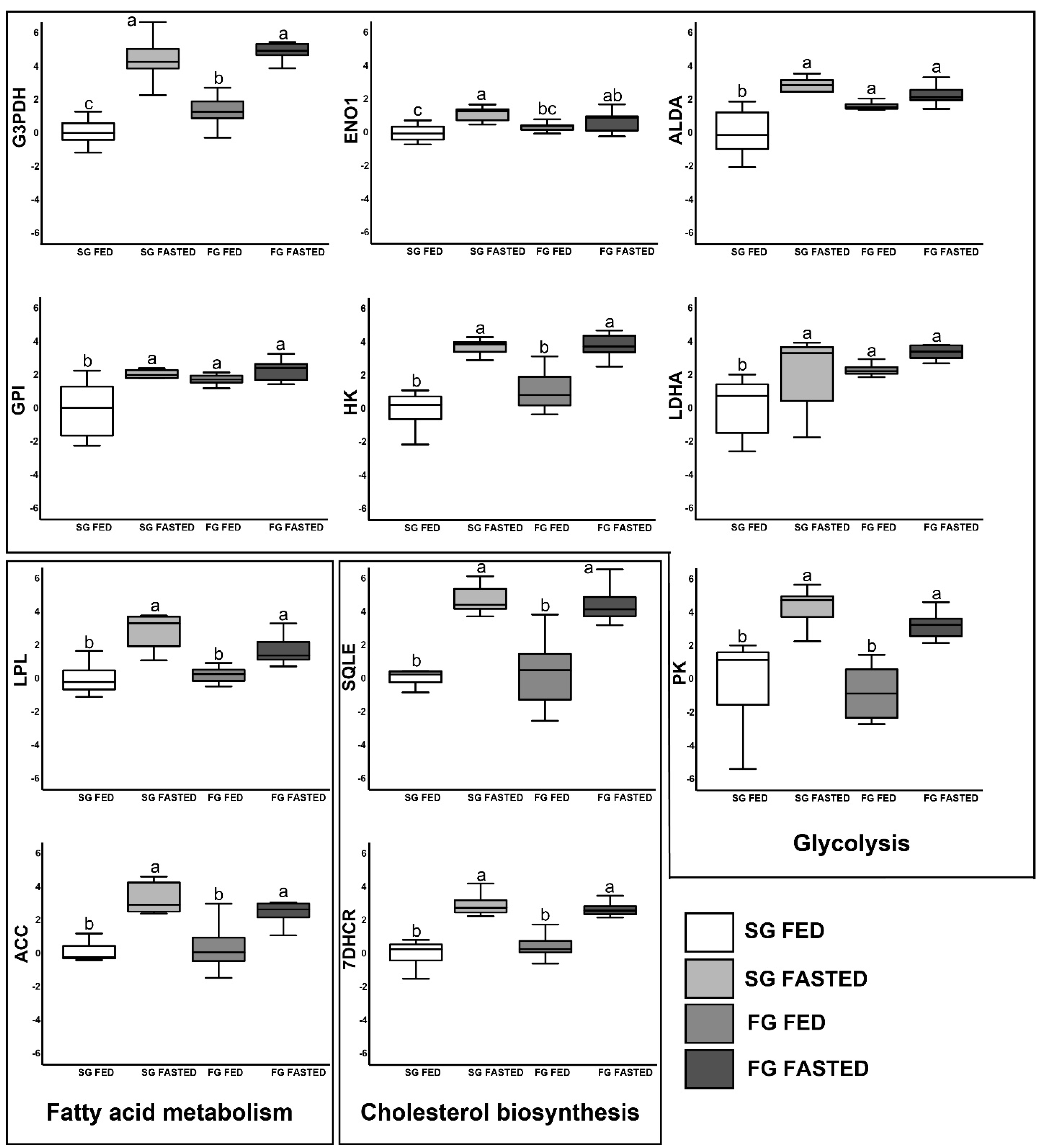
Figure 1. Differential gene expression in muscle. Different letters denote significant difference (p < 0.05).
Fishes 08 00582 g001
Figure 2. Differential gene expression in liver. Different letters denote significant difference (p < 0.05).
Fishes 08 00582 g002
Table 1. Primers specifications for qPCR analysis.
PathwayGene NameNCBI Acc. No.Primer NamePrimer Sequence (5′–3′)Tm (°C)Product Length (bp)
Glucolysisl-lactate dehydrogenase A chainCBXY010014016LDHAFTTGGCCTTAACTCAGCCTGT6086
LDHARATACAGTACACAGAGTATAT58.5
GlucolysisHexokinase ICBXY010013088HKFGATGAGTGCTGCTCCTTTCC6096
HKRGTCTCTGTCTAGTTTCTCTG61
GlucolysisGlucose-6-phosphate isomeraseCBXY010012918GPIFCTCACACAGGACCCCAACTT6087
GPIRTTGTTGAATCTCTCTTTGTCA59
GlucolysisEnolase 1CBXY010009925ENO1FAGATCGTCATTGGCATGGAT6085
ENO1RAGGGGAGATGTAGCGGCTGG60
GlucolysisGlyceraldehyde-3-phosphate dehydrogenaseCBXY010005389G3PDHFTGTAACCCAGCACTCCCTTC6084
G3PDHRGTGGACCTGACATGCCGTCT61
GlucolysisAldolase ACBXY010008418ALDAFCTGTCCGACCACCATGTCTA6080
ALDARGATCTCCTGGTTGCTGTACT60
GlucolysisPyruvate kinaseKF857578PKFGGCGTTCAGAATTTTGAGGA60109
PKRTTGCAGCGTCCAATCATCAT60
Fatty acid metabolismLipoprotein lipaseAM411614LPLFGTAACGGGGATGTTCGAGAG5989
LPLRCTGGTTGGCGCGGGTCAGCC59
Fatty acid metabolismAcetyl-CoA carboxylaseCBXY010003615ACCFAGTACCTGCACAGCCAGGAT6083
ACCRGCAAGTTGACATCAGCCACC60
Cholesterol biosynthesisSqualene epoxidaseCABK01002385SQLEFGAATCGACCGTGATGGAAAG60112
SQLERAGGGTCTGGATGCCCATCTG60
Cholesterol biosynthesis7-dehydrocholesterol reductaseCBXY0100128457DHCRFTCGGCCACATACTCCCATAC60116
7DHCRRGGTAAGGCACAGTGTCTGTG60
Reference geneBeta-actinAY148350ACTBFATCAAGATCATTGCCCCACCT6392
ACTBRTCATACTCCTGCTTGCTGA59
Reference geneElongation factor 1AJ866727EF1FCGCTCTGTGGAAGTTTGAGA59102
EF1RGATCAGCACAGCGCAGTCAG61
Table 2. Growth parameters and proximate composition of the experimental groups. Values represent mean ± standard error (n = 9). Nested ANOVA. T: treatment, G: growth. ns = p > 0.05, * = p < 0.05, ** = p < 0.01, *** = p < 0.001.
SGFGTG
FedFastedFedFasted
Final Weight (g)73.08 ± 4.1284.43 ± 5.00163.88 ± 1.80164.57 ± 2.30ns***
Final Length (cm)20.26 ± 0.0919.77 ± 0.0923.18 ± 0.0722.68 ± 0.08ns***
WGR (%)42.1 ± 2.0639.70 ± 1.7947.53 ± 0.6047.20 ± 0.30ns*
SGR (% day−1)0.86 ± 0.040.83 ± 0.040.97 ± 0.010.97 ± 0.005ns**
CF1.04 ± 0.061.18 ± 0.071.35 ± 0.031.41 ± 0.03ns***
HSI (%)0.48 ± 0.140.51 ± 0.090.76 ± 0.090.58 ± 0.08nsns
VSI (%)2.56 ± 0.152.72 ± 0.222.50 ± 0.102.64 ± 0.18nsns
ISI (%)1.56 ± 0.160.9 ± 0.111.41 ± 0.061.05 ± 0.06*ns
SSI (%)0.12 ± 0.010.10 ± 0.010.11 ± 0.010.11 ± 0.01nsns
IFR (%)1.65 ± 0.672.91 ± 0.675.5 ± 0.455.37 ± 0.63ns***
Moisture (%)68.15 ± 1.7160.73 ± 2.6850.53 ± 1.1360.60 ± 1.35ns***
Crude protein (%)54.77 ± 2.5449.33 ± 2.8641.37 ± 0.9841.06 ± 3.92ns***
Crude lipid (%)25.72 ± 2.6130.86 ± 2.6032.36 ± 0.8433.03 ± 1.28ns*
Ash (%)14.51 ± 1.0411.39 ± 1.417.51 ± 0.3011.21 ± 0.96ns***
Gross energy (KJ/g)22.40 ± 0.5723.38 ± 0.7221.94 ± 0.2824.14 ± 0.53nsns
Table 3. Biochemical indicators of the experimental groups. Values represent means ± standard error (n = 9). Nested ANOVA. T: treatment, G: growth. ns = p > 0.05, * = p < 0.05, ** = p < 0.01.
SGFGTG
FedFastedFedFasted
Glucose (mg/dL)16.9 ± 1.7021.1 ± 3.2027.6 ± 1.1026.4 ± 3.40nsns
Triglycerides (mg/dL)238.0 ± 50.70110.0 ± 47.5117.3 ± 21.0178.1 ± 14*ns
Cholesterol (mg/dL)216.6 ± 20.20161.3 ± 12189.7 ± 23.20157.8 ± 9.10*ns
NEFA (mmol/L)8.5 ± 1.907.9 ± 1.805.9 ± 0.403.7 ± 0.40ns**
LDH (U/L)1348.9 ± 64.60554.6 ± 134.10415.7 ± 127.30541.3 ± 165nsns
Table 4. Fatty acid profiles of experimental groups. Nested ANOVA. T: treatment, G: growth. ns = p > 0.05, * = p < 0.05, ** = p < 0.01, *** = p < 0.001.
SGFGTG
FedFastedFedFasted
14:03.5 ± 0.183.07 ± 0.152.76 ± 0.112.59 ± 0.08ns***
16:017.05 ± 0.3616.83 ± 0.2117.64 ± 0.6916.34 ± 0.51nsns
16:1n-75.21 ± 0.204.98 ± 0.135.02 ± 0.204.55 ± 0.17nsns
18:04.04 ± 0.113.87 ± 0.073.73 ± 0.333.7 ± 0.12nsns
18:1n-925.71 ± 1.0328.57 ± 1.3129.69 ± 2.5731.24 ± 1.05nsns
18:1n-73.16 ± 0.083 ± 0.062.94 ± 0.112.82 ± 0.08nsns
18:2n-69.55 ± 0.4510.53 ± 0.5211.8 ± 0.5011.52 ± 0.41ns**
18:3n-32.08 ± 0.172.46 ± 0.192.93 ± 0.132.87 ± 0.10ns***
18:4n-30.86 ± 0.030.76 ± 0.020.73 ± 0.030.66 ± 0.02ns***
20:1n-93.03 ± 0.102.72 ± 0.122.64 ± 0.122.65 ± 0.08ns*
20:2n-60.73 ± 0.020.66 ± 0.020.69 ± 0.020.76 ± 0.03ns*
20:4n-60.13 ± 0.010.1 ± 0.020.11 ± 0.020.12 ± 0.02nsns
20:5n-35.31 ± 0.334.28 ± 0.203.77 ± 0.163.44 ± 0.12ns***
22:1n-111.68 ± 0.091.45 ± 0.101.09 ± 0.111.19 ± 0.04ns***
22:5n-31.35 ± 0.081.06 ± 0.050.98 ± 0.080.89 ± 0.03ns***
22:6n-37.36 ± 0.465.46 ± 0.414.56 ± 0.174.17 ± 0.13ns***
SFA33.45 ± 0.9631.39 ± 0.4931.4 ± 1.3730.89 ± 0.77nsns
MUFA41.36 ± 0.643.1 ± 0.8143.37 ± 2.0744.67 ± 0.93nsns
Σn-313.42 ± 0.9012.89 ± 0.9911.07 ± 0.6310.3 ± 0.56ns*
Σn-3 HUFA10.12 ± 0.939.37 ± 1.157.15 ± 0.616.54 ± 0.62ns**
Σn-611.08 ± 0.4511.89 ± 0.4813.27 ± 0.4912.99 ± 0.37ns**
EPA + DHA8.14 ± 0.957.68 ± 1.095.61 ± 0.615.12 ± 0.59ns*
SFA/MUFA0.81 ± 0.030.73 ± 0.020.76 ± 0.100.7 ± 0.03nsns
Disclaimer/Publisher’s Note: The statements, opinions and data contained in all publications are solely those of the individual author(s) and contributor(s) and not of MDPI and/or the editor(s). MDPI and/or the editor(s) disclaim responsibility for any injury to people or property resulting from any ideas, methods, instructions or products referred to in the content.