Communication

Histopathological and Immunohistochemical Features and Expression Patterns of Cytochrome p450 1 Family Genes in Black Rockfish (Sebastes schlegelii): Exposure to 2,3,7,8-Tetrachlorodibenzo-p-dioxin and β-Naphthoflavone

Aquaculture Industry Research Division, East Sea Fisheries Research Institute, National Institute of Fisheries Science, Gangneung 25435, Republic of Korea
*
Author to whom correspondence should be addressed.
Fishes 2023, 8(12), 583; https://doi.org/10.3390/fishes8120583
Submission received: 17 November 2023 / Revised: 27 November 2023 / Accepted: 28 November 2023 / Published: 29 November 2023
(This article belongs to the Special Issue Physiological Changes in Fish Exposed to Emerging Stressors)

Abstract

:
The climate crisis and growing petroleum demand have put the health of aquatic animals in jeopardy. Fish are sensitive to chemical pollutants in aquatic environments, such as polycyclic aromatic hydrocarbons, dioxins, and dibenzofurans. This study investigated the effect of 2,3,7,8-tetrachlorodibenzo-p-dioxin (TCDD) and β-naphthoflavone (β-NF) exposure on histopathological and immunohistochemical features and expression patterns of cytochrome P450 1 (CYP1) family genes in black rockfish, Sebastes schlegelii. Histopathological alterations in the liver included congested central vein, sinusoidal dilatation, lymphocyte infiltration, and severe vacuolation within hepatocytes. The most prevalent alterations in TCDD-exposed kidneys were glomerular enlargement, narrowing of tubular lumen, melanomacrophage centers (MMCs), and necrosis. Moreover, CYP1A immunostaining was strong in renal tubules following TCDD exposure. All CYP1 family genes (CYP1A, CYP1B, CYP1C1, and CYP1C2) were significantly increased in the gills, liver, and kidney exposed to TCDD. Similarly, a significant increase of CYP1A mRNA expression in the kidney was observed upon exposure to TCDD (30.9-folds) and β-NF (25.5-folds) compared with that of the control group (p < 0.05). TCDD and β-NF exposure exerted more adverse effects on the kidney than the liver, and TCDD had a greater in vivo toxic effect than β-NF. The combined histopathological, immunohistochemical, and molecular alternations may be helpful for diagnosing chemical contaminant exposure in S. schlegelii.
Key Contribution: TCDD and β-NF exposure exerted more adverse effects on the kidney than on the liver, and TCDD had a greater in vivo toxic effect than β-NF.

1. Introduction

The marine environment is suffering from rising water temperatures, ocean acidification, pollution, dredging, tourism, and overfishing [1,2]. Such radical changes can generate a dramatic impact on sea life populations. To prevent a rise of more than 1.5 degrees Celsius in the average global temperature, numerous countries are implementing energy transition policies. However, developing countries and emerging economies continue to invest in fossil fuels and have problems implementing eco-friendly energy alternatives due to economic feasibility [3,4]. The International Energy Agency reported that oil consumption in Asian countries will reach about 15 million barrels per day by 2040 [5]. Annual global energy demand is currently over 12 billion tons of petroleum fuels [5]. With the forecast that oil demand will increase steadily through 2040, coal and petroleum-related products, heavy metals, and halides have a high probability of being directly released into marine ecosystems.
Polycyclic aromatic hydrocarbons (PAHs) are formed by the oxidation of organic compounds during combustion and constitute about 20% of all crude hydrocarbons [6,7]. PAHs are a product of urban growth and are generated from diverse industrial processes such as incineration, heating, thermoelectric power stations, and landfills [8,9]. PAHs artificially produced by industrial processes are dispersed through various routes, such as aerial, terrestrial, and aquatic, and are deposited in surface water and sediment in remote areas [10]. Prenatal and early life exposure to PAH is associated with adverse cognitive effects on child development and mental health [11]. A recent modeling study in arctic ecosystems reported that PAH concentrations may have increased by up to 13-fold in fish and mussels [12]. PAH contamination in river sediments is particularly important in high-density population environments because PAHs cause carcinogenesis, reproductive toxicity, endocrine disorders, and blue sac disease, a toxic syndrome, in adult and juvenile fish [13]. The biotransformation of ingested or inhaled PAHs increases the risk of mutations that can cause widespread adverse public health and environmental impacts on the food chain.
β-naphthoflavone (β-NF) (also called 5,6-benzoflavone), a synthetically derived flavonoid, is known as an inducer of detoxification enzymes and has a high binding affinity for the aryl hydrocarbon receptor (AhR) [14,15]. β-NF displays potent antitumor activity in breast cancer [16]. However, the mechanism of action is not fully understood. One of the persistent organic pollutants (POPs), 2,3,7,8-tetrachlorodibenzo-p-dioxin (TCDD), which is classified as a “group 1 human carcinogen”, was banned by the International Agency for Research on Cancer in 1997. However, TCDD is an unavoidable byproduct of waste combustion and industrial processes, such as pesticide, disinfectant, and metal production [17], and is a well-known contaminant of the herbicide Agent Orange used during the Vietnam War [18]. The estimated daily intake of TCDD from food in the United States is 34.4 pg [19]. In the United States, the daily intake of TCDD through dairy products, meat, and fish, which are the main sources, has been reported to be 15.9 pg. The World Health Organization (WHO) has established an acceptable daily intake (TDI) of TCDD at 1–4 pg/kg [20].
Exposure to β-NF induced erythrocytic nuclear abnormalities [21], oxidative stress, and genotoxicity in Anguilla anguilla L. [22]. TCDD toxicity showed characteristic signs in teleost fish from the early life stage, and extensive species differences have been reported. The LC(egg)50 for seven species of fish ranged from 539 pg/g for fathead minnow to 2610 pg/g for zebrafish [23]. After injection and waterborne exposure of Oncorhynchus mykiss, eggs were 421 and 439 pg TCDD/g eggs (LD50), respectively [24]. Exposure of O. mykiss eggs to TCDD resulted in half-hatching mortality, sac fry hemorrhage, reticular edema, and yolk sac edema [24,25]. TCDD is detected up to pg g−1 in fish and is strongly adsorbed to suspended and bottom sediments in the aquatic environment due to its hydrophobic characteristics [26,27]. Bioaccumulation of TCDD, because of its stability and lipophilic nature, may pose a potential risk to fish fin regeneration and hyperpigmentation and cause genotoxicity [28,29]. Exposure of Platichthys flesus to 3,3′,4,4′,5 pentachlorobiphenyl (PCB-126) and bis(tri-n-butyltin) oxide (TBTO) caused mortality after 7 to 12 days with gill lesions and reduced the relative thymus volume [30]. Therefore, it is crucial to assess fish health in response to chemical pollutants, such as dioxins and environmental chemicals.
Cytochrome P450 1A (CYP1A) is a hemoprotein superfamily enzyme that plays an important role in phase I metabolic biotransformation of xenobiotics [31]. Studies of CYP1A expression patterns have progressed considerably. Upregulation of CYP1A occurs through activation of the AhR and binding to the responsive elements on DNA. PAHs, polychlorinated dibenzo-dioxins, and certain polynuclear aromatic hydrocarbons serve as ligands that bind to the AhR, translocating it to the nucleus where it forms an active transcription factor complex [32,33]. Due to its inducibility by xenobiotics, CYP1A is widely used as a basic and traditional biomarker in aquatic animals to evaluate POPs, including PAHs and TCDD [34]. A previous study was conducted to evaluate the toxicity of benzo[a]pyrene using CYP1A expression and hepatic ethoxyresorufin-O-deethylase (EROD) activity [35]. Another study revealed target tissues for TCDD exposure in zebrafish with transgenic CYP1A containing an inserted enhanced green fluorescent protein gene (EGFP) [36].
The black rockfish, Sebastes schlegelii, is an economically important farmed fish in Korea, China, and Japan [37]. As of 2021, S. schlegelii farming production accounted for 17,000 tons and USD 168 million in Korea [38]. Recently, S. schlegelii farming has been severely challenged not only by oil pollution accidents such as the Hebei Spirit oil spill incident in 2007 but also soil contamination in the industrial complex. Toxicologic studies to evaluate the health of S. schlegelii are still very limited. In this study, we investigated the histopathological and immunohistochemical features following TCDD and β-NF exposure, as well as mRNA expression patterns of CYP1A in the liver and kidney from S. schlegelii. This combined information may be helpful for diagnosing in vitro under chemical exposure environments against S. schlegelii.

2. Materials and Methods

2.1. Ethics Statement

All procedures for animal management, euthanasia, and surgery complied with the guidelines of the Institutional Animal Care and Use Committee of Pukyong National University. The study was performed in strict accordance with the ethical guidelines published by European Union directive 2010/63/EU guide. Handling of fish and all the experimental protocols were carried out by well-trained scientists who had completed laboratory animal education.

2.2. Animals

S. schlegelii were purchased from a farm in Tongyeong, Korea, with mean body weights of 110 ± 15 g and acclimated to the laboratory conditions in four 600 L capacity tanks (30 fish/tank) with flow rates of approximately 2 L/min for 2 weeks at 18 ± 1 °C. All the fish were hand-fed a commercial extruded pellet (Woosungfeed, Daejeon, Republic of Korea; 50% crude protein and 13% lipid) twice daily at a ratio of 3% of the body weight of the fish at 8 AM and 16 PM for 2 weeks. Also, fish were kept under artificially controlled light conditions for 12 h light and 12 h dark cycles (at 8 AM and 8 PM) with light intensity of 300 lx at water surface. The chemical components of the seawater were analyzed throughout the experiment (Table 1).

2.3. Experimental Design

Following the 2 weeks of acclimation, fish were distributed randomly into 12 of 300 L tanks at a final density of 10 fish per tank (10 fish/tank, 3 replicate tanks/treatment). Group 1 fish (control) were injected with 1 mL of dimethyl sulfoxide. Group 2 fish were injected intraperitoneally (i.p.) with 1 mL of TCDD (1 µg/g body weight), and Group 3 fish were injected with 1 mL of β-NF (50 µg/g body weight) in dimethyl sulfoxide (DMSO). TCDD concentration was adopted according to Andreasen et al. [39] to ensure kidney adverse effects in fish. β-NF concentration was based on their early genotoxicity in fish previously demonstrated by Maria et al. [40]. After 48 h, 10 fish per group (30 fish/chemical treatment) were anesthetized with 50 mg/L of MS-222 (ethyl 3-aminobenzoate methanesulfonate, Sigma, Ronkonkoma, NY, USA) for 2 min by prolonged immersion and sacrificed for further analysis with ethical guidelines. The gills, liver, and head kidney (30 tissues/chemical treatment) were removed aseptically. A portion of the tissues (0.1 g) was immersed in 1 mL of QIAzol (Qiagen, Germantown, MD, USA) and stored at −80 °C until RNA isolation; the remaining tissues were fixed in 10% neutral formalin solution for 48 h for histopathological and immunohistochemical analysis. All tests were conducted in three replications and did not show mortality in all groups.

2.4. Histopathological Analysis

After being fixed in 10% formalin solution, the liver and kidney tissues were transferred to 70% ethanol. After dehydration in a series of ethanol solutions, tissue samples were embedded in paraffin. Then, 5 μm thick sections were prepared using an RM2235 rotary microtome (Leica, New York, NY, USA) and stained with hematoxylin and eosin. The morphological structures of the liver and kidney were observed using a BX53 light microscope (Olympus, Tokyo, Japan).

2.5. Immunohistochemistry (IHC)

IHC was performed to determine the distribution of CYP1A in the liver and kidney from S. schlegelii after exposure to TCDD, β-NF, and control as described previously in Woo (2022b) [35]. Briefly, samples that were 10% formalin-fixed and paraffin-embedded were sectioned at a thickness of 4 μm. We used primary antibodies against CYP1A (1:500, mouse monoclonal, C10-7; Biosense AS, Bergen, Norway), which were diluted in a solution containing 0.25% Triton X-100 in PBS. Then, the primary antibody was mounted on a tissue section for 1 h at room temperature. Tissues were washed three times in PBS (1–2 min each) and incubated for 1 h in biotinylated secondary antibody (1:2000; Goat Anti-Mouse IgG H&L, ab6788, Abcam, Cambridge, MA, USA) with dilution in PBS including 5% horse serum (v/v). After washing three times in PBS (1–2 min each), sections were incubated avidin–biotin complex for 30 min (ABC, Vectastain ABC kit PK-6100; Vector Laboratory, Burlingame, CA, USA). Then, they were washed three times in PBS (1–2 min each) and transferred to a diaminobenzidine (DAB) solution for 10 min, adding peroxidase substrate solution for 1 to 3 min until desired stain intensity develops. Sections were rinsed in PBS, the slices were mounted, and they were dried.

2.6. Real-Time Polymerase Chain Reaction Analysis

Total RNA was extracted from the gills, liver, and kidney samples using an RNeasy Minikit (Qiagen, Madrid, Spain) following the manufacturer’s protocol. RNA was quantified using an Ultrospec 6300 Pro UV spectrophotometer (Thermo Fisher Scientific, Waltham, MA, USA) at 260/280 nm and adjusted to a 1 μg/μL final concentration. Total RNA was converted to cDNA using SuperScript III reverse transcriptase (Invitrogen, Carlsbad, CA, USA) following the manufacturer’s instructions. The real-time polymerase chain reaction was performed using a LightCycler 480 II (Roche Diagnostics, Basel, Switzerland) to assess the expression levels of CYP1A, CYP1B, CYP1C1, and CYP1C2 using gene-specific primers as described previously (Table 2) [41]. The amplification parameters include the following: an initial denaturation step of 95 °C for 5 min; 45 serial cycles of a denaturation step of 95 °C for 10 s, annealing at 58 °C for 10 s, and extension at 72 °C for 10 s; and a final extension step of 72 °C for 5 min. Glyceraldehyde-3-phosphate dehydrogenase (GAPDH) was used as endogenous control. The amplification efficiencies were as follows: CYP1A was 96.5%, CYP1B was 97.2%, CYP1C1 was 96.1%, CYP1C2 was 98.1%, and GAPDH was 95.6%. Gene expression was calculated relative to GAPDH using the 2ΔΔ Ct method [42].

2.7. Statistical Analyses

All results are presented as means ± standard deviation. Prism 9.0 software (GraphPad La Jolla, CA, USA) was used to perform the statistical analyses. Normality and homoscedasticity were verified through Kolmogorov–Smirnov and Levene tests, respectively. All data were subjected to a one-way analysis of variance to determine statistical significance among groups at p < 0.05.

3. Results

3.1. Histopathological Alterations

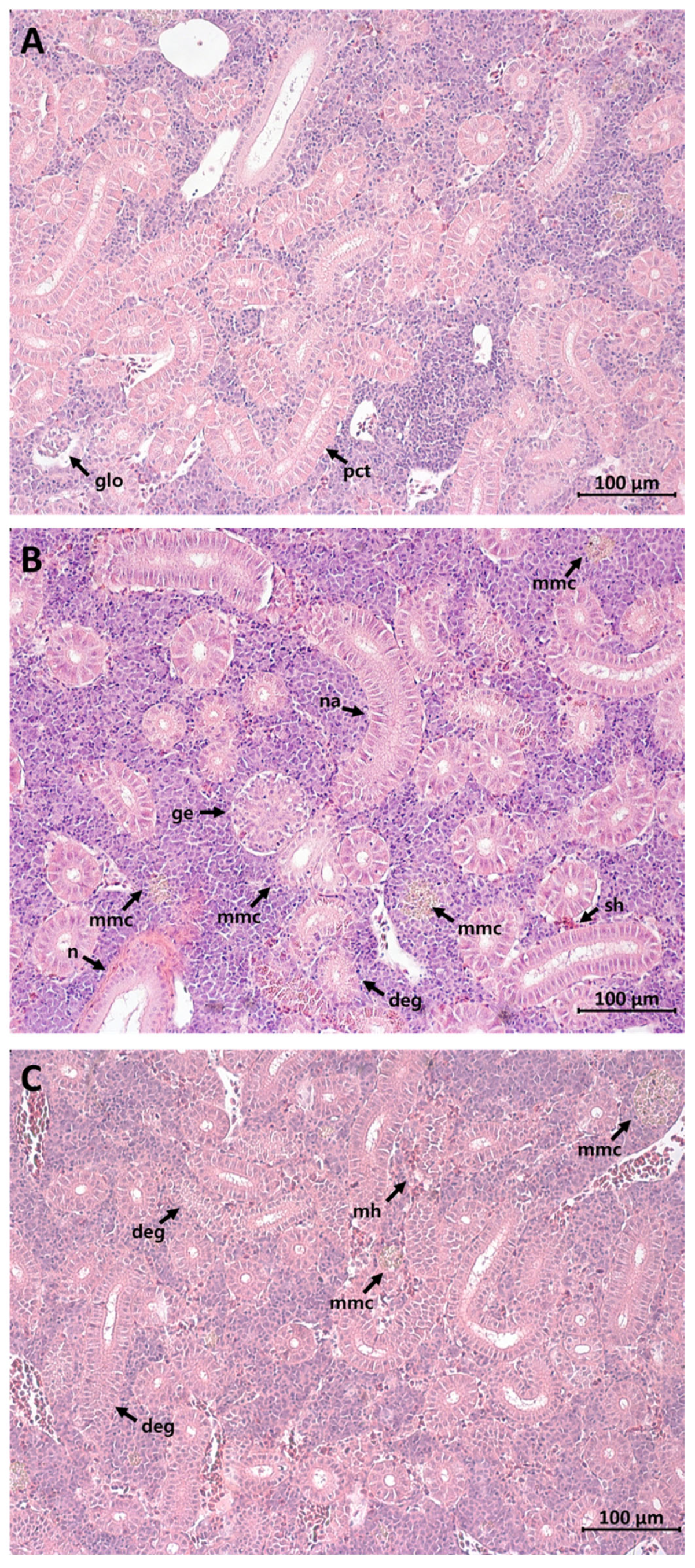
S. schlegelii exposed to DMSO as a control showed the normal structure of the sinusoid (Figure 1A). S. schlegelii exposed to TCDD showed degenerative changes, congested central vein lymphocyte infiltration, and severe vacuolation within the hepatocytes (Figure 1B). Likewise, the group exposed to β-NF exhibited cellular hypertrophy, melanomacrophage centers (MMCs), sinusoidal dilatation, and vacuolation in hepatocytes (Figure 1C). S. schlegelii exposed to DMSO as a control showed normal structure of the glomerulus and proximal convoluted tubule (Figure 2A). The most prevalent alterations in the TCDD-exposed kidneys were glomerular enlargement, narrowing of tubular lumen, MMCs, and necrosis (Figure 2B). In the β-NF exposure group, renal tubule degeneration, multiple hemorrhages, and tubular epithelium degeneration were found (Figure 2C).

3.2. IHC Analysis

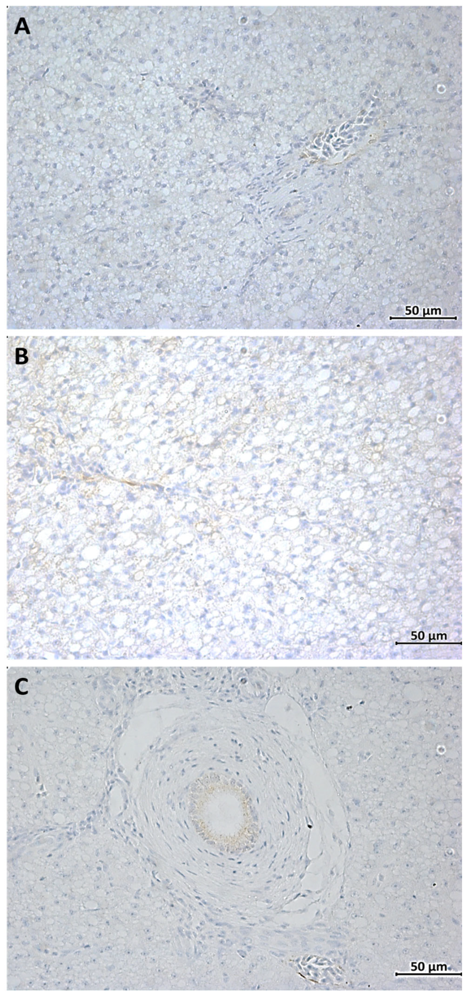
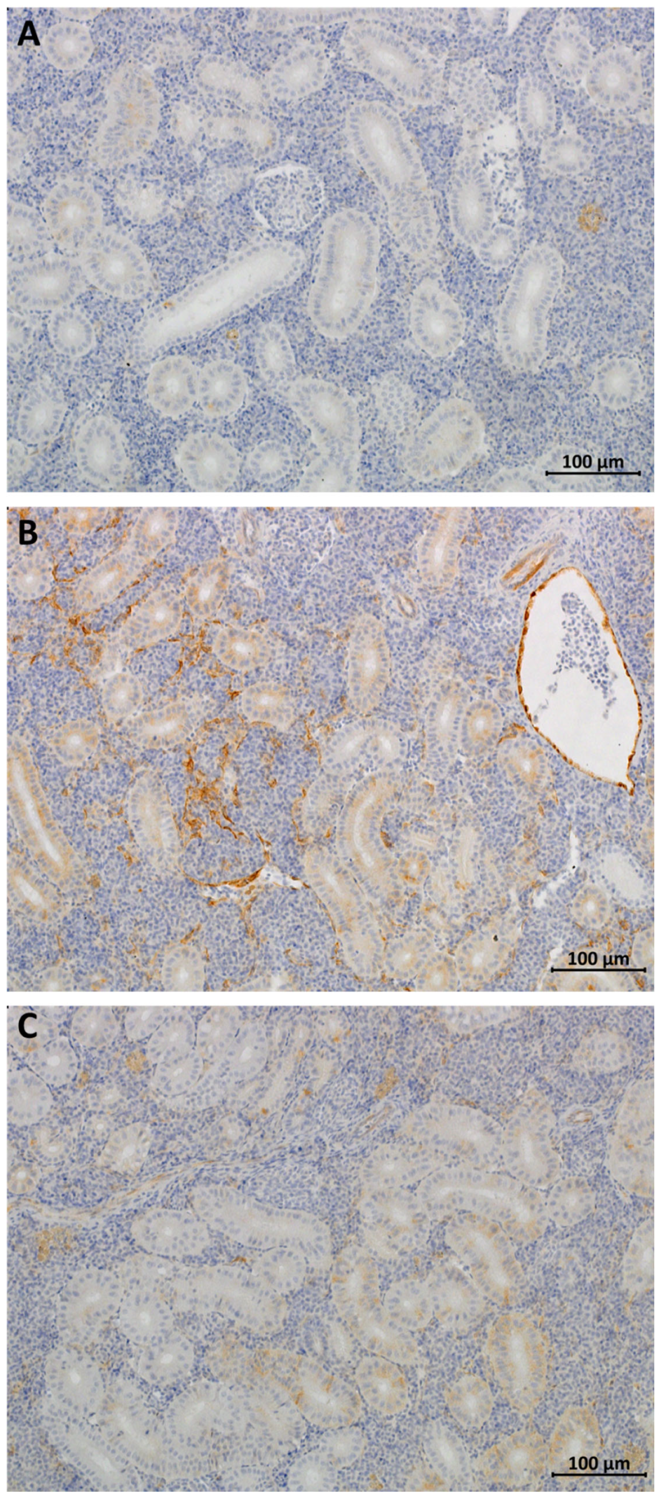
IHC analysis of the liver exposure to DMSO observed very weak staining (Figure 3A), but the TCDD-exposed liver showed that most of the cytoplasm was stained (Figure 3B), and the β-NF exposure group showed the staining of the inside of the blood vessels of the portal vein (Figure 3C). IHC analysis of the kidney exposure to DMSO observed very weak staining (Figure 4A), but the TCDD-exposed kidney showed CYP1A immunostaining in most of the renal cytoplasm and tubules (Figure 4B), and the β-NF exposure group showed the staining of some cytoplasm in the kidney (Figure 4C). IHC staining showed strong CYP1A immunostaining in the renal tubules following TCDD exposure and β-NF; however, it was relatively weak in the liver.

3.3. Expression of CYP1 Family Genes of mRNA

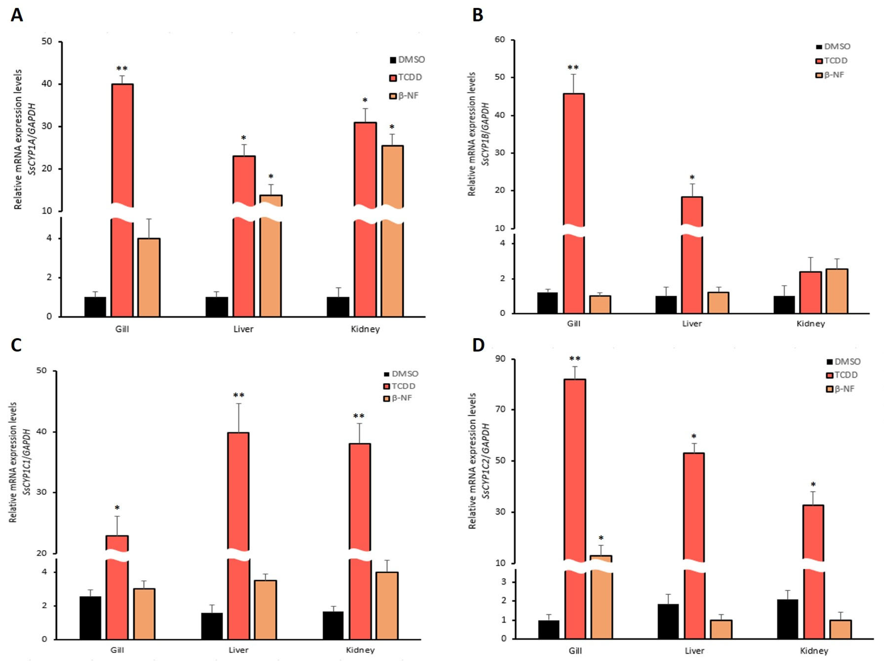
The expression of CYP1A mRNA in the gills (40.1-folds), liver (22.9-folds), and kidney (30.9-folds) was significantly increased in groups exposed to TCDD as compared to that of the control group (p < 0.05) (Figure 5A). The expression of CYP1A mRNA in the gills (13.7-folds) and kidneys (25.5-folds) was significantly increased in groups exposed to β-NF (p < 0.05) (Figure 5A). CYP1B mRNA expression was significantly increased in the gills (45.7-folds) and liver (18.3-folds) exposed to TCDD, while there was no change in β-NF groups (Figure 5B). CYP1C1 mRNA expression was significantly increased in the gills (22.9-folds), liver (39.8-folds), and kidney (38.1-folds) when exposed to TCDD (Figure 5C). The expression of CYP1C2 mRNA in the gills (81.8-folds), liver (53.2-folds), and kidney (32.6-folds) was significantly increased in groups exposed to TCDD as compared to that of the control group (p < 0.05) (Figure 5D). Specifically, CYP1C2 mRNA expression was significantly increased only in the gills following exposure to β-NF groups (p < 0.05) (Figure 5D).

4. Discussion

The occurrence of industrial POPs is a global environmental problem derived from the large-scale development of fishing, agriculture, and urban industries. PAHs and dioxins have diverse toxic potentials in the aquatic environment. PAHs tend to be absorbed mainly through particulate matter suspended in water and accumulate in the sediments over time [43]. Consequently, sediments are considered a major sink for PAHs and a secondary source of contamination for aquatic species. These compounds are detoxified in the liver and excreted by the kidney through biotransformation. However, little information is available on the histopathological and physiological changes resulting from TCDD and β-NF exposure and the induction of CYP1A in the liver and kidney from S. schlegelii.
Fish are in direct contact with pollutants through gills and skin. TCDD causes liver damage, including hepatomegaly, fatty liver, and hepatic atrophy. TCDD exposure in adult Danio rerio results in hypertrophy of hepatocytes and lipidosis [44]. Cyprinus carpio var Jian injected with 0.6 μg/kg TCDD exhibits liver damage and leakage of intracellular enzymes such as alanine aminotransferase, aspartate aminotransferase, and lactate dehydrogenase [45]. Severe tubular and glomerular damage were reported in chicken eggs injected with 0.2 mg/kg bw of TCDD for 4 days [46]; wild-type mice orally administered a total of 200 μg/kg bw of TCDD for 10 weeks had elevated liver weight and reduced thymus weight [47]. Previous studies suggested that dioxin exposure specifically targets the epithelial cell population. The current study showed that a single exposure to TCDD induced histopathological changes, including lymphocyte infiltration, vacuolation, and glomerular enlargement. This study showed prominent glomerular damage against TCDD exposure, suggesting that TCDD caused damage to specific tissues. Also, β-NF induced MMCs, cellular hypertrophy, and sinusoidal dilatation in this study. In addition, the results support that the metabolism of TCDD and β-NF mainly occurs in the liver. Furthermore, the kidney was more vulnerable to exposure to TCDD than β-NF, as shown by histopathological changes such as glomerular enlargement, narrowing of tubular lumen, necrosis, and strong immunostaining of CYP1A. IHC is also widely used in basic research to understand the distribution and localization of biomarkers and differentially expressed proteins in different parts of a biological tissue. Renal EROD activity and CYP1A immunostaining of sea bream were higher in the kidney than in the liver during TCDD exposure [48]. Renal EROD activity at 100 and 200 μg L−1 of benzo[a]pyrene increased earlier than that in the liver and gills, showing tissue differences in induction time.
The accumulation of xenobiotics causes ion exchange changes in renal tubules. This can enhance the entry of water and other molecules into cells, increasing the cell volume and damaging the cell membrane. It is proposed that TCDD and β-NF exposure damages the tubular cell membrane. TCDD remains in the body for a long time because of its high affinity for the AhR, and it is presumed to affect intrinsic functions of the kidney, particularly reabsorption and filtration. Glomerular dilatation, tubular necrosis, and tubular degeneration may occur in response to this situation. To assess damage to the renal tubules, histopathological changes, such as nephron regeneration, have been used as biomarkers in fish [49]. Although there are few serious lesions observed in this study, such as pycnotic nuclei and necrosis, in the liver, the presence of necrosis and hemorrhage in the kidney indicated that TCDD or β-NF exposure had a greater adverse effect on the kidney than liver from S. schlegelii.
CYP1A levels can predict the efficacy and potential toxicity of drugs in vitro and in vivo. CYP1A induction in fish has been widely studied in the liver, spleen, kidney, intestine, and gills [50,51,52]. CYP1A was induced in a few mononuclear cells in hematopoietic tissue of the central kidney and spleen from Platichthys flesus [53]. Several exogenous ligands, such as PAHs, indoles, and dioxin-like compounds, bind the AhR, followed by heterodimerization with the aryl hydrocarbon nuclear translocator protein to elevate CYP1A mRNA expression. In the present study, higher levels of CYP1A mRNA expression were observed in the kidney than in the liver and were more pronounced when S. schlegelii were exposed to TCDD than β-NF. Previous studies also demonstrated higher CYP1A induction in the kidney than in the spleen and liver [50,54]. Furthermore, exposure to halogenated AhR ligands, such as TCDD, resulted in more sustained CYP1A induction, keeping the induced levels stable for a long time [55]. The transcriptional activity of the CYP1B1 gene in fish is controlled by distinct mechanisms in various tissues. There are two types of CYP1B in carp: CYP1B1 is expressed in adult gills, and CYP1B2 is not expressed in normal tissues but is inducted in the gills after treatment with 3-methylcholanthene [56]. Although it has been reported that CYP1B1 transcripts are not involved in the developmental toxicity of dioxin from developing zebrafish [57], our study reported increased CYP1B1 gene expression in the gills and liver. This is presumed to be due to differences in susceptible species or exposure dose, and it needs to be further studied.
Zebrafish and S. schlegelii have two CYP1Cs [58,59]. TCDD and β-NF induced the mRNA expression of two CYP1Cs in the mesencephalic vein of zebrafish, and the knockdown of each CYP1C prevented TCDD-induced mesencephalic vein circulation impairment [60]. When adult zebrafish were exposed to TCDD, the inducible expression of CYP1C1 and CYP1C2 was greater in vascular tissue than in hepatocytes [61]. This supports the finding that CYP1C2 mRNA expression was increased 81-fold in vascularized gills exposed to TCDD. Based on a previous study showing that CYP1C2 regulation is different from CYP1C1, CYP1B1, and CYP1A [62], it is presumed that there is a need for further study of the mechanism of CYP1C2. Although this study is small-scale at the laboratory level, based on the results of this study, we will expand toxicity studies on pollutants in the marine environment. Therefore, it might be assumed that the presence of TCDD and β-NF metabolites was not only toxic to the kidney but also an indicator of CYP1A upregulation.

5. Conclusions

It is crucial to assess fish health in response to chemical pollutants, such as dioxins and environmental chemicals. Our results demonstrate that exposing S. schlegelii to TCDD or β-NF causes histopathological changes, physiological alterations, and CYP1A induction. Tubular dilatation, hemorrhage, and necrosis, as well as strong immunostaining of CYP1A, indicated that TCDD and β-NF exposure have a greater adverse effect on the kidney than the liver from S. schlegelii. Because TCDD has a greater in vivo toxic effect than β-NF, this is an important reference study demonstrating the histopathological and cellular changes in S. schlegelii when exposed to pollutants in aquatic ecosystems.

Author Contributions

Conceptualization, S.-J.W., M.-S.J. and S.-S.K.; methodology, S.-J.W., S.-S.K. and H.-K.Y.; software, M.-S.J. and S.-S.K.; validation, S.-S.K. and J.-J.P.; formal analysis, S.-J.W. and S.-S.K.; investigation, S.-J.W. and M.-S.J.; resources, J.-J.P.; data curation, S.-S.K. and H.-K.Y.; writing—original draft preparation, S.-J.W.; writing—review and editing, S.-J.W. and S.-S.K.; visualization, H.-K.Y.; supervision, J.-J.P.; project administration, J.-J.P.; funding acquisition, J.-J.P. All authors have read and agreed to the published version of the manuscript.

Funding

This research was supported by the National Institute of Fisheries Science, Ministry of Oceans and Fisheries, Republic of Korea (R2023061).

Institutional Review Board Statement

All procedures for animal management, euthanasia, and surgery complied with the guidelines of the Institutional Animal Care and Use Committee of Pukyong National University (PKNUIACUC-2018-02).

Data Availability Statement

The data presented in this study are available on request from the corresponding author. The data used to support the findings of this study are included in the article.

Conflicts of Interest

The authors declare no conflict of interest.

References

  1. Hall, C.M. Trends in ocean and coastal tourism: The end of the last frontier? Ocean. Coast. Manag. 2001, 44, 601–618. [Google Scholar] [CrossRef]
  2. Koch, M.; Bowes, G.; Ross, C.; Zhang, X.H. Climate change and ocean acidification effects on seagrasses and marine macroalgae. Glob. Chang. Biol. 2013, 19, 103–132. [Google Scholar] [CrossRef]
  3. Liu, L.; Cheng, S.Y.; Li, J.B.; Huang, Y.F. Mitigating environmental pollution and impacts from fossil fuels: The role of alternative fuels. Energy Sources A Recovery Util. Environ. Eff. 2007, 29, 1069–1080. [Google Scholar] [CrossRef]
  4. York, R. Do alternative energy sources displace fossil fuels? Nat. Clim. Chang. 2012, 2, 441–443. [Google Scholar] [CrossRef]
  5. Shoar, F.H.; Najafi, B.; Mosavi, A. Effects of triethylene glycol mono methyl ether (TGME) as a novel oxygenated additive on emission and performance of a dual-fuel diesel engine fueled with natural gas-diesel/biodiesel. Energy Rep. 2021, 7, 1172–1189. [Google Scholar] [CrossRef]
  6. Zhang, Z.L.; Hong, H.S.; Zhou, J.L.; Yu, G. Phase association of polycyclic aromatic hydrocarbons in the Minjiang River Estuary, China. Sci. Total Environ. 2004, 323, 71–86. [Google Scholar] [CrossRef]
  7. Feng, J.; Zhai, M.; Sun, J.; Liu, Q. Distribution and sources of polycyclic aromatic hydrocarbons (PAHs) in sediment from the upper reach of Huaihe River, East China. Environ. Sci. Pollut. Res. 2012, 19, 1097–1106. [Google Scholar] [CrossRef]
  8. St-Amand, A.D.; Mayer, P.M.; Blais, J.M. Seasonal trends in vegetation and atmospheric concentrations of PAHs and PBDEs near a sanitary landfill. Atmos. Environ. 2008, 42, 2948–2958. [Google Scholar] [CrossRef]
  9. Kong, S.; Shi, J.; Lu, B.; Qiu, W.; Zhang, B.; Peng, Y.; Zhang, B.; Bai, Z. Characterization of PAHs within PM10 fraction for ashes from coke production, iron smelt, heating station and power plant stacks in Liaoning Province, China. Atmos. Environ. 2011, 45, 3777–3785. [Google Scholar] [CrossRef]
  10. Hwang, H.M.; Wade, T.L. Aerial distribution, temperature-dependent seasonal variation, and sources of polycyclic aromatic hydrocarbons in pine needles from the Houston metropolitan area, Texas, USA. J. Environ. Sci. Health A 2008, 43, 1243–1251. [Google Scholar] [CrossRef]
  11. Pagliaccio, D.; Herbstman, J.B.; Perera, F.; Tang, D.; Goldsmith, J.; Peterson, B.S.; Rauh, V.; Margolis, A.E. Prenatal exposure to polycyclic aromatic hydrocarbons modifies the effects of early life stress on attention and thought problems in late childhood. J. Child. Psychol. Psychiatry 2020, 61, 1253–1265. [Google Scholar] [CrossRef]
  12. Laender, F.D.; Hammer, J.; Hendriks, A.J.; Soetaert, K.; Janssen, C.R. Combining monitoring data and modeling identifies PAHs as emerging contaminants in the Arctic. Environ. Sci. Technol. 2011, 45, 9024–9029. [Google Scholar] [CrossRef]
  13. Eriksson, A.N.; Rigaud, C.; Wincent, E.; Pakkanen, H.; Salonen, P.; Vehniäinen, E.R. Endogenous AhR agonist FICZ accumulates in rainbow trout (Oncorhynchus mykiss) alevins exposed to a mixture of two PAHs, retene and fluoranthene. Ecotoxicology 2022, 31, 1382–1389. [Google Scholar] [CrossRef]
  14. Johnson, B.T. Potential genotoxicity of sediments from the Great Lakes Environ. Toxicol. Water Qual. 1992, 7, 373–390. [Google Scholar] [CrossRef]
  15. Jos, A.; Segner, H.; Herradon, B.; Repetto, G.; Navas, J.M. Induction of EROD activity by 1-phenylimidazole and β-naphthoflavone in rainbow trout cultured hepatocytes: A comparative study. Toxicol. Vitr. 2007, 21, 1307–1310. [Google Scholar] [CrossRef]
  16. Lubet, R.A.; Heckman, B.M.; De Flora, S.L.; Steele, V.E.; Crowell, J.A.; Juliana, M.M.; Grubbs, C.J. Effects of 5,6-benzoflavone, indole-3-carbinol (I3C) and diindolylmethane (DIM) on chemically-induced mammary carcinogenesis: Is DIM a substitute for I3C? Oncol. Rep. 2011, 26, 731–736. [Google Scholar] [CrossRef]
  17. White, S.S.; Birnbaum, L.S. An overview of the effects of dioxins and dioxin-like compounds on vertebrates, as documented in human and ecological epidemiology. J. Environ. Sci. Health C Toxicol. Carcinog. 2009, 27, 197–211. [Google Scholar] [CrossRef]
  18. Schecter, A.; Birnbaum, L.; Ryan, J.J.; Constable, J.D. Dioxins: An overview. Environ. Res. 2006, 101, 419–428. [Google Scholar] [CrossRef]
  19. EFSA Panel on Contaminants in the Food Chain (CONTAM); Knutsen, H.K.; Alexander, J.; Barregård, L.; Bignami, M.; Brüschweiler, B.; Hoogenboom, L. Risk for animal and human health related to the presence of dioxins and dioxin-like PCBs in feed and food. Efsa J. 2018, 16, e05333. [Google Scholar] [CrossRef]
  20. Yang, J.; Shin, D.; Park, S.; Chang, Y.; Kim, D.; Ikonomou, M.G. PCDDs, PCDFs, and PCBs concentrations in breast milk from two areas in Korea: Body burden of mothers and implications for feeding infants. Chemosphere 2002, 46, 419–428. [Google Scholar] [CrossRef]
  21. Pacheco, M.; Santos, M.A. Induction of Liver EROD and Erythrocytic Nuclear Abnormalities by Cyclophosphamide and PAHs in Anguilla anguilla L. Ecotoxicol. Environ. Saf. 1998, 40, 71–76. [Google Scholar] [CrossRef]
  22. Ahmad, I.; Maria, V.L.; Oliveira, M.; Pacheco, M.; Santos, M.A. Oxidative stress and genotoxic effects in gill and kidney of Anguilla anguilla L. exposed to chromium with or without pre-exposure to β-naphthoflavone. Mutat. Res. Genet. Toxicol. Environ. Mutagen. 2006, 608, 16–28. [Google Scholar] [CrossRef]
  23. Elonen, G.E.; Spehar, R.L.; Holcombe, G.W.; Johnson, R.D.; Fernandez, J.D.; Erickson, R.J.; Cook, P.M. Comparative toxicity of 2,3,7,8-tetrachlorodibenzo-p-dioxin to seven freshwater fish species during early life-stage development. Environ. Toxicol. Chem. 1998, 17, 472–483. [Google Scholar] [CrossRef]
  24. Walker, M.K.; Hufnagle, L.C., Jr.; Clayton, M.K.; Peterson, R.E. An egg injection method for assessing early life stage mortality of polychlorinated dibenzo-p-dioxins, dibenzofurans, and biphenyls in rainbow trout, (Oncorhynchus mykiss). Aquat. Toxicol. 1992, 22, 15–37. [Google Scholar] [CrossRef]
  25. Giesy, J.P.; Jones, P.D.; Kannan, K.; Newsted, J.L.; Tillitt, D.E.; Williams, L.L. Effects of chronic dietary exposure to environmentally relevant concentrations to 2,3,7,8-tetrachlorodibenzo-p-dioxin on survival, growth, reproduction and biochemical responses of female rainbow trout (Oncorhynchus mykiss). Aquat. Toxicol. 2002, 59, 35–53. [Google Scholar] [CrossRef]
  26. Greco, L.; Serrano, R.; Blanes, M.A.; Serrano, E.; Capri, E. Bioaccumulation markers and biochemical responses in European sea bass (Dicentrarchus labrax) raised under different environmental conditions. Ecotoxicol. Environ. Saf. 2010, 73, 38–45. [Google Scholar] [CrossRef]
  27. Nunes, M.; Marchand, P.; Vernisseau, A.; Le Bizec, B.; Ramos, F.; Pardal, M.A. PCDD/Fs and dioxin-like PCBs in sediment and biota from the Mondego estuary (Portugal). Chemosphere 2011, 83, 1345–1352. [Google Scholar] [CrossRef]
  28. Zodrow, J.M.; Tanguay, R.L. 2,3,7,8-tetrachlorodibenzo-p-dioxin inhibits zebrafish caudal fin regeneration. Toxicol. Sci. 2003, 76, 151–161. [Google Scholar] [CrossRef]
  29. Della Torre, C.; Buonocore, F.; Frenzilli, G.; Corsolini, S.; Brunelli, A.; Guidi, P.; Kocan, A.; Mariottini, M.; Mottola, F.; Nigro, M.; et al. Influence of titanium dioxide nanoparticles on 2,3,7,8-tetrachlorodibenzo-p-dioxin bioconcentration and toxicity in the marine fish European sea bass (Dicentrarchus labrax). Environ. Pollut. 2015, 196, 185–193. [Google Scholar] [CrossRef]
  30. Grinwis, G.C.M.; Vethaak, A.D.; Wester, P.W.; Vos, J.G. Toxicology of environmental chemicals in the flounder (Platichthys flesus) with emphasis on the immune system: Field, semi-field (mesocosm) and laboratory studies. Toxicol. Lett. 2000, 112, 289–301. [Google Scholar] [CrossRef]
  31. Isin, E.M.; Guengerich, F.P. Complex reactions catalyzed by cytochrome P450 enzymes. Biochim. Biophys. Acta—Gen. Subj. 2007, 1770, 314–329. [Google Scholar] [CrossRef]
  32. Beedanagari, S.R.; Bebenek, I.; Bui, P.; Hankinson, O. Resveratrol inhibits dioxin-induced expression of human CYP1A1 and CYP1B1 by inhibiting recruitment of the aryl hydrocarbon receptor complex and RNA polymerase II to the regulatory regions of the corresponding genes. Toxicol. Sci. 2009, 110, 61–67. [Google Scholar] [CrossRef]
  33. Goldstone, J.V.; Jönsson, M.E.; Behrendt, L.; Woodin, B.R.; Jenny, M.J.; Nelson, D.R.; Stegeman, J.J. Cytochrome P450 1D1: A novel CYP1A-related gene that is not transcriptionally activated by PCB126 or TCDD. Arch. Biochem. Biophys. 2009, 482, 7–16. [Google Scholar] [CrossRef]
  34. Bucheli, T.D.; Fent, K. Induction of cytochrome P450 as a biomarker for environmental contamination in aquatic ecosystems. Crit. Rev. Environ. Sci. Technol. 1995, 25, 201–268. [Google Scholar] [CrossRef]
  35. Woo, S.J. Effects of benzo [a] pyrene exposure on black rockfish (Sebastes schlegelii): EROD activity, CYP1A protein, and immunohistochemical and histopathological alterations. Environ. Sci. Pollut. Res. 2022, 29, 4033–4043. [Google Scholar] [CrossRef]
  36. Kim, K.H.; Park, H.J.; Kim, J.H.; Kim, S.; Williams, D.R.; Kim, M.K.; Jung, Y.D.; Teraoka, H.; Park, H.C.; Choy, H.E.; et al. Cyp1a reporter zebrafish reveals target tissues for dioxin. Aquat. Toxicol. 2013, 134, 57–65. [Google Scholar] [CrossRef]
  37. Xi, D.; Zhang, X.; Lü, H.; Zhang, Z. Cannibalism in juvenile black rockfish, Sebastes schlegelii (Hilgendorf, 1880), reared under controlled conditions. Aquaculture 2017, 479, 682–689. [Google Scholar] [CrossRef]
  38. Statistics Korea. Fishery Production Survey. Available online: https://www.kostat.go.kr (accessed on 5 May 2022).
  39. Andreasen, E.A.; Hahn, M.E.; Heideman, W.; Peterson, R.E.; Tanguay, R.L. The zebrafish (Danio rerio) aryl hydrocarbon receptor type 1 is a novel vertebrate receptor. Mol. Pharmacol. 2002, 62, 234–249. [Google Scholar] [CrossRef]
  40. Maria, V.L.; Correia, A.C.; Santos, M.A. Anguilla anguilla L. blood and liver DNA strand breaks after beta-naphthoflavone exposure. Fresenius Environ. Bull. 2004, 13, 93–97. [Google Scholar]
  41. Woo, S.J. Molecular characterization of the aryl hydrocarbon receptor 2 gene in black rockfish, Sebastes schlegelii, and its expression patterns upon exposure to benzo [a] pyrene, 2,3,7,8-tetrachlorodibenzo-p-dioxin, and β-naphthoflavone. J. Appl. Toxicol. 2022, 42, 638–650. [Google Scholar] [CrossRef]
  42. Livak, K.J.; Schmittgen, T.D. Analysis of relative gene expression data using real-time quantitative PCR and the 2−ΔΔCT method. Methods 2001, 25, 402–408. [Google Scholar] [CrossRef] [PubMed]
  43. Hylland, K. Polycyclic aromatic hydrocarbon (PAH) ecotoxicology in marine ecosystems. J. Toxicol. Environ. Health Part A 2006, 69, 109–123. [Google Scholar] [CrossRef] [PubMed]
  44. Zodrow, J.M.; Stegeman, J.J.; Tanguay, R.L. Histological analysis of acute toxicity of 2,3,7,8-tetrachlorodibenzo-p-dioxin (TCDD) in zebrafish. Aquat. Toxicol. 2004, 66, 25–38. [Google Scholar] [CrossRef] [PubMed]
  45. Du, J.; Cao, L.; Jia, R.; Yin, G. Hepatoprotective and antioxidant effects of dietary Glycyrrhiza polysaccharide against TCDD-induced hepatic injury and RT-PCR quantification of AHR2, ARNT2, CYP1A mRNA in Jian Carp (Cyprinus carpio var. Jian). J. Environ. Sci. 2017, 51, 181–190. [Google Scholar] [CrossRef] [PubMed]
  46. Al-Musawi, M.T.; Ali, A.E.M.H.; Humadi, A.A.; Al-Kaisei, B.I. Nephropathy Effects of 2,3,7,8-Tetrachlorodibenzo-P-Dioxin (TCDD) Toxicity in Ova Injected Chicken. Ann. Rom. Soc. Cell Biol. 2021, 25, 4418–4429. [Google Scholar]
  47. Esteban, J.; Sánchez-Pérez, I.; Hamscher, G.; Miettinen, H.M.; Korkalainen, M.; Viluksela, M.; Pohjanvirta, R.; Håkansson, H. Role of aryl hydrocarbon receptor (AHR) in overall retinoid metabolism: Response comparisons to 2,3,7,8-tetrachlorodibenzo-p-dioxin (TCDD) exposure between wild-type and AHR knockout mice. Reprod. Toxicol. 2021, 101, 33–49. [Google Scholar] [CrossRef] [PubMed]
  48. Ortiz-Delgado, J.B.; Behrens, A.; Segner, H.; Sarasquete, C. Tissue-specific induction of EROD activity and CYP1A protein in Sparus aurata exposed to B(a)P and TCDD. Ecotoxicol. Environ. Saf. 2008, 69, 80–88. [Google Scholar] [CrossRef] [PubMed]
  49. Reimschuessel, R. A fish model of renal regeneration and development. ILAR J. 2001, 42, 285–291. [Google Scholar] [CrossRef]
  50. Carlson, E.A.; Li, Y.; Zelikoff, J.T. Benzo [a] pyrene-induced immunotoxicity in Japanese medaka (Oryzias latipes): Relationship between lymphoid CYP1A activity and humoral immune suppression. Toxicol. Appl. Pharmacol. 2004, 201, 40–52. [Google Scholar] [CrossRef]
  51. Reynaud, S.; Deschaux, P. The effects of 3-methylcholanthrene on lymphocyte proliferation in the common carp (Cyprinus carpio L.). Toxicology 2005, 211, 156–164. [Google Scholar] [CrossRef]
  52. Reynaud, S.; Deschaux, P. The effects of polycyclic aromatic hydrocarbons on the immune system of fish: A review. Aquat. Toxicol. 2006, 77, 229–238. [Google Scholar] [CrossRef] [PubMed]
  53. Grinwis, G.C.M.; Besselink, H.T.; Van den Brandhof, E.J.; Bulder, A.S.; Engelsma, M.Y.; Kuiper, R.V.; Wester, P.W.; Vaal, M.A.; Vethaak, A.D.; Vos, J.G. Toxicity of TCDD in European flounder (Platichthys flesus) with emphasis on histopathology and cytochrome P450 1A induction in several organ systems. Aquat. Toxicol. 2000, 50, 387–401. [Google Scholar] [CrossRef] [PubMed]
  54. Carlson, E.A.; Li, Y.; Zelikoff, J.T. Suppressive effects of benzo [a] pyrene upon fish immune function: Evolutionarily conserved cellular mechanisms of immunotoxicity. Mar. Environ. Res. 2004, 58, 731–734. [Google Scholar] [CrossRef] [PubMed]
  55. Whyte, J.J.; Jung, R.E.; Schmitt, C.J.; Tillitt, D.E. Ethoxyresorufin-O-deethylase (EROD) activity in fish as a biomarker of chemical exposure. Crit. Rev. Toxicol. 2000, 30, 347–570. [Google Scholar] [CrossRef] [PubMed]
  56. El-Kady, M.A.; Mitsuo, R.; Kaminishi, Y.; Itakura, T. Isolation of cDNA of novel cytochrome P450 1B gene, CYP1B2, from Carp (Cyprinus carpio) and its induced expression in gills. Environ. Sci. 2004, 11, 345–354. [Google Scholar] [PubMed]
  57. Yin, H.C.; Tseng, H.P.; Chung, H.Y.; Ko, C.Y.; Tzou, W.S.; Buhler, D.R.; Hu, C.H. Influence of TCDD on zebrafish CYP1B1 transcription during development. Toxicol. Sci. 2008, 103, 158–168. [Google Scholar] [CrossRef] [PubMed]
  58. Godard, C.A.; Goldstone, J.V.; Said, M.R.; Dickerson, R.L.; Woodin, B.R.; Stegeman, J.J. The new vertebrate CYP1C family: Cloning of new subfamily members and phylogenetic analysis. Biochem. Biophys. Res. Commun. 2005, 331, 1016–1024. [Google Scholar] [CrossRef]
  59. Woo, S.J.; Chung, J.K. Cytochrome P450 1 enzymes in black rockfish, Sebastes schlegelii: Molecular characterization and expression patterns after exposure to benzo [a] pyrene. Aquat. Toxicol. 2020, 226, 105566. [Google Scholar] [CrossRef]
  60. Kubota, A.; Stegeman, J.J.; Woodin, B.R.; Iwanaga, T.; Harano, R.; Peterson, R.E.; Hiraga, K.; Teraoka, H. Role of zebrafish cytochrome P450 CYP1C genes in the reduced mesencephalic vein blood flow caused by activation of AHR2. Toxicol. Appl. Pharmacol. 2011, 253, 244–252. [Google Scholar] [CrossRef]
  61. Bugiak, B.; Weber, L.P. Hepatic and vascular mRNA expression in adult zebrafish (Danio rerio) following exposure to benzo-a-pyrene and 2,3,7,8-tetrachlorodibenzo-p-dioxin. Aquat. Toxicol. 2009, 95, 299–306. [Google Scholar] [CrossRef]
  62. Zanette, J.; Jenny, M.J.; Goldstone, J.V.; Woodin, B.R.; Watka, L.A.; Bainy, A.C.; Stegeman, J.J. New cytochrome P450 1B1, 1C2 and 1D1 genes in the killifish Fundulus heteroclitus: Basal expression and response of five killifish CYP1s to the AHR agonist PCB126. Aquat. Toxicol. 2009, 93, 234–243. [Google Scholar] [CrossRef] [PubMed]
Figure 1. Histopathological alterations in liver from Sebastes schlegelii. (A) Exposure to dimethyl sulfoxide (1 mL; control); normal structure of the sinusoid. (B) Exposure to 2,3,7,8-tetrachlorodibenzo-p-dioxin (TCDD) (1 µg/g body weight); degenerative changes, congested central vein, lymphocyte infiltration, and vacuolation. (C) Exposure to β-naphthoflavone (β-NF) (50 µg/g body weight); cellular hypertrophy, melanomacrophage centers, sinusoidal dilatation and vacuolation (hematoxylin and eosin staining, 200×). The scale bars indicate 100 µm. ch, cellular hypertrophy; con, congested central vein; dc, degenerative changes; lym, lymphocyte infiltration; mmc, melanomacrophage centers; s, sinusoid; sd, sinusoidal dilatation; vac, vacuolation.
Fishes 08 00583 g001
Figure 2. Histopathological alterations in kidney from Sebastes schlegelii. (A) Exposure to dimethyl sulfoxide (1 mL; control); normal structure of the glomerulus and proximal convoluted tubule. (B) Exposure to 2,3,7,8-tetrachlorodibenzo-p-dioxin (TCDD) (1 µg/g body weight); glomerular enlargement, small hemorrhage, narrowing of tubular lumen, and presence of melanomacrophage centers; (C) Exposure to β-naphthoflavone (β-NF) (50 µg/g body weight); degeneration of renal tubule, multiple melanomacrophage centers, and multiple hemorrhage (hematoxylin and eosin staining, 200×). The scale bars indicate 100 µm. deg, degeneration of renal tubule; ge, glomerular enlargement; glo, glomerulus; mh, multiple hemorrhage; n, necrosis; na, narrowing of tubular lumen; mmc, melanomacrophage centers; pct, proximal convoluted tubule; sh, small hemorrhage.
Fishes 08 00583 g002
Figure 3. Immunohistochemical analysis in liver from Sebastes schlegelii using a CYP1A antibody. (A) Exposure to dimethyl sulfoxide (1 mL; control). (B) Exposure to 2,3,7,8-tetrachlorodibenzo-p-dioxin (TCDD) (1 µg/g body weight); staining of most cytoplasm in hepatocyte. (C) Exposure to β-naphthoflavone (β-NF) (50 µg/g body weight) staining of the inside of the blood vessels of the portal vein.
Fishes 08 00583 g003
Figure 4. Immunohistochemical analysis in kidney from Sebastes schlegelii using a CYP1A antibody. (A) Exposure to dimethyl sulfoxide (1 mL; control). (B) Exposure to 2,3,7,8-tetrachlorodibenzo-p-dioxin (TCDD) (1 µg/g body weight); staining of the renal cytoplasm and tubules. (C) Exposure to β-naphthoflavone (β-NF) (50 µg/g body weight); staining of some cytoplasm in the kidney.
Fishes 08 00583 g004
Figure 5. mRNA expression of CYP1 genes family of gills, liver, and kidney of S. schlegelii post-exposure to dimethyl sulfoxide, 2,3,7,8-tetrachlorodibenzo-p-dioxin (1 µg/g body weight), β-naphthoflavone (50 µg/g body weight). (A) CYP1A, (B) CYP1B, (C) CYP1C1, and (D) CYP1C2 were normalized to GAPDH values. Data are expressed as means ± standard deviation. Significant differences are indicated by asterisks (* p < 0.05, ** p < 0.01).
Fishes 08 00583 g005
Table 1. Chemical composition of seawater and experimental conditions.
ComponentsValue
Temperature (°C)18 ± 1.0
pH8.2 ± 0.5
Salinity (‰)33.1 ± 0.5
Light photoperiod (h)12
Dark photoperiod (h)12
Dissolved oxygen (mg/L)7.6 ± 0.3
Chemical oxygen demand (mg/L)1.2 ± 0.1
Ammonia (µg/L)11.3 ± 0.5
Nitrite (µg/L)1.5 ± 0.2
Nitrate (µg/L)10.1 ± 1.0
Table 2. Oligonucleotide primers used for quantitative real-time PCR.
GeneAccession NumberSequence (5′−3′)Product Size (bp)Reference
GAPDHKF430617.1F: AGTACGACTCCACTCACGGC112This study
R: TTGGAAGGGTCCTTCTCGTG
CYP1AMK331129.1F: GACACCTGCGTCTTCATCAATCA118[38]
R: GCTTGATGACTTCGGTGCCATC
CYP1BMK331130.1F: GCACCATCAGGGACATGACA133
R: AGTGTGTCTTGACTTGCTCCAA
CYP1C1MK331131.1F: CGCGCTTCCTGGATGGAAAC111
R: CTTCCACCTTGGCGATCTGG
CYP1C2MK331132.1F: GGCTCCCTAGCAAGGATCTCA126
R: GCATTGGTGGATAGAATCGCG
Disclaimer/Publisher’s Note: The statements, opinions and data contained in all publications are solely those of the individual author(s) and contributor(s) and not of MDPI and/or the editor(s). MDPI and/or the editor(s) disclaim responsibility for any injury to people or property resulting from any ideas, methods, instructions or products referred to in the content.