Article

Cu/Zn Superoxide Dismutase and Catalase of Yangtze Sturgeon, Acipenser dabryanus: Molecular Cloning, Tissue Distribution and Response to Fasting and Refeeding

1
Key Laboratory of Sichuan Province for Fishes Conservation and Utilization in the Upper Reaches of the Yangtze River, Neijiang Normal University, Neijiang 641000, China
2
The Fishery Institute of the Sichuan Academy of Agricultural Sciences, Chengdu 611730, China
*
Author to whom correspondence should be addressed.
Fishes 2022, 7(1), 35; https://doi.org/10.3390/fishes7010035
Submission received: 21 November 2021 / Revised: 16 January 2022 / Accepted: 18 January 2022 / Published: 1 February 2022
(This article belongs to the Special Issue Advances in Nutrition and Feeding of Fish)

Abstract

:
Superoxide dismutase and catalase are two major antioxidant enzymes in the fish antioxidant defense system, which can remove excess reactive oxygen species and protect fish from stress-induced oxidative damage. The present study aimed to clone the sequences of Yangtze sturgeon, Acipenser dabryanus, Cu/Zn superoxide dismutase (AdCu/Zn-SOD) and catalase (AdCAT), and to explore changes of gene expression in the liver and intestine during fasting and refeeding. A total of 120 fish were exposed to four fasting and refeeding protocols (fasting for 0, 3, 7, or 14 d and then refeeding for 14 d). The coding sequences of AdCu/Zn-SOD and AdCAT encoded 155 and 526 amino acid proteins, respectively, both of which were expressed mainly in the liver. During fasting, when compared to the control group, liver AdCu/Zn-SOD expression was significantly higher in the 3- and 14-d groups, whereas its intestinal expression increased significantly only in the 7-d group. Liver AdCAT expression increased significantly in the 3-, 7-, and 14-d groups. During refeeding, liver AdCu/Zn-SOD expression increased significantly in the 3-, 7-, and 14-d groups compared with those in the control group. Similarly, intestinal AdCu/Zn-SOD expression increased significantly in the 3- and 7-d groups. Moreover, intestinal AdCAT expression was significantly higher in the 3-d group than in the control group, but decreased significantly in the 14-d group. Our findings indicated that AdCu/Zn-SOD and AdCAT play important roles in protecting fish against starvation-induced oxidative stress. Yangtze sturgeon exhibited the potential to adapt to a starvation and refeeding regime.

1. Introduction

Starvation is a common phenomenon when fishes lack sufficient foods to meet nutritional requirements [1]. Many fish species are routinely affected by periods of starvation of variable length. Starvation is considered to be one of the most important factors affecting the normal growth, development, reproduction, and survival of fishes [2,3]. In contrast to other vertebrates, who can only resist a short period of starvation, teleosts can survive long-term food deprivation by utilizing stored energy through oxidative pathways [4,5,6]. Starvation not only induces cessation of growth, but also causes stress in fishes [7]. Several studies have revealed that the pro-oxidant effects mediated by starvation are responsible for most of the negative effects, because the reactive oxygen species (ROS) generated by sustained aerobic metabolism are not adequately neutralized by antioxidant systems [8,9,10].
ROS are continuously produced and eliminated by living organisms and are normally maintained at certain steady-state levels. However, overproduction of ROS causes damage to cellular macromolecules (e.g., membrane lipids, DNA, carbohydrates, and proteins) [11,12]. To cope with the continuous generation of ROS, organisms have evolved a cellular antioxidant detoxification system consisting of enzymatic antioxidants and non-enzymatic small molecules. In particular, the major antioxidant enzymes, superoxide dismutase (SOD), catalase (CAT), glutathione peroxidase (GPX), and peroxidase bear the most responsibility for ROS elimination [13]. Among these enzymes, SODs play an essential role in cellular defense systems against oxidative stress by catalyzing the dismutation of superoxide anions to hydrogen peroxide and molecular oxygen [14]. SODs are ubiquitous and have been classified into three groups according to their metal ion cofactor in the active site: (1) copper/zinc-SOD (Cu/Zn-SOD), (2) manganese SOD (Mn-SOD), and (3) iron-SOD (Fe-SOD). Cu/Zn-SOD, because of its wide distribution in intracellular and extracellular compartments of both prokaryotes and eukaryotes, plays a particularly important role in protection against oxidative tissue injury caused by excessive ROS [15,16]. CAT is a major oxidoreductase enzyme that converts H2O2 to H2O and O2. In addition, at low H2O2 concentration, catalase can also oxidize toxins such as alcohols, phenols, formaldehyde and formic acid via the peroxidatic pathway, by using one molecule of H2O2 [17]. Cu/Zn-SOD has been cloned from several fish species including the cyprinid, Onychostoma macrolepis [18], Pengze crucian carp, Carassius auratus var. Pengze [19], marbled eel, Anguilla marmorata [20], and large yellow croaker, Pseudosciaena crocea [21]. CAT gene sequences have been reported for turbot, Scophthalmus maximus [22], rock bream, Oplegnathus fasciatus [23], and Chinese black sleeper, Bostrychus sinensis [24]. So far, the Cu/Zn SOD and CAT from Yangtze sturgeon have not been elucidated. Understanding the identification and characteristics of these two antioxidant enzymes will be helpful to uncover underlying molecular mechanism for further improving stress tolerance in Yangtze sturgeon in the future.
Yangtze sturgeon, Acipenser dabryanus, also known as Dabry’s sturgeon, is an important and rare native species in China. Since 2000, the Yangtze sturgeon has not reproduced naturally due to changes in hydrological conditions caused by human activities, and there is currently no natural population in the Yangtze River [25,26]. Releasing captive-bred populations into appropriate reaches is the only way to restore the natural Yangtze sturgeon population. However, similar to other fish species, sturgeons entering natural habitats for the first time will inevitably face the threat of starvation, especially during breeding and migration periods [27]. The fish is valued and rare; research involving the Yangtze sturgeon has mainly focused on its population genetics [28,29,30] and on cloning and expression profiles of immune-related genes [31,32,33,34], while only a few studies regarding the effect of starvation on the antioxidant function of Yangtze sturgeon are available. Previous study found that Yangtze sturgeon could alter the level of their antioxidant defense to cope with oxidative stress under starvation and refeeding [10]. Additionally, in our earlier study, we found that the starvation-induced oxidative stress in Yangtze sturgeon could be alleviated by refeeding [35]. However, these results were obtained by changes of antioxidant enzyme activity, and limited information is reported regarding the expression of antioxidant enzymes involved in starvation and refeeding in Yangtze sturgeon. To gain a deeper understanding of the mechanism of physiological adaptation to nutritional restriction stress in Yangtze sturgeon, we cloned and characterized the Cu/Zn-SOD and CAT genes from Yangtze sturgeon (designated as AdCu/Zn-SOD and AdCAT, respectively). We also detected their mRNA expression levels in response to different feeding statuses.

2. Materials and Methods

2.1. Experimental Design

The F2 generation of Yangtze sturgeon juveniles were obtained from the Fishery Institute of the Sichuan Academy of Agricultural Sciences (Sichuan, China), and were cultured for two weeks to acclimatize to the experimental conditions. A total of 120 fish, with an approximate mean initial body weight of 60.53 ± 0.28 g, were distributed randomly into four groups, and each group contained 3 tanks (1.5 m diameter, 0.8 m height). The groups were as follows: The 0 d group (control): starved for 0 d; the 3 d group: starved for 3 d; the 7 d group: starved for 7 d; and the 14 d group: starved for 14 d. At the end of the starvation test, all groups were refed for 14 d. The fish were fed twice a day (at 08:00 and 18:00 h) using a commercial feed with 41.31% crude protein and 10% lipid (Supplementary Materials Table S1). About 30% of the water in each tank was replaced daily to avoid accumulation of waste products (uneaten feed and feces). During the experimental period, the water temperature was maintained at 18.5 ± 2 °C, the dissolved oxygen content was 6.24 ± 0.53 mg L−1, the pH was 7.0 ± 0.5, the NH4+-N content was 0.066 ± 0.003 mg L−1, and the NO2-N content was 0.001 ± 0.000 mg L−1. The feeding trial was conducted under natural light and dark cycles. All experimental protocols were carried out following the Guide for the Care and Use of Laboratory Animals (Ministry of Science and Technology of China, 2006) and were approved by the Animal Care Advisory Committee of the Sichuan Academy of Agricultural Sciences (Approval No.: 20180929001A).

2.2. Sample Collection

Tissues for cloning (liver) and tissue distribution analysis (three fish after temporary culture) were sampled before the trial, including heart, muscle, intestine, stomach, eye, skin, gill, liver, spleen, and brain tissues. The fish were euthanized using 3-aminobenzoic acid ethyl ester methanesulfonate (MS-222, 60 mg L−1) and then sampled immediately. The liver and intestine were collected from two fish of each tank to analyze the expression of Cu/Zn-SOD and CAT at the end of fasting and at 14 d of refeeding, and stored at −80 °C until analysis.

2.3. Molecular Cloning of Cu/Zn-SOD and CAT from Yangtze Sturgeon

Total RNA was extracted from the above-mentioned tissues using the Trizol reagent (Invitrogen, Waltham, MA, USA) and electrophoresed on a 1% agarose gel to test the quality of the RNA. Then, the RNA was reverse transcribed to cDNA using a PrimeScript™ RT Reagent Kit (Takara, Dalian, China) based on the manufacturer’s protocol. The partial Cu/Zn-SOD and CAT cDNA sequences were obtained from the transcriptome of Yangtze sturgeon, which was generated in our laboratory using high-throughput DNA sequencing (data not shown). The open reading frame (ORF) sequences encoding AdCu/Zn-SOD and AdCAT were then amplified by PCR using gene-specific primers. The cycling conditions of the PCR reactions were the same as those reported by Qin et al. [36]. Next, the PCR products were purified using a Universal DNA Purification Kit (TIANGEN, Beijing, China) and then inserted into vector pMD-19 T using a cloning kit (Takara, Dalian, China). Finally, the clones were sequenced by the Tsingke Biotechnology Co., Ltd. (Chengdu, China). The specific PCR primers for cloning are shown in Table 1.

2.4. Sequence Analysis and Phylogenetic Analysis

The ORFs were analyzed using online software (https://www.ncbi.nlm.nih.gov/gorf/gorf.html, accessed on 18 February 2021), and the deduced amino acid sequences of AdCu/Zn-SOD and AdCAT were aligned with their counterparts from other species using DNAMAN software (Lynnon Biosoft, San Ramon, CA, USA). Protein sequence identities (%) were obtained using the Basic Local Alignment Search Tool (BLAST). Amino acid sequences of AdCu/Zn-SOD and AdCAT from Yangtze sturgeon and other organisms were used to construct a phylogenetic tree. The Neighbor-Joining phylogenetic tree was generated using the MEGA 7.0 program. Furthermore, 1000 bootstrap replicates were assessed to guarantee the reliability of the tree. All protein sequence IDs used in the present study are shown in Supplementary Materials Tables S2 and S3.

2.5. Quantitative Real-Time PCR (qPCR)

Primers for the AdCu/Zn-SOD and AdCAT mRNA, as well as those for the β-actin gene, are shown in Table 1. All primers were designed using the Primer 5.0 software (PREMIER Biosoft International, San Francisco, CA, USA). The qPCR reaction was performed using a Bio-Rad CFX Connect System (Bio-Rad, Hercules, CA, USA). The PCR reaction was conducted with a total volume of 20 μL containing 10 μL of 2 × TB Green Premix Ex TaqII (Takara, Shiga, Japan), 2 μL of cDNA template, 1 μL of 10 μmol L−1 each forward and reverse primer, and 6 μL of RNase-free dH2O. The cycling conditions were: 95 °C for 3 min, followed by 40 cycles of 95 °C for 5 s, annealing at different temperatures (Table 1) for each gene for 10 s, and 72 °C for 30 s. Each transcript was analyzed in triplicate (n = 3 for each group). The target and housekeeping gene amplification efficiencies were calculated according to the specific gene standard curves generated from 10-fold serial dilutions. The 2−ΔΔCT method was used to calculate the expression results.

2.6. Statistical Analysis

All data were tested for statistical significance using one-way analysis of variance (ANOVA) followed by Duncan’s multiple range tests using the SPSS statistical package version 22.0 (IBM Corp., Armonk, NY, USA), and are shown as the mean ± the standard error of the mean (SEM). Significant differences were confirmed at p < 0.05.

3. Results

3.1. Cloning, Characterization, and Phylogenetic Analysis of AdCu/Zn-SOD and AdCAT

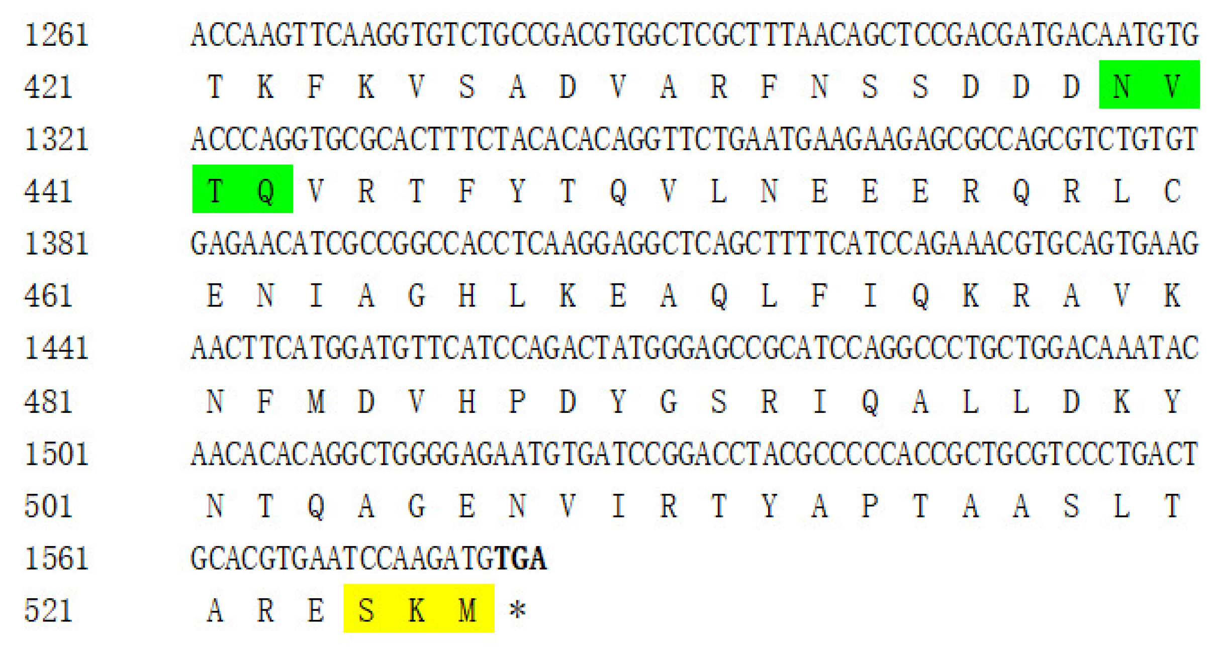
The nucleotide sequences of the AdCu/Zn-SOD and AdCAT genes were obtained and characterized from Yangtze sturgeon, and have been deposited in GenBank (accession no. MZ004926 and MZ004927). The ORFs of AdCu/Zn-SOD and AdCAT were 468 bp and 1581 bp, encoding proteins of 155 and 526 amino acids, respectively. Each nucleotide sequence consisted of an initiation codon and a stop codon (Figure 1 and Figure 2).
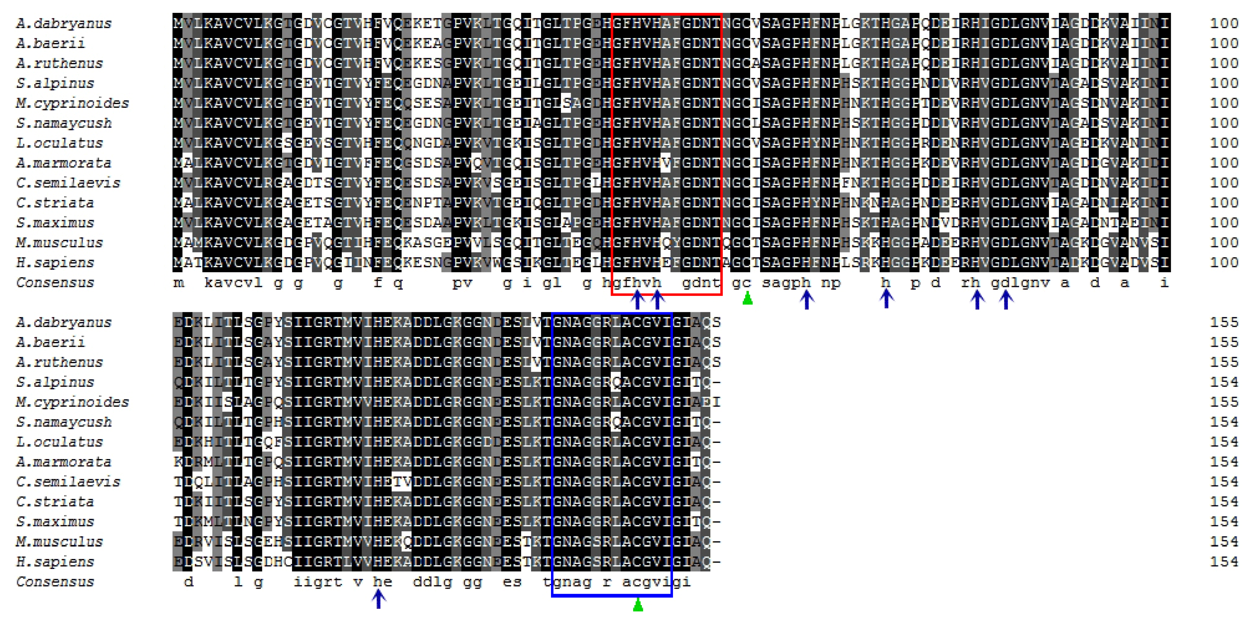
A BLASTX search showed that the protein sequence of AdCu/Zn-SOD was highly similar to many other teleost Cu/Zn-SODs, such as that from sterlet sturgeon, Acipenser ruthenus (98.06%), Arctic char, Salvelinus alpinus (81.17%), lake trout, Salvelinus namaycush (80.52%), nine-spined stickleback, Pungitius pungitius (79.87%), and marbled eel (79.22%) (Supplementary Materials Table S2), which revealed the high conservation of Cu/Zn-SOD in teleosts. Notably, two conserved signature sequences of the Cu/Zn-SOD family were found in AdCu/Zn-SOD: signature 1 (GFHVHAFGDNT) from position 45 to 55 and signature 2 (GNAGGRLACGVI) from position 139 to 150 residues. In addition, we also detected a single disulfide bond formed by four Cu2 + -binding active-site residues (His47, His49, His64, and His121), four Zn2 + -binding active-site residues (His64, His72, His81 and Asp84), and two cysteine residues (Cys58 and Cys147) from the deduced AdCu/Zn-SOD protein (Figure 1 and Figure 3). According to the results of the phylogenetic analysis, AdCu/Zn-SOD was clustered with Cu/Zn-SODs from other teleosts, but was most closely related to the Cu/Zn-SOD from sterlet sturgeon (Figure 4).
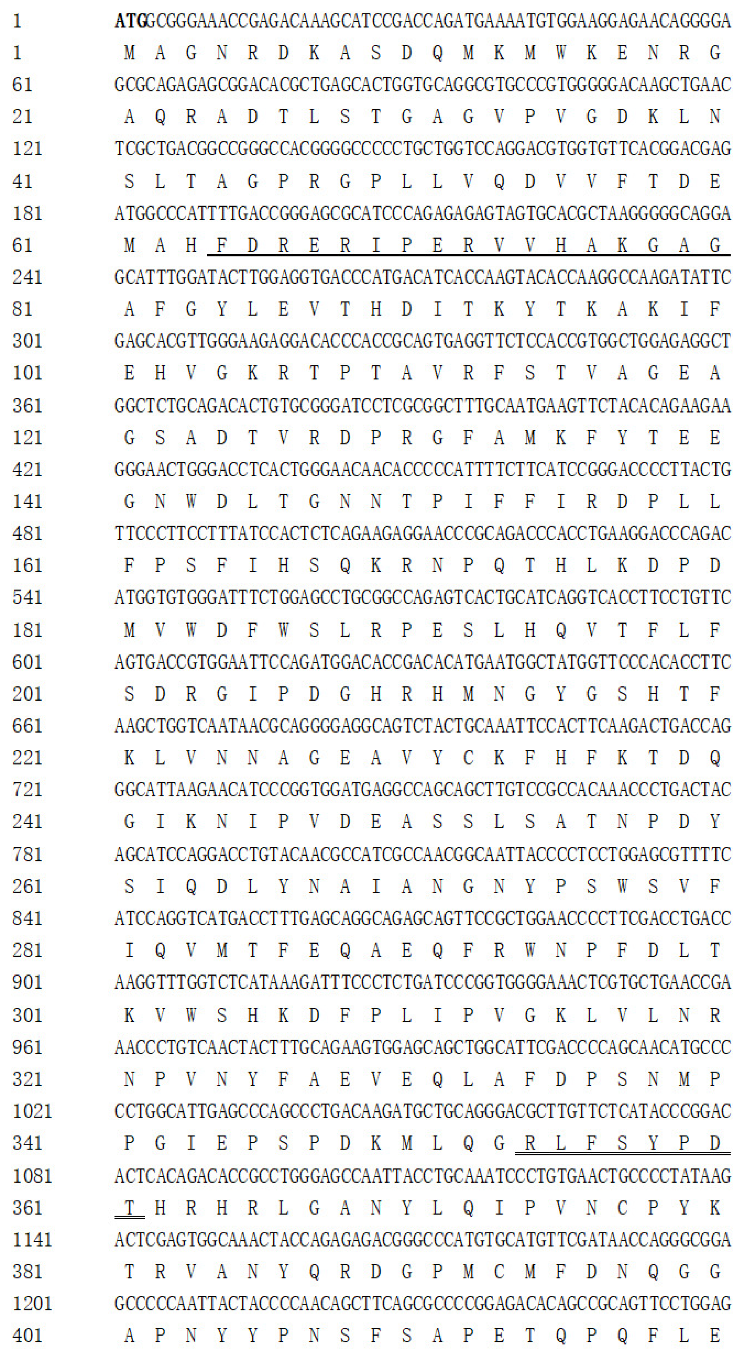
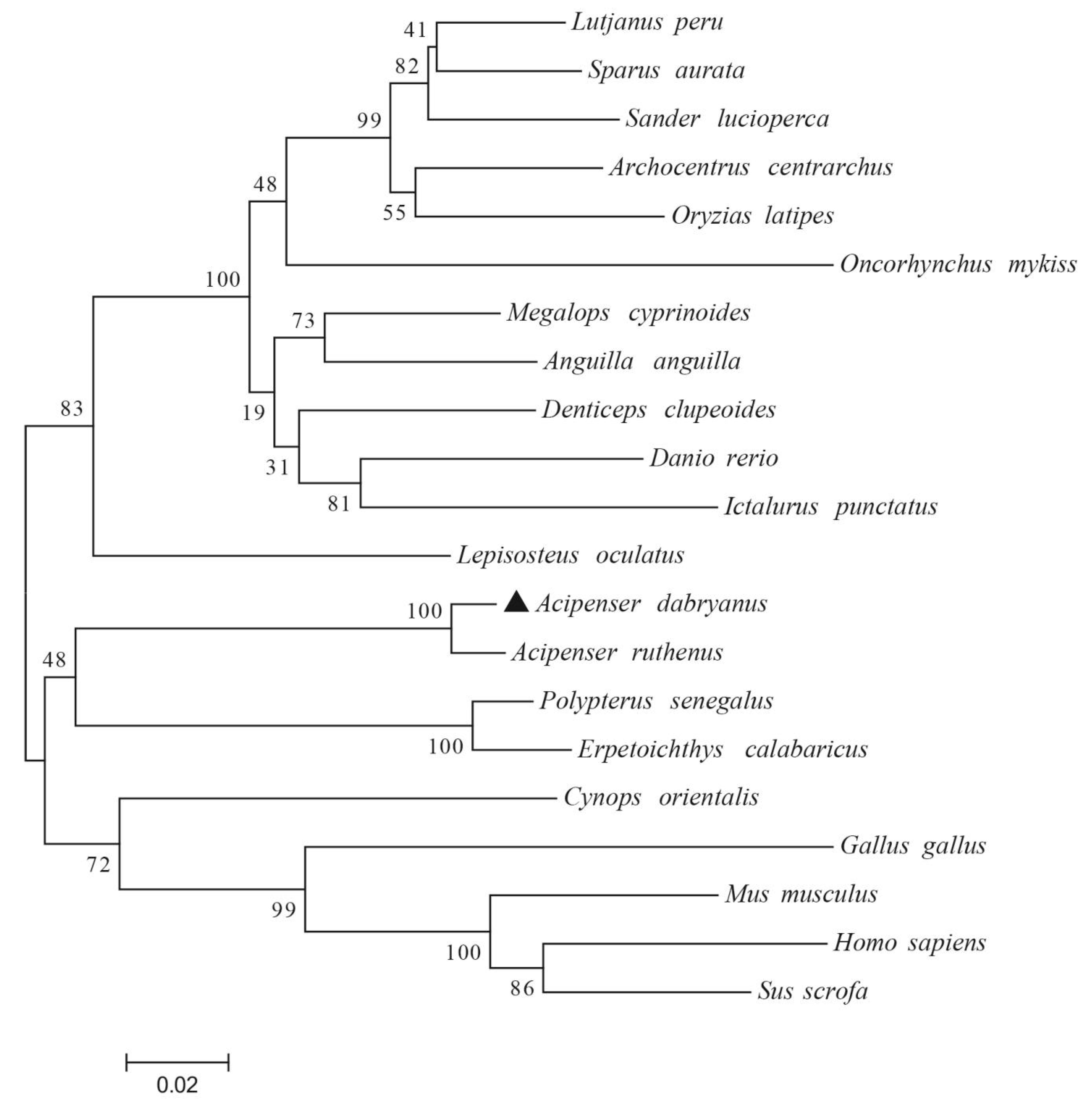
Regarding the deduced AdCAT protein sequence, the BLASTX search showed that it shared high affinities with many other teleost CATs deposited in GenBank, including those from sterlet sturgeon (98.10%), spotted gar, Lepisosteus oculatus (85.93%), senegal bichir, Polypterus senegalus (84.41%), ropefish, Erpetoichthys calabaricus (84.03%), and Pacific red snapper, Lutjanus peru (83.11%) (Supplementary Materials Table S3), which revealed high conservation of CAT in teleosts. In addition, we found typical catalase family signatures, including an active-site signature (FDRERIPERVVHAKGAG) from position 64 to 80 and a proximal heme-ligand signature motif (RLFSYPDTH) from position 354 to 361, which are known to be highly conserved among teleosts. Additionally, a well-conserved potential N-glycosylation site (NVTQ) from position 439 to 442 and a predicted peroxisome targeting signal (SKM) from position 524 to 526 were found in the derived amino acid sequence of AdCAT (Figure 2 and Supplementary Materials Figure S1). The phylogenetic analysis showed the AdCAT forms a clade with CAT sequences from other teleosts, and was most closely related to CAT from sterlet sturgeon (Figure 5).

3.2. Tissue Distribution of AdCu/Zn-SOD and AdCAT mRNA Expression

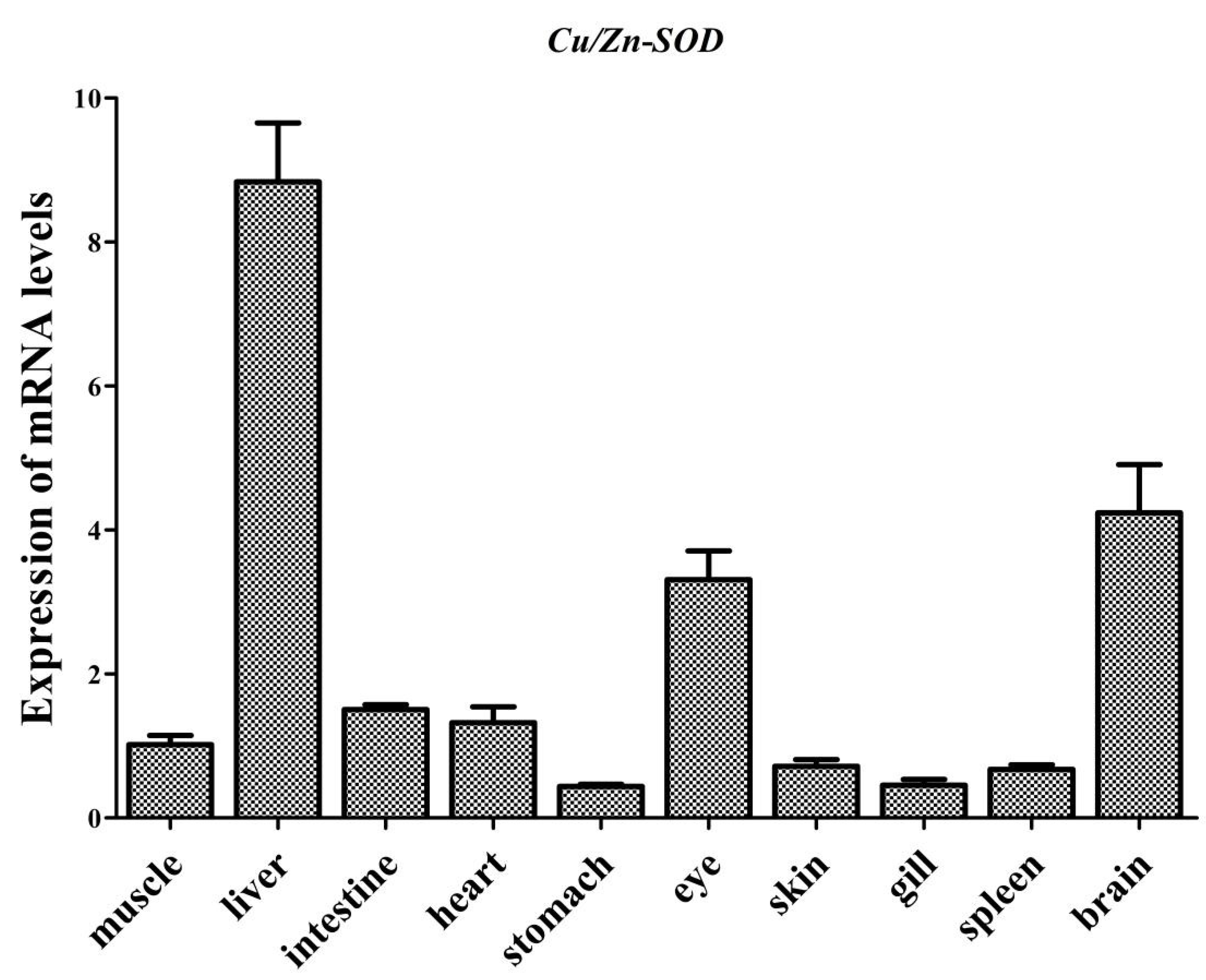
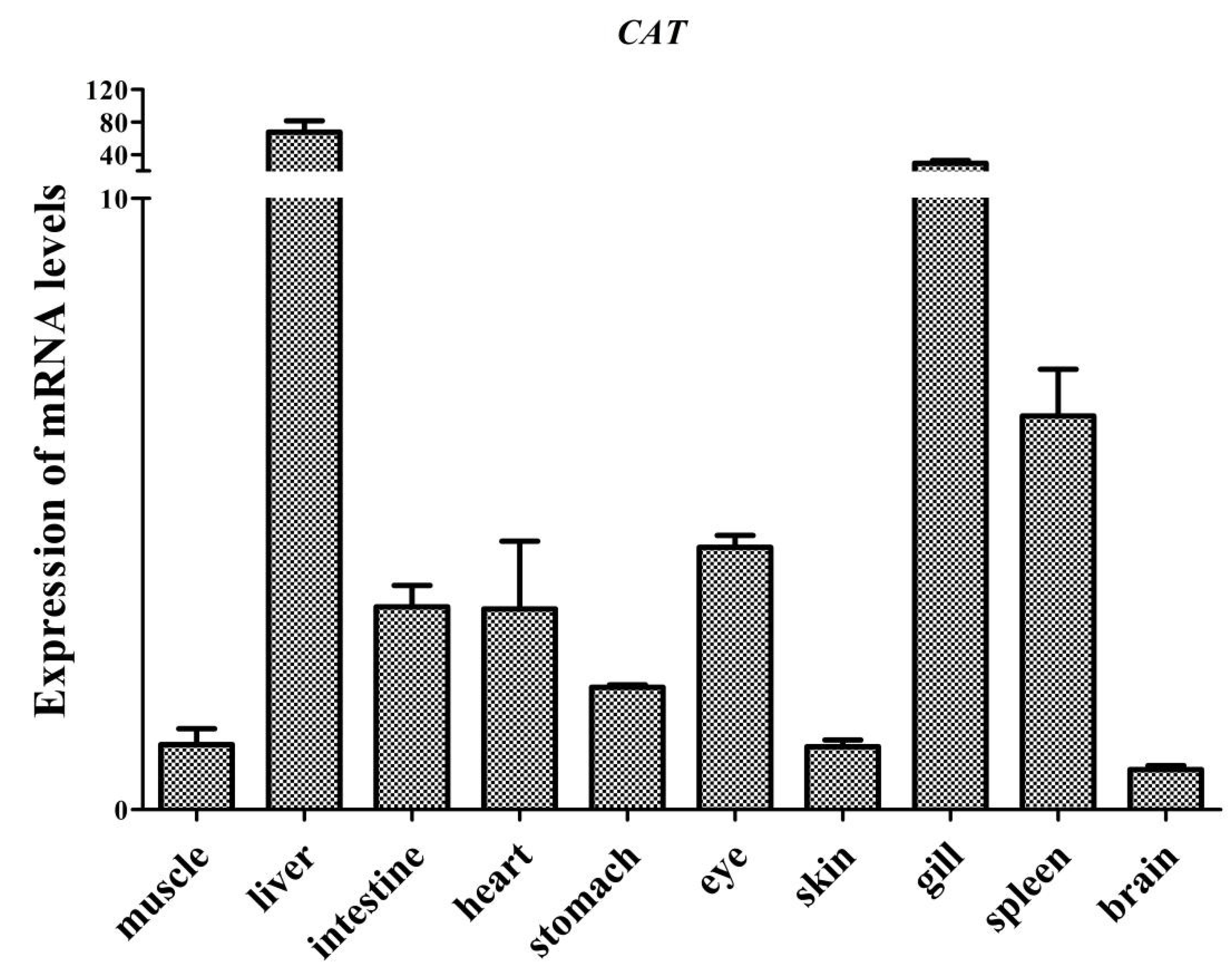
Tissue distributions of AdCu/Zn-SOD and AdCAT mRNA expression are shown in Figure 6 and Figure 7. They were expressed in all detected tissues but showed some variations. AdCu/Zn-SOD mRNA was highly expressed in the liver, brain, and eye, but showed low expression levels in the stomach, gill, spleen, skin, and muscle. AdCAT mRNA was also detected in all examined tissues, with higher expression in the brain, gill, and spleen, and lower expression in muscle, skin, brain, and stomach.

3.3. Effect of Fasting and Refeeding on Yangtze Sturgeon Liver and Intestine AdCu/Zn-SOD Expression

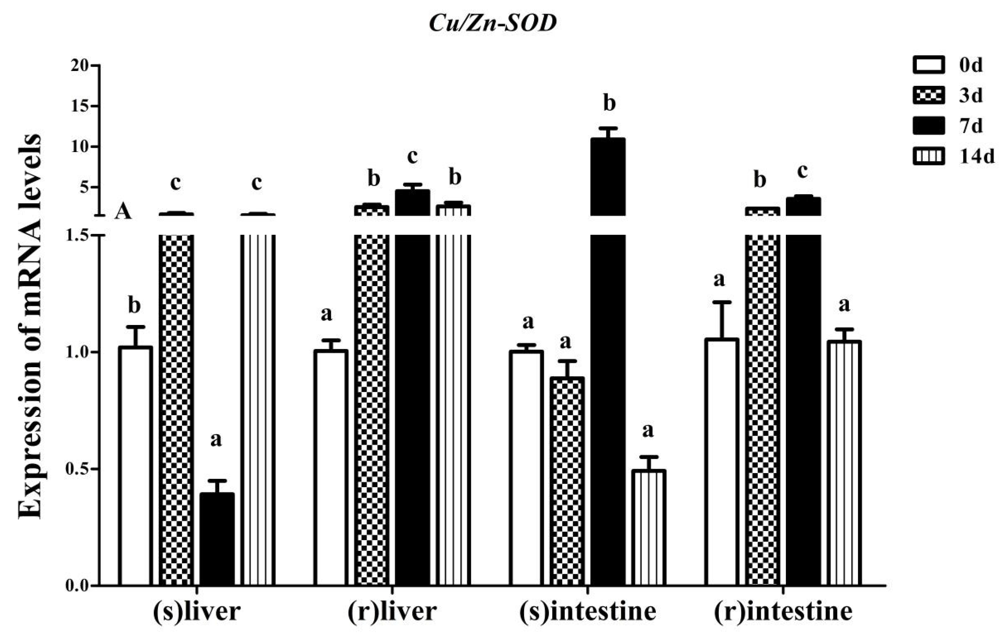
Transcriptional changes of AdCu/Zn-SOD in the liver and intestine in response to the feeding regime in Yangtze sturgeon are displayed in Figure 8. During fasting, the expression levels of AdCu/Zn-SOD in the liver were significantly higher in the 3- and 14-d groups (p < 0.05), but significantly lower in the 7-d group compared with that in the control group (p < 0.05). The AdCu/Zn-SOD mRNA level in the intestine increased significantly only in the 7-d group in comparison with that in the control group (p < 0.05). During refeeding, the expression levels of AdCu/Zn-SOD in the liver increased significantly in the 3-, 7-, and 14-d groups compared with those in the control group (p < 0.05). Similarly, the AdCu/Zn-SOD mRNA levels in the intestine increased significantly in the 3- and 7-d groups (p < 0.05).

3.4. Effect of Fasting and Refeeding on Yangtze Sturgeon Liver and Intestine AdCAT Expression

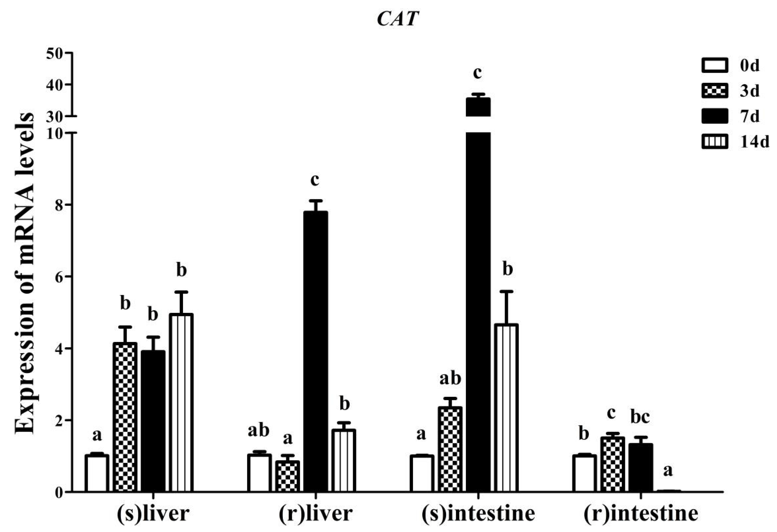
The effects of fasting and refeeding on the liver and intestine AdCAT mRNA expression in Yangtze sturgeon are shown in Figure 9. During fasting, the liver AdCAT transcript levels increased significantly in the 3-, 7-, and 14-d groups (p < 0.05). The intestinal AdCAT mRNA level was significantly higher in the 7-d group than in the other groups (p < 0.05), the expression levels of AdCAT mRNA were increased significantly in the 14-d group compared with that in the control group (p < 0.05), while there was no significant difference between the 14-d and the control group. During refeeding, the mRNA level of the liver AdCAT was significantly higher only in the 7-d group compared with that in the control group (p < 0.05). In the intestine of Yangtze sturgeon, the mRNA expression of AdCAT was significantly higher in the 3-d group than in the control group (p < 0.05), whereas a significant decrease was found in the 14-d group (p < 0.05).

4. Discussion

SOD and CAT are two major antioxidant enzymes in the fish antioxidant defense system. Their transcript levels are considered to accurately reflect a fish’s antioxidant capacity to the exclusion of interference of the biochemical origin of the oxidant stressors [16]. To provide a better understanding of the molecular mechanism through which starvation and refeeding regime affect the antioxidant function of Yangtze sturgeon, the present study investigated the mRNA profiles of Cu/Zn-SOD and CAT. Initially, we cloned and sequenced the ORFs of AdCu/Zn-SOD and AdCAT. The obtained amino acid sequences of AdCu/Zn-SOD showed several highly conserved sequences, including Cu2+ active sites and Zn2+ active sites, which were highly consistent with Cu/Zn-SODs from other teleosts. No signal peptide was found, suggesting that it would be localized in the cytoplasm, as supported by the cytoplasmic location of Cu/Zn-SODs in other teleosts, such as large yellow croaker [21], Pengze crucian carp [19], and silver carp, Hypophthalmichthys molitrix [37]. The sequence homology analysis demonstrated that Yangtze sturgeon Cu/Zn-SOD showed high similarity (66–98%) to Cu/Zn-SOD proteins from other teleosts. The phylogenetic analysis also indicated that AdCu/Zn-SOD clustered together with SODs from most teleosts, particularly that from sterlet sturgeon. Collectively, these analyses strongly suggest that AdCu/Zn-SOD plays similar roles to other SODs based on its possession of the essential characteristics of known SODs orthologs.
For AdCAT, multiple alignment showed that AdCAT shared high identity (79–98%) with CATs from other teleosts, which suggested that AdCAT might be present as a homotetramer like most CATs. There was no signal peptide in AdCAT, indicating that CAT is an intracellular protein, similar to most CAT homologs [38]. Moreover, the AdCAT protein sequence contains several conserved sequences and characteristic motifs, such as a proximal active site signature domain, a peroxisome targeting signal, and a heme-ligand signature motif, representing the typical characteristics of CAT family proteins. These findings were in line with several previous reports on CATs [19,24,38], strongly suggesting that AdCAT has similar functions to CATs from other species. The phylogenetic analysis demonstrated that AdCAT formed a clade with CATs from other teleosts, and was closely related to CAT from sterlet sturgeon. Taken together, these analyses indicated that AdCAT is a member of the CAT family, and it was expected to be a peroxisomal protein as in other teleosts.
The mRNA expression levels of AdCu/Zn-SOD and AdCAT in various tissues were detected using qRT-PCR. The results showed that both genes were expressed in all tissues tested. Notably, both AdCu/Zn-SOD and AdCAT mRNAs showed their highest expression in the liver. This was consistent with earlier research on Pengze crucian carp [19]. To date, many studies have revealed that the liver is a vital organ for the expression of antioxidant genes in teleosts [24,38,39,40]. The high expression of the antioxidant genes might be related to the fact that the liver is the key organ in which oxidative reactions and antioxidant defenses occur [39]. In fishes, the gills contact xenobiotics most directly and provide the first line of antioxidant defense [41]. Thus, it was not surprising that higher mRNA expression of AdCAT was found in the gills. Besides, we also noted high expression of AdCu/Zn-SOD mRNA in the brain. This supported the view that the brain is highly vulnerable to the effect of ROS because of its high oxygen consumption [42]. Therefore, the strong expression of the two antioxidant genes in the liver, gill, and brain suggested that they are closely related to oxidative reactions and antioxidant defenses of Yangtze sturgeon.
In the present study, the expression levels of AdCu/Zn-SOD and AdCAT were detected in the liver and intestine during starvation. The AdCu/Zn-SOD and AdCAT expression levels were increased significantly in the intestine of Yangtze sturgeon exposed to 7 d of starvation compared with that in the control group, while the liver AdCAT expression was always high during starvation. This indicated that starvation causes oxidative stress at the cellular level by creating energy deficits. Similar to our results, both SOD and CAT expression levels increased in the liver of roho, Labeo rohita fingerlings during starvation for a week [1], in the right lobe of the liver of large yellow croaker for 4 d [43], and in the liver of Nile tilapia, Oreochromis niloticus for 21 d [44]. In contrast to our results, Wang et al. [45] found that food deprivation did not significantly affect SOD and CAT expression levels in the gill, spleen, liver, and kidney of Schizothorax wangchiachii. The discrepancies among these data might, in part, be caused by differences in life-history stages, experimental conditions, fish species, starvation duration, food availability, and target tissues examined. Besides, the intestinal expression levels of AdCu/Zn-SOD and AdCAT decreased significantly during starvation for 14 d compared with that of 7 d. This was in line with a previous report that juvenile sterlet sturgeon showed decreased total antioxidant capacity when exposed to long-term starvation [46]. This phenomenon is associated with the elimination of fish’s resistance to persistent production of intracellular ROS. With increasing time of stress, the production of ROS exceeds the scavenging ability of endogenous antioxidants, resulting in oxidative damage to tissues. Notably, the liver AdCu/Zn-SOD expression level was significantly higher in the 14-d group than in the 7-d group. This agreed with the report of Lin et al. [47], in which the CytMnSOD and MtMnSOD expression levels in hemocytes of white shrimp, Litopenaeus vannamei, decreased significantly after starvation for 5 d, and then were markedly elevated after 14 d of fasting. One possible explanation could be that antioxidant function in fishes might be indirectly reactivated by the strengthened immunity that develops with increasing stress time. As was found in white shrimp, higher antioxidant gene expression levels (CytMnSOD and MtMnSOD) were observed together with a higher immune capability during long-term fasting [47]. However, the mechanisms are still not well understood and require further investigation. Interestingly, the expression levels of AdCu/Zn-SOD and AdCAT were all increased significantly in the intestine of Yangtze sturgeon exposed to 7 d of starvation compared with those in the liver, suggesting that the intestine of Yangtze sturgeon juveniles seems to be more sensitive to lipid peroxidation mediated by ROS that were induced during starvation and refeeding regimes than the liver subjected to the same regimes. A previous report showed that exposure of stellate sturgeon, Acipenser stellatus, to starvation for 7 d and 14 d resulted in differential change of the SOD and CAT activities in the liver and intestine [48]. The discrepancy could be attributed to the different tissue distributions of the genes, their protein functions, and their physiological functions. As is widely known, the liver and intestine play crucial roles in maintaining normal physiological functions. The liver is a major site of detoxification and xenobiotic metabolism, and the intestine plays a central role in digestion and absorption [49].
We also evaluated whether the oxidative stress induced by starvation could be relieved or eliminated by refeeding. In the present study, the mRNA levels of the liver and intestine AdCu/Zn-SOD in the 3- and 7-d groups as well as the expression of the liver AdCAT in the 7-d group increased significantly compared with those in the control group after 14 d of refeeding. Previously, we found that the activities of CAT increased significantly in the 3- and 7-d groups compared with those in the control group after 14 d of refeeding, which were consistent with the gene expression patterns in liver and intestine [35]. In addition, we also observed that the intestine, malondialdehyde (MDA) contents in the 3- and 7-d groups were reduced to the level of the control group, or were even lower than that of the control group, after 14 d of refeeding [35]. MDA is a stable product of lipid peroxidation that is produced after ROS-mediated oxidation of the polyunsaturated fatty acids (PUFA) [50]. Decreased MDA in fish exposed to stress is a sign of increasing antioxidant activity. Therefore, it might be considered that the production level of ROS after refeeding could be diminished in comparison to the level induced by starvation. On the other hand, the antioxidant function in liver and intestine was enhanced by refeeding. In agreement with our results, the activities of SOD and CAT were significantly increased after refeeding for 7 d or 14 d in the liver and intestine of stellate sturgeon [48,51], in the intestine of European sea bass, Dicentrarchus labrax [52], and in the liver of brown trout, Salmo trutta refed for 21 d [3]. In contrast, Yang et al. [10] found that SOD and CAT activities recovered to control values in the serum of Yangtze sturgeon after refeeding for 8 weeks. The apparent discrepancy might be related to the various tissues, fish ages, and refeeding periods. Furthermore, the liver and intestine AdCu/Zn-SOD and AdCAT mRNA levels decreased markedly in the 14-d group compared with those in the 7-d group after 14 d of refeeding. Similar to the results reported by Su et al. [53] that the SOD activity of the liver in blunt snout bream, Megalobrama amblycephala, decreased significantly during 3 weeks of refeeding after fasting for 20 d compared with that fasting for 15 d. One possibility was that the starvation period might have been too long or the refeeding period might have been too short to fully restore oxidative stress biomarkers and antioxidant genes expressions in the liver and intestine of Yangtze sturgeon.
The present work extends a previous study conducted by our research group on Yangtze sturgeon liver and intestine regarding the adaptability of this species to a starvation and refeeding regime. Our previous data showed that Yangtze sturgeon subjected to 3-d fasting and 14-d refeeding regime presented complete compensatory growth and were able to efficiently alleviate the oxidative stress by enhancing activities of the antioxidant enzymes in their liver and intestine. We concluded that this alternative feeding regime is worthy of further investigation. Therefore, Yangtze sturgeon juveniles possess a potential to adapt to a short starvation period introduced in the feeding schedule.

5. Conclusions

We successfully identified and characterized the Cu/Zn-SOD and CAT genes from Yangtze sturgeon, and AdCu/Zn-SOD and AdCAT were highly conserved in Yangtze sturgeon. The amino acid sequences of AdCu/Zn-SOD and AdCAT were also highly homologous with those of sterlet sturgeon. AdCu/Zn-SOD and AdCAT were expressed in all tissues examined, with the highest expression level in liver. Our results indicated that AdCu/Zn-SOD and AdCAT play important roles in protecting fish against starvation-induced oxidative stress. Moreover, this research revealed that the expression of liver and intestine AdCu/Zn-SOD and AdCAT in the 3- and 7-d groups increased significantly during starvation and refeeding. However, after a long period of fasting (14 d), the level of expression of AdCu/Zn-SOD was inhibited; even after refeeding for 14 d, the expression still did not recover. According to our previous and current data, Yangtze sturgeon juveniles possess the potential to adapt to a short starvation period introduced in the feeding regime. Our findings not only provide information regarding the physiological status of Yangtze sturgeon, but also promote the development and optimization of protective strategies for Yangtze sturgeon.

Supplementary Materials

The following are available online at https://www.mdpi.com/article/10.3390/fishes7010035/s1: Figure S1: Alignment of the full amino acid sequences of AdCAT with its homologs from other species; Table S1: Nutrients content of diets; Table S2: List of AdCu/Zn-SOD sequences used in this study. Amino acid identities (%) of Cu/Zn-SOD proteins from other species with AdCu/Zn-SOD; and Table S3: List of AdCAT sequences used in this study. Amino acid identities (%) of CAT proteins from other species with AdCAT.

Author Contributions

Writing—original draft, Q.S.; conceptualization, Q.S.; methodology, X.W.; software, Z.W. and X.W.; validation, X.W., formal analysis, Z.Z.; investigation, C.Q.; data curation, X.W.; writing—review and editing, Q.S.; visualization, X.X. and R.L.; supervision, Q.G.; project administration, X.W.; funding acquisition, X.W. All authors have read and agreed to the published version of the manuscript.

Funding

This research was funded by the foundation of Key Laboratory of Sichuan Province for Fishes Conservation and Utilization in the Upper Reaches of the Yangtze River (Grant No. NJTCCJSYSYS07) and Sichuan Science and Technology Program (Grant No. 2019YJ0398, 2021YFN0028).

Institutional Review Board Statement

The experimental animals used in this experiment were carried out following the Guide for the Care and Use of Laboratory Animals (Ministry of Science and Technology of China, 2006) and were approved by the Animal Care Advisory Committee of the Sichuan Academy of Agricultural Sciences (Approval No. 20180929001A).

Data Availability Statement

The data used during the current study are available from the corresponding author on reasonable request.

Conflicts of Interest

The authors declare no conflict of interest.

References

  1. Dar, S.A.; Srivastava, P.P.; Varghese, T.; Nazir, M.I.; Gupta, S.; Krishna, G. Temporal changes in superoxide dismutase, catalase, and heat shock protein 70 gene expression, cortisol and antioxidant enzymes activity of Labeo rohita fingerlings subjected to starvation and refeeding. Gene 2019, 692, 94–101. [Google Scholar] [CrossRef] [PubMed]
  2. Furné, M.; Sanz, A.; García-Gallego, M.; Hidalgo, M.C.; Domezain, A.; Domezain, J.; Morales, A.E. Metabolic organization of the sturgeon Acipenser naccarii: A comparative study with rainbow trout Oncorhynchus mykiss. Aquaculture 2009, 289, 161–166. [Google Scholar] [CrossRef]
  3. Bayir, A.; Sirkecioglu, A.N.; Bayir, M.; Haliloglu, H.I.; Kocaman, E.M.; Aras, N.M. Metabolic responses to prolonged starvation, food restriction, and refeeding in the brown trout, Salmo trutta: Oxidative stress and antioxidant defenses. Comp. Biochem. Physiol. B 2011, 159, 191–196. [Google Scholar] [CrossRef] [PubMed]
  4. Rios, F.S.; Kalinin, A.L.; Rantin, F.T. The effects of long-term food deprivation on respiration and haematology of the neotropical fish Hoplias malabaricus. J. Fish Biol. 2002, 61, 85–95. [Google Scholar] [CrossRef]
  5. Furné, M.; Morales, A.E.; Trenzado, C.E.; García-Gallego, M.; Carmen Hidalgo, M.; Domezain, A.; Sanz Rus, A. The metabolic effects of prolonged starvation and refeeding in sturgeon and rainbow trout. J. Comp. Physiol. B 2012, 182, 63–76. [Google Scholar] [CrossRef]
  6. Slater, J.J.; Lankford, T.E.; Buckel, J.A. Overwintering ability of young-of-the-year bluefish Pomatomus saltatrix: Effect of ration and cohort of origin on survival. Mar. Ecol. Prog. Ser 2007, 339, 259–269. [Google Scholar] [CrossRef] [Green Version]
  7. Azodi, M.; Ebrahimi, E.; Motaghi, E.; Morshedi, V. Metabolic responses to short starvation and re-feeding in rainbow trout (Oncorhynchus mykiss). Ichthyol. Res 2015, 62, 177–183. [Google Scholar] [CrossRef]
  8. Naisbitt, C.; Davies, S. Starvation, exercise and the stress response. Anaesth. Intensive Care 2017, 18, 508–512. [Google Scholar] [CrossRef]
  9. Wu, X.Y.; Chen, Y.Y.; Lai, J.S.; Liu, Y.; Long, Z.H. Effects of starvation and refeeding on growth performace, appetite, GH-IGFs axis levels and digestive function of Acipenser dabryanus. Brit. J. Nutr. 2020, 126, 1–39. [Google Scholar]
  10. Yang, S.; He, K.; Yan, T.; Wu, H.; Zhou, J.; Zhao, L.L.; Wang, Y.; Gong, Q. Effect of starvation and refeeding on oxidative stress and antioxidant defenses in Yangtze sturgeon (Acipenser dabryanus). Fish Physiol. Biochem 2019, 45, 987–995. [Google Scholar] [CrossRef]
  11. Choi, C.Y.; Choi, J.Y.; Choi, Y.J.; Yoo, J.H. Physiological effects of various light spectra on oxidative stress by starvation in olive flounder, Paralichthys olivaceus. Mol. Cell. Toxicol. 2018, 14, 399–408. [Google Scholar] [CrossRef]
  12. Regoli, F.; Giuliani, M.E. Oxidative pathways of chemical toxicity and oxidative stress biomarkers in marine organisms. Mar. Environ. Res. 2014, 93, 106–117. [Google Scholar] [CrossRef]
  13. Nikahval, B.; Hosseini, Z.; Nazifi, S.; Foroud, M. Evaluation of antioxidant enzymes changes in synovial fluid and blood, following experimental osteoarthritis in dogs. Comp. Clin. Path. 2015, 24, 23–27. [Google Scholar] [CrossRef]
  14. Tiwari, M.K.; Hägglund, P.M.; Møller, I.M.; Davies, M.J.; Bjerrum, M.J. Copper ion/H2O2 oxidation of Cu/Zn-Superoxide dismutase: Implications for enzymatic activity and antioxidant action. Redox. Biol. 2019, 26, 101262. [Google Scholar] [CrossRef] [PubMed]
  15. Gómez-Anduro, G.A.; Barillas-Mury, C.V.; Peregrino-Uriarte, A.B.; Gupta, L.; Gollas-Galván, T.; Hernández-López, J.; Yepiz-Plascencia, G. The cytosolic manganese superoxide dismutase from the shrimp Litopenaeus vannamei: Molecular cloning and expression. Dev. Comp. Immunol. 2006, 30, 893–900. [Google Scholar] [CrossRef] [PubMed]
  16. Martins, D.; English, A.M. SOD1 oxidation and formation of soluble aggregates in yeast: Relevance to sporadic ALS development. Redox. Biol. 2014, 2, 632–639. [Google Scholar] [CrossRef] [Green Version]
  17. Olson, K.R.; Gao, Y.; DeLeon, E.R.; Arif, M.; Arif, F.; Arora, N.; Straub, K.D. Catalase as a sulfide-sulfur oxido-reductase: An ancient (and modern?) regulator of reactive sulfur species (RSS). Redox. Biol. 2017, 12, 325–339. [Google Scholar] [CrossRef]
  18. Yu, H.; Deng, W.; Zhang, D.; Gao, Y.; Ji, H. Antioxidant defenses of Onychostoma macrolepis in response to thermal stress: Insight from mRNA expression and activity of superoxide dismutase and catalase. Fish Shellfish Immunol. 2017, 66, 50–61. [Google Scholar] [CrossRef]
  19. Li, M.; Zheng, Y.; Liang, H.; Zou, L.; Sun, J.; Zhang, Y.; Qin, F.; Liu, S.; Wang, Z. Molecular cloning and characterization of cat, gpx1 and Cu/Zn-sod genes in pengze crucian carp (Carassius auratus var. Pengze) and antioxidant enzyme modulation induced by hexavalent chromium in juveniles. Comp. Biochem. Physiol. C 2013, 157, 310–321. [Google Scholar] [CrossRef]
  20. Wang, X.; Wang, L.; Ren, Q.; Yin, S.; Liang, F.; Jia, Y. Two superoxide dismutases (SODs) respond to bacterial challenge identified in the marbled eel Anguilla marmorata. Aquaculture 2016, 451, 316–325. [Google Scholar] [CrossRef]
  21. Liu, H.; He, J.; Chi, C.; Gu, Y. Identification and analysis of icCu/Zn-SOD, Mn-SOD and ecCu/Zn-SOD in superoxide dismutase multigene family of Pseudosciaena crocea. Fish Shellfish Immunol. 2015, 43, 491–501. [Google Scholar] [CrossRef] [PubMed]
  22. Ren, H.; Li, J.; Liu, P.; Ren, X.; Song, T.; Gao, G.; Li, D.; Liu, S. Cloning of catalase gene and antioxidant genes in Scophthalmus maximus response to metalloprotease of Vibrio anguillarum stress. J. Oceanol. Limnol. 2021. [Google Scholar] [CrossRef]
  23. Elvitigala, D.A.S.; Premachandra, H.K.A.; Whang, I.; Priyathilaka, T.T.; Kim, E.; Lim, B.S.; Jung, H.B.; Yeo, S.Y.; Park, H.C.; Lee, J. Marine teleost ortholog of catalase from rock bream (Oplegnathus fasciatus): Molecular perspectives from genomic organization to enzymatic behavior with respect to its potent antioxidant properties. Fish Shellfish Immunol. 2013, 35, 1086–1096. [Google Scholar] [CrossRef] [PubMed]
  24. Shen, B.; Wei, K.; Ding, Y.; Zhang, J. Molecular cloning, mRNA expression and functional characterization of a catalase from Chinese black sleeper (Bostrychus sinensis). Fish Shellfish Immunol. 2020, 103, 310–320. [Google Scholar] [CrossRef] [PubMed]
  25. Zhang, H.; Wei, Q.W.; Du, H.; Li, L.X. Present status and risk for extinction of the Dabry’s sturgeon (Acipenser dabryanus) in the Yangtze River watershed: A concern for intensified rehabilitation needs. J. Appl. Ichthyol. 2011, 27, 181–185. [Google Scholar] [CrossRef]
  26. Wu, J.M.; Wei, Q.W.; Du, H.; Wang, C.Y.; Zhang, H. Initial evaluation of the release programme for Dabry’s sturgeon (Acipenser dabryanus Duméril, 1868) in the upper Yangtze River. J. Appl. Ichthyol. 2014, 30, 1423–1427. [Google Scholar] [CrossRef]
  27. Zhuang, P.; Ke, F.E.; Wei, Q.; He, X.; Cen, Y. Biology and life history of Dabry’s sturgeon, Acipenser dabryanus, in the Yangtze River. Environ. Biol. Fish 1997, 48, 257–264. [Google Scholar] [CrossRef]
  28. Liu, Y.; Chen, Y.; Quan, G.; Lai, J.; Deng, X. Paternity assignment in the polyploid Acipenser dabryanus based on a novel microsatellite marker system. PLoS ONE 2017, 12, e0185280. [Google Scholar] [CrossRef] [Green Version]
  29. Hu, Y.; Liu, X.; Yang, J.; Xiao, K.; Wang, B.; Du, H. Development of 17 novel cross-Species microsatellite markers for Dabry’s Sturgeon (Acipenser dabryanus) from Chinese sturgeon (Acipenser sinensis) via next-generation sequencing. Pak. J. Zool. 2019, 51, 2381–2384. [Google Scholar] [CrossRef]
  30. Ruan, R.; Feng, T.; Li, Y.; Yue, H.; Ye, H.; Du, H.; Liu, Q.; Ruan, L.; Li, H.; Wei, Q. Screening and identification of female-specific DNA sequences in octaploid sturgeon using comparative genomics with high-throughput sequencing. Genomics 2021, 113, 4237–4244. [Google Scholar] [CrossRef]
  31. Chen, Y.; Gong, Q.; Song, M.; Lai, J.; Sun, J.; Liu, Y. Identification and characterization of three novel antimicrobial peptides from Acipenser dabryanus. Fish Shellfish Immunol. 2019, 88, 207–216. [Google Scholar] [CrossRef] [PubMed]
  32. Chen, Y.; Liu, Y.; Du, J.; Song, M.; Lai, J.; Gong, Q. Isolation and characterization of 47 SNP markers in the critically endangered Acipenser dabryanus. Conserv. Genet. Resour 2020, 12, 9–12. [Google Scholar] [CrossRef]
  33. Chen, Y.; Wu, X.; Lai, J.; Liu, Y.; Song, M.; Li, F.; Gong, Q. Full-length transcriptome sequencing and identification and immune response of TRIM genes in Dabry’s sturgeon (Acipenser dabryanus). Aquaculture 2021, 538, 736599. [Google Scholar] [CrossRef]
  34. Han, P.; Wang, S.; Zhang, Q.; Zhang, S.; Shao, R.; Xu, W.; Zhang, W.; Xu, Q.; Wei, Q.; Qi, Z. Molecular characterization and expression analysis of TLR1 and TLR4 from the endangered fish Dabry’s sturgeon (Acipenser dabryanus). Dev. Comp. Immunol. 2018, 86, 180–188. [Google Scholar] [CrossRef]
  35. Wu, X.Y.; Chen, Y.Y.; Lai, J.S.; Liu, Y.; Song, M.J.; Gong, Q.; Long, Z.H. Effects of starvation and re-feeding on antioxidative function of liver, intestine and muscle in Acipenser dabryanus. J. South. Agric. 2020, 17, 100370. [Google Scholar]
  36. Qin, C.J.; Wen, Z.Y.; Wang, J.; He, Y.; Yuan, D.Y.; Li, R. Uncoupling protein 1 in snakehead (Channa argus): Cloning, tissue distribution, and its expression in response to fasting and refeeding. Comp. Biochem. Physiol. A 2018, 225, 1–6. [Google Scholar] [CrossRef]
  37. Zhang, Z.W.; Li, Z.; Liang, H.W.; Li, L.; Luo, X.Z.; Zou, G.W. Molecular cloning and differential expression patterns of copper/zinc superoxide dismutase and manganese superoxide dismutase in Hypophthalmichthys molitrix. Fish Shellfish Immunol. 2011, 30, 473–479. [Google Scholar] [CrossRef]
  38. Ren, H.; Li, J.; Li, J.; Ying, Y.; Ge, H.; Li, D.; Yu, T. Cloning of catalase and expression patterns of catalase and selenium-dependent glutathione peroxidase from Exopalaemon carinicauda in response to low salinity stress. Acta. Oceanol. Sin. 2015, 34, 52–61. [Google Scholar] [CrossRef]
  39. Shi, Q.; Rong, H.; Hao, M.; Zhu, D.; Aweya, J.J.; Li, S.; Wen, X. Effects of dietary Sargassum horneri on growth performance, serum biochemical parameters, hepatic antioxidant status, and immune responses of juvenile black sea bream Acanthopagrus schlegelii. J. Appl. Phycol. 2019, 31, 2103–2113. [Google Scholar] [CrossRef]
  40. Choi, C.Y.; Shin, H.S.; Choi, Y.J.; Kim, N.N.; Lee, J.; Kil, G.S. Effect of LED light spectra on starvation-induced oxidative stress in the cinnamon clownfish Amphiprion melanopus. Comp. Biochem. Physiol. A 2012, 163, 357–363. [Google Scholar] [CrossRef]
  41. Martínez-Álvarez, R.M.; Morales, A.E.; Sanz, A. Antioxidant defenses in fish: Biotic and abiotic factors. Rev. Fish Biol. Fish. 2005, 15, 75–88. [Google Scholar] [CrossRef]
  42. Bouayed, J.; Rammal, H.; Soulimani, R. Oxidative stress and anxiety: Relationship and cellular pathways. Oxid. Med. Cell. Longev. 2009, 2, 63–67. [Google Scholar] [CrossRef] [PubMed]
  43. Zheng, J.L.; Zhu, Q.L.; Shen, B.; Zeng, L.; Zhu, A.Y.; Wu, C.W. Effects of starvation on lipid accumulation and antioxidant response in the right and left lobes of liver in large yellow croaker Pseudosciaena crocea. Ecol. Indic. 2016, 66, 269–274. [Google Scholar] [CrossRef]
  44. Sakyi, M.E.; Cai, J.; Tang, J.; Abarike, E.D.; Xia, L.; Li, P.; Kuebutornye, F.K.A.; Zou, Z.; Liang, Z.; Jian, J. Effects of starvation and subsequent re-feeding on intestinal microbiota, and metabolic responses in Nile tilapia, Oreochromis niloticus. Aquacult. Rep. 2020, 17, 100370. [Google Scholar] [CrossRef]
  45. Wang, C.; Liang, Y.; Fang, Y.; Chang, X. Effects of cyclical short-term food deprivation and refeeding on compensatory growth and gene expression of SOD, GPX and HSP70 in Schizothorax wangchiachii. Fish Shellfish Immunol. 2019, 94, 628–633. [Google Scholar] [CrossRef]
  46. Feng, G.; Shi, X.; Huang, X.; Zhuang, P. Oxidative stress and antioxidant defenses after long-term fasting in blood of Chinese sturgeon (Acipenser sinensis). Procedia Environ. Sci. 2011, 8, 469–475. [Google Scholar] [CrossRef] [Green Version]
  47. Lin, Y.C.; Chen, J.C.; Man, S.C.; Morni, W.W.; Shahrir, N.; Cheng, S.Y.; Hu, C.H. Modulation of innate immunity and gene expressions in white shrimp Litopenaeus vannamei following long-term starvation and re-feeding. Res. Immunol. 2012, 2, 148–156. [Google Scholar] [CrossRef] [Green Version]
  48. Florescu, I.E.; Georgescu, S.E.; Dudu, A.; Balaș, M.; Voicu, S.; Grecu, I.; Dediu, L.; Dinischiotu, A.; Costache, M. Oxidative stress and antioxidant defense mechanisms in response to starvation and refeeding in the intestine of stellate sturgeon (Acipenser stellatus) Juveniles from Aquaculture. Animals 2021, 11, 76. [Google Scholar] [CrossRef]
  49. Ticho, A.L.; Malhotra, P.; Dudeja, P.K.; Gill, R.K.; Alrefai, W.A. Bile acid receptors and gastrointestinal functions. Liver Res. 2019, 3, 31–39. [Google Scholar] [CrossRef]
  50. Datta, S.; Kundu, S.; Ghosh, P.; De, S.; Ghosh, A.; Chatterjee, M. Correlation of oxidant status with oxidative tissue damage in patients with rheumatoid arthritis. Clin. Rheumatol. 2014, 33, 1557–1564. [Google Scholar] [CrossRef]
  51. Florescu, I.E.; Burcea, A.; Popa, G.O.; Dudu, A.; Georgescu, S.E.; Balas, M.; Dinescu, S.; Voicu, S.; Grecu, I.; Dediu, L.; et al. Effects of starvation and refeeding on growth performance and stress defense mechanisms of stellate sturgeon Acipenser stellatus juveniles from aquaculture. Acta Biochim. Pol. 2019, 66, 1–13. [Google Scholar] [CrossRef] [PubMed] [Green Version]
  52. Antonopoulou, E.; Kentepozidou, E.; Feidantsis, K.; Roufidou, C.; Despoti, S.; Chatzifotis, S. Starvation and re-feeding affect Hsp expression, MAPK activation and antioxidant enzymes activity of European sea bass (Dicentrarchus labrax). Comp. Biochem. Physiol. A 2013, 165, 79–88. [Google Scholar] [CrossRef] [PubMed]
  53. Su, Y.L.; Ge, X.P.; Sun, S.M.; Zhu, J.; Zhang, W.X.; Yu, H. Effects of compensatory feeding after starvation on growth performance, serum biochemical indexes, intestinal digestive enzyme activities and hepatic antioxidant enzyme activities of juvenile blunt snout bream (Megalobrama amblycephal) in Summer. Chinese. J. Animal. Nutr. 2017, 29, 4198–4206. [Google Scholar]
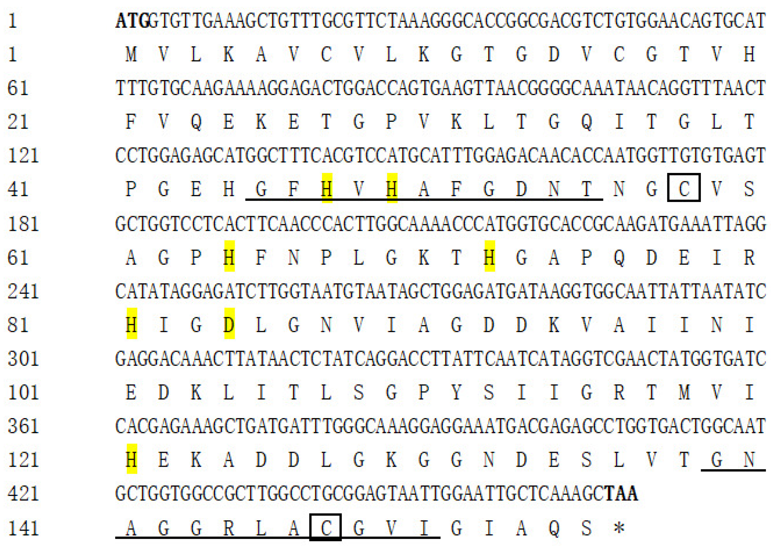
Figure 1. The nucleotide and deduced amino acid sequence of AdCu/Zn-SOD (Cu/Zn superoxide dismutase from Yangtze sturgeon). Two conserved signature sequences are marked by underlining; four Cu2+-binding active sites (His47, His49, His64, and His121) and four Zn2+-binding active sites (His64, His72, His81 and Asp84) are highlighted in yellow; and two cysteine residues (Cys58 and Cys147) are boxed, * indicates a stop codon.
Fishes 07 00035 g001
Figure 2. The nucleotide and deduced amino acid sequence of AdCAT (Catalase from Yangtze sturgeon). The catalase proximal active-site signature domain is marked by single underlining, the catalase proximal heme-ligand signature domain is marked by double underlining, the N-glycosylation site is highlighted in green, and the peroxisome targeting signal is highlighted in yellow, * indicates a stop codon.
Fishes 07 00035 g002aFishes 07 00035 g002b
Figure 3. Alignment of the full amino acid sequences of AdCu/Zn-SOD with its homologs from other species. The two conserved signature sequences of AdCu/Zn-SOD are highlighted by red and blue boxes, respectively. Four Cu2+-binding active sites (His47, His49, His64, and His121) and four Zn2 + -binding active sites (His64, His72, His81 and Asp84) are marked by arrows. Two cysteine residues (Cys58 and Cys147) are marked with green triangles.
Fishes 07 00035 g003
Figure 4. Phylogenetic tree of the amino acid sequence of the AdCu/Zn-SOD protein. ▲ was used to highlight Acipenser dabryanus.
Fishes 07 00035 g004
Figure 5. Phylogenetic tree of the amino acid sequence of the AdCAT protein. ▲ was used to highlight Acipenser dabryanus.
Fishes 07 00035 g005
Figure 6. Distribution of AdCu/Zn-SOD gene expression in different tissues of Yangtze sturgeon.
Fishes 07 00035 g006
Figure 7. Distribution of AdCAT gene expression in different tissues of Yangtze sturgeon.
Fishes 07 00035 g007
Figure 8. Relative expression of AdCu/Zn-SOD in the liver of Yangtze sturgeon during fasting and refeeding, s: starvation, r: refeeding. Data represent means ± SEM of six replicates. The different lowercase letters represent significant differences (p < 0.05).
Fishes 07 00035 g008
Figure 9. Relative expression of AdCAT in the intestine of Yangtze sturgeon during fasting and refeeding, s: starvation, r: refeeding. Data represent means ± SEM of six replicates. The different lowercase letters represent significant differences (p < 0.05).
Fishes 07 00035 g009
Table 1. PCR primers and optimal annealing temperatures (OATs) used for cloning and gene expression studies.
PrimerSequence (5′ to 3′)OAT (°C)
AdCu/Zn-SOD-FATGGTGTTGAAAGCTGTTTGCG51
AdCu/Zn-SOD-RGACAGAAACACTGAAGATTAGC
AdCAT-FATGGCGGGAAACCGAGAC51
AdCAT-RTCACATCTTGGATTCACGTGCA
AdCu/Zn-SOD-qFAAACTTATAACTCTATCAGGACCTTATTCA54.9
AdCu/Zn-SOD-qRCAGTCACCAGGCTCTCGTCAT
AdCAT-qFCCTGTGAACTGCCCCTAT60.4
AdCAT-qRACATTGTCATCGTCGGAG
β-actin-qFGACCGAGGCACCCCTGAAC54.9
β-actin-qRGATGGGCACTGTGTGTGTGAC
Publisher’s Note: MDPI stays neutral with regard to jurisdictional claims in published maps and institutional affiliations.