1. Introduction
The American eel (
Anguilla rostrata) is a typical economic fish species that migrates to the sea to spawn. Its life cycle encompasses the yellow eel stage, which grows in freshwater, and the silver eel stage, which reproduces in the ocean. It is widely distributed in the freshwater basins of the West Indies and the Caribbean Sea region in western Venezuela, South America [
1,
2]. Mature individuals migrate to the Sargasso Sea (19.5°–29.0° N, 52°–79° W) to complete spawning, after which the parent eels die due to energy depletion [
3,
4]. Currently, full artificial breeding technology for American eels has not yet been achieved. One of the core bottlenecks is the unclear synergistic regulation mechanism between nutrient supply, growth and development, and gonadal maturation during the maturation process. Existing research has mostly focused on the effects of hormones on gonadal development [
5,
6], ignoring the synergistic effects of nutritional conditions (such as feeding/starvation) on morphological characteristics and gonadal development. This makes it difficult to simultaneously improve breeding performance and product quality through nutritional regulation in practical production.
Existing research has confirmed significant gender differences in the life history of American eels. Female eels adopt a “maximizing body size” strategy, with metamorphic age positively correlated with latitude, and a growth rate significantly higher than that of male eels. Male eels, on the other hand, tend to adopt a “minimizing time” strategy, entering the migratory breeding stage earlier [
7].
Anguilla fish have a unique life history of deep-sea migration. During the process of migrating from freshwater habitats to marine breeding grounds (which may last for 3–6 months or even longer in nature), parent eels undergo silvering metamorphosis, during which their digestive tract naturally shrinks, feeding behavior stops, and they rely entirely on the consumption of muscle and liver reserves to maintain the enormous energy consumption required for long-distance migration and gonadal development [
8]. Studies have shown that American eels have strong hunger resistance. Walsh et al. [
9] confirmed that they can maintain normal metabolic levels even after fasting for 6 months at low temperatures.
During the artificial maturation process of American eels, hunger stress can disrupt the reproductive endocrine homeostasis of the body, disrupt hormone secretion balance, alter the normal external morphological characteristics of eels, and inhibit gonadal proliferation and development; feeding high-protein, lipid-rich fresh prey can stabilize the function of the reproductive endocrine axis, maintain the normal external morphological characteristics of eels, ensure the orderly regulation of gonadal differentiation, development, and maturation by hormones, and provide key physiological support for the normal development of gonads during artificial maturation. From the perspective of artificial breeding, orderly gonadal maturation guarantees gamete quality, synchronizes the development of male and female individuals to facilitate artificial spawning induction, reduces damage caused by excessive exogenous hormone stimulation, improves fertilization and larval survival rates, cuts breeding costs, and is highly beneficial for large-scale reproduction.
However, there is still a lack of systematic research on the synchronous changes in the external morphology and gonadal development of American eels during the maturation period mediated by hormone regulation of feeding and starvation status. It is difficult to accurately elucidate the correlation mechanism between nutritional levels, endocrine hormones, external morphology, and gonadal development, and provide precise theoretical support and technical guidance for the nutritional control and artificial maturation optimization of American eels in fully artificial reproduction.
The purpose of this work is to study the regulation of exogenous nutrition input on the reproductive endocrine level, external morphological characteristics and gonadal development of the American eel under artificial ripening stress by setting the starvation group, simulating the natural migration and fasting state of eels, as the control group, and the feeding group as the experimental intervention group. This study systematically analyzed the external morphological changes, oocyte development, reproductive endocrine hormone expression characteristics, and gonadal development differences during the maturation stage of the American eel under feeding conditions. The aim of this study is to elucidate the role of exogenous nutrition in maintaining endocrine homeostasis and promoting gonadal development during artificial maturation of American eels by comparing the synergistic changes and regulatory differences in the levels of “external reproductive hormone gonadal development” between two groups of eels. This study also aims to elucidate the intrinsic relationship between nutrition, hormones, morphology, and gonadal development, providing important theoretical basis and data support for the management strategies of parent-fish nutrition enhancement and artificial maturation optimization in the fully artificial reproduction of American eels.
2. Materials and Methods
2.1. Experimental Materials
This study was conducted from January 2024 to September 2024 at the Ninghai Research Center in Zhejiang. The period from January 2024 to June 2024 was the stage of nutritional strengthening, and the period from July to September 2024 was the stage of hormone-induced artificial maturing. The experimental materials were all taken from the Zhejiang Ninghai Research Center of the East China Sea Fisheries Research Institute, Chinese Academy of Fishery Sciences. A total of 48 healthy American eels with normal appearance were selected for the experiment, with 24 in the feeding group and 24 in the starvation group. The initial weight of the feeding group was (308.43 ± 36.99 g), and the initial weight of the starvation group was (291.67 ± 63.31 g). The American eels from the feeding and starvation group were placed in an indoor circulating-water nutrient-enhanced freshwater aquaculture pool at the base, which is 4 m by 8 m by 2 m long and has a water level of 1 m. The indoor cement pool is equipped with cooling and heating machines to control the temperature, with the water temperature controlled at (15 ± 1 °C). Black sunshade net is used for shading. Both the feeding and starvation group used female American eels as experimental fish. During the subsequent collection process, the starvation group fasted throughout the entire artificial maturation process.
2.2. Experimental Design and Feeding Management
The experimental process included two functional stages: nutrient-enhanced cultivation and exogenous propagation for artificial ripening. The first stage was the nutritional enhancement period, which lasted for 6 months. During the nutritional enhancement cultivation period, the feeding group was fed with white leg shrimp, Litopenaeus vannamei, every 72 h to simulate the freshwater accumulation process of the American eel; the starvation group fasted for 6 months to simulate their natural reproductive migration energy-consumption state. In addition, L. vannamei shrimp undergo freezing treatment before feeding to ensure that their nutritional components remain relatively consistent across different batches and to reduce the impact of fluctuations in feed quality on the experimental results.
Before the first injection, in order to eliminate transport stress and achieve metabolic homeostasis, the experimental fish in the feeding group underwent adaptive feeding for 2 weeks. In contrast, the hunger group strictly fasted throughout the entire adaptation period to maintain their hunger state. During the temporary cultivation period, the water temperature was controlled at (15 ± 1 °C), and then salinity adaptation was carried out. The seawater was changed every 3 days at a depth of about 0.2–0.3 m, and the salinity was increased by 3–4‰ until pure local seawater aquaculture was achieved, with a final salinity of (20 ± 1‰). After the adaptation period ended, the second stage of ripening hormone injections began.
The second stage was the artificial ripening period, which lasted for 3 months. The ripening test was carried out in a self-designed indoor circulating water culture system. The experimental system was centered on an indoor cement pool with dimensions of 4 m × 8 m × 2 m, fully equipped with constant temperature controllers and a matched independent biological filtration device to stably maintain all water quality indicators within the suitable range for experimental organisms. The filter adopts a box-type integrated structure with an effective filtration volume of 2.5 m3 and a circulating water treatment capacity of 8–10 m3 per hour.
The water temperature was controlled at (15 ± 1 °C), and the salinity was adjusted using salt brine, increasing by 5–6‰ until it reached the required salinity for the experiment, which was (30 ± 1‰). The salinity of brine is 250‰, purchased from Baicheng Salt Field in Zhangzhou City. The ripening hormone consisted of a mixture of carp pituitary extract (CPE) and human chorionic gonadotropin (HCG) in a homogenized form (CPE: 6.5 mg, HCG: 200 IU/mL). CPE was purchased from Hunan Fisheries Research Institute, and HCG was purchased from Ningbo Sansheng Pharmaceutical. This was administered via injection every 7 days, for a total of 12 injections of 1 mL per fish.
To simulate the fasting physiological state during the marine migration of parental eels, the feeding was stopped throughout the entire hormone injection cycle and a fasting state was maintained across the entire process, identical to the starving groups.
Sampling was conducted at 0, 4, 8, and 12 doses, which corresponded to the initial stage of ripening, early yolk generation, early yolk accumulation, and late yolk accumulation stages, respectively. Within 24 h of the conclusion of the experimental period, six fish were randomly selected from the feeding group and the starvation group. The selected American eels were anesthetized with 0.1 mg/L eugenol. Subsequently, morphometric measurements were recorded, and gonadal tissues were immediately excised. The tissue samples were flash-frozen in liquid nitrogen and stored at −80 °C for subsequent hormone analysis.
2.3. Morphological Index Determination and Other Sample Collection
Female eels were anesthetized with 0.1 mg/L eugenol, and morphological indicators such as body mass, total length, anal length, vertical diameter of the left eye, horizontal diameter of the left eye, liver mass, gonadal mass, and digestive tract mass were measured.
The gonadal index (GSI), hepatosomatic index (HSI), digestive tract index (DI), and eye diameter index (OI) were calculated based on the data measured above. The calculation formulas are as follows:
In the formula: GSI is the gonadal index; MG is gonadal mass, g; MTBW is body mass, g; HSI is liver body index; ML is liver mass, g; DI is digestive tract index; MD stands for digestive tract mass, g; EI stands for eye diameter index; LDE is the vertical diameter of the left eye, cm; and LTL is the total length, cm.
2.4. Histological Observation
Gonadal tissues were dehydrated via a gradient alcohol series, cleared in xylene, embedded in paraffin, and sectioned into 4 μm thick slices. Subsequently, the sections were stained with hematoxylin and eosin (H.E) and mounted with neutral resin glue. Sample observation and imaging were performed using an optical microscope (the brand is Leica, the model is DM 4B). The diameters of oocytes, nuclei, oil globules, and nucleoli were measured via Image J software 1.54f, with average values calculated from 20 oocytes at the identical developmental stage. Oocytes of the American eel were categorized into distinct developmental stages according to the criteria described by Lin et al. [
10].
Ovarian developmental stages were ultimately determined when oocytes of a single developmental stage accounted for more than 70% of the total oocyte population.
2.5. Hormone Assay
This study measured the levels of five sex hormones, namely follicle stimulating hormone (FSH), luteinizing hormone (LH), estradiol (E2), testosterone (T), and 11-ketotestosterone (11-KT), using the enzyme-linked immunosorbent assay (ELISA). The test kit was purchased from Qingdao Standco Creation Testing Co., Ltd. (Qingdao, China). The determination principle and calculation method are as follows: (1) The target hormone content in the sample is determined by the double antibody sandwich method, and the purified target hormone antibody or antigen is completely attached to the microplate to prepare a solid-phase antibody; (2) Add target hormones into micropores and bind with HR enzyme-labeled target hormone antibodies to form antibody–antigen enzyme-labeled antibody complexes; (3) After washing and removing impurities, the antigen–antibody complex on the surface of the solid carrier is added to the TMB substrate working solution for color development. It turns blue under the catalysis of HRP enzyme and yellow under the action of acid. The depth of color is positively correlated with the target hormone content in the sample; (4) Measure the absorbance (OD value) at a wavelength of 450 nm using an enzyme-linked immunosorbent assay (ELISA) reader, and calculate the target hormone content in the sample using a standard curve.
2.6. Data Analysis
The experimental data were first processed using Excel 2019, and then two-way ANOVA was performed using SPSS 24.0. Duncan’s multiple comparison method was used to analyze the differences between different treatment groups, with a significance level of 0.05. All data are presented as mean ± standard error (mean ± SE).
3. Results
3.1. Morphological Characteristics of American Eel Fed and Starved Groups Under Different Injection Doses
During the experiment, the survival rate of American eels in both the feeding and starvation group was 100%. The number of injections exerted a significant effect on the body length and body weight of eels in both groups (
p < 0.05) (
Table 1). For body length, the feeding group exhibited a significant increase relative to the initial value (
p < 0.05) at the fourth injection (early yolk initiation stage), after which body length stabilized within the range of 67–68 cm. The body length of the starvation group remained steady at approximately 66 cm, with no significant fluctuations observed from the 4th to the 12th injection, and values were lower than those of the feeding group at identical injection time points. Upon entering the maturation stage, the feeding group exhibited a significantly higher body mass compared to the starvation group, with an initial weight of (308.43 ± 36.99 g). As the maturation process proceeded, their body mass increased and reached (873.00 ± 99.81 g) at the 12th injection. In contrast, the starvation group presented a declining or stagnant trend in body mass, with a final weight of only (297.68 ± 86.68 g) at the 12th injection, which was significantly lower than that of the feeding group (
p < 0.05).
Table 1.
Basic morphological parameters of American eel with different injection doses in each feeding group. (N = 6).
Table 1.
Basic morphological parameters of American eel with different injection doses in each feeding group. (N = 6).
| Morphological Indicators | Group | Basic Morphological Parameters of American Eel (Mean ± SE) |
|---|
| 0th Injection | 4th Injection | 8th Injection | 12th Injection |
|---|
| Body length (cm) | Feeding group | 63.39 ± 1.88 Bb | 67.41 ± 1.91 Aa | 67.87 ± 2.25 Aab | 68.15 ± 4.85 Aab |
| | Starvation group | 59.47 ± 12.70 Bb | 66.29 ± 0.66 Aa | 66.76 ± 3.2 Aab | 66.05 ± 3.55 Aab |
| Body weight (g) | Feeding group | 308.43 ± 36.99 Cb | 579.46 ± 118.19 Bab | 568.00 ± 72.95 Bb | 873.00 ± 99.81 Aa |
| | Starvation group | 291.67 ± 63.31 Bc | 356.43 ± 53.96 Ab | 347.67 ± 46.75 Ab | 297.68 ± 86.68 Bc |
3.2. Morphological Parameters of Central American Eel in Feeding and Starvation Group Under Different Needle Doses
The number of injections significantly affected the chest circumference (PC), pectoral fin index (PI), eye diameter index (EI), gonadal index (GSI), liver index (HSI), and digestive tract index (DI) of both groups of eels (
p < 0.05)
Table 2.
The chest circumference (PC) and pectoral fin index (PI) of the feeding group continued to increase with the number of injections. At the 12th injection, the chest circumference (PC) reached (13.57 ± 0.81 cm), which was significantly higher than that of the starvation group at the same time (9.71 ± 0.46 cm). The gonadal index (GSI) of both groups showed an upward trend, with significant differences in their growth rates. The gonadal index (GSI) of the feeding group increased exponentially and reached its peak at the 12th dose (32.72 ± 7.01%); the gonadal index (GSI) of the starvation group increased slowly, recording only (4.35 ± 2.48%) at the 12th dose, which was significantly lower than that of the feeding group (p < 0.05).
The liver index (HSI) of the feeding group exhibited an increasing trend followed by a decreasing trend, peaked at the 8th dose (1.98 ± 0.23%), and then declined. By contrast, the liver index (HSI) of the starvation group remained consistently low throughout the experiment. The digestive tract index (DI) of the feeding group also rose first and then fell, reaching a peak of (1.18 ± 0.27%) at the 8th dose and dropping to the lowest level of (0.82 ± 0.5%) at the 12th dose. The digestive tract index (DI) of the starvation group also fluctuated during the treatment, but its overall values were generally lower than those of the feeding group at the same injection doses. Its peak value (1.07 ± 0.43%) occurred at the 8th injection, and it decreased to a minimum of (0.64 ± 0.29%) at the 12th injection.
Table 2.
Effects of different injection times on the morphological indices of female American eels in the feeding and starvation group. (N = 6).
Table 2.
Effects of different injection times on the morphological indices of female American eels in the feeding and starvation group. (N = 6).
| Morphological Parameters | Group | Correlation Analysis of Morphological Index of American Eel (Mean ± SE) |
|---|
| 0th Injection | 4th Injection | 8th Injection | 12th Injection |
|---|
| PC/cm | Feeding group | 10.27 ± 0.31 Bc | 11.76 ± 0.23 Abc | 12.73 ± 0.74 Ab | 13.57 ± 0.81 Aa |
| | Starvation group | 9.5 ± 0.24 Ba | 9.58 ± 0.67 Ba | 9.6 ± 0.58 Ba | 9.71 ± 0.46 Ba |
| PI/% | Feeding group | 5.00 ± 0.03 Ac | 5.57 ± 0.09 Aab | 5.80 ± 0.08 Aa | 5.84 ± 0.30 ABa |
| | Starvation group | 4.98 ± 0.26 Ac | 5.13 ± 0.06 Abc | 5.21 ± 0.11 Abc | 5.63 ± 0.13 Bab |
| EI/% | Feeding group | 0.82 ± 0.03 Abc | 1.04 ± 0.07 Abc | 1.06 ± 0.12 Abc | 1.09 ± 0.02 Ab |
| | Starvation group | 0.8 ± 0.04 Ac | 0.86 ± 0.05 Abc | 1.05 ± 0.04 Abc | 1.57 ± 0.2 Ba |
| GSI/% | Feeding group | 1.1 ± 0.06 Ac | 4.83 ± 2.45 Ac | 11.83 ± 1.57 Ab | 32.72 ± 7.01 Aa |
| | Starvation group | 0.7 ± 0.2 Ac | 3.1 ± 0.02 Ab | 3.48 ± 2.57 Bb | 4.35 ± 2.48 Ba |
| HSI/% | Feeding group | 1.11 ± 0.11 Ab | 1.11 ± 0.02 Ab | 1.98 ± 0.23 Aa | 1.2 ± 0.21 Ab |
| | Starvation group | 0.98 ± 0.11 Ac | 1.41 ± 0.09 Abc | 1.37 ± 0.05 Abc | 1.45 ± 0.14 Ab |
| DI/% | Feeding group | 1.03 ± 0.02 Aa | 1.4 ± 0.1 Aa | 1.18 ± 0.27 Aab | 0.82 ± 0.5 Ab |
| | Starvation group | 0.9 ± 0.02 Ab | 0.85 ± 0.08 Ab | 1.07 ± 0.43 Aa | 0.64 ± 0.29 Ac |
3.3. Effects of Nutritional Status and Hormone Injection Frequency on American Eel Oocyte Development
The diameter of oocytes in the feeding group did not change significantly (286.91–311.70 μm) between the 0th and 8th injections, but increased sharply to 725.34 μm after the 12th injection. The oocytes in the starvation group showed no significant growth with the increase in injection frequency, and measured only 298.17 μm at the 12th injection, which was significantly lower than that in the feeding group (p < 0.05).
The diameter of oocyte nuclei in both groups exhibited an overall upward trend as the number of injections increased. At the 12th injection, the nuclear diameter reached (80.62 ± 22.81 μm) in the feeding group and (76.53 ± 26.68 μm) in the starvation group.
The data regarding nucleolus numbers were presented in interval form. The feeding group had a higher number of nucleoli during the middle stage of injection treatment (4th and 8th injections), with values ranging from 12 to 27 and 15 to 26, respectively, before declining to 9–12 at the 12th injection. The number of nucleoli in the starvation group increased gradually with the rising number of injections, ranging from 7–10 at the initial stage to 15–21 at the 12th injection.
The diameter of oil droplets in both groups increased continuously as the induction process proceeded. In the feeding group, the oil droplet diameter rose from (8.2 ± 1.65 μm) at the 0th injection to (14.60 ± 4.68 μm) at the 12th injection. In the starvation group, it increased from (9.29 ± 0.6 μm) at the 0th injection to (13.84 ± 4.68 μm) at the 12th injection. At the 8th and 12th injections, the feeding group presented slightly higher oil droplet diameter values than the starvation group (
Table 3).
Table 3.
Effects of different injection times on the developmental pattern of oocytes in American eels in the feeding and starvation group.
Table 3.
Effects of different injection times on the developmental pattern of oocytes in American eels in the feeding and starvation group.
| Sampling Time for the Experimental Group | Treatment Groups | The Developmental Pattern of Oocytes in American Eels (Mean ± SE) (n = 6) |
|---|
| 0th Injection | 4th Injection | 8th Injection | 12th Injection |
|---|
| Oocyte diameter (μm) | Feeding group | 286.91 ± 97.55 Bb | 257.96 ± 140.90 Bb | 311.70 ± 114.37 Ba | 725.34 ± 4.85 Aa |
| | Starvation group | 292.02 ± 133.82 Aa | 353.54 ± 147.51 Aa | 264.73 ± 41.56 Ab | 298.17 ± 3.55 Bb |
| Nuclear diameter (μm) | Feeding group | 57.04 ± 31.74 Ba | 60.36 ± 34.21 Aa | 61.01 ± 25.37 Aa | 80.62 ± 22.81 Ab |
| | Starvation group | 69.29 ± 33.46 Aab | 50.94 ± 28.32 Ba | 60.93 ± 28.99 Aab | 76.53 ± 26.68 Ab |
| Nucleolus number | Feeding group | 10–12 | 12–27 | 15–26 | 9–12 |
| | Starvation group | 7–10 | 11–18 | 11–15 | 15–21 |
| Oil drop average diameter (μm) | Feeding group | 8.23 ± 1.65 Aa | 9.89 ± 1.65 Ab | 14.4 ± 0.57 Ac | 14.60 ± 4.68 Ac |
| | Starvation group | 9.29 ± 0.6 Aa | 9.64 ± 1.73 Aa | 10.93 ±1.75 Bb | 13.84 ± 4.68 Bc |
3.4. Comparison of Gonadotropin Content in Ovaries Under Feeding and Non-Feeding Conditions
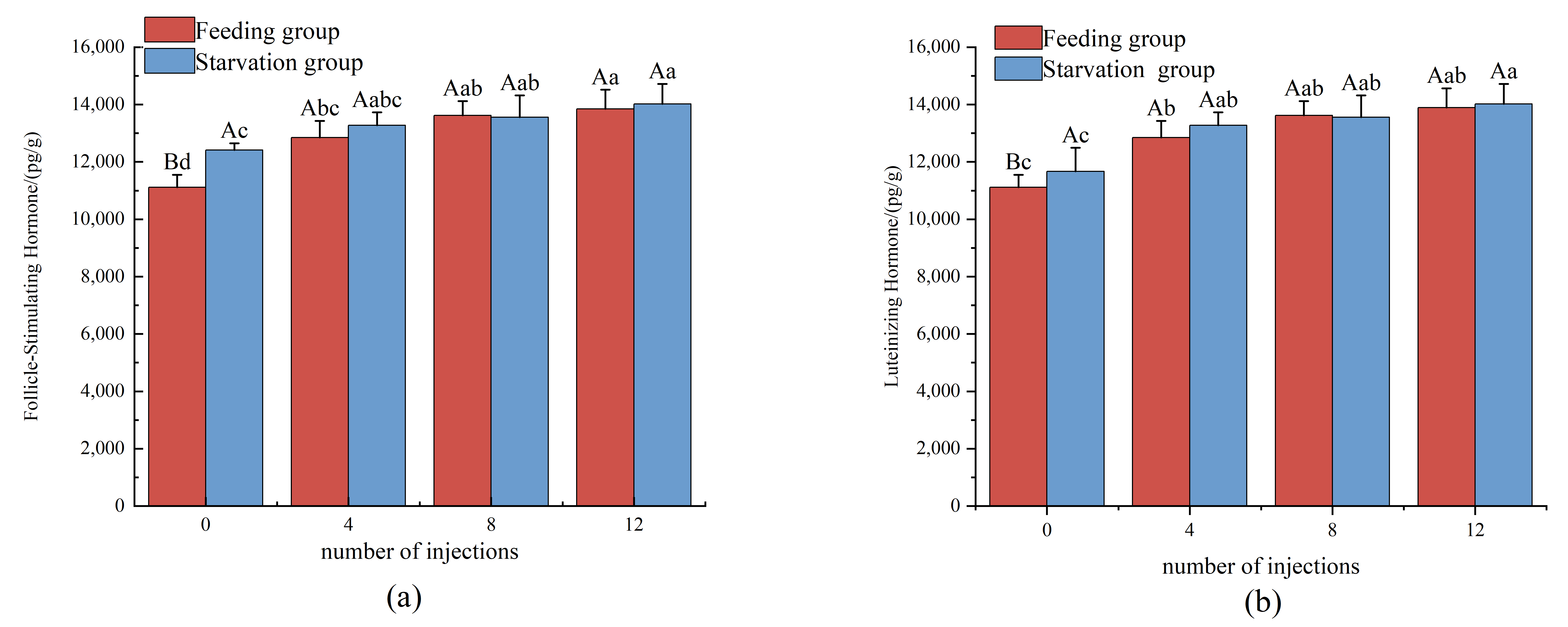
Follicle stimulating hormone (FSH) is an important reproductive regulatory hormone secreted by the pituitary gland, which plays a key role in the proliferation of follicular granulosa cells and the growth of oocytes in the early stages of ovarian development. The FSH content in the ovaries of American eels during feeding and different maturation stages is shown in
Figure 1a. With the increase in injection doses, the FSH content in the ovaries of American eels in the feeding and starvation group showed an upward trend, and reached its peak at 12 doses (late yolk accumulation stage). In the initial stage of development (0 injections), the content of follicle stimulating hormone (FSH) in the starvation group was significantly higher than that in the feeding group (
p < 0.05), which might be a stress-induced endocrine compensatory response of the body under starvation. However, as the number of injections increased to four, eight, and 12, the growth rate of follicle stimulating hormone (FSH) in the feeding group was faster, which gradually narrowed the gap with the starvation group. There was no significant difference between the two groups at 12 injections (late yolk accumulation stage) (
p > 0.05).
Luteinizing hormone (LH) is an important reproductive hormone secreted by the pituitary gland. During the late stage of follicular development and yolk accumulation, it induces meiosis of oocytes and triggers the final maturation and release of gametes. In this experiment, the changes in LH content in the ovaries of American eels at different maturation and feeding stages are presented in
Figure 1b. With the increase in injection times, the overall content of luteinizing hormone (LH) in the ovaries of American eels in each feeding group showed an upward trend. At the initial developmental stage (0 injections), the luteinizing hormone (LH) content in the feeding group was significantly lower than that in the starvation group (
p < 0.05). When the number of injections increased to four (the early stage of yolk formation), the starvation group exhibited a higher luteinizing hormone (LH) content than the feeding group. As the number of injections further increased to eight and 12, the significant difference in luteinizing hormone (LH) levels between the two groups gradually diminished. At 12 injections (the late stage of yolk accumulation), the LH level in the starvation group was slightly higher.
3.5. Comparison of Changes in Ovarian-Neutral Steroid Hormone Levels Between Fed and Non-Fed States
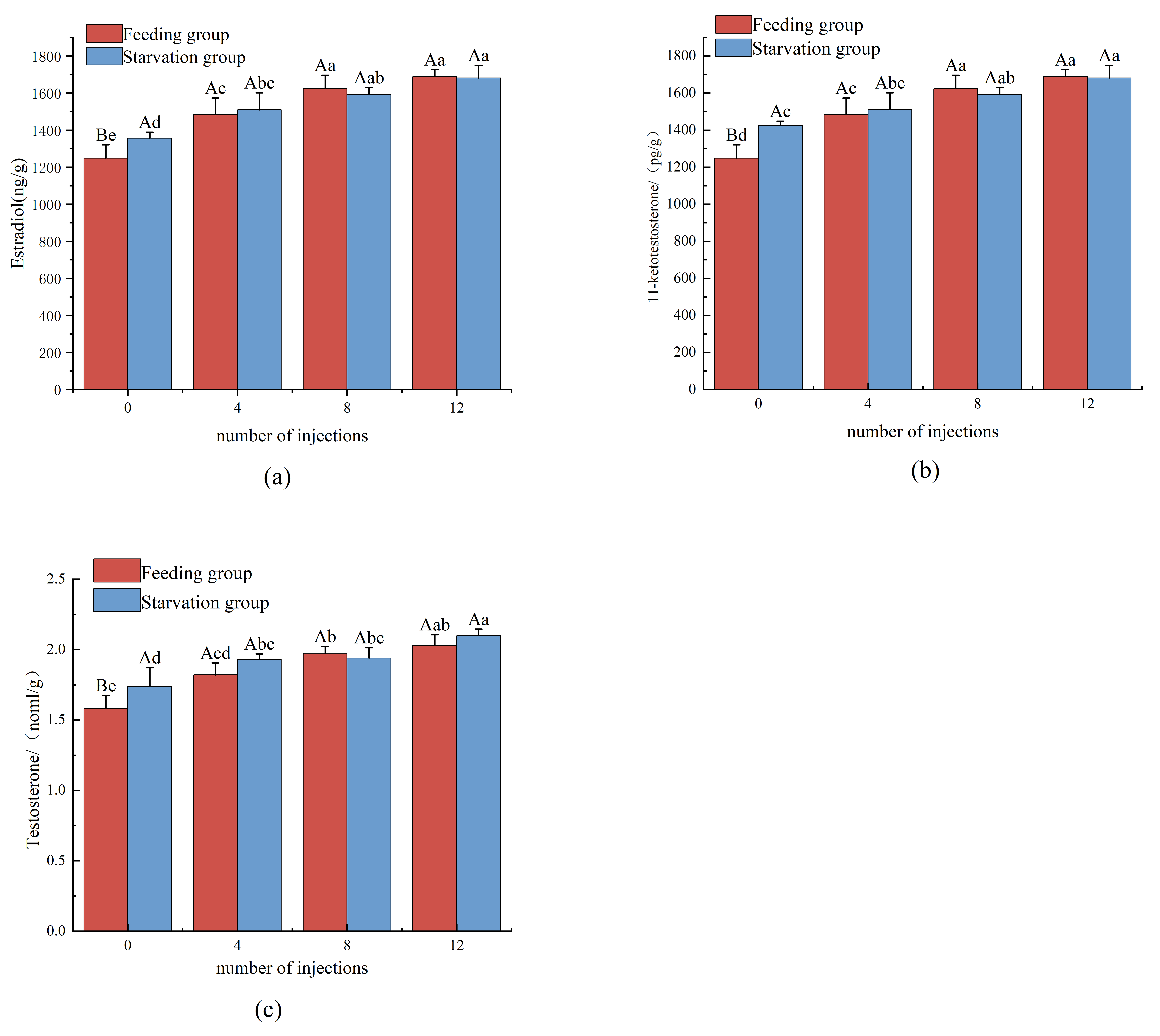
Estradiol (E
2) is the main estrogen secreted by the ovaries, which plays a central role in reproductive development and physiological homeostasis, and reaches its peak secretion level during yolk production. The changes in estradiol (E
2) content in the ovaries of American eels during different maturation stages with and without feeding are shown in
Figure 2c. With the increase in injection doses, the estradiol (E
2) content in the ovaries of both the feeding and starvation group exhibited a significant upward trend, which indicates that injection stimulation effectively activated the estrogen synthesis pathway in the ovaries and provided key endocrine support for follicle development and oocyte maturation. At the initial stage of development (0 injections), the starvation group had a significantly higher ovarian estradiol (E
2) content than the feeding group (
p < 0.05), which suggested that during starvation at this developmental stage, the organism preferentially initiated estrogen synthesis via endocrine compensation mechanisms to facilitate early gonadal development. When the number of injections increased to four, the estradiol (E
2) content in the starvation group remained slightly higher than that in the feeding group, though the difference narrowed moderately. As the number of injections further increased to eight and 12, the estradiol (E
2) content in the feeding group gradually approximated that in the starvation group, and no significant difference was observed between the two groups at 12 injections (
p > 0.05).
Figure 2d showed the changes in the content of 11 ketotestosterone (11-KT) in the ovaries of American eels across different feeding groups and maturation stages. The dynamic changes in ovarian 11 ketotestosterone (11-KT) exhibited a clear nutrition-dependent reversal pattern. In the early stages of maturation (0 injections), the level of 11 ketotestosterone (11-KT) in the starvation group was significantly higher than that in the feeding group (
p < 0.05), which reflected a compensatory endocrine response to energy stress. However, with the continuous intake of exogenous nutrients, the rate of hormone synthesis increased significantly from the fourth injection onwards. During the critical period of yolk protein accumulation (eighth and 12th injections), the level of 11 ketotestosterone (11-KT) in the feeding group was higher than that in the starvation group. This sustained increase indicated that exogenous nutrition provided essential substrates (such as cholesterol) for steroidogenesis, thereby supporting the androgen-mediated lipid accumulation required for oocyte development.
Testosterone (T) is not only an important sex hormone, but also a precursor to estradiol (E
2) synthesis. Its content is closely related to ovarian development and egg maturation.
Figure 2e shows the changes in testosterone (T) content in the ovaries of American eels across different feeding groups and different maturation stages. At the initial stage of development (0 injections), the testosterone (T) content in the starvation group was significantly higher than that in the feeding group (
p < 0.05), but starting from the fourth injection (early yolk formation initiation stage), the growth rate of testosterone (T) in the feeding group accelerated significantly. At the 12th injection (late yolk accumulation stage), although the testosterone (T) content in the feeding group was slightly lower than that in the starved group, it still remained at a high level.
4. Discussion
4.1. The Effects of Feeding and Starvation on the Morphological Characteristics and Indicators of the American Eel After Hormone Injection
The results of this study indicate that pre-experimental feeding reinforcement could alleviate energy limitation and morphological stunting in female eels during the subsequent hormone-induced reproductive stage [
11,
12]. Anguillids exhibit a typical catadromous life history. Wild broodstock cease feeding during silvering and seaward migration, relying entirely on the catabolism of endogenous somatic tissues (muscle and lipid reserves) to sustain basal metabolism and gonadal development. In the present experiment, all individuals in the feeding group received six months of freshwater feeding reinforcement prior to hormone induction, followed by complete food deprivation throughout the entire hormone injection cycle. By contrast, the starvation group received no supplementary feeding either before or during hormone treatment.
The starvation group exhibited stagnant body mass and restricted growth in chest circumference (PC), demonstrating that eels adopt a survival-first metabolic strategy under severe energy deficiency: somatic tissue proliferation is fully inhibited to cut energy expenditure, which further suppresses the increase in the gonadosomatic index (GSI). The feeding group possessed superior morphological indicators at the initiation of hormone induction. At the end of the pre-induction feeding period, their body weight reached 873 g, and chest circumference rose from 10.27 cm to 13.57 cm. The prominent intergroup difference in body shape was accumulated during the pre-induction feeding phase, rather than deriving from de novo somatic tissue synthesis supported by exogenous nutrition during fasting maturation. Abundant energy reserves accumulated via long-term exogenous nutrition during freshwater reinforcement which established a robust somatic foundation, enabling simultaneous gonadal development during the subsequent fasting ripening stage.
Second, an increase in absolute chest circumference reflects expanded visceral cavity volume, which provides essential physical space for ovarian development. In this study, the feeding group retained higher chest circumference acquired from prior feeding, while total body length remained relatively stable. The enlarged abdominal cavity was highly consistent with the sharp elevation of GSI during hormone induction. In comparison, fish in the starvation group sustained a low chest circumference of approximately 9.5 cm due to continuous consumption of internal energy reserves, and such morphological restriction mechanically impeded ovarian expansion. These findings verify that sufficient pre-experimental feeding reinforcement not only accumulates nutritional substrates but also enlarges visceral cavity volume through abdominal muscle deposition, providing critical structural support for efficient reproduction during the subsequent fasting maturation period. Moreover, the high eye diameter index (1.57%) observed in late-stage starved eels indicates a massive breakdown of endogenous energy reserves, representing a typical allometric adaptive trait under prolonged food deprivation.
Finally, visceral organ indices reflected distinct metabolic adaptation strategies determined by the pre-experimental feeding status. Beyond whole-body morphology, organ-level indices further illustrated intergroup adaptive differences at the tissue level. The reduced digestive index (DI) in the starvation group stemmed from long-term disuse atrophy of the digestive tract, a typical characteristic of silver eels during natural migration. The feeding group maintained a high digestive index after reinforcement, with intact digestive absorption structures that allowed substantial energy storage before fasting induction. The intact digestive system during the feeding phase served as the structural basis for energy accumulation in fish.
In conclusion, long-term freshwater feeding reinforcement before hormone induction built favorable somatic conditions for American eels by increasing body mass, expanding visceral cavity volume and preserving digestive tract function. The energy and morphological advantages acquired in this feeding phase are prerequisites for breaking the inherent energy bottleneck of wild silver eels and achieving high-quality gonadal maturation throughout the subsequent fasting.
4.2. The Developmental Patterns of Oocytes in Fed American Eels with Different Injection Frequencies and Starved Experimental Groups
Our findings demonstrate that prolonged hunger stress imposes a highly compartmentalized inhibitory effect on American eel oocytes, characterized by a more pronounced suppression of cytoplasmic expansion than nuclear development. During exogenous gonadotropin induction, the steady increase in both egg and nucleus diameters in the feeding group reflects a harmonized vitellogenic growth, aligned with classic teleost maturation dynamics [
13,
14,
15]. Conversely, the starvation group exhibited significant structural asynchrony, while cytoplasmic growth was severely arrested due to nutrient deficits, and the nuclear diameter maintained a relatively stable, albeit sluggish, growth trajectory. This discrepancy indicates a higher physiological tolerance of nuclear development to nutrient deprivation. In fish oogenesis, the nucleus houses critical maternal transcription machinery; thus, the preferential preservation of nuclear growth under energetic constraints suggests a strategic metabolic prioritization. The maternal organism likely shunts its limited endogenous reserves toward maintaining genomic and transcriptional integrity rather than structural cytoplasmic scaling, a survival adaptation previously documented in other migratory teleosts under energetic stress.
Mechanistically, the asynchronous temporal patterns of nucleolus numbers and oil droplet diameters further elucidate how nutritional deprivation disrupts oocyte homeostasis. In the feeding group, the initial proliferation followed by a late-stage regression of nucleoli perfectly mirrors the normal physiological rhythm of ribosomal RNA (rRNA) transcriptional activation and subsequent cessation prior to germinal vesicle breakdown (GVBD). However, the persistent elevation of nucleolus numbers in the starvation group at the 12th injection signifies a severe disruption of this developmental clock. This failure to undergo timely nucleolar regression indicates a transcriptional arrest or delayed silencing of maternal genes, potentially interfering with standard yolk protein synthesis [
16].
Furthermore, the striking lag in oil droplet expansion observed in starved eels underscores the critical role of exogenous dietary lipids. Eel oocytes require a massive influx of highly unsaturated fatty acids (HUFAs) and neutral lipids for oil droplet fusion and vitellogenesis [
17]. Feeding on
L. vannamei provided immediate, highly bioavailable lipid substrates that facilitated early droplet maturation. In contrast, hunger stress forces the oocytes to rely on depleted endogenous somatic reserves, thereby delaying lipid deposition and shifting the timing of oil droplet coalescence. Taken together, these asynchronous disruptions at both the transcriptional (nucleoli) and metabolic (oil droplets) levels provide a comprehensive explanation for the impaired oocyte quality under hunger stress.
4.3. Regulation of the Secretion Pattern of Ovarian Gonadotropins (FSH/LH) by Feeding Status
Gonadotropins are key hormones in the hypothalamic–pituitary–gonadal endocrine axis (HPG) of bony fish. The hypothalamus secretes gonadotropin-releasing hormone (GnRH), which acts on the pituitary gland. Under GnRH stimulation, the pituitary gland secretes gonadotropins (GTHs). In fish and some lower vertebrates, gonadotropins (GTHs) are mainly divided into two types: GTH-1 and GTH-2 [
18,
19]. They mimic follicle-stimulating hormone (FSH) and luteinizing hormone (LH) in mammals via the conservation of the alpha subunit and the functional differentiation of the beta subunit, respectively [
20]. Their main function is to stimulate the growth and development of sperm, egg, and gonadal cells, as well as the production of sex steroid hormones in reproductive regulation. The produced sex steroid hormones can promote the synthesis activity of gonadotropin cells in the hypothalamus and pituitary gland through positive feedback [
21,
22,
23].
Exogenous carp pituitary extract (CPE) and teleost HPG axis-mimicking hormones (FSH/LH) activated gonadal function either directly or indirectly and accelerated gamete maturation and ovulation by bypassing hypothalamic control. In teleosts, FSH stimulated the secretion of sex steroid hormones including estradiol (E
2), testosterone (T), and 11-ketotestosterone (11-KT) during the early phase of gonadal development, thereby modulating gonadal growth and gametogenesis [
24,
25]. Luteinizing hormone (LH) triggered the synthesis of DHP during vitellogenesis in the late gonadal developmental stage, which facilitated oocyte maturation, ovulation, and sperm ejaculation [
26].
The present study found that nutritional status exerted markedly time-dependent effects on HPG axis regulation in American eels. During the early maturation phase, concentrations of follicle-stimulating hormone (FSH) and luteinizing hormone (LH) in the starvation group were significantly higher than those measured in the feeding group, a phenomenon that might be associated with the unique migratory life-history strategy of eels [
15].
In their natural state, eels underwent silvering and migration accompanied by the cessation of feeding and atrophy of the digestive tract [
27]. Hunger stress might have served as a physiological signal that simulated the metabolic state during migration, thereby preferentially activating the secretion of gonadotropins through the hypothalamic–pituitary axis in the early stages to accelerate reproductive initiation. In contrast, the feeding group received sufficient exogenous nutrition in the early stages, and the body’s metabolic center tended toward somatic cell growth. High levels of growth-axis hormones (such as GH/IGF-1) might have temporarily inhibited the vigorous activation of the reproductive axis via antagonistic effects. However, as development entered the late stage of yolk accumulation (8–12 injections), follicle-stimulating hormone (FSH) levels in the feeding group surpassed those in the starvation group and remained elevated, whereas gonadal development in the starvation group slowed. This indicated that exogenous nutrient input was crucial for sustaining long-term, high-intensity reproductive activity. Continuous follicle-stimulating hormone (FSH) synthesis required substantial energy support, and abundant energy reserves within the feeding group might have upregulated the gene expression of pituitary gonadotropin subunits. Furthermore, although luteinizing hormone (LH) levels showed no significant inter-group difference in the late developmental phase, the feeding group secured the sustained hormonal signals required for oocyte development through the synergistic action of the “nutrient–reproductive axis”, and avoided potential endocrine dysfunction triggered by energy depletion in the starvation group [
28,
29].
4.4. The Effect of Changes in the Content of Ovarian Steroid Hormones
During the development and maturation of eel gonads, gonadotropins mainly promote the development and maturation of germ cells by regulating sex steroid hormones [
30,
31,
32], and sex steroid hormones play an important role in promoting ovarian maturation and final ovulation in female eels [
33]. The sex steroid hormones in eels are mainly convert from cholesterol. Membrane cells convert cholesterol to progesterone through cholesterol side-chain lyase, while stromal cells catalyze progesterone to produce testosterone (T) through CYP17A1. Testosterone (T) is further convert to estradiol (E
2) by aromatase. Estradiol (E
2) stimulates the synthesis of vitellogenin in the liver of female eels and releases it into the bloodstream, but cannot directly enter the egg cells [
34]. Testosterone transports vitellogenin from the blood to the egg cells, promoting a large accumulation of yolk inside the egg cells [
35].
Estradiol (E
2), as the main estrogen in fish, plays two physiological roles in reproductive regulation: one is to regulate the growth and maturation of oocytes, inducing a decrease in the number of oocyte divisions and an increase in their diameter; the second is to promote the synthesis of vitellogenin (VTG) in oocytes, thereby accelerating the accumulation of yolk granules in the egg [
36]. In this study, the levels of estradiol (E
2) and 11 ketotestosterone (11-KT) in the feeding group were both elevated in the late developmental stage (8–12 injections) compared to the starvation group, which confirmed the crucial substrate support role of exogenous nutrition in maintaining steroidogenesis. Cholesterol was a common precursor for sex steroid hormone synthesis [
37]. Feeding not only directly supplemented exogenous cholesterol, but also promoted the synthesis and transport of vitellogenin by enhancing liver fat metabolism [
38]. In contrast, the hungry group mainly relied on consuming their own muscle and liver lipid reserves during the late yolk accumulation period. As the endogenous energy pool was depleted, the activity of their steroid synthase system might be limited due to substrate scarcity [
39].
Of particular note, the feeding group showed a significant increase in 11 ketotestosterone (11-KT) during the late stage of yolk accumulation. Although 11 ketotestosterone (11-KT) was traditionally considered a male hormone, it played an important regulatory role in early oocyte growth and lipid accumulation in female eels [
40]. The higher levels of 11 ketotestosterone (11-KT) in the feeding group in this study may have adapted to their more vigorous lipid metabolism needs [
41]. Hormones assisted the body in efficiently converting ingested nutrients into lipid reserves required for ovarian development. As for the decline in testosterone (T) levels in the late stage, it may have been related to the increased conversion efficiency of testosterone (T) to estradiol (E
2). Adequate aromatase substrate flux in the feeding group accelerated the conversion of testosterone (T) to estradiol (E
2), thereby maintaining higher estrogen levels to support yolk production, which was consistent with the regulatory model of steroid hormone synthesis proposed by Izquierdo et al. [
39] for fish nutrition.
While frozen white leg shrimp (Litopenaeus vannamei) was successfully utilized as an accessible exogenous nutrient source, its fatty acid, lipid, and carotenoid profiles may deviate from the diverse marine/estuarine prey types wild silver eels consume prior to their marine migration.
5. Conclusions
This study investigated the impacts of feeding regimes on the morphology, muscle nutrition, and reproductive performance of artificially matured American eels, clarifying stage-specific nutritional effects. All eels achieved 100% survival. The greater body weight, chest circumference and gonadal index in the feeding group reflected somatic advantages accumulated during the 6-month freshwater reinforcement phase, which established an elevated pre-hormone-induction baseline.
Nutrition markedly modulated hormone-induced oocyte development. Pre-established adequate nutrition supported normal increases in egg diameter, ordered oil droplet accumulation and regular nucleolar rhythms. In contrast, maturation-stage starvation inhibited cytoplasmic and oil droplet growth, disturbed nucleolar dynamics, and minimally affected nuclei. Thus, sufficient nutritional baseline accumulation is essential for complete oocyte maturation.
Nutritional regulation of the HPG axis was stage-dependent. The starvation group exhibited compensatory hormonal elevation during early reproductive development, whereas the feeding group with a superior nutritional baseline maintained higher reproductive hormone levels at the late developmental stage.
In summary, feeding during the freshwater reinforcement phase builds a favorable somatic and nutritional baseline, which improves eel morphological traits, facilitates orderly oocyte development, optimizes reproductive hormone profiles and enhances reproductive performance. These findings provide a theoretical basis for optimizing nutritional strategies in American eel artificial breeding.