Article

Tissue Distribution and Depletion of Praziquantel and Its Main Metabolites in Grass Carp (Ctenopharyngodon idella) Following 24 h Bath Administration

1
Department of Animal Protection and Welfare and Veterinary Public Health, Faculty of Veterinary Hygiene and Ecology, University of Veterinary Sciences Brno, 612 42 Brno, Czech Republic
2
Department of Animal Breeding, Animal Nutrition and Biochemistry, Faculty of Veterinary Hygiene and Ecology, University of Veterinary Sciences Brno, 612 42 Brno, Czech Republic
*
Author to whom correspondence should be addressed.
Fishes 2026, 11(7), 403; https://doi.org/10.3390/fishes11070403
Submission received: 22 May 2026 / Revised: 2 July 2026 / Accepted: 6 July 2026 / Published: 7 July 2026
(This article belongs to the Section Welfare, Health and Disease)

Abstract

Praziquantel (PZQ) is a widely used antiparasitic drug in aquaculture; however, data on its environmental fate, tissue distribution, and metabolite kinetics in fish following bath treatment remain limited, particularly with respect to its metabolites. This study investigated the long-term elimination of PZQ and its two major metabolites, trans-4-hydroxypraziquantel (TPZQ) and cis-4-hydroxypraziquantel (CPZQ), in water and selected biological matrices of grass carp (Ctenopharyngodon idella) following a 24 h therapeutic bath with PZQ administered at 4 mg/L. Results showed a rapid decrease in PZQ in water after the transfer of fish to clean water, accompanied by a transient increase in its metabolites, with TPZQ as the predominant metabolite. PZQ tissue residues were highest in the hepatopancreas, caudal kidney, and skin, where the parent compound remained detectable up to day 14 of the experiment. Among the metabolites, higher concentrations of CPZQ were detected across all examined tissues; however, its elimination rate was faster than that of TPZQ. This difference was most pronounced in plasma and skin, where TPZQ remained detectable until day 7 of the experiment, whereas the cis-form was detected only up to day 3. Similar to the parent compound, the highest concentrations of both monitored metabolites were observed in the hepatopancreas and caudal kidney, where they remained detectable until day 7 of the experiment. These findings provide comprehensive insight into the distribution and elimination of PZQ in grass carp following 24 h bath administration and contribute valuable data that may support environmental risk assessment and future withdrawal-period evaluation in aquaculture.
Key Contribution: Bath administration of praziquantel in grass carp resulted in rapid water elimination but prolonged persistence of praziquantel and its metabolites in hepatopancreas, kidney, and skin, providing important data for environmental risk assessment and withdrawal period determination in aquaculture.

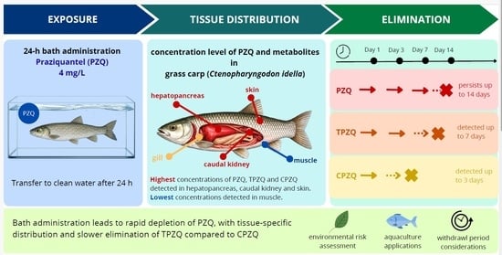
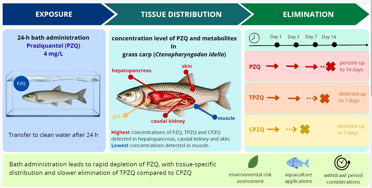
Graphical Abstract

1. Introduction

Praziquantel (PZQ) is a synthetic antiparasitic drug from the pyrazinoisoquinoline group that is highly effective against flatworms (Platyhelminthes), including tapeworms (cestodes) and flukes (trematodes) [1,2,3]. Owing to its high efficiency, favorable safety profile, and low cost, PZQ is included on the World Health Organization’s List of Essential Medicines, highlighting its importance in the treatment of parasitic infections in human medicine [4]. Since its introduction in the 1970s, it has become one of the most widely used antiparasitic agents for treating diseases caused by flukes and tapeworms in humans and animals, such as schistosomiasis, clonorchiasis, and taeniasis [5]. In veterinary medicine and aquaculture, PZQ is also applied to control parasitic flatworms in fish, particularly in ornamental and farmed species [1,6,7].
However, within the European Union, PZQ has long lacked formal registration for use in aquaculture, and its application in fish was therefore restricted to the veterinary medicinal product cascade, as defined by Regulation (EU) 2019/6 [8], in cases where no authorised veterinary medicinal product was available. The use of PZQ in food-producing fish has also been closely linked to residue considerations regulated under Commission Regulation (EU) No. 37/2010. Recently, the maximum residue limit (MRL) for PZQ (sum of isomers) in muscle and skin in natural proportions in finfish has been established at 20 μg/kg, following the opinion of the EMA Committee for Veterinary Medicinal Products and its inclusion in Annex I of Commission Implementing Regulation (EU) 2023/981 [9]. Despite this regulatory progress, commercial formulations specifically designed for fish are still unavailable, and standardized treatment guidelines for aquaculture remain limited [2,10].
PZQ can be administered in several ways depending on the target species. In mammals, it is given orally or by injection [11]. In aquaculture, where large groups of fish are treated simultaneously, the most practical options are medicated feed or therapeutic baths [2,3,10]. Typical oral doses range from 50 to 200 mg/kg of body weight (b.w.) for single treatments and 7 to 75 mg/kg b.w. for repeated doses. Bath treatments are used at concentrations of 0.25–50 mg/L, depending on exposure time and parasite species [12]. Short “dip” treatments employ higher concentrations for a few minutes, whereas long baths involve lower concentrations for several hours. In laboratory studies, precise dosing may be achieved by oral gavage or injection, but these methods are unsuitable for large-scale fish production due to handling stress [6,7]. A withdrawal period of 500 degree-days has been reported in the literature for food fish under cascade therapy principles (i.e., “off-label” use of veterinary drugs) to ensure consumer safety [1]. The mechanism of action of PZQ is not yet fully understood, but research shows that it mainly affects calcium homeostasis in parasite cells. The drug increases the influx of calcium ions into the parasite, resulting in muscle contractions, paralysis, and damage to the outer body surface (i.e., tegument) [5,13,14]. Tegumental damage caused by praziquantel leads to the exposure of parasite surface antigens, facilitating recognition by the host immune system [15]. In Schistosoma worms, this leads to spastic paralysis and detachment from host tissues, after which the worms are transported via the hepatic portal circulation to the liver, where they become susceptible to host immune-mediated clearance [5]. Additional studies suggest that this calcium influx may be mediated through specific transient receptor potential (TRP) calcium channels in the parasite’s nervous system [6]. Only the R-(−) enantiomer of the drug is active against parasites, while the S-(+) form mainly contributes to the bitter taste and mild side effects [5,7].
After administration, PZQ is rapidly absorbed and widely distributed in the body. In fish, this drug and its metabolites are primarily detected in metabolically active tissues, particularly the liver/hepatopancreas, kidney, gill, and skin, reflecting the central role of hepatic cytochrome P450 enzymes in PZQ biotransformation [3,7]. The major metabolites of PZQ are cis- and trans-4-hydroxypraziquantel (CPZQ and TPZQ, respectively), which are formed via stereoselective cytochrome P450–mediated hydroxylation. Their relative abundance varies among fish species, experimental systems, and routes of administration. PZQ and its metabolites are predominantly excreted via the kidney, with elimination in fish typically occurring within several days and being influenced by environmental factors such as temperature and salinity [3,6,16,17]. Beyond their formation kinetics, TPZQ and CPZQ may also differ from the parent compound in their biological activity, pharmacokinetic behaviour, and contribution to the overall efficacy and safety of praziquantel treatment. Although their antiparasitic activity is generally lower than that of PZQ, both metabolites are considered biologically relevant and may contribute to interspecies differences in PZQ disposition and pharmacological effects [18,19].
This study aimed to investigate the long-term elimination of PZQ and its two major metabolites, CPZQ and TPZQ, in grass carp (Ctenopharyngodon idella) following a single 24 h bath exposure at a dose of 4 mg/L. After exposure, fish were transferred to clean water, and the concentrations of PZQ and its metabolites were monitored over a total 35-day depletion period to characterize their persistence in blood plasma and selected tissues. To the best of our knowledge, this is the first study to provide a comprehensive evaluation of both PZQ and its major metabolites across multiple tissues, including hepatopancreas, caudal kidney, gill, muscle, skin, and blood plasma, enabling a detailed assessment of tissue distribution and depletion. While most previous studies have focused solely on the parent compound, the present work offers a detailed, multi-tissue evaluation of both PZQ and its metabolites following bath administration.

2. Materials and Methods

2.1. Experimental Design and Sampling

A total of 84 juvenile grass carp (C. idella) were used in the experiment. The fish were two years old, with a mean total length of 21.6 ± 1.8 cm and a mean body weight of 93.2 ± 25.4 g. All fish originated from a commercial fish farm (Lnáře Fishery Ltd., Lnáře, Czech Republic). Fish were randomly distributed into 7 glass aquaria with a volume of 200 L, each containing 12 individuals. Throughout the experiment, water quality parameters were monitored once daily during the morning feeding period, including temperature (19.1 ± 1.3 °C), dissolved oxygen (89–96%), and pH (7.6 ± 0.3). Fish were fed a commercial pelleted diet at a daily ration of 2% of body weight. Before the experiment, fish were acclimated for 10 days under laboratory conditions at the Faculty of Fisheries and Protection of Waters (University of South Bohemia, Czech Republic).
A therapeutic bath containing PZQ at a target concentration of 4 mg/L was prepared by dissolving PZQ in ethanol and subsequently diluting it with water. The final ethanol concentration in the bath was 0.005%. Fish were exposed to the praziquantel bath for 24 h. Following the 24 h bath exposure, PZQ and its two major metabolites (TPZQ and CPZQ) were monitored in water, blood plasma, and selected tissues of grass carp at defined time intervals. At each sampling time point, seven fish were randomly selected and sampled (one fish from each aquarium). Blood was collected from the caudal vein into heparinized tubes. Plasma was obtained by centrifugation at 800× g for 10 min at 4 °C and stored at −80 °C until analysis. Immediately after blood collection, the fish were stunned by a blunt blow to the head and subsequently euthanized by severing the gill arches. Tissue samples, including hepatopancreas, gill, caudal kidney, muscle, and skin, were collected and stored at −80 °C until further analysis. Simultaneously, one 1.5 L water sample was collected from each aquarium (n = 7) before the scheduled water exchange, resulting in seven water samples at each sampling time point. Water samples were stored at −20 °C until analysis.
The first sampling (0 h) was performed immediately after preparation of the praziquantel bath, before fish exposure. Fish were then placed in the treatment bath for 24 h. After completion of the exposure period, the second sampling was conducted (24 h), and the remaining fish were transferred to clean, praziquantel-free water. Subsequent samplings were performed at 24 h post-exposure (2 d), 48 h post-exposure (3 d), 6 days post-exposure (7 d), 13 days post-exposure (14 d), 20 days post-exposure (21 d), 27 days post-exposure (28 d), and 34 days post-exposure (35 d). The first water exchange was performed on day 2 of the experiment; thereafter, aquarium water was changed once per week until the end of the experiment.

2.2. LC–MS/MS Analysis of Praziquantel and Its Metabolites

The determination of praziquantel (PZQ) and its main metabolites (CPZQ and TPZQ) in biological matrices was performed using liquid chromatography coupled with tandem mass spectrometry (Thermo Scientific, Waltham, MA, USA) according to a previously published method [3]. The analysed matrices included water, blood plasma, and selected tissues (hepatopancreas, caudal kidney, muscle, gill, and skin).
Before analysis, all samples were spiked with isotopically labelled internal standards. Water and plasma samples were purified using solid-phase extraction (SPEC C18 AR cartridges, 3 mL, 30 mg; Agilent, Santa Clara, CA, USA). Tissue samples (0.5 g) were homogenised with methanol (2 × 3 mL; tissue-to-solvent ratio 1:12, w/v) using an immersion blender Schütthomgen (Schütt Labortechnik, Göttingen, Germany). The homogenates were centrifuged (800× g, 15 min, 20 °C), and the supernatants were subsequently purified by solid-phase extraction (SPEC C18 AR cartridges, 3 mL, 30 mg; Agilent, Santa Clara, CA, USA). After evaporation to dryness under a gentle stream of nitrogen, the residues were reconstituted in deionised water and analysed by liquid chromatography coupled with tandem mass spectrometry. Chromatographic separation was achieved on a reversed-phase C18 column using gradient elution with water containing formic acid and acetonitrile. Detection was carried out on a triple quadrupole mass spectrometer equipped with a heated electrospray ionisation source operated in positive mode. Quantification was performed using the internal standard method in multiple reaction monitoring mode. Limits of detection (LOD) for individual analytes and matrices are provided in Table 1.

2.3. Data Analysis

Statistical analyses were conducted using Unistat for Excel 6.5 (Unistat Ltd., London, UK). Temporal variations in the concentrations of the target analytes were assessed for blood plasma and selected tissues (hepatopancreas, caudal kidney, muscle, gill, and skin). Data normality was evaluated using the Shapiro–Wilk test. As the datasets deviated from a normal distribution, non-parametric methods were applied, specifically the multiple-sample median test. Measurements below LOD (see Table 1) were imputed as half of the corresponding LOD for the calculation of mean values and the following statistical evaluation. This substitution approach is commonly applied in toxicological studies for handling left-censored data and is consistent with US EPA recommendations for data quality assessment and statistical treatment of non-detects [20]. Statistical significance was defined at p < 0.05. For each analyte (PZQ, CPZQ, TPZQ) and each tissue, statistical comparisons were performed among sampling time points. For graphical presentation, concentrations were additionally displayed on a logarithmic (log10) scale to improve visualization of data spanning several orders of magnitude.

3. Results

3.1. Water Analysis

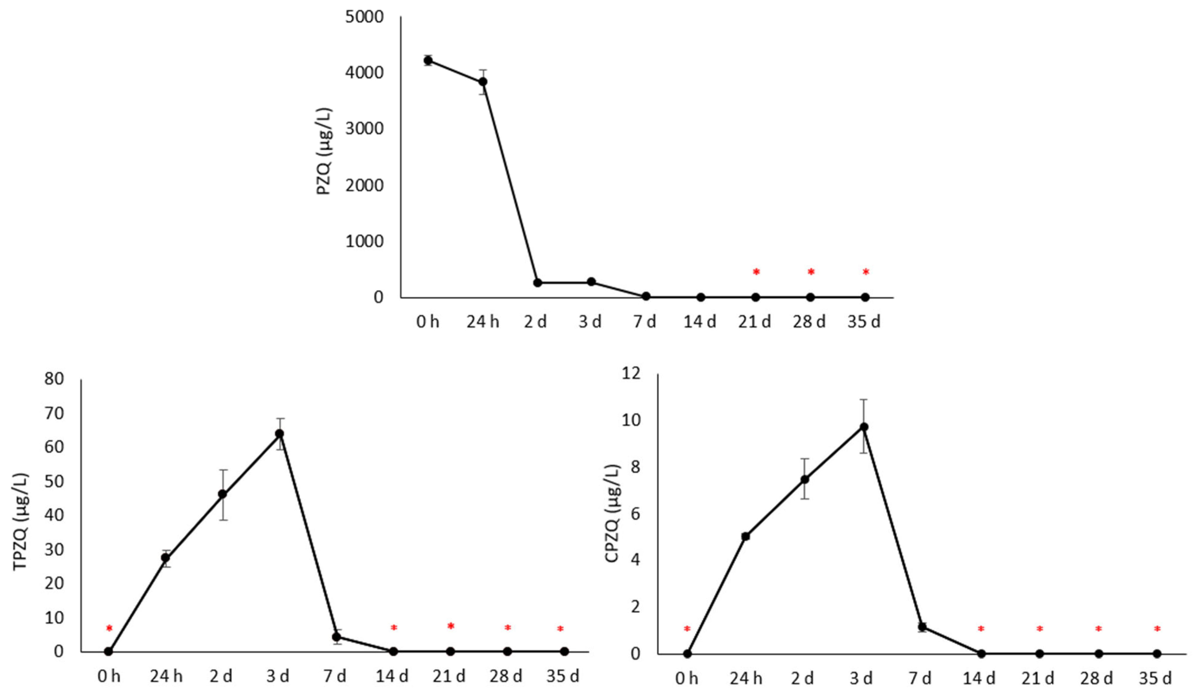
Praziquantel and its two major metabolites (TPZQ, CPZQ) were monitored in water (Figure 1). The water sample collected at 0 h represents the solution immediately after the bath was prepared, before the introduction of the experimental fish. The subsequent sample (24 h) was taken at the end of the bath. Afterwards, the fish were transferred to clean water, and the kinetics of the individual analytes were monitored. The figures show that after two days, when the fish had been transferred to clean water, the parent compound decreased rapidly, accompanied by increases in both metabolites. The major metabolite was TPZQ. At day 14 of the experiment, the concentration of the parent compound was very low (0.8 ± 0.2 µg/L), while both metabolites were below the LOD. From day 21 onwards, the PZQ concentration was also found below the LOD.

3.2. Tissue Analysis

Residues of PZQ and its metabolites were analyzed in six different tissues: the hepatopancreas, caudal kidney, skin, gill, muscle, and blood plasma. As expected, the highest concentrations were observed for the parent compound PZQ, predominantly in the hepatopancreas and caudal kidney, where levels remained above the LOD even on day 14 of the experiment. Among the metabolites, higher concentrations were observed for CPZQ, primarily in the hepatopancreas and caudal kidney; however, levels declined below the LOD by day 14. The elimination pattern of TPZQ was similar to that of CPZQ. Nevertheless, TPZQ in plasma and skin remained detectable up to day 7 of the experiment, whereas the cis-metabolite fell below the LOD by day 7. Detailed results for each tissue and sampling time are presented in the following sections. It should be noted that limits of detection varied among analytes and matrices, particularly for TPZQ in selected tissues; therefore, minor differences in detectability near the LOD should be interpreted with caution.

3.2.1. Hepatopancreas

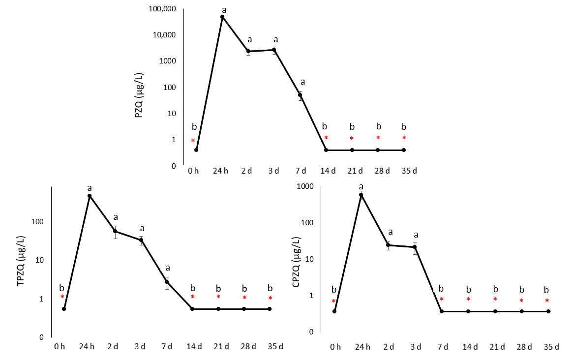
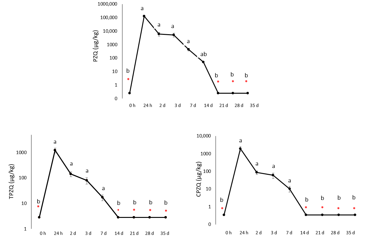
The hepatopancreas exhibited the greatest accumulation of the parent compound and its metabolites (Figure 2), with peak levels detected immediately after the 24 h bath (126,143 µg/kg for PZQ, 1236 µg/kg for TPZQ, and 1911 µg/kg for CPZQ). A rapid decline followed this in the concentrations of all monitored compounds. The parent compound remained detectable up to day 14 (49 µg/kg) but fell below the LOD from day 21 onward. Positive findings of both metabolites were recorded up to 7 days (17 µg/kg for TPZQ and 11 µg/kg for CPZQ); thereafter, their concentrations were below the LOD.

3.2.2. Caudal Kidney

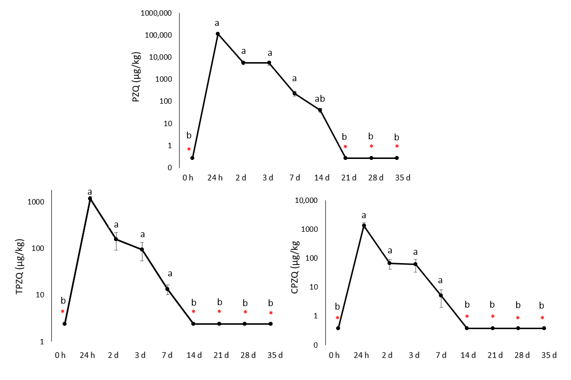
The caudal kidney was the second tissue with the highest detected concentrations of PZQ residue and its metabolites. Similar to the hepatopancreas, the highest levels were observed after the 24 h bath exposure (113,857 µg/kg for PZQ, 1171 µg/kg for TPZQ, and 1363 µg/kg for CPZQ), followed by a rapid decline in the concentrations of the monitored analytes. Positive PZQ findings were still detected on day 14 of the experiment (40 µg/kg), whereas its metabolites were detected only on day 7 (13 µg/kg for TPZQ and 5 µg/kg for CPZQ). Subsequently, all analytes fell below the LOD (Figure 3).

3.2.3. Skin

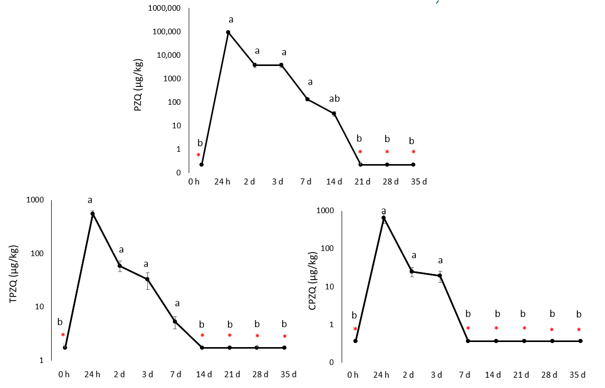
The skin was the third tissue with the highest residue levels detected. Immediately after the bath exposure, the concentrations of the monitored analytes were 94,771 µg/kg for PZQ, 551 µg/kg for TPZQ, and 655 µg/kg for CPZQ. As observed in other tissues, rapid elimination of the xenobiotics occurred. The parent compound PZQ remained detectable until day 14 of the experiment (33 µg/kg), while TPZQ was detected up to day 7 (5 µg/kg). In contrast, CPZQ was eliminated more rapidly, with positive findings observed only until day 3 of the experiment (19 µg/kg). At all subsequent sampling times, concentrations in all samples were below the LOD. An overview of the detailed results is provided in Figure 4.

3.2.4. Gill

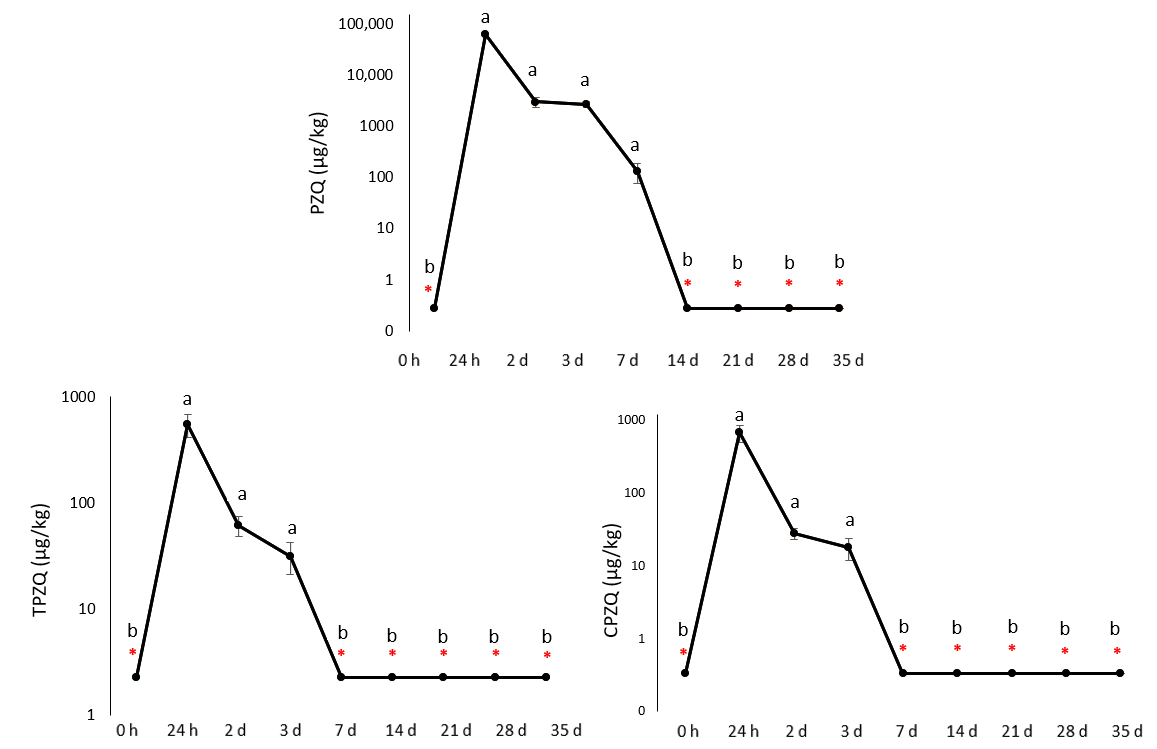
Concentrations of PZQ residue and its metabolites in the gill again peaked immediately after the bath exposure. The concentration of the parent compound was 62,985 µg/kg. In contrast, metabolite concentrations were approximately two orders of magnitude lower, reaching mean values of 551 and 676 µg/kg for TPZQ and CPZQ, respectively. Compared to the organs described above (i.e., hepatopancreas, caudal kidney, and skin), elimination of all analytes from the gill was faster, as positive PZQ findings were detected only on day 7 of the experiment (131 µg/kg). Metabolites were detected only on day 3 (32 and 18 µg/kg for TPZQ and CPZQ, respectively). At all subsequent sampling times, concentrations of monitored analytes were below the LOD. Detailed results are presented in Figure 5.

3.2.5. Muscle

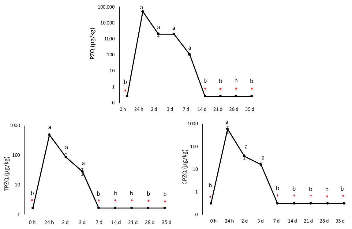
Another analyzed tissue was muscle, where the concentrations of the monitored analytes were approximately half those in the hepatopancreas and similar to those in the gill and plasma samples. As expected, the highest concentrations were detected immediately after the bath, reaching 54,000, 493, and 583 µg/kg for PZQ, TPZQ, and CPZQ, respectively. Subsequently, significant decreases in all analytes were observed; for the parent compound, positive findings were detected only up to day 7 of the experiment (111 µg/kg), while for the metabolites, this was limited to day 3 of the experiment (28 and 16 µg/kg for TPZQ and CPZQ, respectively). In the following days, concentrations were below the LOD in all cases. Detailed results are shown in Figure 6.

3.2.6. Blood Plasma

In the blood plasma, an elimination pattern comparable to that observed in the analyzed tissues was recorded. Immediately after the bath exposure, concentrations reached their maximum values (48,229; 466, and 587 µg/L for PZQ, TPZQ, and CPZQ, respectively), followed by a rapid decline in the subsequent days. Positive detections of PZQ and TPZQ persisted up to day 7 of the experiment (52 and 3 µg/L, respectively). In contrast, CPZQ was eliminated more rapidly, with detectable concentrations observed only up to day 3 (22 µg/L). Thereafter, all analyzed blood plasma samples were below the LOD. Detailed results are presented in Figure 7.

4. Discussion

Praziquantel (PZQ) is widely recognized as an effective antiparasitic agent in aquaculture; however, information on its long-term tissue distribution and residue depletion following bath administration, particularly with respect to its major metabolites, remains limited. The present study provides a comprehensive evaluation of the tissue distribution and depletion profiles of PZQ and its major metabolites following a 24 h bath administration in grass carp (C. idella). By monitoring the parent compound together with its major metabolites across multiple biological matrices over 35 days, this study provides new insights into their persistence and tissue-specific distribution following therapeutic bath exposure.
In general, after administration, PZQ is rapidly absorbed and distributed throughout the body. In mammals, the highest concentrations are found in the liver and kidneys, while smaller amounts are present in the lungs, pancreas, and other organs [14]. A similar distribution pattern has also been reported in fish, where PZQ and its metabolites occur in various tissues (e.g., blood plasma, liver/hepatopancreas, caudal kidney, gill, muscle, and skin), with the highest concentrations typically observed in the hepatopancreas, gill, and skin following oral administration [3]. These findings are consistent with the central role of the liver in PZQ metabolism, as this organ exhibits high activity of cytochrome P450 enzymes responsible for the compound’s oxidative biotransformation [7,14].
The main metabolic products of PZQ are cis- and trans-4-hydroxypraziquantel (CPZQ and TPZQ, respectively), which differ in stereochemistry and are formed in varying proportions across animal species and experimental conditions. These metabolites arise primarily through cytochrome P450-mediated hydroxylation, and their formation is stereoselective, showing a strong dependence on both the parent PZQ enantiomer and the metabolic system involved [18,21,22]. In humans, the trans-isomer is the predominant metabolite. It has also been identified as the major metabolite in fish, whereas the cis-isomer is present at relatively higher proportions in rodents, particularly under in vitro conditions [3,18]. Notably, differences between in vitro and in vivo metabolism have been repeatedly described. While CPZQ often predominates in microsomal or hepatocyte-based assays, whole-organism studies typically report higher levels of TPZQ [3,19,22]. In contrast to these findings, the results of the present study revealed a somewhat different pattern. Following bath exposure, the relative proportions of the monitored metabolites were comparable, with a slight predominance of CPZQ. However, CPZQ was eliminated more rapidly from the skin and plasma than TPZQ, resulting in a longer persistence of the trans-isomer in the examined matrices. The observed differences in the relative proportions of CPZQ and TPZQ in our study are likely attributable to the route of administration and species-specific metabolic characteristics. Previous studies on other veterinary drugs in fish have demonstrated that bath administration results in substantial uptake across the gills and skin, leading to pharmacokinetic profiles that differ from those observed after oral administration [23,24]. Consistent with these general pharmacokinetic principles, bath exposure to PZQ may partially reduce the influence of hepatic first-pass metabolism, resulting in a more balanced formation of both cis- and trans-metabolites. In contrast, peroral or parenteral administration typically favours initial hepatic metabolism, which can enhance stereoselective formation of TPZQ [3].
Following systemic distribution, PZQ undergoes rapid metabolism, and the resulting metabolites are primarily eliminated via renal excretion, with this process generally completed within a few days in fish. However, environmental factors such as temperature and salinity can significantly influence elimination rates; higher temperatures tend to enhance drug absorption, whereas increased salinity may accelerate its elimination [6,16]. It should be noted that the present study was conducted at a single water temperature (approximately 19 °C), which may influence the observed elimination dynamics and should be considered when extrapolating the results to variable aquaculture conditions. In the present study, the dynamics of PZQ and its metabolites were also monitored in the water. After transfer of the fish to clean water, the concentration of the parent compound decreased rapidly, accompanied by a transient increase in both monitored metabolites, with TPZQ as the predominant form. By day 14, both metabolites were below the LOD, while PZQ was no longer detectable from day 21 onwards. Compared with oral administration, bath treatment resulted in a more pronounced short-term release and transient accumulation of metabolites in the aquatic environment [3]. From an environmental perspective, this pattern indicates that exposure to CPZQ and TPZQ in aquaculture effluents is short-lived and closely associated with the treatment window. Although specific ecotoxicological data for these metabolites remain limited, available evidence suggests that PZQ exposure in fish induces only transient physiological responses without sustained systemic effects [10], which is consistent with a limited duration of biological impact under short-term exposure scenarios. Nevertheless, due to the lack of dedicated ecotoxicological studies for CPZQ and TPZQ, their potential effects on non-target aquatic organisms cannot be fully excluded and warrant further investigation.
In the present study, tissue residues of PZQ declined rapidly following the transfer of fish to clean water, reflecting the absence of continued external exposure. While a similar tissue distribution pattern has been reported in the study by Dobsikova et al. [3] based on peroral administration, a more prolonged persistence of residues was observed in this case, likely due to ongoing gastrointestinal absorption. In our experiment, the highest concentrations of PZQ were detected in the hepatopancreas, caudal kidney, and skin, suggesting that these tissues play a key role in the compound’s distribution and elimination. The parent compound remained detectable in these tissues until day 14. In contrast, muscle tissue exhibited considerably lower concentrations and more rapid depletion. A comparable distribution pattern was observed following oral administration of PZQ in grass carp (C. idella), where the hepatopancreas represented the most PZQ-burdened tissue, reaching a maximum concentration of 4318.6 µg/kg on day 1 after a single oral dose of 50 mg/kg. Muscle concentrations were substantially lower (maximum 709.4 µg/kg); however, residues in both muscle and skin remained above the maximum residue limit (20 µg/kg) until day 16 post-administration [3]. In our study, muscle concentrations declined below the maximum residue limit by day 14, with all samples falling below the LOD, whereas skin concentrations dropped below this threshold by day 21, which may be relevant in the context of residue depletion and regulatory assessment. These differences are most likely attributable to the route of administration, as bath exposure and oral dosing result in distinct absorption and distribution kinetics of PZQ. Preferential accumulation of PZQ in metabolically active organs has also been reported in other fish species. In rice field eels (Monopterus albus), kidney tissue exhibited the longest elimination half-life (54.1 h), whereas muscle tissue showed faster elimination (20.2 h) following repeated oral dosing [25]. Similarly, in rainbow trout (Oncorhynchus mykiss), the highest overall drug exposure was observed in gill tissue, while muscle displayed the fastest elimination, with a half-life of 13.38 h [26]. Taken together, these findings suggest that PZQ distribution is strongly influenced by tissue-specific metabolic and excretory functions, while the route of administration also plays a key role.
An important contribution of the present study is the quantitative characterization of PZQ metabolites following bath exposure, which has been scarcely addressed in the literature. Immediately after treatment, CPZQ was detected at slightly higher concentrations than TPZQ in all examined tissues; however, it was eliminated more rapidly. As with the parent compound, the highest concentrations were observed in the hepatopancreas and caudal kidney. Detectable levels of both monitored metabolites were still present in these two tissues on day 7 of the experiment; by day 14, concentrations had fallen below the LOD in all cases. In other tissues, metabolites were, in most cases, detectable only on day 3, and by day 7 and onwards, they were already below the LOD. The exception was plasma and skin, where TPZQ was still detectable on day 7; however, by day 14, it had also declined below the LOD. Given the analyte- and matrix-specific differences in limits of detection, particularly for TPZQ, minor differences in detectability at the final sampling points should be interpreted with caution. Our metabolite pattern differs from observations reported after oral administration in grass carp (C. idella), where TPZQ predominated over CPZQ in monitored matrices and, in most cases, exhibited longer persistence. Maximum concentrations were observed on the day following oral administration (i.e., day 1), and TPZQ concentrations were, in most cases, nearly tenfold higher than those of CPZQ [3]. The authors further reported the highest TPZQ concentrations in gill tissue, reaching 1161.9–1440.9 µg/kg during days 1–4; however, by day 10 of the experiment, levels had already fallen below the LOD. In contrast, the longest persistence of PZQ metabolites was observed in the hepatopancreas, where CPZQ was detected until day 23 of the experiment, while TPZQ was detectable only until day 16. Such differences indicate that the route of administration can substantially influence PZQ biotransformation pathways and the relative abundance of metabolites.
Comparable metabolic trends have also been reported in terrestrial food-producing animals. In black goats receiving a single oral dose of PZQ (35 mg/kg), both cis- and trans-4-hydroxypraziquantel were rapidly formed and eliminated, with elimination half-lives generally not exceeding 13 h across most monitored tissues [27]. In that study, TPZQ predominated in most tissues and was proposed as a suitable marker residue due to its higher concentrations and longer persistence. Although the present study involved fish and bath exposure rather than oral administration, the formation of hydroxylated metabolites across vertebrate taxa is consistent with a conserved role of oxidative biotransformation in praziquantel metabolism and clearance. However, the comparatively shorter metabolite persistence observed following bath exposure highlights the influence of exposure route and duration on metabolite kinetics.
It should be noted that the limits of detection differed among the analysed compounds (PZQ, CPZQ, and TPZQ) in the investigated biological matrices, with LOD values ranging from 0.45–0.81 µg/L for PZQ, 0.63–0.75 µg/L for CPZQ, and 1.1–5.6 µg/L for TPZQ, depending on the matrix. However, the measured concentrations in the present study were generally several orders of magnitude above these limits during most of the experimental period. Therefore, these differences in analytical sensitivity are considered to have no meaningful impact on the interpretation of the overall depletion trends and tissue distribution patterns, and are primarily relevant only for the late depletion phase when concentrations approached the detection limits. In this context, the interpretation of the final detectable time points should be made with caution.
Taken together, the present study confirms previously reported observations describing preferential accumulation of PZQ in metabolically active tissues and the more rapid decline in hydroxylated metabolites compared to the parent compound. At the same time, it provides novel quantitative evidence that bath administration of PZQ results in distinct metabolite profiles and tissue-specific residue depletion patterns over time, as well as measurable post-exposure dynamics in the aquatic environment. These findings highlight the importance of considering the administration route when interpreting tissue residue behaviour and support the inclusion of both parent compound and metabolite monitoring when assessing residue depletion and potential environmental exposure in aquaculture settings.

5. Conclusions

This study provides a detailed characterization of the tissue distribution and depletion of PZQ and its major metabolites (TPZQ and CPZQ) in grass carp following a 24 h therapeutic bath with PZQ administered at 4 mg/L. PZQ was rapidly eliminated from the aquatic environment after transfer to clean water, while its metabolites showed distinct tissue-specific elimination kinetics. The hepatopancreas, caudal kidney, and skin were identified as the primary sites of PZQ accumulation, with the parent compound remaining detectable for up to 14 days. Among the metabolites, TPZQ exhibited a slower depletion than CPZQ, particularly in plasma and skin, indicating differences in metabolic and clearance pathways. When evaluating drug depletion in fish, the persistence of both metabolites in metabolically active tissues highlights the importance of considering not only the parent compound but also its transformation products. Overall, these findings enhance the current understanding of the tissue distribution and residue depletion of PZQ following bath administration and provide valuable data that may support environmental risk assessment and future residue depletion evaluation in aquaculture. Extrapolation of these findings to other fish species, environmental conditions (e.g., temperature), doses, or treatment durations should therefore be made with caution.

Author Contributions

Conceptualization, R.D. and J.B.; methodology, R.D.; validation, P.M.; investigation, P.M.; data curation, J.B. and R.D.; writing—original draft preparation, A.S. and J.B.; writing—review and editing, R.D. and P.M.; visualization, J.B.; supervision, R.D.; project administration, R.D.; funding acquisition, R.D. All authors have read and agreed to the published version of the manuscript.

Funding

Supported by the Ministry of Agriculture of the Czech Republic (Project No. QK21010113).

Institutional Review Board Statement

All experimental procedures were conducted in accordance with applicable national legislation and were approved by the Ministry of Education, Youth and Sports of the Czech Republic on 23 March 2021 (Project No. 3126/2021-3-MSMT).

Informed Consent Statement

Not applicable.

Data Availability Statement

The original contributions presented in this study are included in the article. Further inquiries can be directed to the corresponding author.

Acknowledgments

The authors would like to thank colleagues from the Faculty of Fisheries and Protection of Waters, University of South Bohemia in České Budějovice (Czechia), for their assistance with the experiments. During the preparation of this manuscript, the authors used ChatGPT (OpenAI, GPT-5.5) and Grammarly for English language editing and proofreading. The authors reviewed and edited the output and take full responsibility for the content of this publication.

Conflicts of Interest

The authors declare no conflicts of interest.

Abbreviations

b.w.body weight
CPZQcis-4-hydroxypraziquantel
LC–MS/MSliquid chromatography–tandem mass spectrometry
LODlimit of detection
MRLmaximum residue limit
PZQpraziquantel
TPZQtrans-4-hydroxypraziquantel
TRPtransient receptor potential

References

  1. Velisek, J.; Zuskova, E.; Kubec, J.; Sandova, M.; Stara, A. Effects of praziquantel on common carp embryos and larvae. Sci. Rep. 2022, 12, 17290. [Google Scholar] [CrossRef] [Scilit] [PubMed]
  2. Zuskova, E.; Stara, A.; Velisek, J. New Approaches to the Treatment of Parasitoses Caused by Flukes and Tapeworms in Fish in the Czech Republic; Metodiky; Faculty of Fisheries and Protection of Waters: Vodňany, Czech Republic, 2024; Volume 206, ISBN 978-80-7514-208-5. (In Czech) [Google Scholar]
  3. Dobsikova, R.; Blahova, J.; Marsalek, P.; Doubkova, V.; Zuskova, E.; Velisek, J. Pharmacokinetics of praziquantel and its metabolites in grass carp (Ctenopharyngodon idella) following the oral administration of a single bolus. Vet. Med. 2024, 69, 52–60. [Google Scholar] [CrossRef] [Scilit] [PubMed]
  4. World Health Organization. Electronic Essential Medicines List (eEML). 2023. Available online: https://list.essentialmeds.org (accessed on 6 April 2026).
  5. Nogueira, R.A.; Lira, M.G.S.; Lica, I.C.L.; Frazao, G.C.C.; dos Santos, V.A.; Filho, A.C.C.M.; Rodrigues, J.G.M.; Miranda, G.S.; Carvalho, R.C.; Nascimento, F.R.F. Praziquantel: An update on the mechanism of its action against schistosomiasis and new therapeutic perspectives. Mol. Biochem. Parasitol. 2022, 252, 111531. [Google Scholar] [CrossRef] [Scilit] [PubMed]
  6. Norbury, L.J.; Shirakashi, S.; Power, C.; Nowak, B.F.; Bott, N.J. Praziquantel use in aquaculture—Current status and emerging issues. Int. J. Parasitol. Drugs Drug Resist. 2022, 18, 87–102. [Google Scholar] [CrossRef] [Scilit] [PubMed]
  7. Zhang, Y.; He, L.; Li, X.; Wang, Y.; Xie, J.; Lee, J.T.; Armstrong, D.W. Selective depletion of chiral 4-hydroxypraziquantel metabolites in three types of aquaculture fish by LC-MS/MS. J. Agric. Food Chem. 2019, 67, 4098–4104. [Google Scholar] [CrossRef] [Scilit] [PubMed]
  8. European Parliament and Council. Regulation (EU) 2019/6 on Veterinary Medicinal Products and Repealing Directive 2001/82/EC. 2019. Official Journal of the European Union. Available online: https://eur-lex.europa.eu/legal-content/EN/TXT/?uri=CELEX:32019R0006 (accessed on 7 April 2026).
  9. European Commission. Commission Implementing Regulation (EU) 2023/981 of 17 May 2023 amending Regulation (EU) No 37/2010 as regards the classification of the substance praziquantel with respect to maximum residue limit in foodstuffs of animal origin. 2023. Official Journal of the European Union. Available online: https://eur-lex.europa.eu/legal-content/EN/TXT/?uri=CELEX:32023R0981 (accessed on 7 April 2026).
  10. Dobsikova, R.; Marsalek, P.; Velisek, J.; Blahova, J. Effect of praziquantel’s 24-hour bath on haematological and biochemical profile and selected parameters of oxidative stress in grass carp (Ctenopharyngodon idella). Vet. Med. 2026, 71, 147–155. [Google Scholar] [CrossRef] [Scilit] [PubMed]
  11. Vermaak, C.; Boshoff, C. The use of intramuscular praziquantel in South-African ruminants against cestode parasites: A first pilot study. Pharmacol. Pharm. 2022, 13, 417–428. [Google Scholar] [CrossRef]
  12. Bader, C.; Starling, D.E.; Jones, D.E.; Brewer, M.T. Use of praziquantel to control platyhelminth parasites of fish. J. Vet. Pharmacol. Ther. 2019, 42, 139–153. [Google Scholar] [CrossRef] [Scilit] [PubMed]
  13. Chan, J.D.; Zarowiecki, M.; Marchant, J.S. Ca2+ channels and praziquantel: A view from the free world. Parasitol. Int. 2013, 62, 619–628. [Google Scholar] [CrossRef] [Scilit] [PubMed]
  14. Olliaro, P.; Delgado-Romero, P.; Keiser, J. The little we know about the pharmacokinetics and pharmacodynamics of praziquantel (racemate and R-enantiomer). J. Antimicrob. Chemother. 2014, 69, 863–870. [Google Scholar] [CrossRef] [Scilit] [PubMed]
  15. Xiao, S.-H.; Sun, J.; Chen, M.-G. Pharmacological and immunological effects of praziquantel against Schistosoma japonicum: A scoping review of experimental studies. Infect. Dis. Poverty 2018, 7, 9. [Google Scholar] [CrossRef] [Scilit] [PubMed]
  16. Xie, X.; Zhao, Y.; Yang, X.; Hu, K. Comparison of praziquantel pharmacokinetics and tissue distribution in fresh and brackish water cultured grass carp (Ctenopharyngodon idella) after oral administration of single bolus. BMC Vet. Res. 2015, 11, 84. [Google Scholar] [CrossRef] [Scilit] [PubMed]
  17. Kogiannou, D.; Nikoloudaki, C.; Valsamidi, V.; Kotsiri, M.; Golomazou, E.; Rigos, G. Pharmacokinetics of praziquantel in gilthead seabream (Sparus aurata) plasma and gills. Aquaculture 2023, 577, 739975. [Google Scholar] [CrossRef] [Scilit]
  18. Meister, I.; Ingram-Sieber, K.; Cowan, N.; Todd, M.H.; Robertson, M.N.; Meli, C.; Patra, M.; Gasser, G.; Keiser, J. Activity of praziquantel enantiomers and main metabolites against Schistosoma mansoni. Antimicrob. Agents Chemother. 2014, 58, 5466–5472. [Google Scholar] [CrossRef] [Scilit] [PubMed]
  19. Barry, A.; Kabatende, J.; Telele, N.F.; Mnkugwe, R.H.; Mugisha, M.; Ntirenganya, L.; Bienvenu, E.; Aklillu, E. Effect of pharmacogenetic variations on praziquantel plasma concentration and safety outcomes among school children in Rwanda. Sci. Rep. 2023, 13, 1446. [Google Scholar] [CrossRef] [Scilit] [PubMed]
  20. US EPA. Guidance for Data Quality Assessment: Practical Methods for Data Analysis. Practical Method for Data Analysis; EPA QA/G-9; United States Environmental Protection Agency: Washington, DC, USA, 2000; 219p. Available online: https://www.epa.gov/sites/default/files/2015-06/documents/g9-final.pdf (accessed on 7 April 2026).
  21. Huang, J.; Bathena, S.P.R.; Alnouti, Y. Metabolite profiling of praziquantel and its analogs during the analysis of in vitro metabolic stability using information-dependent acquisition on a hybrid triple quadrupole linear ion trap mass spectrometer. Drug Metab. Pharmacokinet. 2010, 25, 487–499. [Google Scholar] [CrossRef] [Scilit] [PubMed]
  22. Kapungu, N.N.; Li, X.; Nhachi, C.; Masimirembwa, C.; Thelingwani, R.S. In vitro and in vivo human metabolism and pharmacokinetics of S- and R-praziquantel. Pharmacol. Res. Perspect. 2020, 8, e00618. [Google Scholar] [CrossRef] [Scilit] [PubMed]
  23. Lebwart, G.; Vaden, S.; Deen, J.; Manaugh, C.; Whitt, D.; Doi, A.; Smith, T.; Flammer, K. Pharmacokinetics of enrofloxacin in the red pacu (Colossoma brachypomum) after intramuscular, oral and bath administration. J. Vet. Pharmacol. Ther. 1998, 20, 124–128. [Google Scholar] [CrossRef] [Scilit] [PubMed]
  24. Rigos, G.; Smith, P. A critical approach on pharmacokinetics, pharmacodynamics, dose optimisation and withdrawal times of oxytetracycline in aquaculture. Rev. Aquac. 2015, 7, 77–106. [Google Scholar] [CrossRef] [Scilit]
  25. Xu, N.; Dong, J.; Yang, Y.; Ai, X. Pharmacokinetics and residue depletion of praziquantel in rice field eels (Monopterus albus). Dis. Aquat. Org. 2016, 119, 67–74. [Google Scholar] [CrossRef] [Scilit] [PubMed]
  26. Mallik, S.K.; Pathak, R.; Kala, K.; Patil, P.K.; Shahi, N.; Tandel, R.S.; Chatterjee, N.S.; Nadella, R.K.; Kunal, K. Pharmacokinetics of praziquantel in rainbow trout (Oncorhynchus mykiss) following a single dose oral administration. Vet. Res. Commun. 2025, 49, 341. [Google Scholar] [CrossRef] [Scilit] [PubMed]
  27. Ding, T.Y.; Shu, X.G.; Xiong, R.P.; Qiu, J.L.; Li, L.; He, L.M. Simultaneous determination of praziquantel and its main metabolites in the tissues of black goats and their residue depletion. Food Addit. Contam. Part A 2022, 39, 666–677. [Google Scholar] [CrossRef] [Scilit] [PubMed]
Figure 1. Time-dependent concentration profiles of praziquantel (PZQ) and its main metabolites (cis-4-hydroxypraziquantel—CPZQ, trans-4-hydroxypraziquantel—TPZQ) in water during and after 24 h bath exposure (mean ± standard deviation, n = 7). Red asterisks (*) indicate values below the limit of detection.
Fishes 11 00403 g001
Figure 2. Time-dependent concentration profiles of praziquantel (PZQ) and its main metabolites (cis-4-hydroxypraziquantel—CPZQ, trans-4-hydroxypraziquantel—TPZQ) in the hepatopancreas during and after 24 h bath exposure (mean ± standard deviation; n = 7). Different letters indicate statistically significant differences among sampling time points within the same analyte (p < 0.05). The y-axis is presented on a logarithmic (log10) scale. Red asterisks (*) indicate values below the limit of detection.
Fishes 11 00403 g002
Figure 3. Time-dependent concentration profiles of praziquantel (PZQ) and its main metabolites (cis-4-hydroxypraziquantel—CPZQ, trans-4-hydroxypraziquantel—TPZQ) in the caudal kidney during and after 24 h bath exposure (mean ± standard deviation; n = 7). Different letters indicate statistically significant differences among sampling time points within the same analyte (p < 0.05). The y-axis is presented on a logarithmic (log10) scale. Red asterisks (*) indicate values below the limit of detection.
Fishes 11 00403 g003
Figure 4. Time-dependent concentration profiles of praziquantel (PZQ) and its main metabolites (cis-4-hydroxypraziquantel—CPZQ, trans-4-hydroxypraziquantel—TPZQ) in the skin during and after 24 h bath exposure (mean ± standard deviation; n = 7). Different letters indicate statistically significant differences among sampling time points within the same analyte (p < 0.05). The y-axis is presented on a logarithmic (log10) scale. Red asterisks (*) indicate values below the limit of detection.
Fishes 11 00403 g004
Figure 5. Time-dependent concentration profiles of praziquantel (PZQ) and its main metabolites (cis-4-hydroxypraziquantel—CPZQ, trans-4-hydroxypraziquantel—TPZQ) in the gill during and after 24 h bath exposure (mean ± standard deviation; n = 7). Different letters indicate statistically significant differences among sampling time points within the same analyte (p < 0.05). The y-axis is presented on a logarithmic (log10) scale. Red asterisks (*) indicate values below the limit of detection.
Fishes 11 00403 g005
Figure 6. Time-dependent concentration profiles of praziquantel (PZQ) and its main metabolites (cis-4-hydroxypraziquantel—CPZQ, trans-4-hydroxypraziquantel—TPZQ) in the muscle during and after 24 h bath exposure (mean ± standard deviation; n = 7). Different letters indicate statistically significant differences among sampling time points within the same analyte (p < 0.05). The y-axis is presented on a logarithmic (log10) scale. Red asterisks (*) indicate values below the limit of detection.
Fishes 11 00403 g006
Figure 7. Time-dependent concentration profiles of praziquantel (PZQ) and its main metabolites (cis-4-hydroxypraziquantel—CPZQ, trans-4-hydroxypraziquantel—TPZQ) in the blood plasma during and after 24 h bath exposure (mean ± standard deviation; n = 7). Different letters indicate statistically significant differences among sampling time points within the same analyte (p < 0.05). The y-axis is presented on a logarithmic (log10) scale. Red asterisks (*) indicate values below the limit of detection.
Fishes 11 00403 g007
Table 1. Limits of detection (LOD) of praziquantel (PZQ) and its main metabolites (cis-4-hydroxypraziquantel—CPZQ, trans-4-hydroxypraziquantel—TPZQ) in different matrices. Values are given in µg/L (for water and plasma samples) or µg/kg (for tissue samples).
Matrix PZQ TPZQ CPZQ
Water 0.19 0.26 0.029 
Plasma 0.81 1.1 0.73 
Hepatopancreas 0.49 5.6 0.69 
Caudal kidney 0.53 4.8 0.75 
Gill 0.55 4.5 0.67 
Muscle 0.51 3.3 0.63 
Skin 0.45 3.4 0.73 
Disclaimer/Publisher’s Note: The statements, opinions and data contained in all publications are solely those of the individual author(s) and contributor(s) and not of MDPI and/or the editor(s). MDPI and/or the editor(s) disclaim responsibility for any injury to people or property resulting from any ideas, methods, instructions or products referred to in the content.