Article

Pseudostellaria heterophylla Extract Enhances the Immune Responses in Larimichthys crocea Against Pseudomonas plecoglossicida Infection

1
Engineering Research Center of Mindong Aquatic Product Deep-Processing, College of Marine Sciences, Ningde Normal University, Ningde 352100, China
2
State Key Laboratory of Mariculture Breeding, Key Laboratory of Healthy Mariculture for the East China Sea, Ministry of Agriculture and Rural Affairs, Fisheries College, Jimei University, Xiamen 361021, China
3
School of Marine Biology, Xiamen Ocean Vocational College, Applied Technology Engineering Center of Fujian Provincial Higher Education for Marine Resource Protection and Ecological Governance, Xiamen Key Laboratory of Intelligent Fishery, Xiamen 361100, China
4
Ningde Yiye Marine Industry Development Co., Ltd., Ningde 352103, China
5
Fisheries Technical Extension Station of Zhangzhou, Zhangzhou 363000, China
*
Authors to whom correspondence should be addressed.
Fishes 2026, 11(6), 371; https://doi.org/10.3390/fishes11060371
Submission received: 2 May 2026 / Revised: 10 June 2026 / Accepted: 17 June 2026 / Published: 22 June 2026
(This article belongs to the Special Issue Recent Studies on Pathogen-Host Interaction of Aquatic Animals)

Abstract

Visceral white spot disease caused by Pseudomonas plecoglossicida poses a severe threat to large yellow croaker (Larimichthys crocea) aquaculture. This study investigated the immunomodulatory effects and underlying mechanisms of Pseudostellaria heterophylla extract against P. plecoglossicida infection in L. crocea. Fish were fed a basal diet supplemented with 1% P. heterophylla extract for 30, 45, and 60 days, followed by intraperitoneal injection with 200 μL P. plecoglossicida (1 × 10⁴ CFU/mL). When the control group reached about 50% mortality, transcriptome sequencing of head kidney tissues was performed on the 45 and 60 days post-feeding of the treatment groups to analyze gene expression changes following bacterial infection. Survival rates of the treatment groups were 33.33%, 13.33%, and 20% higher than those of the control group at 30, 45, and 60 days post-feeding, respectively. Transcriptomic analysis revealed time-dependent transcriptional responses: in one group, 45 days post-feeding, 10 differentially expressed genes (DEGs) were identified (2 up-regulated and 8 down-regulated), whereas in another group, 60 days post-feeding, 893 DEGs were detected (417 up-regulated and 476 down-regulated). Functional enrichment analysis (GO, KEGG, and GSEA) demonstrated that DEGs were significantly enriched in immune-related pathways, including Toll-like receptor signaling, chemokine activity, Th1 and Th2 cell differentiation, hematopoietic cell lineage, and cytokine–cytokine receptor interaction. Key immune genes, including chemokines, Toll-like receptors, and T cell regulators, were significantly up-regulated. These findings indicate that P. heterophylla extract enhances both the specific and non-specific immune capabilities of L. crocea in a time-dependent manner, with prolonged supplementation eliciting more robust transcriptional activation of immune defense pathways. This study provides a scientific foundation for developing immunological prevention strategies against P. plecoglossicida infection in aquaculture.
Key Contribution: This study demonstrates that dietary supplementation with Pseudostellaria heterophylla extract enhances both the nonspecific and specific immune capabilities of Larimichthys crocea in a time-dependent manner, with prolonged feeding eliciting more robust transcriptional activation of immune defense pathways against Pseudomonas plecoglossicida infection.

1. Introduction

Large yellow croaker (Larimichthys crocea) is an economically important marine aquaculture species in China, renowned for its nutritional value and tender flesh, with the highest annual production among all marine cultured fish [1]. However, the industry faces significant economic losses due to visceral white spot disease (VWSD) during low-temperature periods (15–20 °C) [2,3]. VWSD, caused primarily by Pseudomonas plecoglossicida, is a major disease affecting this species [4]. P. plecoglossicida is a rod-shaped, Gram-negative, flagellated bacterium [5] that causes high mortality in various marine fish, including large yellow croaker [6], grouper (Epinephelus coioides) [7], yellow drum (Nibea albiflora) [8], and rainbow trout (Oncorhynchus mykiss), posing a serious threat to aquaculture [9]. Notably, P. plecoglossicida is a psychrophilic pathogen, with optimal infection temperatures ranging from 15 to 20 °C [3].
Pseudostellaria heterophylla is a medicinal herb rich in bioactive components, including polysaccharides, cyclic peptides, saponins, amino acids, and trace elements [10]. Polysaccharides and cyclic peptides are considered the primary bioactive constituents [11,12]. P. heterophylla exhibits diverse pharmacological properties, including anti-inflammatory, immunomodulatory [13], antidiabetic [14], and antitumor effects [15]. Notably, P. heterophylla polysaccharides enhance exercise endurance and confer protection against oxidative stress [16,17]. While P. heterophylla effectively enhances immune function in organisms [18], its effects on aquatic species remain understudied.
Given the therapeutic efficacy of herbal medicines against specific diseases in large yellow croaker, investigating the immunomodulatory effects of P. heterophylla extract in this species holds significant theoretical and practical value. This study evaluated the immunomodulatory effects of dietary P. heterophylla extract following intraperitoneal challenge with P. plecoglossicida in large yellow croaker, with feeding durations of 30, 45, and 60 days representing short-term, medium-term, and long-term exposure, respectively. Based on the evaluation, RNA-seq and bioinformatics analyses were performed to further investigate the transcriptomic changes in the head kidney tissue following 45 and 60 days of dietary supplementation with P. heterophylla extract. These findings provide a scientific foundation for understanding whether P. heterophylla modulates immune responses to P. plecoglossicida infection and contribute to disease prevention strategies in aquaculture.

2. Materials and Methods

2.1. Preparation of P. heterophylla Extract

P. heterophylla root material was purchased from Fujian West Bank Biotechnology Co., Ltd. (Ningde, China), in April 2023. The root material was then washed, air-dried at room temperature, ground to a powder, and sieved through a 60-mesh screen. Hot water extraction was performed by combining 8.75 g of powder with 200 mL distilled water, followed by incubation in a water bath at 95 °C for 4 h. The extract was filtered, concentrated, sterilized by boiling, adjusted to a final volume of 350 mL (40 mg/mL), and then stored at 4 °C until use. Regarding preparation reproducibility, the extraction protocol (hot water extraction at 95 °C for 4 h) was strictly followed for all batches, and the extract was prepared in a single batch to ensure consistency.

2.2. Bacterium, Fish and Sample Collection

P. plecoglossicida, originally isolated from diseased large yellow croaker in the laboratory, was cultured in trypticase soy broth (TSB) medium at 28 °C with constant shaking (200 r/min) for 12 h. Additionally, the pathogenicity and the strain were confirmed through infection and sequencing methods. The resulting bacterial suspension was subsequently standardized to a concentration of 1 × 10⁹ colony-forming units (CFU)/mL and stored for subsequent experiments.
The large yellow croakers (n = 300, mean ± SD total length: 16.0 ± 5.5 cm, body weight: 55.3 ± 3.2 g) were sourced from Ningde City Shengsheng Fishery Technology Co., Ltd., (Ningde, Fujian Province, China), and randomly assigned to a control group and an experimental group (n = 150 per group). Fish were reared in a cement tank (10 m × 3 m × 2 m) under controlled conditions (temperature: 18.5 ± 0.5°C, salinity: 25.7 ± 0.6, dissolved oxygen 6.4 ± 0.7 mg/L, and pH 7.6 ± 0.5). The experimental group received a basal diet (a commercial marine fish feed, crude protein ≥ 45%, crude lipid ≥ 10%, crude ash ≤ 16%, crude fiber ≤ 5%) supplemented with 1.0% P. heterophylla extract, while the control group was fed the basal diet only.
Following 30, 45 and 60 days of feeding with/without 1.0% P. heterophylla extract, the fish were intraperitoneally injected with P. plecoglossicida (200 μL, 1 × 10⁴ CFU/mL). When the control group reached about 50% mortality, head kidneys from nine fish per group were euthanized with MS-222 (100 mg/L), aseptically excised, flash-frozen in liquid nitrogen, and stored at −80 °C. Samples were designated CHK-1/CHK-2 (control, 45/60 days of feeding a basal diet) and EHK-1/EHK-2 (experimental, 45/60 days of feeding a basal diet supplemented with 1.0% P. heterophylla extract), with three biological replicates per group for transcriptomic analysis.

2.3. RNA Extraction and Transcriptome Sequencing

Total RNA was extracted through the steps of lysis, binding, column passage, washing, and elution using a SPARKeasy Tissue/Cell RNA Rapid Extraction Kit (SparkJade, Jinan, China) according to the manufacturer’s protocol. Eukaryotic mRNA was enriched by Oligo(dT) selection, fragmented, and reverse-transcribed into cDNA using the NEBNext Ultra RNA Library Prep Kit for Illumina (New England Biolabs, Ipswich, MA, USA). The cDNA was purified, end-repaired, adapter-ligated, and PCR-amplified to generate sequencing libraries. Library construction and sequencing were performed by Guangzhou Kidio Biotechnology Co., Ltd. (Guangzhou, China) on the Illumina NovaSeq 6000 platform.

2.4. Sequencing Data Analysis and Transcript Assembly

Raw sequencing data from 12 samples were processed using fastp (v0.18.0) [19] for quality control. Low-quality reads, adapter-containing sequences, reads with >10% ambiguous bases (N), poly-A sequences, and reads with >50% of bases having Q ≤ 20 were removed. The resulting clean reads were aligned to the L. crocea reference genome (GCF_000972845.2_L_crocea_2.0) using HISAT2 [20], a splice-aware aligner optimized for RNA-seq data.

2.5. Identification of Differentially Expressed Genes

Differentially expressed genes (DEGs) were identified using DESeq2 (v1.24) [21] and edgeR (v3.12.1) [22], with p values adjusted for false discovery rate (FDR). Genes with FDR < 0.05 and |log2 fold change| > 2 were considered statistically significant. These DEGs were subsequently subjected to functional enrichment analysis (GO and KEGG) and immune-related gene screening.

2.6. Functional Annotation and Signaling Pathway Analysis of DEGs

GO enrichment analysis was performed using Goatools (v1.4.12) with Fisher’s exact test, comparing DEGs against the Gene Ontology database (http://www.geneontology.org, accessed on 12 October 2024). KEGG pathway analysis was conducted using KOBAS (v3.0) software [23] with Fisher’s exact test against the Kyoto Encyclopedia of Genes and Genomes database (http://www.genome.jp/kegg, accessed on 12 October 2024). Significant enrichment was defined as FDR < 0.05, with highly significant enrichment set at FDR < 0.01.

2.7. Gene Set Enrichment Analysis

Unlike traditional hypergeometric tests that rely solely on significantly differentially expressed genes, GSEA [24] evaluates all genes based on their ranked expression changes, thereby detecting subtle but coordinated pathway alterations. GSEA was performed on all samples to identify enriched pathways, GO terms, and functional units. Significant enrichment was defined as |normalized enrichment score (NES)| > 1, nominal p-value < 0.05, and FDR Q-value < 0.25.

2.8. Protein–Protein Interaction Network Analysis

Protein–protein interaction (PPI) networks were constructed using the STRING database (http://string-db.org, accessed on 15 October 2024) [25]. For species with available data, DEGs were mapped directly to STRING and visualized using Cytoscape (v1.2.0) [26]. For species not represented in STRING, target gene sequences were first aligned to reference species proteins using BLASTX (https://blast.ncbi.nlm.nih.gov, accessed on 15 October 2024), and interaction networks were inferred based on orthologous relationships.

2.9. Quantitative Real-Time PCR (qPCR) Validation

Selected DEGs were validated by qPCR using SYBR Premix Ex Taq II (Takara, Kyoto, Japan) on a real-time PCR system (Table 1). cDNA templates were diluted 100-fold and optimized to achieve amplification efficiencies (E) of 0.9–1.0 for both target and reference genes (β-actin), with inter-group efficiency differences <5% and Ct values of 15–35. Primer specificity was confirmed by BLASTn (https://blast.ncbi.nlm.nih.gov, accessed on 20 October 2024) against the L. crocea reference genome (GCF_000972845.2). All primer pairs showed 100% identity to their target gene sequences with an E-value = 0 and no significant off-target matches (identity < 80% for non-target genes). Relative expression was calculated using the 2−ΔΔCt method [27] with three technical replicates per gene.

3. Results

3.1. Effects of P. heterophylla on Survival of P. plecoglossicida-Infected L. crocea

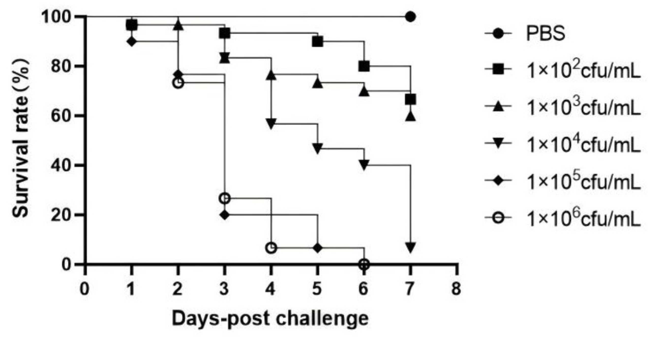
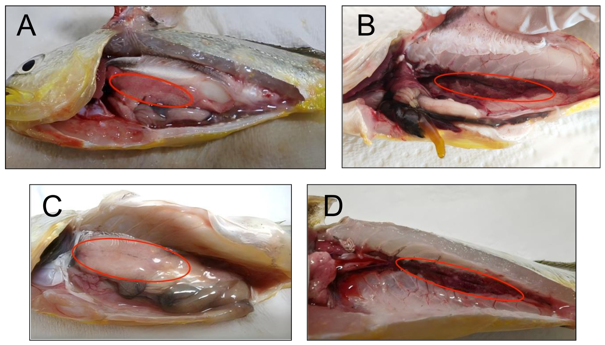
To determine the optimal challenge dose, fish were intraperitoneally injected with P. plecoglossicida at concentrations ranging from 1 × 10² to 1 × 10⁶ CFU/mL and monitored for 7 days. Post-mortem examination revealed characteristic visceral white spot disease pathology, including ascites, hepatomegaly with pallor, and renal white nodules (Figure 1). Acute mortality occurred at higher doses (1 × 10⁵ and 1 × 10⁶ CFU/mL), reaching 80.0% and 73.3% by day 3, with 100% mortality by day 6. The 1 × 10⁴ CFU/mL dose produced 53.3% mortality on day 5 and 93.3% by day 7, while lower doses (1 × 10² and 1 × 10³ CFU/mL) resulted in slower kinetics (33.3% and 40.0% cumulative mortality, respectively). The LD50 at 7 days post-infection was calculated as 3.1 × 10⁴ CFU/kg using the Reed–Muench method. Based on these kinetics, intraperitoneal injection with P. plecoglossicida (200 μL, 1 × 10⁴ CFU/mL) was selected for subsequent challenge experiments (Figure 2).
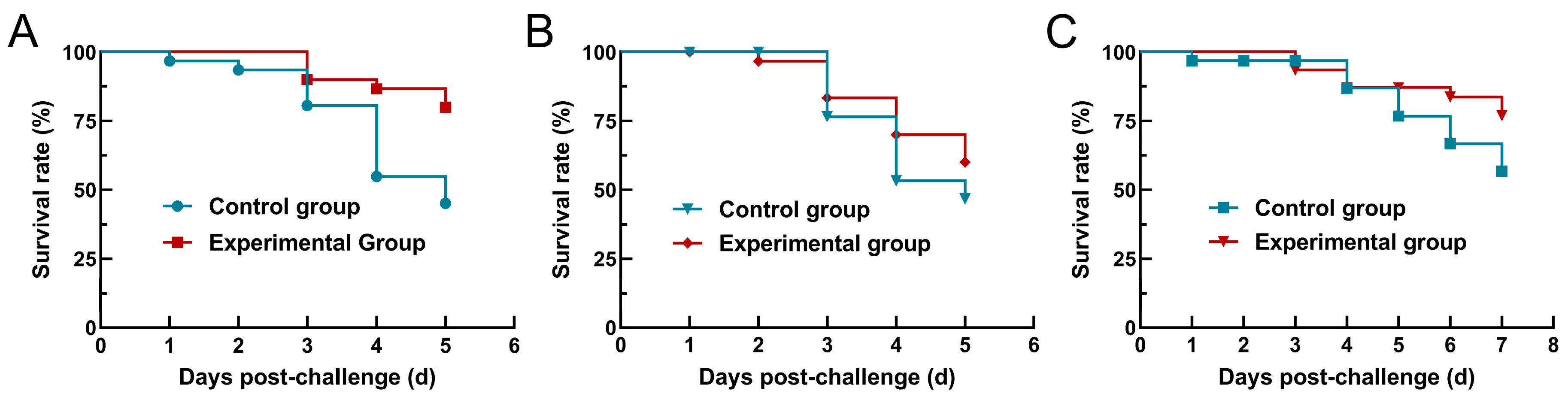
Dietary supplementation with P. heterophylla extract significantly improved survival following bacterial challenge (Figure 3). After 30 days of feeding, survival rates on day 5 post-infection increased from 46.67% (14/30) in controls to 80.00% (24/30) in treated fish, representing a 33.33% improvement (Figure 3A). Following 45 days of supplementation, survival improved from 46.67% to 60.00% (13.33% increase; Figure 3B), and after 60 days of feeding, survival rates on day 7 post-infection increased from 56.67% to 76.67% (20.00% increase; Figure 3C). Notably, the survival rate at 45 days post-feeding (60.00%) was lower than that at 30 days post-feeding (80.00%) and 60 days post-feeding (76.67%). This non-monotonic pattern may reflect a dynamic immunomodulatory effect of P. heterophylla extract. The initial high survival at 30 days suggests an early activation of innate immunity. The relative decline at 45 days may indicate a transient regulatory phase or immune adaptation, while the subsequent increase at 60 days likely reflects the establishment of more sustained and coordinated immune responses.

3.2. RNA-Seq Data Quality and Alignment

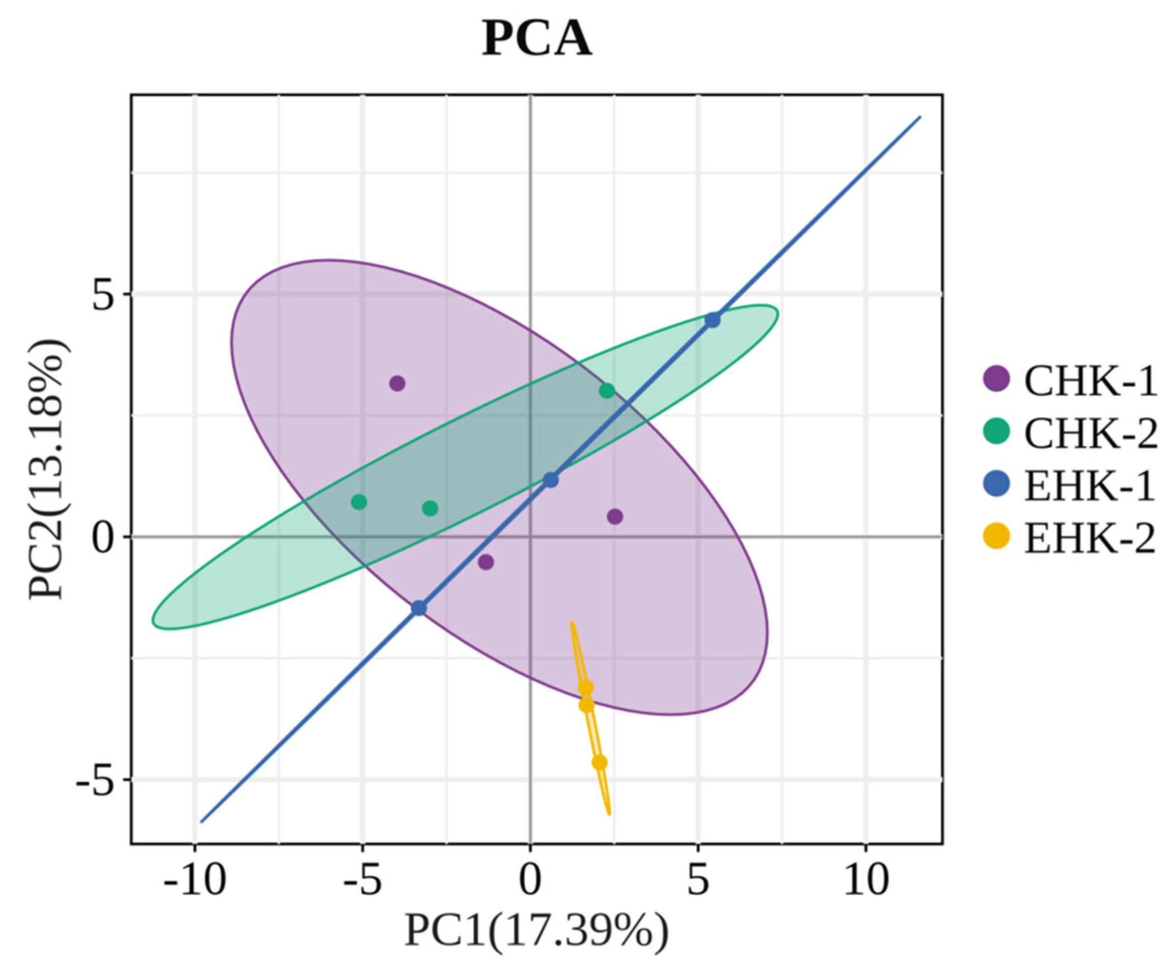
Transcriptomic libraries were constructed from 12 head kidney samples and sequenced on the Illumina NovaSeq 6000 platform. Raw reads were processed using Trimmomatic v0.39 to remove adapters and low-quality sequences, yielding ≥52.97 million high-quality reads per sample with Q20 ≥ 97.27% and Q30 ≥ 92.30% (Table 2). Alignment to the L. crocea reference genome (GCF_000972845.2) using HISAT2 v2.2.1 achieved total mapping rates of 82.76–87.43% and unique mapping rates of 75.06–80.12%. Principal component analysis (PCA) confirmed clustering by treatment group with no significant outliers, indicating high reproducibility (Figure 4).

3.3. Differential Gene Expression Analysis

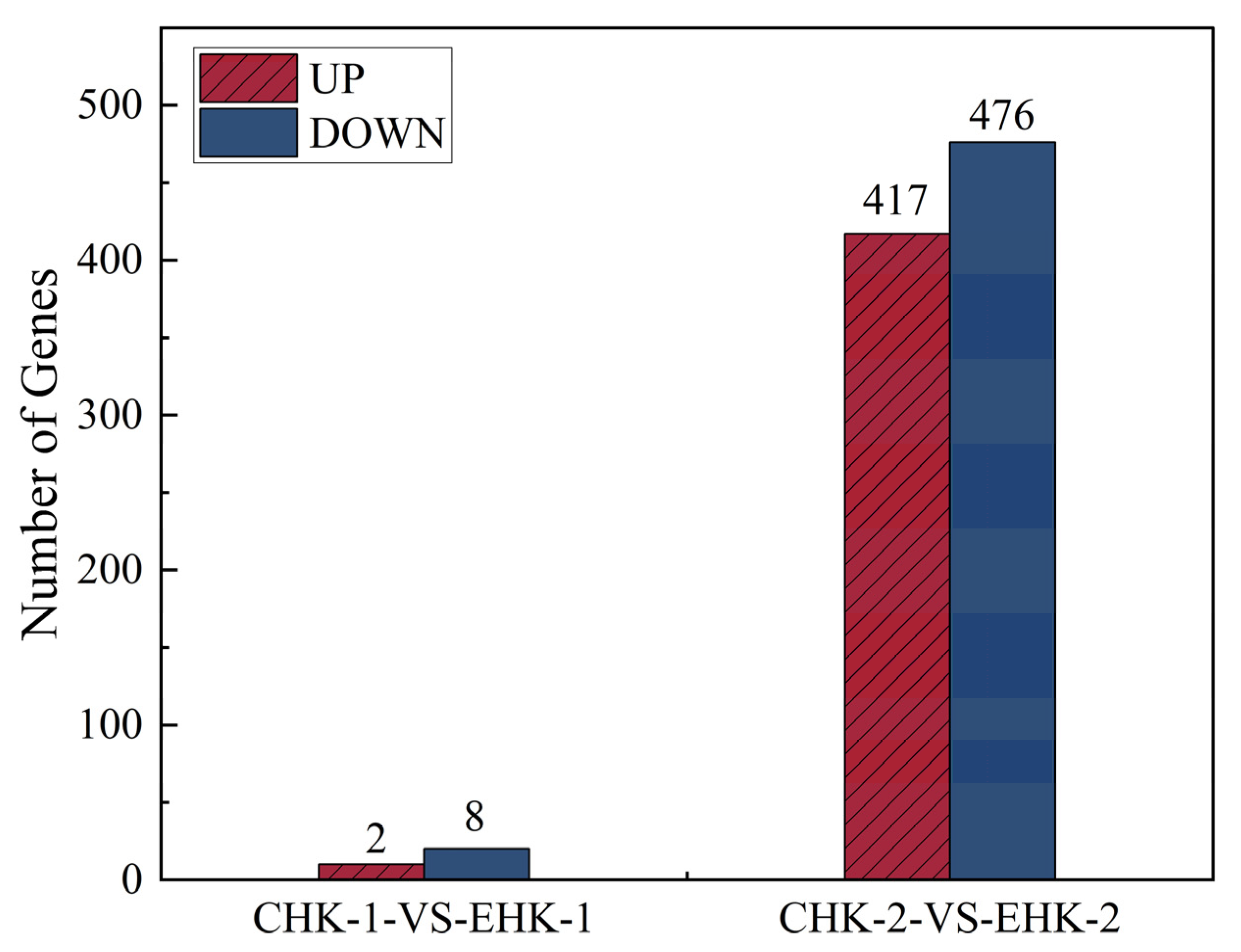
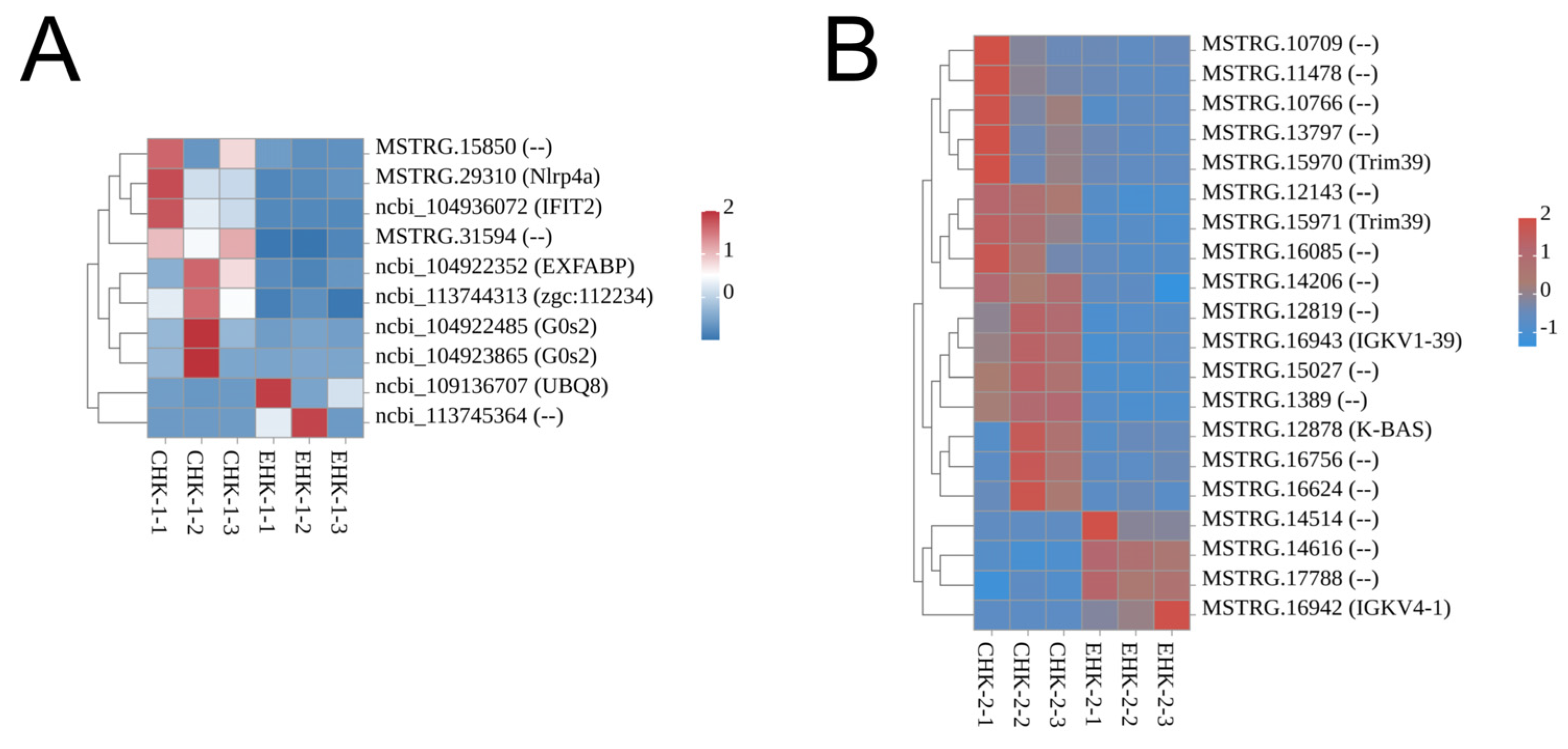
Pairwise comparisons between treatment and control groups at 45 and 60 days post-feeding revealed time-dependent transcriptional responses to P. plecoglossicida infection (Figure 5). At 45 days, the treatment group exhibited 10 DEGs (2 up-regulated, 8 down-regulated), whereas at 60 days, 893 DEGs were identified (417 up-regulated, 476 down-regulated), indicating progressive enhancement of immunomodulatory effects with prolonged P. heterophylla supplementation. The results of significantly higher number of DEGs (893) further support the enriched adaptive immune pathways at this time point. Hierarchical clustering analysis demonstrated consistent expression patterns among biological replicates and distinct transcriptional profiles between treatment and control groups (Figure 6).

3.4. GO Functional Enrichment Analysis

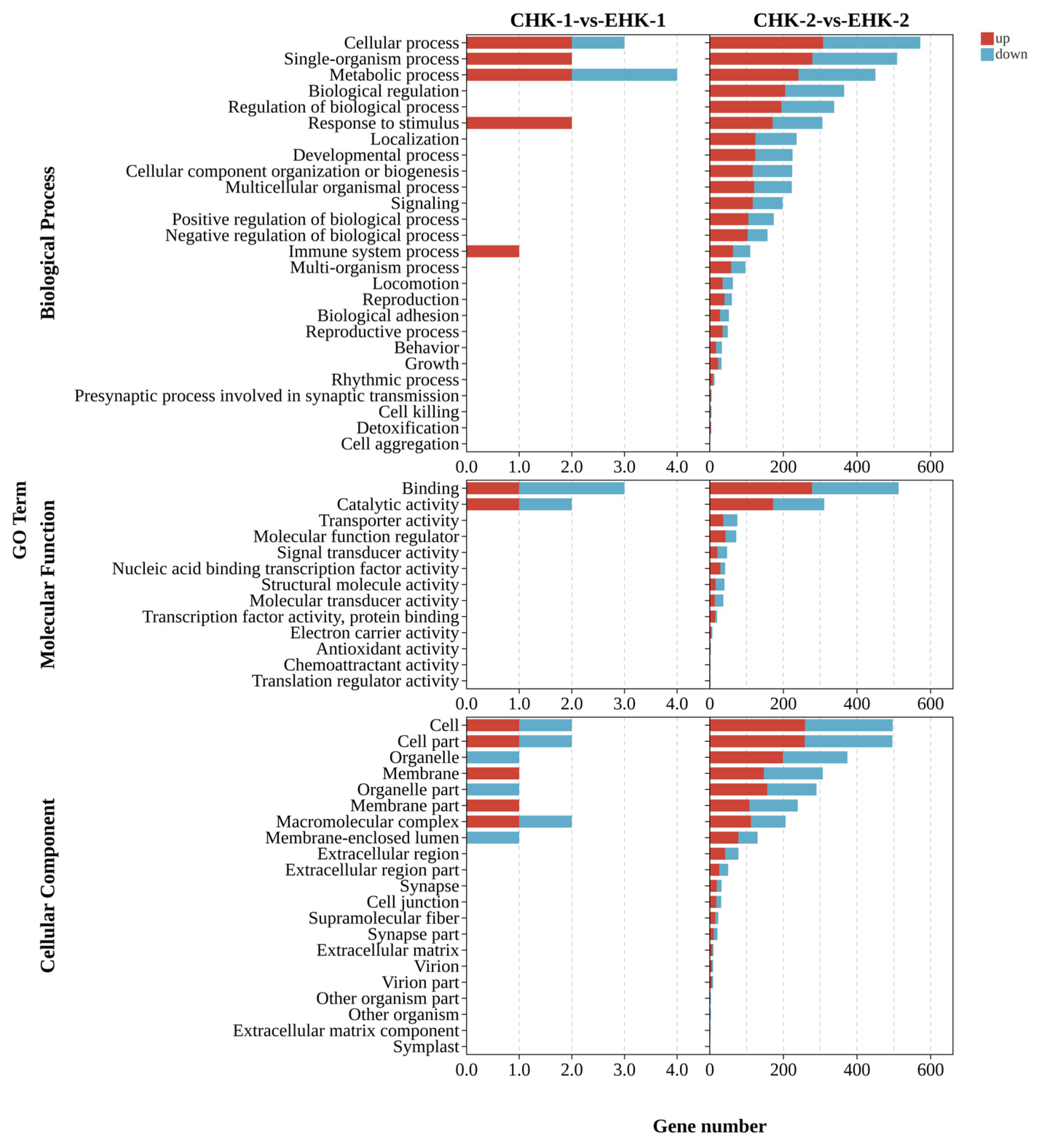
GO enrichment analysis was performed on DEGs from pairwise comparisons between treatment and control groups at 45 and 60 days post-feeding (Figure 7). At 45 days, 28 GO annotation entries were mapped to GO terms, predominantly in the biological process (BP, n = 12), cellular component (CC, n = 11), and molecular function (MF, n = 5) categories. Notably, three immune-related entries were identified, all of which up-regulated. Significant enrichment (p < 0.05) was observed in 14 GO terms, with 4 terms showing highly significant enrichment (p < 0.01). At 60 days, 8483 entries were annotated, with the majority classified under BP (n = 4501), CC (n = 2811), and MF (n = 1171). This included 139 immune-related entries (81 up-regulated, 58 down-regulated). Significant enrichment was detected in 1022 GO terms, with 328 terms being highly significant (p < 0.01). Immune-related GO terms, including immune system process, immune response, cytokine activity, chemokine activity, and chemokine receptor binding, were significantly enriched at both timepoints, indicating enhanced immunomodulatory effects of P. heterophylla supplementation.
GSEA identified 7305 gene sets, with 731 up-regulated sets enriched at 45 days post-infection (80 significant at FDR < 0.25; 133 at p < 0.05). The top enriched sets included regulation of leukocyte-mediated cytotoxicity, natural killer cell-mediated immunity, TRIF-dependent toll-like receptor signaling pathway, and regulation of interferon-gamma production (Supplementary Table S1). At 60 days, 5640 up-regulated sets were identified (1240 significant at FDR < 0.25), with prominent enrichment in tumor necrosis factor secretion, B cell activation, somatic diversification of immunoglobulins, and positive regulation of myeloid leukocyte differentiation (Supplementary Table S2). These results consistently revealed enrichment in lymphocyte differentiation, chemokine activity, leukocyte chemotaxis, and interleukin-10 production pathways.

3.5. KEGG Pathway Enrichment Analysis

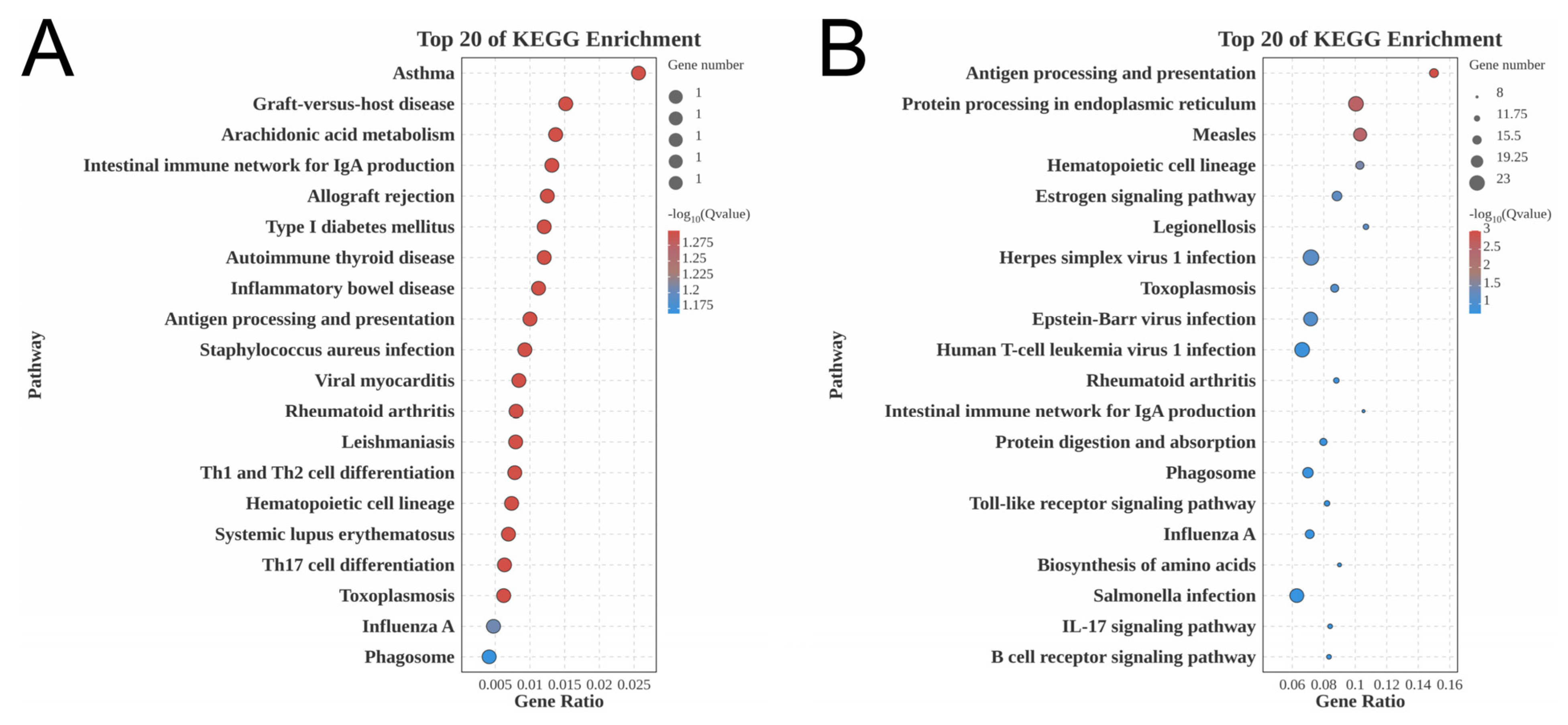
DEGs from pairwise comparisons (CHK-1 vs. EHK-1, CHK-2 vs. EHK-2) were subjected to KEGG pathway enrichment analysis (Figure 8). At 45 days, 19 pathways significantly enriched (p < 0.05). Among the top 20 pathways, 19 were immunity- and disease-related, including Staphylococcus aureus infection, Epstein–Barr virus infection, herpes simplex virus infection, human T-cell leukemia virus infection, hematopoietic cell lineage, and Th1/Th2 and Th17 cell differentiation (Figure 8A). At 60 days, 41 pathways were significantly enriched (p < 0.05). The top 20 pathways included 16 immunity- and disease-related pathways, such as herpes simplex virus infection, human T-cell leukemia virus infection, Salmonella infection, hematopoietic cell lineage, Toll-like receptor signaling, IL-17 signaling, and B cell receptor signaling (Figure 8B). These results demonstrate that P. heterophylla supplementation modulates immune-related signaling pathways in a time-dependent manner, with enhanced pathway diversity at 60 days compared to 45 days.
GSEA of KEGG pathways identified 328 gene sets, with 64 up-regulated sets enriched at 45 days (45 significant at FDR < 0.25; 42 at p < 0.05). The top enriched pathways included viral protein interaction with cytokine and cytokine receptor, cytokine–cytokine receptor interaction, the Toll-like receptor signaling pathway, and Th1 and Th2 cell differentiation (Supplementary Table S3). At 60 days, 247 up-regulated sets were identified (107 significant at FDR < 0.25), with prominent enrichment in the C-type lectin receptor signaling pathway, mTOR signaling pathway, TNF signaling pathway, B cell receptor signaling pathway, and chemokine signaling pathway (Supplementary Table S4). These results consistently demonstrated enrichment in cytokine-cytokine receptor interactions, chemokine signaling, and T/B cell differentiation pathways.

3.6. Protein–Protein Interaction Network Analysis

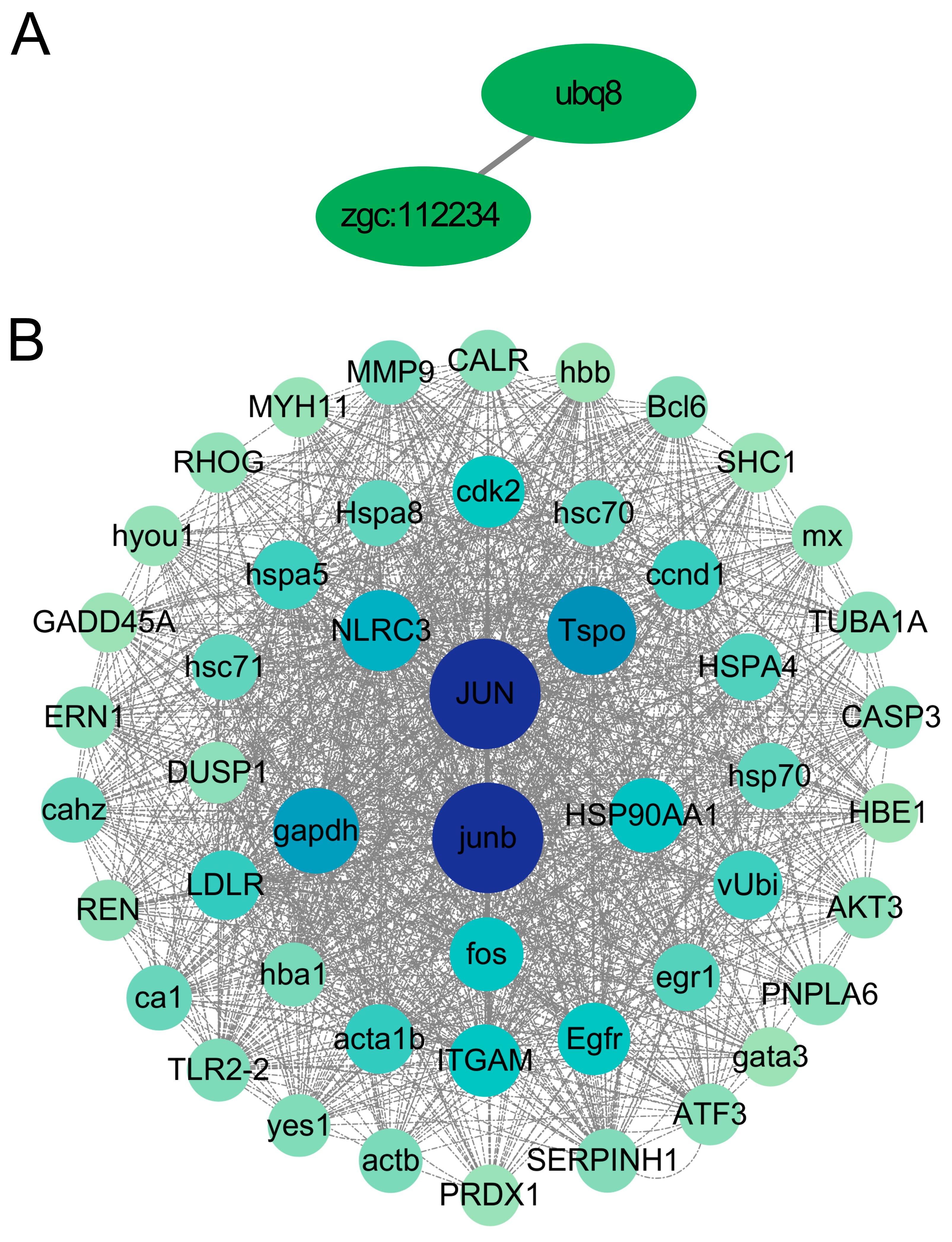
PPI networks were constructed using the STRING database and visualized with Cytoscape. At 45 days, the network comprised a single interacting node pair, ubq8 (NCBI: 109136707) and zgc:112234 (NCBI: 113744313), both encoding polyubiquitin proteins (Figure 9A). At 60 days, the network expanded to 678 proteins with 10,894 interactions, forming five distinct modules (Figure 9B). The two largest modules included genes such as tspo (ncbi_104931334), gapdh (ncbi_104936375), jun (ncbi_104937023), and junb (ncbi_104932219), which were significantly enriched in signaling molecule and infectious disease pathways. Notably, junb is implicated in Epstein–Barr virus infection.

3.7. Validation of DEGs by Quantitative Real-Time PCR

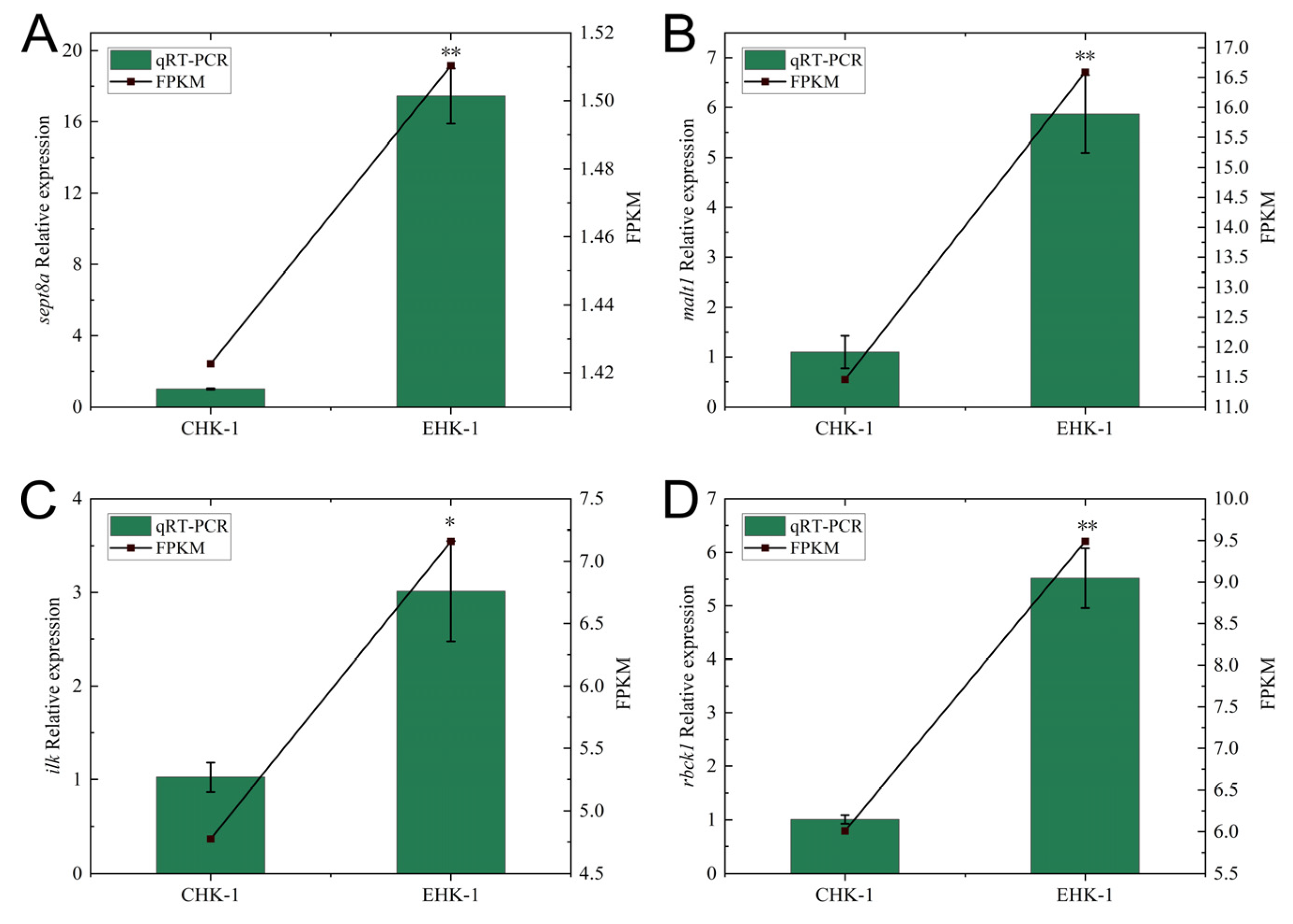
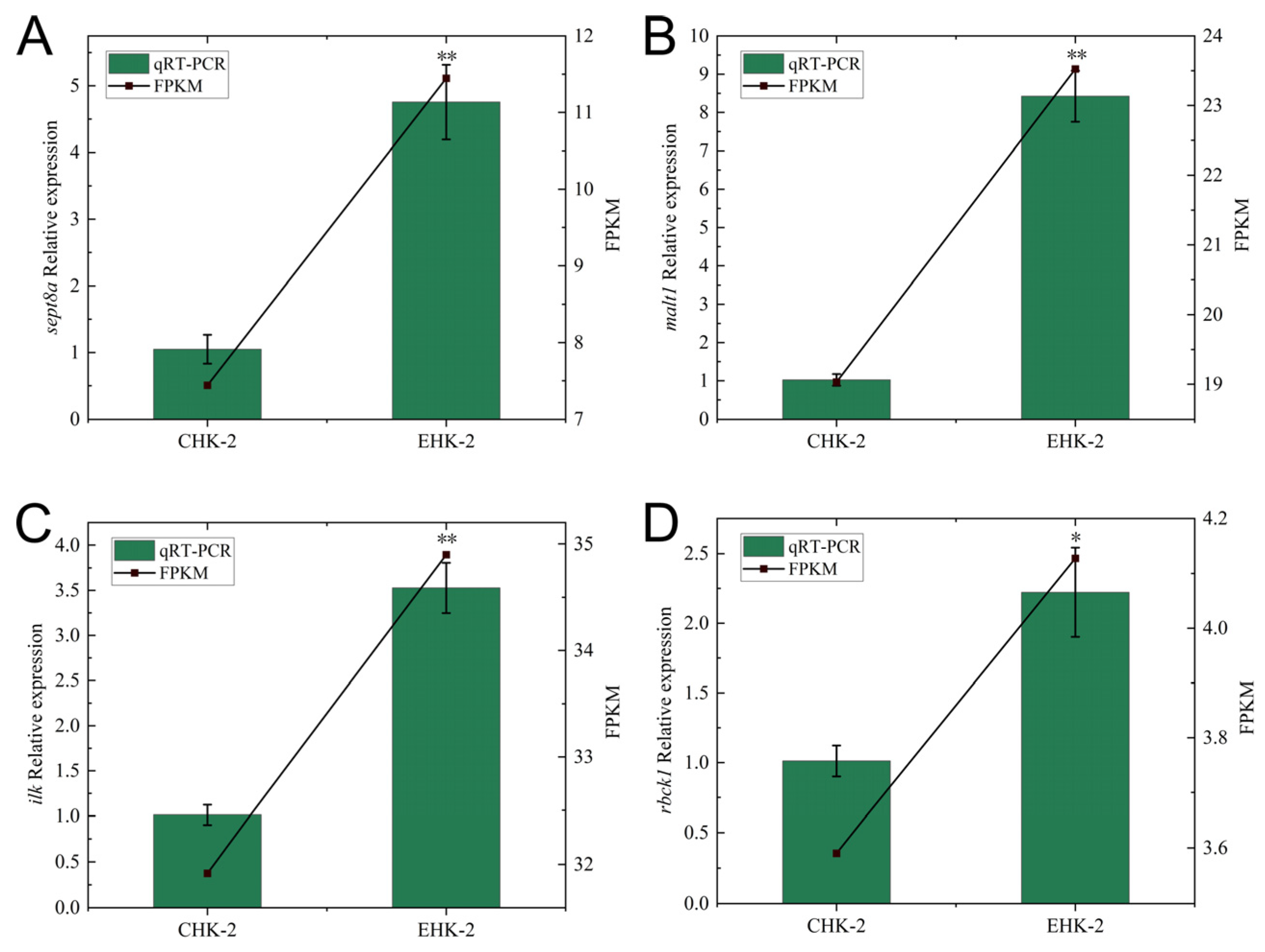
Four DEGs (sept8a, ilk, rbck1, malt1) were validated by qRT-PCR using β-actin as the reference gene. RNA samples were extracted from head kidney tissues of six fish per group, with three technical replicates per sample. At 45 days, all four genes were significantly up-regulated in the treatment group compared to controls (p < 0.05; Figure 10). At 60 days, sept8a, ilk, rbck1, and malt1 also showed significant up-regulation (p < 0.05, Figure 11). The expression patterns were consistent with RNA-seq data, confirming the reliability of transcriptome results.

4. Discussion

The head kidney serves as the primary immune organ in teleost fish, functioning as both a central and peripheral immune organ responsible for the generation, differentiation, and proliferation of macrophages, granulocytes, and B lymphocytes [28]. In large yellow croaker, the head kidney is the first immune organ to develop during ontogeny, making it a critical target for investigating immunomodulatory effects.
Chemokine activity and Toll-like receptor (TLR) signaling pathways constitute the primary mechanisms of nonspecific immunity in fish during bacterial infection, playing essential roles in maintaining immune homeostasis. TLRs function not only as core protein molecules in nonspecific immune responses but also serve as critical bridges connecting nonspecific and specific immunity [29]. Toll-like receptor 3 (TLR3), a member of the TLR family, mediates transcriptional induction of type I interferons (IFNs), proinflammatory cytokines, and chemokines, thereby collectively establishing the host’s antiviral defense [30]. In the present study, TLR3 expression was significantly up-regulated in the head kidney of large yellow croaker fed with P. heterophylla extract, consistent with previous findings that plant herbal extracts act as immunostimulants to enhance disease resistance in farmed fish by activating proinflammatory responses, TLR9/IRF7 signaling, and IFN-β protein expression [31,32,33].
Interleukin-17 (IL-17) is a multifunctional cytokine produced by various immune cells that plays a pivotal role in host defense against pathogens [34]. It stimulates T cell proliferation and differentiation, mediates bacterial clearance, and promotes Th1 cell development to eliminate invasive bacteria [35]. IL-17 exerts its protective effects by binding to specific receptors, including IL-17 receptor and IL-17R [36]. Previous studies have demonstrated that plant polysaccharides can enhance immune responses; for example, licorice polysaccharides fed to chickens increased the expression of IL-2, IL-4, IFN-γ, and IL-17 cytokines, confirming the immunostimulatory potential of herbal extracts [37]. In the present study, expression levels of multiple immune factor genes in head kidney tissue were significantly elevated in P. heterophylla-fed fish compared to controls, with IL-17 showing the most pronounced up-regulation. These findings demonstrate that P. heterophylla extract effectively triggers nonspecific immune responses in large yellow croaker by significantly up-regulating key immune factors including TLR3 and IL-17.
Compared with other herbal immunostimulants such as the extracts of Glycyrrhiza glabra (liquorice) and Astragalus membranaceus (AM), P. heterophylla extract exhibits unique advantages in enhancing both innate and adaptive immunity in fish [38]. While licorice polysaccharides primarily up-regulate IL-2, IL-4, and IFN-γ, P. heterophylla extract significantly up-regulates TLR3, IL-17, and chemokines, indicating broader activation of both Toll-like receptor signaling and T cell differentiation pathways [39]. Furthermore, the time-dependent transcriptional activation observed in this study (from 10 DEGs at 45 days to 893 DEGs at 60 days) suggests that P. heterophylla may require prolonged administration to fully exert its immunomodulatory effects, a feature not extensively reported for other herbal extracts in fish.

5. Conclusions

In summary, this study analyzed transcriptomic changes in the head kidney tissue of large yellow croaker fed with P. heterophylla extract for 45 and 60 days following P. plecoglossicida challenge. The results demonstrated significant up-regulation of genes associated with Toll-like receptors, chemokines, interleukins, and specific immune cell proliferation. Notably, both nonspecific and specific immunity-related pathways showed time-dependent enhancement, with more pronounced up-regulation at 60 days compared to 45 days. These findings indicate that P. heterophylla extract effectively enhances resistance to P. plecoglossicida infection in large yellow croaker, providing a scientific foundation for immunological prevention and control strategies against visceral white spot disease in aquaculture.

Supplementary Materials

The following supporting information can be downloaded at https://www.mdpi.com/article/10.3390/fishes11060371/s1. Table S1. The top 20 gene sets of differentially expressed gene GSEA (CHK-1-vs-EHK-1.GO.GSEA) in the head kidney that up-regulated at 45 days-post challenge; Table S2. The top 20 gene sets of differentially expressed gene GSEA (CHK-2-vs-EHK-2.GO.GSEA) in the head kidney that up-regulated at 60 days-post challenge; Table S3. The top 20 gene sets of differentially expressed gene GSEA (CHK-1-vs-EHK-1. KEGG.GSEA) in the head kidney that up-regulated at 45 days-post challenge; Table S4. The top 20 gene sets of differentially expressed gene GSEA (CHK-2-vs-EHK-2. KEGG.GSEA) in the head kidney that up-regulated at 60 days-post challenge.

Author Contributions

K.H.H. conceived and designed the study. Z.M.L. contributed to the experimental work and original data preparation. L.Z. contributed to the manuscript writing and revision. D.L.Z., Z.H.S., J.C., Z.D.L. and Y.B.D. contributed with the experimental materials. Y.L. and P.F.Z. contributed to the manuscript writing (review and editing). All authors have read and agreed to the published version of the manuscript.

Funding

The work was supported by the National Key Research and Development Program of China (2025YFD2400302), Fujian Provincial Science and Technology Plan Project (2025S2003, 2020J02011), Scientific Research Program of Ningde Normal University (2025ZX091), and Industry-University-Research Cooperation Project of Jiaocheng District, Ningde City (2025C002).

Institutional Review Board Statement

The animal study protocol was approved by the Ethics Committee of Ningde Normal University (protocol code NDNU-LL-202508 and approval date: 25 February 2025).

Informed Consent Statement

Not applicable.

Data Availability Statement

The original contributions presented in this study are included in the article and Supplementary Material. Further inquiries can be directed to the corresponding authors.

Conflicts of Interest

Author Zhao Han Sun and Jia Chen were employed by the Ningde Yiye Marine Industry Development Co., Ltd. The remaining authors declare that the research was conducted in the absence of any commercial or financial relationships that could be construed as a potential conflict of interest.

References

  1. Zhang, H.; Wang, J.; Jing, Y. Larimichthys crocea (large yellow croaker): A bibliometric study. Heliyon 2024, 10, e37393. [Google Scholar] [CrossRef]
  2. Wang, L.-Y.; Liu, Z.-X.; Zhao, L.-M.; Huang, L.-X.; Qin, Y.-X.; Su, Y.-Q.; Zheng, W.-Q.; Wang, F.; Yan, Q.-P. Dual RNA-seq provides novel insight into the roles of dksA from Pseudomonas plecoglossicida in pathogen-host interactions with large yellow croakers (Larimichthys crocea). Zool. Res. 2020, 41, 410–422. [Google Scholar] [CrossRef]
  3. Huang, L.; Zhao, L.; Su, Y.; Yan, Q. Genome Sequence of Pseudomonas plecoglossicida Strain NZBD9. Genome Announc. 2018, 6, 10-1128. [Google Scholar] [CrossRef] [PubMed]
  4. Huang, L.; Zuo, Y.; Jiang, Q.; Su, Y.; Qin, Y.; Xu, X.; Zhao, L.; Yan, Q. A metabolomic investigation into the temperature-dependent virulence of Pseudomonas plecoglossicida from large yellow croaker (Pseudosciaena crocea). J. Fish Dis. 2019, 42, 431–446. [Google Scholar] [CrossRef] [PubMed]
  5. Li, K.; Li, W.; Chen, X.; Luo, T.; Mu, Y.; Chen, X. Molecular and functional identification of a β-defensin homolog in large yellow croaker (Larimichthys crocea). J. Fish Dis. 2021, 44, 391–400. [Google Scholar] [CrossRef] [PubMed]
  6. Zhang, J.T.; Zhou, S.M.; An, S.W.; Chen, L.; Wang, G.L. Visceral granulomas in farmed large yellow croaker, Larimichthys crocea (Richardson), caused by a bacterial pathogen, Pseudomonas plecoglossicida. J. Fish Dis. 2014, 37, 113–121. [Google Scholar] [CrossRef] [PubMed]
  7. Zhang, B.; Luo, G.; Zhao, L.; Huang, L.; Qin, Y.; Su, Y.; Yan, Q. Integration of RNAi and RNA-seq uncovers the immune responses of Epinephelus coioides to L321_RS19110 gene of Pseudomonas plecoglossicida. Fish Shellfish Immunol. 2018, 81, 121–129. [Google Scholar] [CrossRef] [PubMed]
  8. Xiang, J.; Chen, R.; Xu, D.; Sun, Y.; Liu, H. Characterization of pathological changes and immune-related gene expression in yellow drum (Nibea albiflora) in response to Pseudomonas plecoglossicida and poly I:C challenge. Aquac. Rep. 2020, 17, 100350. [Google Scholar] [CrossRef]
  9. Sun, Y.; Luo, G.; Zhao, L.; Huang, L.; Qin, Y.; Su, Y.; Yan, Q. Integration of RNAi and RNA-seq Reveals the Immune Responses of Epinephelus coioides to sigX Gene of Pseudomonas plecoglossicida. Front. Immunol. 2018, 9, 1624. [Google Scholar] [CrossRef] [PubMed]
  10. Wang, Z.; Liao, S.-G.; He, Y.; Li, J.; Zhong, R.-F.; He, X.; Liu, Y.; Xiao, T.-T.; Lan, Y.-Y.; Long, Q.-D.; et al. Protective effects of fractions from Pseudostellaria heterophylla against cobalt chloride-induced hypoxic injury in H9c2 cell. J. Ethnopharmacol. 2013, 147, 540–545. [Google Scholar] [CrossRef] [PubMed]
  11. Yang, J.; Wang, D.-Q.; Yao, Y.; Jin, X.-F. Observation on biological characteristics of wild Pseudostellaria heterophylla. J. Chin. Med. Mater. 2011, 34, 1323–1328. [Google Scholar] [PubMed]
  12. Wang, J.; Li, J.; Li, H.; Wu, X.; Gao, W. HPLC–ESI–MSn Analysis, Fed-Batch Cultivation Enhances Bioactive Compound Biosynthesis and Immune-Regulative Effect of Adventitious Roots in Pseudostellaria heterophylla. Appl. Biochem. Biotechnol. 2015, 177, 63–75. [Google Scholar] [CrossRef] [PubMed]
  13. Yang, Q.; Cai, X.; Huang, M.; Jia, L.; Wang, S. Immunomodulatory effects of Pseudostellaria heterophylla peptide on spleen lymphocytes via a Ca2+/CaN/NFATc1/IFN-γ pathway. Food Funct. 2019, 10, 3466–3476. [Google Scholar] [CrossRef] [PubMed]
  14. Fang, Z.-H.; Duan, X.-C.; Zhao, J.-D.; Wu, Y.-J.; Liu, M.-M. Novel Polysaccharide H-1-2 from Pseudostellaria Heterophylla Alleviates Type 2 Diabetes Mellitus. Cell. Physiol. Biochem. 2018, 49, 1037–1047. [Google Scholar] [CrossRef] [PubMed]
  15. Sun, H.; Shi, K.; Qi, K.; Kong, H.; He, Q.; Zhou, M. Pseudostellaria heterophylla Extract Polysaccharide H-1-2 Suppresses Pancreatic Cancer by Inhibiting Hypoxia-Induced AG2. Mol. Ther. Oncolytics 2020, 17, 61–69. [Google Scholar] [CrossRef] [PubMed]
  16. Chuanlong, Z.; Xiaoxia, Z. Effects of polysaccharides from Pseudostellaria heterophylla on exercise endurance capacity and oxidative stress in forced swimming rats. Sci. Res. Essays 2011, 6, 2360–2365. [Google Scholar]
  17. Chen, Z.; Li, S.; Wang, X.; Zhang, C.L. Protective effects of Radix Pseudostellariae polysaccharides against exercise-induced oxidative stress in male rats. Exp. Ther. Med. 2013, 5, 1089–1092. [Google Scholar] [CrossRef] [PubMed]
  18. Li, H.; Liu, Z.; Liu, Q.; Zhang, X.; Li, S.; Tang, F.; Zhang, L.; Yang, Q.; Wang, Q.; Yang, S.; et al. Extraction of Polysaccharides from Root of Pseudostellaria heterophylla (Miq.) Pax. and the Effects of Ultrasound Treatment on Its Properties and Antioxidant and Immune Activities. Molecules 2023, 29, 142. [Google Scholar] [CrossRef] [PubMed]
  19. Chen, S.; Zhou, Y.; Chen, Y.; Gu, J. fastp: An ultra-fast all-in-one FASTQ preprocessor. Bioinformatics 2018, 34, i884–i890. [Google Scholar] [CrossRef] [PubMed]
  20. Kim, D.; Langmead, B.; Salzberg, S.L. HISAT: A fast spliced aligner with low memory requirements. Nat. Methods 2015, 12, 357–360. [Google Scholar] [CrossRef] [PubMed]
  21. Love, M.I.; Huber, W.; Anders, S. Moderated estimation of fold change and dispersion for RNA-seq data with DESeq2. Genome Biol. 2014, 15, 550. [Google Scholar] [CrossRef] [PubMed]
  22. Robinson, M.D.; McCarthy, D.J.; Smyth, G.K. EdgeR: A Bioconductor package for differential expression analysis of digital gene expression data. Bioinformatics 2010, 26, 139–140. [Google Scholar] [CrossRef] [PubMed]
  23. Kanehisa, M.; Goto, S. KEGG: Kyoto Encyclopedia of Genes and Genomes. Nucleic Acids Res. 2000, 28, 27–30. [Google Scholar] [CrossRef] [PubMed]
  24. Subramanian, A.; Tamayo, P.; Mootha, V.K.; Mukherjee, S.; Ebert, B.L.; Gillette, M.A.; Paulovich, A.; Pomeroy, S.L.; Golub, T.R.; Lander, E.S.; et al. Gene set enrichment analysis: A knowledge-based approach for interpreting genome-wide expression profiles. Proc. Natl. Acad. Sci. USA 2005, 102, 15545–15550. [Google Scholar] [CrossRef] [PubMed]
  25. Szklarczyk, D.; Franceschini, A.; Wyder, S.; Forslund, K.; Heller, D.; Huerta-Cepas, J.; Simonovic, M.; Roth, A.; Santos, A.; Tsafou, K.P.; et al. STRING v10: Protein–protein interaction networks, integrated over the tree of life. Nucleic Acids Res. 2015, 43, D447–D452. [Google Scholar] [CrossRef] [PubMed]
  26. Shannon, P.; Markiel, A.; Ozier, O.; Baliga, N.S.; Wang, J.T.; Ramage, D.; Amin, N.; Schwikowski, B.; Ideker, T. Cytoscape: A software environment for integrated models of Biomolecular Interaction Networks. Genome Res. 2003, 13, 2498–2504. [Google Scholar] [CrossRef] [PubMed]
  27. Schmittgen, T.D.; Livak, K.J. Analyzing real-time PCR data by the comparative CT method. Nat. Protoc. 2008, 3, 1101–1108. [Google Scholar] [CrossRef] [PubMed]
  28. Morelli, A.B.; Becher, D.; Koernig, S.; Silva, A.; Drane, D.; Maraskovsky, E. ISCOMATRIX: A novel adjuvant for use in prophylactic and therapeutic vaccines against infectious diseases. J. Med. Microbiol. 2012, 61, 935–943. [Google Scholar] [CrossRef] [PubMed]
  29. Sahoo, B.R. Structure of fish Toll-like receptors (TLR) and NOD-like receptors (NLR). Int. J. Biol. Macromol. 2020, 161, 1602–1617. [Google Scholar] [CrossRef] [PubMed]
  30. Chen, Y.; Lin, J.; Zhao, Y.; Ma, X.; Yi, H. Toll-like receptor 3 (TLR3) regulation mechanisms and roles in antiviral innate immune responses. J. Zhejiang Univ. B 2021, 22, 609–632. [Google Scholar] [CrossRef] [PubMed]
  31. Vallejos-Vidal, E.; Reyes-López, F.; Teles, M.; MacKenzie, S. The response of fish to immunostimulant diets. Fish Shellfish Immunol. 2016, 56, 34–69. [Google Scholar] [CrossRef] [PubMed]
  32. Ghochikyan, A.; Pichugin, A.; Bagaev, A.; Davtyan, A.; Hovakimyan, A.; Tukhvatulin, A.; Davtyan, H.; Shcheblyakov, D.; Logunov, D.; Chulkina, M.; et al. Targeting TLR-4 with a novel pharmaceutical grade plant derived agonist, Immunomax®, as a therapeutic strategy for metastatic breast cancer. J. Transl. Med. 2014, 12, 322. [Google Scholar] [CrossRef] [PubMed]
  33. Lin, C.-J.; Lin, H.-J.; Chen, T.-H.; Hsu, Y.-A.; Liu, C.-S.; Hwang, G.-Y.; Wan, L. Polygonum cuspidatum and Its Active Components Inhibit Replication of the Influenza Virus through Toll-Like Receptor 9-Induced Interferon Beta Expression. PLoS ONE 2015, 10, e0117602. [Google Scholar] [CrossRef] [PubMed]
  34. Xue, T.; Liu, Y.; Cao, M.; Zhang, X.; Fu, Q.; Yang, N.; Li, C. Genome-wide identification of interleukin-17 (IL-17)/interleukin-17 receptor (IL- 17R) in turbot (Scophthalmus maximus) and expression pattern analysis after Vibrio anguillarum infection. Dev. Comp. Immunol. 2021, 121, 104070. [Google Scholar] [CrossRef] [PubMed]
  35. Mu, P.; Huo, J.; Li, X.; Li, W.; Li, X.; Ao, J.; Chen, X. IL-2 Signaling Couples the MAPK and mTORC1 Axes to Promote T Cell Proliferation and Differentiation in Teleosts. J. Immunol. 2022, 208, 1616–1631. [Google Scholar] [CrossRef] [PubMed]
  36. Cua, D.J.; Tato, C.M. Innate IL-17-producing cells: The sentinels of the immune system. Nat. Rev. Immunol. 2010, 10, 479–489. [Google Scholar] [CrossRef] [PubMed]
  37. Wu, Y.; Li, N.; Zhang, T.; Che, Y.; Duan, K.; Wang, Y.; Zhou, H.; Wan, X.; Lei, H.; Nguyễn, A.D.; et al. Glycyrrhiza polysaccharides can improve and prolong the response of chickens to the Newcastle disease vaccine. Poult. Sci. 2022, 101, 101549. [Google Scholar] [CrossRef] [PubMed]
  38. Elabd, H.; Wang, H.-P.; Shaheen, A.; Yao, H.; Abbass, A. Feeding Glycyrrhiza glabra (liquorice) and Astragalus membranaceus (AM) alters innate immune and physiological responses in yellow perch (Perca flavescens). Fish Shellfish Immunol. 2016, 54, 374–384. [Google Scholar] [CrossRef] [PubMed]
  39. Wu, Y.; Zhou, H.; Wei, K.; Zhang, T.; Che, Y.; Nguyễn, A.D.; Pandita, S.; Wan, X.; Cui, X.; Zhou, B.; et al. Structure of a new glycyrrhiza polysaccharide and its immunomodulatory activity. Front. Immunol. 2022, 13, 1007186. [Google Scholar] [CrossRef] [PubMed]
Figure 1. Visceral lesions of large yellow croaker following experimental infection. Representative images showing pathological changes in internal organs. The corresponding tissues are indicated by circles. (A) Liver from healthy fish; (B) head kidney from healthy fish; (C) liver from diseased fish; (D) head kidney from diseased fish.
Fishes 11 00371 g001
Figure 2. Survival of large yellow croaker following intraperitoneal challenge with P. plecoglossicida at different doses. Five groups of large yellow croakers were infected by intraperitoneal injection with 200 μL P. plecoglossicida at 1 × 10⁶ CFU/mL, 1 × 10⁵ CFU/mL, 1 × 10⁴ CFU/mL, 1 × 10³ CFU/mL, and 1 × 10² CFU/mL, respectively. The mortality of the fish was monitored and recorded for 7 days.
Fishes 11 00371 g002
Figure 3. Survival rates of large yellow croaker challenged with P. plecoglossicida following different acclimation periods. Survival of control and experimental groups with intraperitoneal injection of 200 μL 1 × 10⁴ CFU/mL P. plecoglossicida after temporary feeding with P. heterophylla extract for (A) 30 days, (B) 45 days, and (C) 60 days.
Fishes 11 00371 g003
Figure 4. Principal component analysis (PCA) of transcriptomic profiles. PCA plot illustrating the clustering patterns of head kidney samples from control and experimental groups based on gene expression profiles.
Fishes 11 00371 g004
Figure 5. Statistical analysis of differentially expressed genes (DEGs) in head kidney tissues of L. crocea. Bar chart summarizing the numbers of upregulated (red bars) and downregulated (blue bars) DEGs of control group (CHK) fed with the basal diet only and experimental group (EHK) supplemented with 1.0% P. heterophylla extract for 45 or 60 days.
Fishes 11 00371 g005
Figure 6. Clustering diagram showing the expression patterns of DEGs in head kidney tissues of L. crocea. (A) The expression of the control group (CHK) fed with the basal diet only and experimental group (EHK) supplemented with 1.0% P. heterophylla at 45 days post-feeding. (B) The expression of CHK and EHK at 60 days post-feeding. The color scale on the far right of the heatmap represents expression, whereby red, blue, and white indicate up-regulated, down-regulated and unaltered expression, respectively.
Fishes 11 00371 g006
Figure 7. Gene Ontology (GO) enrichment analysis of DEGs in head kidney tissues of L. crocea. Comparison between control (CHK) and experimental (EHK) head kidney tissues at 45 and 60 days post-feeding with P. heterophylla extract.
Fishes 11 00371 g007
Figure 8. Top 20 pathways of the Kyoto Encyclopedia of Genes and Genomes (KEGG) classification. Top 20 pathways of the KEGG classification of DEGs between CHK and EHK at (A) 45 and (B) 60 days post-feeding with P. heterophylla extract.
Fishes 11 00371 g008
Figure 9. Protein–protein interaction (PPI) network analysis of DEGs in the head kidney of L. crocea at (A) 45 and (B) 60 days post-feeding with P. heterophylla extract. Nodes represent proteins, and edges indicate predicted interactions. Node size reflects the degree of connectivity, and color intensity denotes differential expression levels.
Fishes 11 00371 g009
Figure 10. qRT-PCR verification of DEGs in head kidney tissue of L. crocea at 45 days post-feeding with P. heterophylla extract. Expression levels of the target genes including sept8a (A), malt1 (B), ilk (C), and rbck1 (D) were normalized to β-actin and are presented as mean ± SE (n = 6). Statistical significance was determined by Student’s t-test. * p < 0.05, ** p < 0.01.
Fishes 11 00371 g010
Figure 11. qRT-PCR verification of DEGs in head kidney tissue of L. crocea at 60 days post-feeding with P. heterophylla extract. Expression levels of the target genes including sept8a (A), malt1 (B), ilk (C), and rbck1 (D) were normalized to β-actin and are presented as mean ± SE (n = 6). Statistical significance was determined by Student’s t-test. * p < 0.05, ** p < 0.01.
Fishes 11 00371 g011
Table 1. Primer information for qRT-PCR.
GenePrimersAmplicon Size (bp)Primer Efficiency (%)
sept8aForward primerATACTCAAGTGTCGACGGCAG17899.5
Reverse primerTTGGCGCATTTCTTCTTCGTG
ilkForward primerCACGGAGTTCCCTTCTCGAC21598.8
Reverse primerACCTTTCTTTCCCCAGGCTAC
rbck1Forward primerGTTGCATTTTACAGGGCAAAGGT17796.8
Reverse primerTGGCAATTTTGGCAGTTGGG
malt1Forward primerAACGATGCAGGAGATTGACGA18999.8
Reverse primerTTTTTCATCAAAGCCCCCGTT
β-actinForward primerTCGTCGGTCGTCCCAGGCATCAG16798.8
Reverse primerATGGCGTGGGGCAGAGCGTAACC
Table 2. Summary of sequencing data.
SampleRaw Data (bp)Clean Data (bp)Q20 (%)Q30 (%)GC (%)Unique_Mapped (%)Total_Mapped (%)
CHK-1-17,632,059,7007,277,674,43597.6993.7745.9779.8784.93
CHK-1-28,430,991,2008,001,031,88997.6293.5846.6381.1586.04
CHK-1-37,013,979,3006,681,684,22897.6993.7146.5475.0682.76
CHK-2-17,406,268,0007,045,258,94498.0894.6746.6480.5186.88
CHK-2-26,890,075,7006,581,480,96298.3695.0448.3078.5989.38
CHK-2-35,451,673,8005,296,742,37098.3295.0748.8381.0091.44
EHK-1-16,243,928,2005,852,180,62997.2792.3046.5881.2686.33
EHK-1-27,058,998,5006,752,416,22698.1994.8845.7077.8184.57
EHK-1-38,679,359,7008,299,852,93597.6593.5046.2778.1584.79
EHK-2-16,162,554,1006,015,304,30897.6993.4946.9578.0788.07
EHK-2-25,814,360,3005,673,353,83797.9794.2448.5580.5391.00
EHK-2-36,454,137,9006,282,802,33898.2394.8548.4280.3790.64
Disclaimer/Publisher’s Note: The statements, opinions and data contained in all publications are solely those of the individual author(s) and contributor(s) and not of MDPI and/or the editor(s). MDPI and/or the editor(s) disclaim responsibility for any injury to people or property resulting from any ideas, methods, instructions or products referred to in the content.