Article

Neuroimmune Interactions in the Intestine of Goldfish (Carassius auratus): Insights into Enteric Neurons and Immune Cell Organization

by
Doaa M. Mokhtar
1,2,*,
Eugenia Rita Lauriano
3,
Marialuisa Aragona
4,
Hailah M. Almohaimeed
5,
Nashmiah S. Alshammari
6,
Anthea Miller
7,
Tahani A. Al-Matrafi
8,
Maria Cristina Guerrera
4,
Giorgia Pia Lombardo
3,
Adriana Nunnari
3 and
Giacomo Zaccone
9,*
1
Department of Cell and Tissues, Faculty of Veterinary Medicine, Assiut University, Assiut 71526, Egypt
2
Department of Histology and Anatomy, School of Veterinary Medicine, Badr University in Assiut, Assiut 71511, Egypt
3
Department of Chemical, Biological, Pharmaceutical and Environmental Sciences, University of Messina, 98166 Messina, Italy
4
Zebrafish Neuromorphology Lab, Department of Veterinary Sciences, University of Messina, 98168 Messina, Italy
5
Department of Basic Science, College of Medicine, Princess Nourah Bint Abdulrahman University, P.O. Box 84428, Riyadh 11671, Saudi Arabia
6
Department of Biology, College of Sciences, University of Ha’il, Ha’il 81411, Saudi Arabia
7
Department for the Promotion of Human Sciences and Quality of Life, San Raffaele University, 00166 Rome, Italy
8
Anatomy Department, College of Medicine, King Saudi University, Riyadh 11461, Saudi Arabia
9
Department of Veterinary Sciences, University of Messina, 98168 Messina, Italy
*
Authors to whom correspondence should be addressed.
Fishes 2026, 11(6), 359; https://doi.org/10.3390/fishes11060359
Submission received: 11 May 2026 / Revised: 7 June 2026 / Accepted: 11 June 2026 / Published: 16 June 2026

Abstract

Neuroimmune interactions in the intestine are essential for maintaining tissue homeostasis, yet they remain poorly understood in teleost fish. This study investigated the structural and cellular organization of enteric neurons and immune cells in the intestine of goldfish (Carassius auratus) using semithin histology, transmission electron microscopy (TEM), and confocal immunofluorescence. Histological observations revealed a well-organized epithelium composed of enterocytes and goblet cells, with numerous lymphocytes located in the basal epithelium. Prominent gut-associated lymphoid tissue (GALT) was identified in both scattered and aggregated forms within the lamina propria and submucosa. Macrophages were widely distributed throughout all intestinal layers and were consistently found in close proximity to enteric neurons and nerve fibers. Ultrastructural analysis confirmed direct contacts between macrophages and neuronal elements. These macrophages exhibited typical phagocytic features, including lysosomes, vacuoles, and engulfed material, particularly in association with myenteric nerve fibers. Immunofluorescence analysis revealed strong expression of toll-like receptor 2 (TLR2) and major histocompatibility complex class II (MHC II) in macrophages and enterocytes, suggesting an active role in antigen recognition. Langerin-positive dendritic-like cells were identified in the submucosa, while CD4-positive lymphocytes showed partial colocalization with serotonin (5-HT). S100-positive cells also exhibited partial overlap with 5-HT, and goblet cells demonstrated serotonin immunoreactivity. In addition, inducible nitric oxide synthase (iNOS) colocalized with TLR2 in submucosal immune cells. These findings demonstrate a close structural and functional association between enteric neurons and immune cells, highlighting an integrated neuroimmune network in the goldfish intestine.
Key Contribution: This study provides the first detailed ultrastructural and immunofluorescence investigation describing the spatial organization of enteric neurons and immune cell populations in the intestine of goldfish. The study also provides new evidence for the coexistence of TLR2-, MHC II-, Langerin-, and serotonin-related immune signaling within the teleost intestinal neuroimmune network.

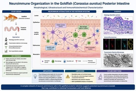
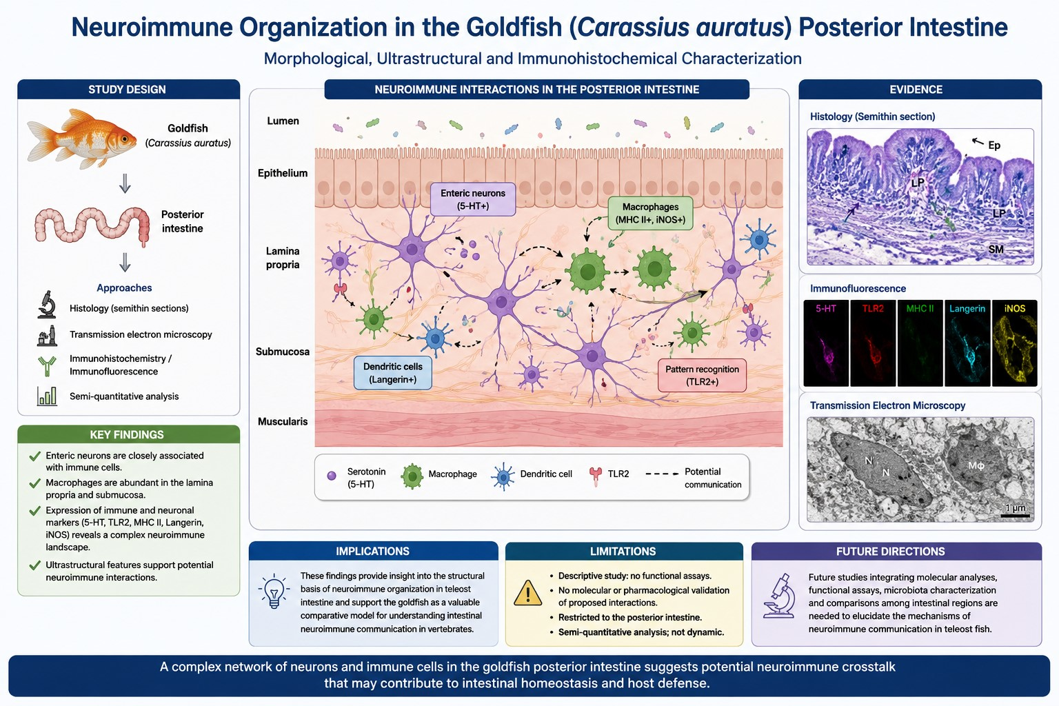
Graphical Abstract

1. Introduction

The intestine represents one of the most active interfaces between the external environment and the internal milieu of vertebrates. In addition to its digestive and absorptive functions, the intestinal wall contains highly coordinated nervous and immune components that work together to preserve tissue homeostasis and protect against pathogens [1]. The enteric nervous system (ENS), often referred to as the intrinsic nervous system of the gut, regulates motility, secretion, epithelial permeability, and vascular activity through extensive neuronal networks distributed within the intestinal wall [2]. Beyond these classical functions, the ENS also participates in immune regulation through continuous bidirectional communication with resident immune cells [3,4]. In mammals, interactions between enteric neurons, macrophages, lymphocytes, and glial cells form an integrated neuroimmune network that plays an essential role in intestinal defense and inflammatory regulation [5].
In teleost fish, increasing evidence indicates that the intestine possesses a complex mucosal immune system composed of both innate and adaptive components. Gut-associated lymphoid tissue (GALT) represents the main immunological barrier against environmental antigens and microorganisms [6]. Unlike the encapsulated lymphoid organs of mammals, fish GALT is mainly composed of diffuse and aggregated immune cell populations distributed throughout the epithelium, lamina propria, and submucosa [7]. These immune structures are continuously exposed to waterborne pathogens and microbial communities, making the intestinal mucosa a highly active immunological environment. Previous studies in several teleost species have demonstrated the presence of macrophages, lymphocytes, dendritic-like cells, and antigen-presenting cells within the intestinal wall, highlighting the importance of GALT in immune surveillance and pathogen defense [8].
To investigate the cellular components involved in intestinal neuroimmune interactions, markers associated with both immune and neural functions were selected. MHC II was employed to identify antigen-presenting cells involved in adaptive immune responses [9], whereas TLR2 was used as a marker of innate immune recognition and microbial sensing [10]. Langerin was selected to characterize dendritic-like cells associated with antigen capture and processing [11], while serotonin (5-HT) was examined because of its established role in enteric neurotransmission and immune modulation [12]. Together, these markers provide complementary information on the organization of immune surveillance and neuroimmune communication within the intestinal wall.
Recent histological and ultrastructural studies have shown that intestinal immune cells are not randomly distributed within the gut wall. Instead, macrophages, lymphocytes, and dendritic-like cells are frequently observed in close proximity to enteric neurons and nerve fibers, suggesting direct anatomical and functional communication between the nervous and immune systems [13]. Macrophages, in particular, exhibit structural characteristics and phagocytic activity that support their involvement in tissue surveillance and neuroimmune signaling [14]. Neuroactive mediators such as serotonin (5-hydroxytryptamine, 5-HT) further contribute to this integrated network. In addition to its well-known role in regulating gut motility and secretion, serotonin can modulate immune cell activity, indicating a bidirectional relationship in which neuronal mediators influence immune responses while immune-derived signals affect neuronal function [15].
Although enteric neurons and immune cells have been described separately in teleost intestines, their precise spatial organization and ultrastructural interactions remain poorly characterized. In particular, direct associations between neuronal elements and immune cell populations, including macrophages, lymphocytes, and dendritic-like cells, have received limited attention. This study represents one of the first detailed histological, ultrastructural, and immunofluorescence investigations of neuroimmune interactions in the goldfish intestine, providing evidence of close spatial associations between enteric neurons and diverse immune cell populations. Understanding these relationships is important because the fish intestine functions as a major interface between the host and a microbe-rich aquatic environment, requiring coordinated neural and immune responses to maintain intestinal homeostasis. The study also provides new evidence for the coexistence of TLR2-, MHC II, Langerin-, and serotonin-related immune signaling within the teleost intestinal neuroimmune network.
The goldfish (Carassius auratus) represents a suitable model for this investigation because it possesses a well-developed enteric nervous system, a distinct gut-associated lymphoid tissue, and a diverse intestinal immune cell population [1,16]. In addition, goldfish have been widely used in comparative anatomical, physiological, and immunological studies, making them valuable for exploring conserved mechanisms of intestinal regulation and host defense among vertebrates [17,18]. Accordingly, the present study aimed to investigate the spatial organization and interactions between enteric neurons and immune cell populations in the goldfish intestine using histological, ultrastructural, and confocal immunofluorescence approaches. Understanding neuroimmune interactions in fish is important not only from a comparative perspective but also for insights into the evolution of gut immunity and its regulatory mechanisms across vertebrates.

2. Materials and Methods

2.1. Ethics Statement

All experimental procedures involving animals were performed in accordance with the ethical standards of the European Union Directive 2010/63/EU governing the use of animals for scientific purposes and followed the ARRIVE guidelines. Ethical approval for the study was obtained from the Ethics Committee of the University of Messina.

2.2. Experimental Protocol

Eighteen healthy adult male and female goldfish (Carassius auratus), measuring 7.0–8.0 cm in total length, were obtained from the laboratory fish facility. All fish originated from the same stock population and were maintained in glass aquaria under standard laboratory conditions for four weeks prior to sampling. Fish were kept at 25 °C under a 12 h light/12 h dark photoperiod with a stable pH of 7.0 to 7.5 and dissolved oxygen at 6–7 mg/L using continuous aeration, while total water hardness ranged from 100 to 250 mg/L. Stocking density was maintained at 1 fish per 5 L to prevent crowding stress. The water-exchange regime consisted of a 20% to 30% water change twice a week using fresh dechlorinated water of the same temperature, while toxic total ammonia and nitrite levels remained strictly at 0 mg/L. Fish were fed ad libitum twice daily with a commercial goldfish diet (Goldfish Flakes, Tetra, Germany). According to the manufacturer’s specifications, the diet contained approximately crude protein (42%), crude fat (11%), crude fiber (2%), and moisture (6.5%). Prior to tissue collection, fish were fasted for 24 h to minimize the influence of recent feeding on intestinal physiological and immunological parameters. The fish were monitored daily and exhibited normal swimming behavior, feeding activity, and no visible signs of disease before tissue collection. Before sampling, the fish were euthanized by immersion in MS-222 (3% tricaine) for 15 min. Intestinal tissue samples were collected and paraffin-embedded according to standard histological procedures to obtain ideal preparations for microscopic examination.

2.3. Immunofluorescence

To determine the localization of anti-TLR2, serotonin (5-HT), MHCII, Langerin/CD207, S100 protein, and CD4, an immunohistochemical analysis was performed on intestinal tissue. Deparaffinized and rehydrated serial sections (10 μm thick) were washed in Tris–HCl buffer (0.05 M, pH 7.5) containing 0.1% bovine serum albumin and 0.2% Triton X-100. Sections were then treated with 0.3% H2O2 in PBS to block endogenous peroxidase activity. After rinsing, fetal bovine serum (F7524, Sigma-Aldrich, St. Louis, MO, USA) was applied for 30 min to reduce nonspecific binding, followed by incubation with primary antibodies. Double-labeling experiments were carried out using polyclonal anti-TLR2 and anti-serotonin (5-HT) antibodies, while monoclonal antibodies were employed for MHCII, Langerin/CD207, S100 protein, and CD4 (see Table 1 for details). Incubation was conducted overnight at 4 °C in a humidified chamber. Subsequently, sections were washed in buffer and incubated for 40 min at room temperature with Alexa Fluor IgG (H+L) secondary antibodies (see Table 1) in a dark, humid chamber. Finally, sections were mounted using Fluoromount aqueous mounting medium (Sigma-Aldrich, Burlington, MA, USA).
Negative controls were performed by omitting the primary antibodies (Supplementary File). Details of all antibodies, including source, host species, and working dilutions, are provided in Table 1. All primary antibodies were selected based on reported cross-reactivity in teleost studies and their use in previous morphological analyses of fish immune and intestinal tissues [8].

2.4. Laser Confocal Immunofluorescence

Sections were analyzed and images were acquired using a Zeiss LSM DUO confocal laser scanning microscope equipped with a META module (Carl Zeiss MicroImaging GmbH, Jena, Germany) and an argon laser (458, 488 nm) together with two helium–neon lasers (543 and 633 nm). All images were digitized at 8-bit resolution with a frame size of 2048 × 2048 pixels. Optical sections of fluorescent samples were obtained using the 543 nm helium–neon laser and the 458 nm argon laser, with a scanning speed of 1 min 2 s and up to eight averages. Sections of approximately 1.50 µm thickness were collected using a pinhole setting of 250. The acquired images were processed with Zen 2011 software (LSM 700, Zeiss, Jena, Germany). To limit photobleaching, images were captured as rapidly as possible. Digital images were subsequently cropped and assembled into figure panels using Adobe Photoshop CC (Adobe Systems, San Jose, CA, USA).

2.5. Semithin Sections and Transmission Electron Microscopy (TEM)

Intestinal tissue samples were fixed for 24 h in a 2.5% paraformaldehyde–glutaraldehyde solution [19]. Following primary fixation, the specimens were post-fixed in 1% osmium tetroxide prepared in 0.1 M sodium cacodylate buffer (pH 7.3) and subsequently rinsed in 0.1 M phosphate buffer. The samples were dehydrated through a graded ethanol series, cleared in propylene oxide, and embedded in Araldite resin. Semithin sections (1 µm thick) were prepared using a Reichert Ultracut microtome (Leica, Nussloch, Germany) and stained with toluidine blue for light microscopic examination [20]. Ultrathin sections (approximately 70 nm thick) were obtained using an Ultratom VRV ultramicrotome (LKB Bromma, Stockholm, Germany), contrasted with uranyl acetate and lead citrate, and examined with a JEOL 100CX II transmission electron microscope at the Electron Microscopy Unit, Assiut University (Assiut, Egypt).

2.6. Semi-Quantitative Immunofluorescence Analysis

Semi-quantitative analysis of immunofluorescence labeling was performed to evaluate the relative distribution of macrophage-associated immunoreactive cells within different intestinal regions of goldfish. Images were captured using confocal laser scanning microscopy under identical acquisition settings for all examined sections. The density of immunopositive macrophage-like cells was assessed in the epithelium, submucosa, and muscular layer using randomly selected microscopic fields from different intestinal sections. Cells showing positive immunoreactivity for TLR2 and MHC II were identified as macrophage-associated cells according to their morphology and localization. For semi-quantitative evaluation, confocal images were analyzed using ImageJ software (v.1.54p, National Institutes of Health, Bethesda, MD, USA). Three non-consecutive sections were examined from each fish (n = 18), and five randomly selected microscopic fields were analyzed per section. Immunopositive cells were manually counted, and the mean number of positive cells per field was calculated for each intestinal layer. The data were used for descriptive comparison of immunolabeled cell distribution and were presented graphically as mean values. No inferential statistical analyses were performed because the assessment was intended to provide a semi-quantitative description of staining patterns.

3. Results

3.1. Histological Observations

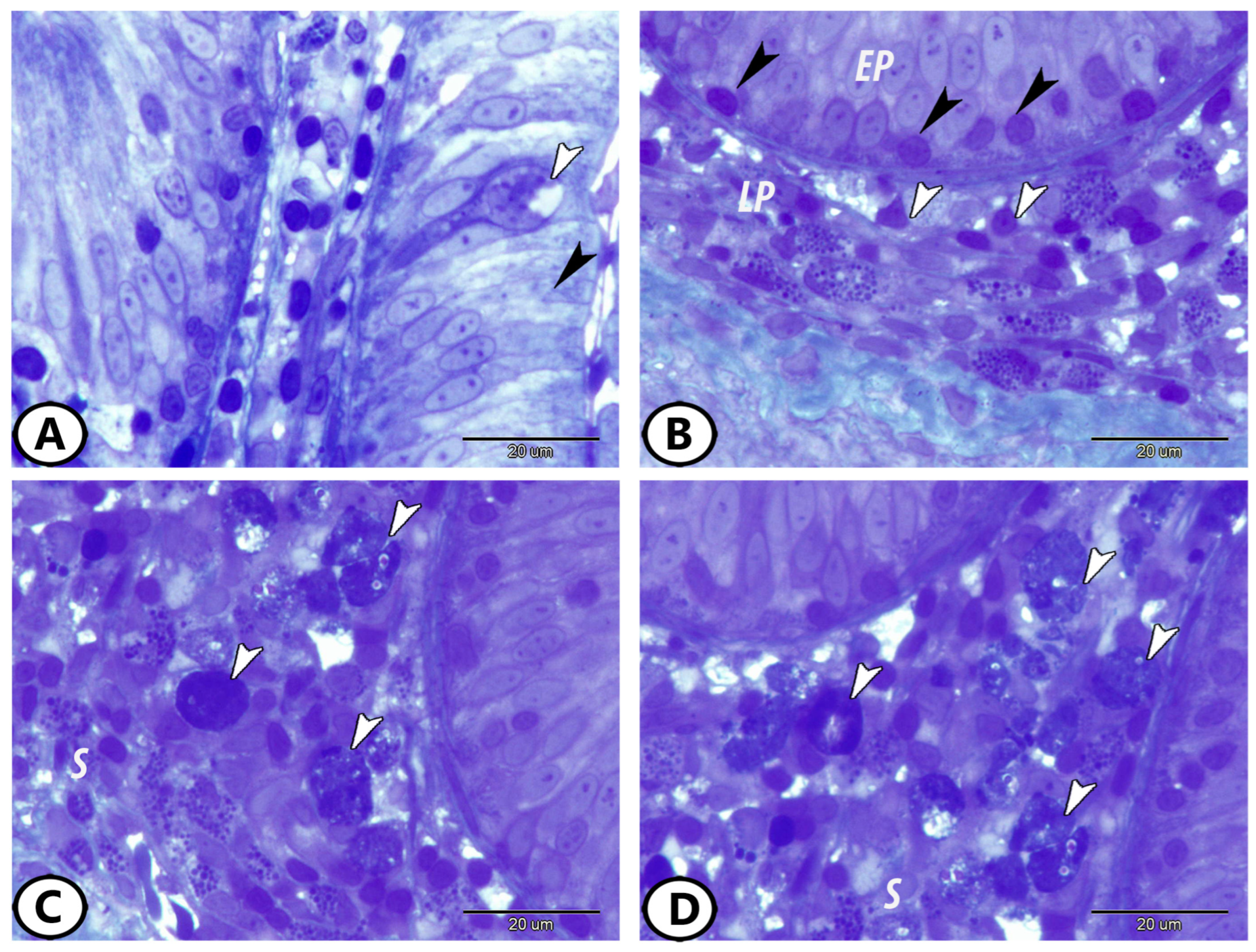
Semithin sections of the posterior intestine of goldfish, stained with toluidine blue, showed that the epithelium was composed of enterocytes and goblet cells (Figure 1A). Numerous lymphocytes were observed within the basal region of the epithelium (Figure 1B). Prominent aggregates of immune cells were also evident. Gut-associated lymphoid tissue (GALT) was present in both scattered (Figure 1B) and aggregated forms (Figure 1C,D) within the lamina propria and submucosa. These lymphoid structures consisted mainly of densely packed lymphocytes of varying sizes, along with macrophages.
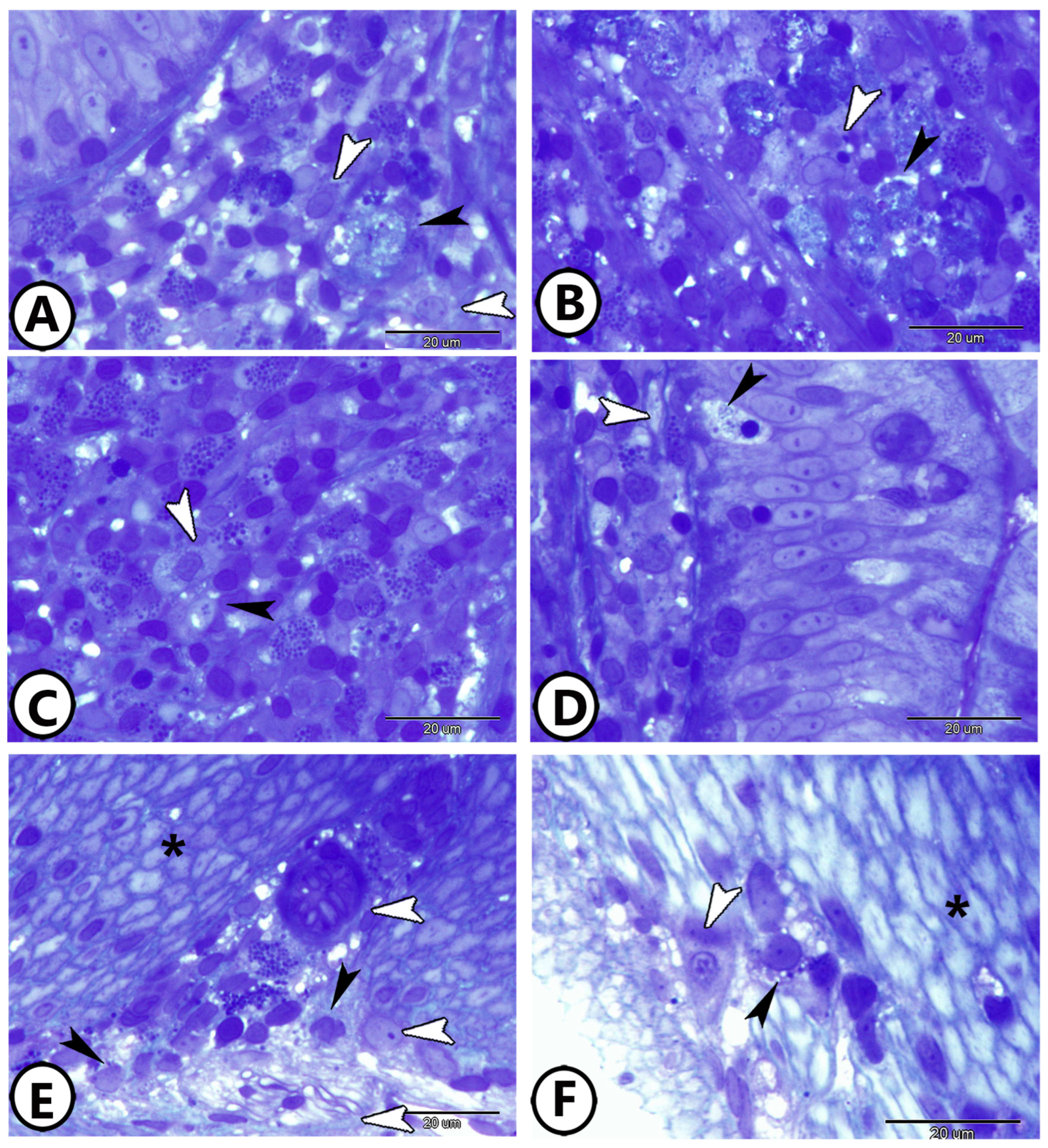
A wide distribution of immune cells across the different intestinal layers was observed. Macrophages were frequently detected in the submucosa, where they were closely associated with enteric neurons and nerve fibers (Figure 2A–C). In the epithelial layer, macrophages were also found in direct contact with underlying nerve fibers (Figure 2D). Within the muscular layer, numerous macrophages were identified in close proximity to nerve fibers and neuronal cell bodies, indicating a consistent spatial relationship throughout the intestinal wall (Figure 2E,F).

3.2. Ultrastructural Findings

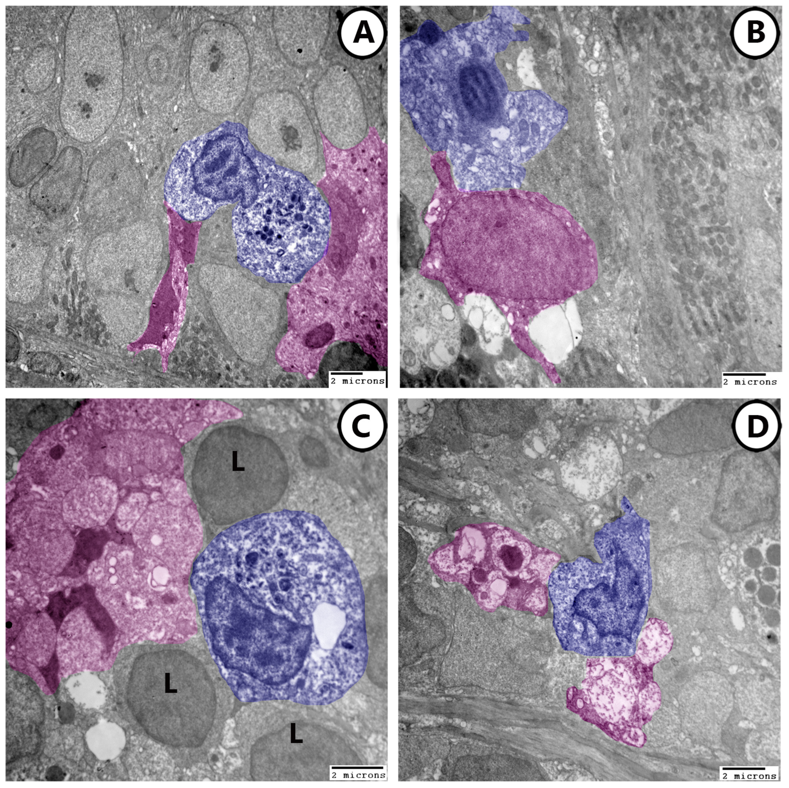
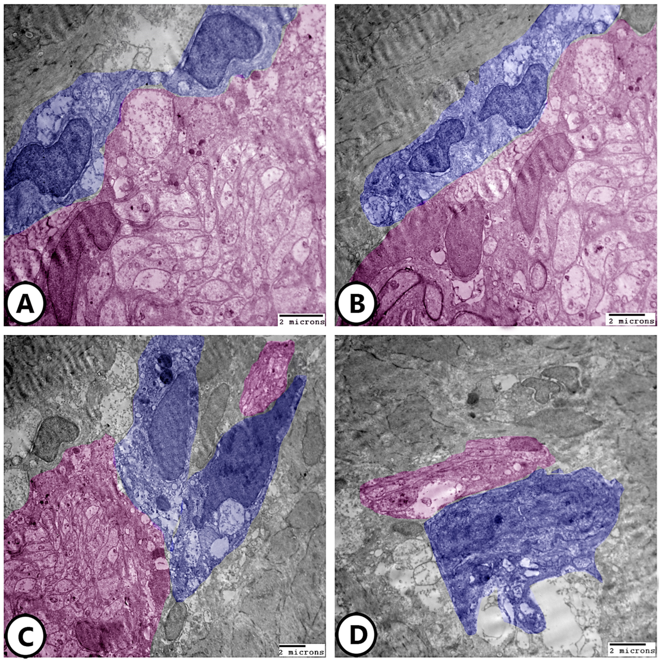
Transmission electron microscopy confirmed the close association between immune cells and neural elements. In both the epithelium and submucosa, macrophages were observed in direct contact with nerve fibers and enteric neurons (Figure 3A–D). Numerous lymphocytes were also present in these regions. In the muscular layer, macrophages containing lysosomes, vacuoles, and phagocytosed material were frequently seen establishing intimate contacts with myenteric nerve fibers (Figure 4A–D), supporting their active functional state.

3.3. Immunofluorescence Analysis

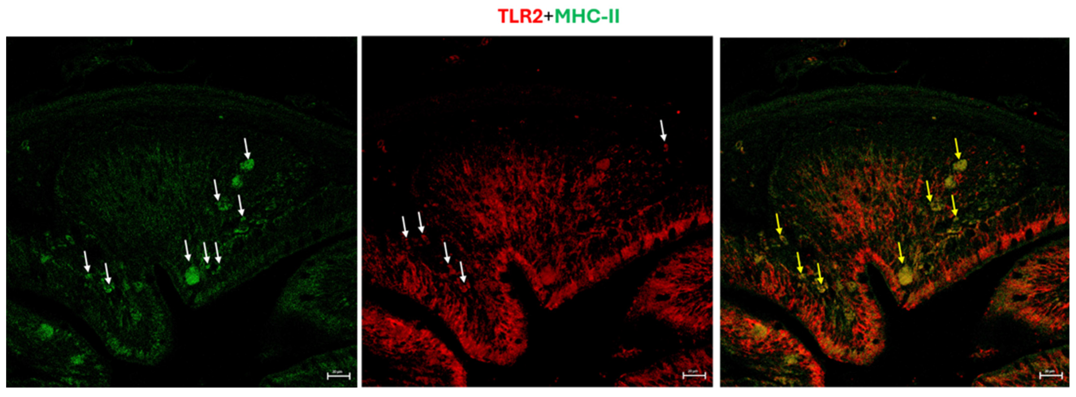
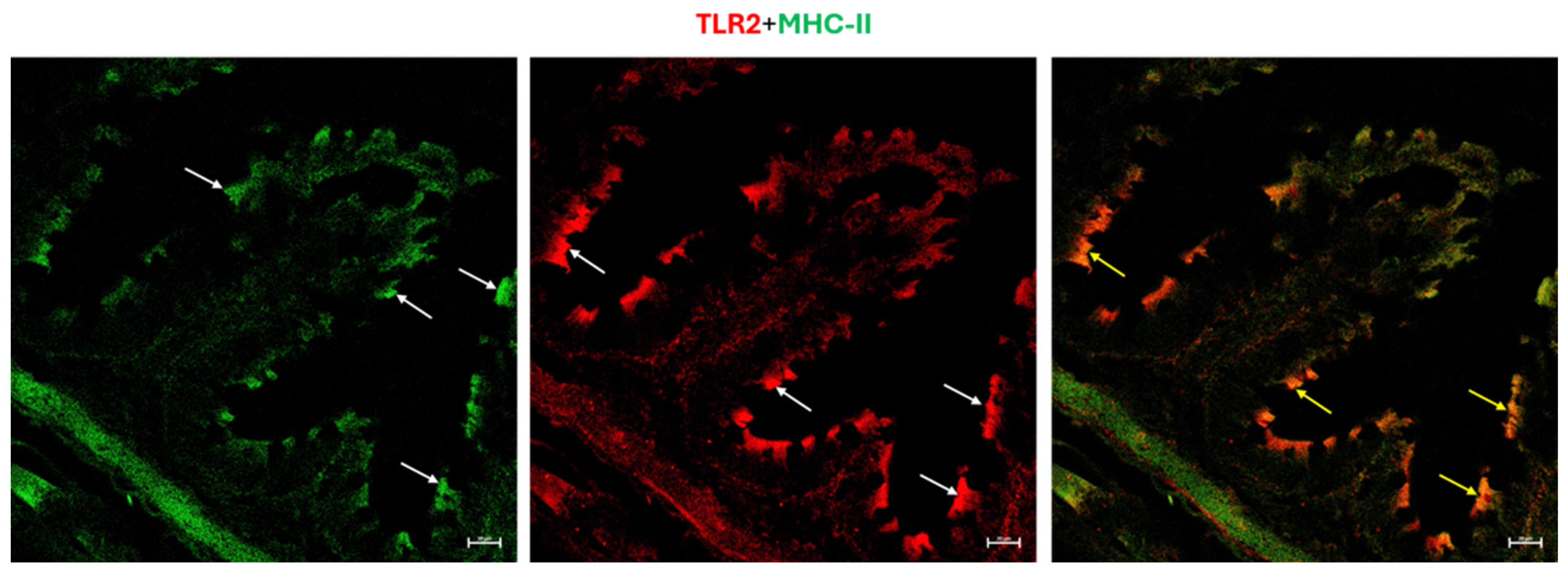
Confocal immunofluorescence demonstrated distinct distribution patterns of immune and neural markers. Double labeling for toll-like receptor 2 (TLR2) and major histocompatibility complex class II (MHC II) revealed numerous positive macrophages within lymphoid aggregates (Figure 5). In addition, enterocytes exhibited immunoreactivity for both TLR2 and MHC II, predominantly localized to their apical region (Figure 6).
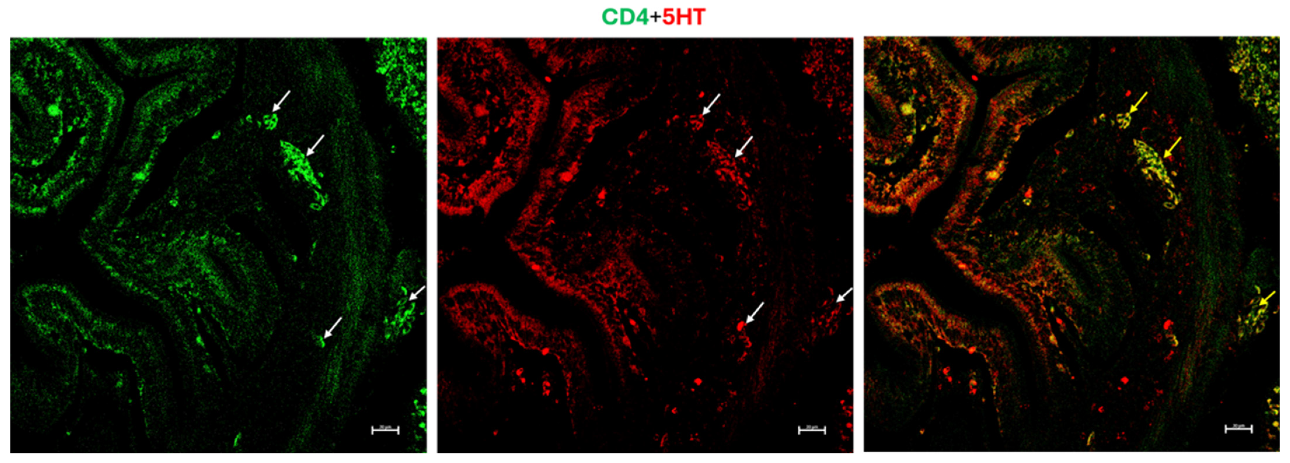
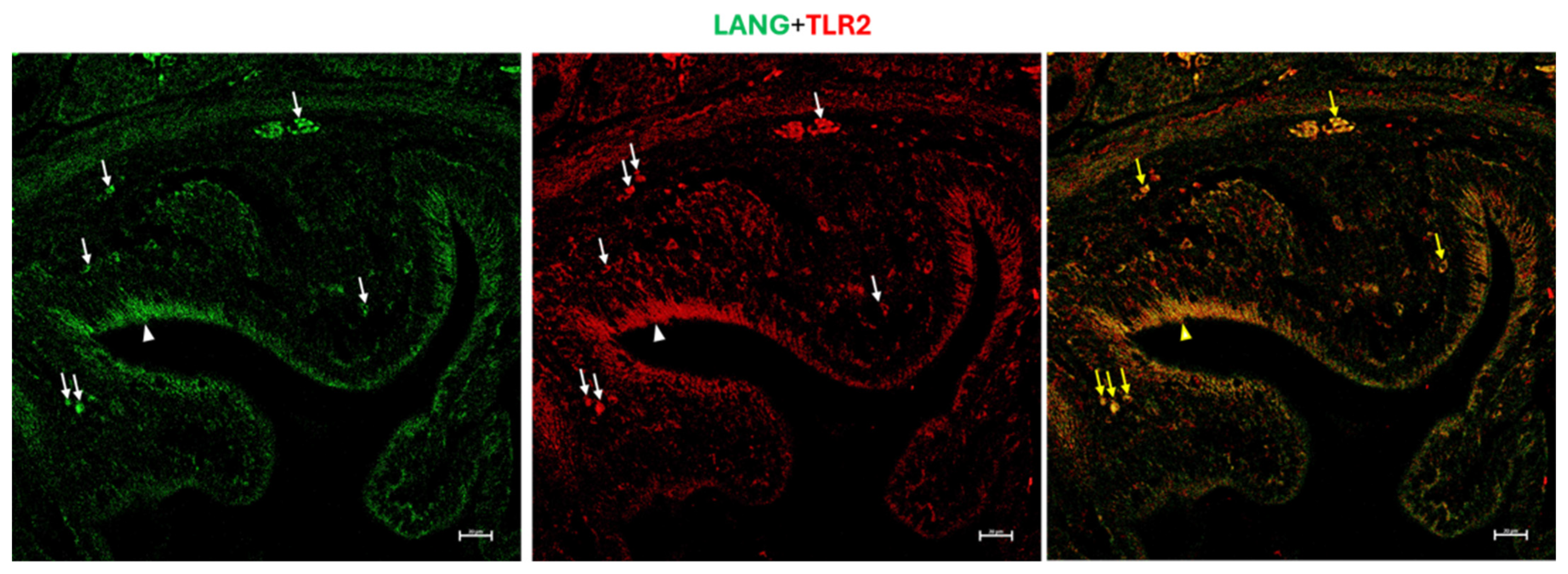
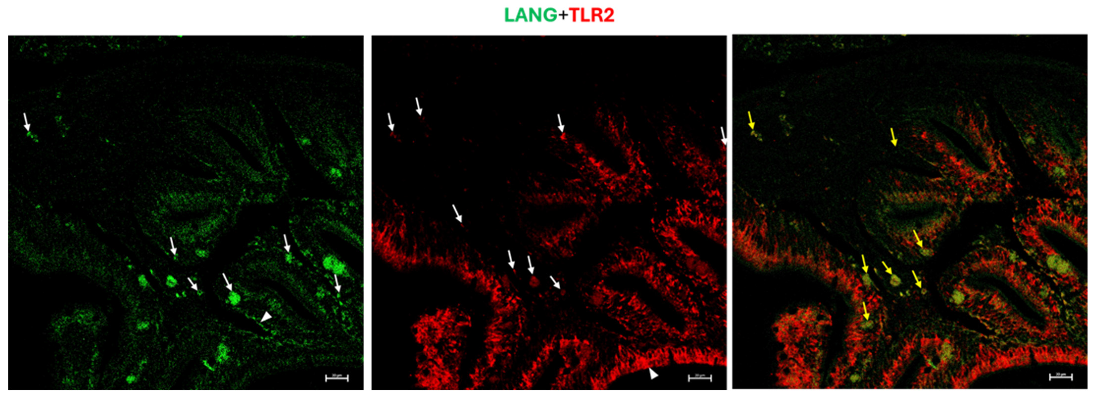
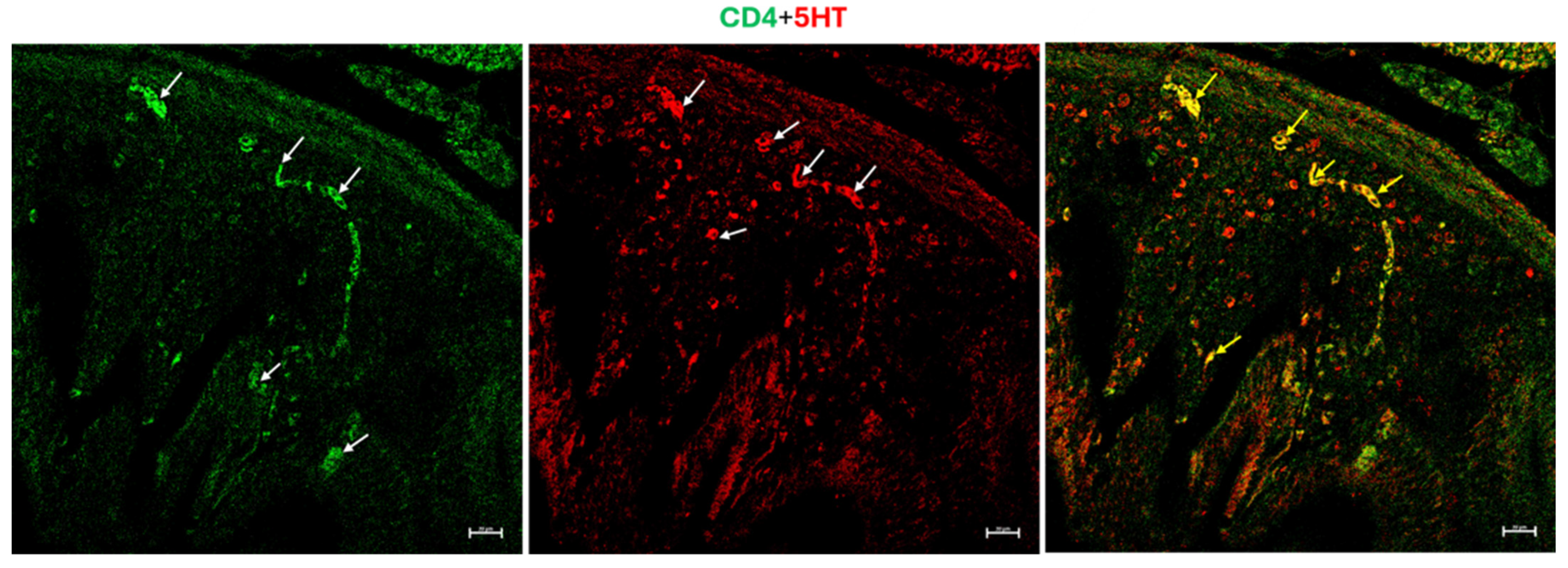
Immunofluorescence staining also showed positivity for Langerin and TLR2 in the intestinal epithelium, particularly within enterocytes. In the submucosa, clusters of dendritic cell-like (DC-like) cells displaying immunoreactivity for both Langerin and TLR2 were observed, with clear colocalization between the two markers (Figure 7 and Figure 8). A marked aggregation of densely packed lymphocytes was observed in the submucosa, highlighted by CD4 and 5-HT immunoreactivity, with partial colocalization between the two markers (Figure 9 and Figure 10).
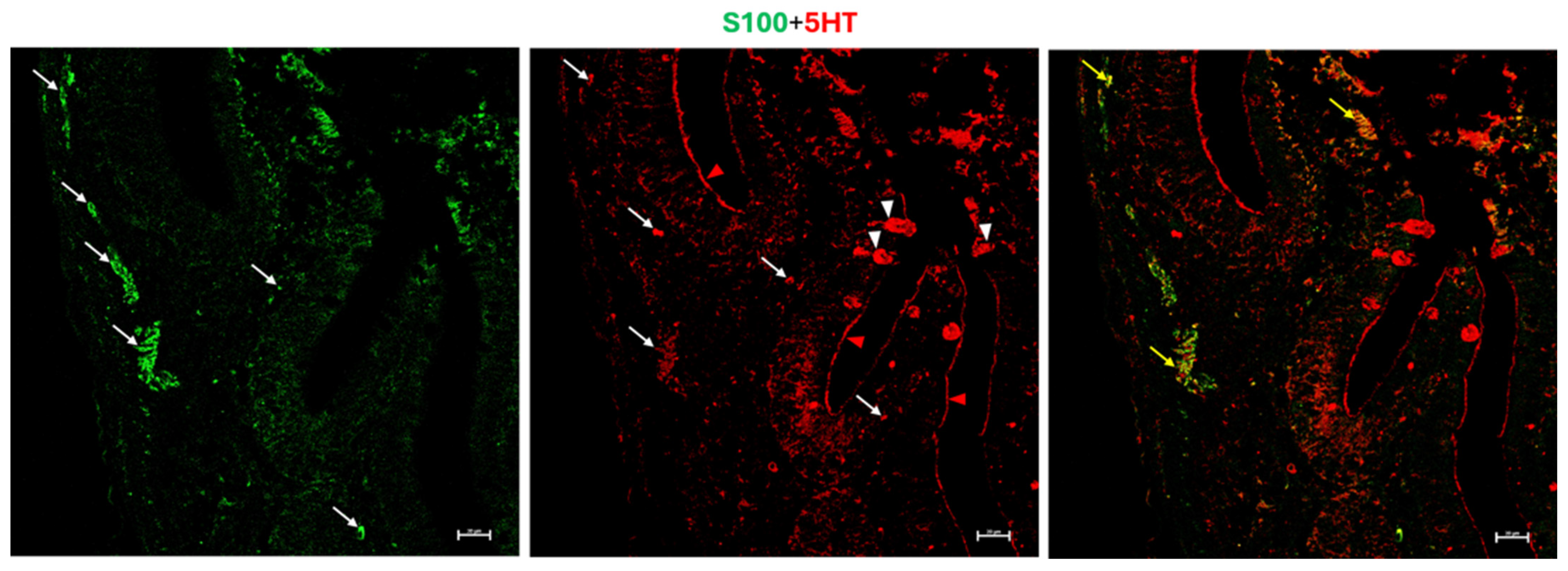
S100-positive immune cells were detected in both the submucosa and lamina propria, with a subset also exhibiting 5-HT positivity. Clear colocalization between these markers was evident. Strong 5-HT immunoreactivity was observed in epithelial structures morphologically compatible with goblet cells and along the apical microvillar border, although the precise cellular source of the signal could not be definitively established (Figure 11).
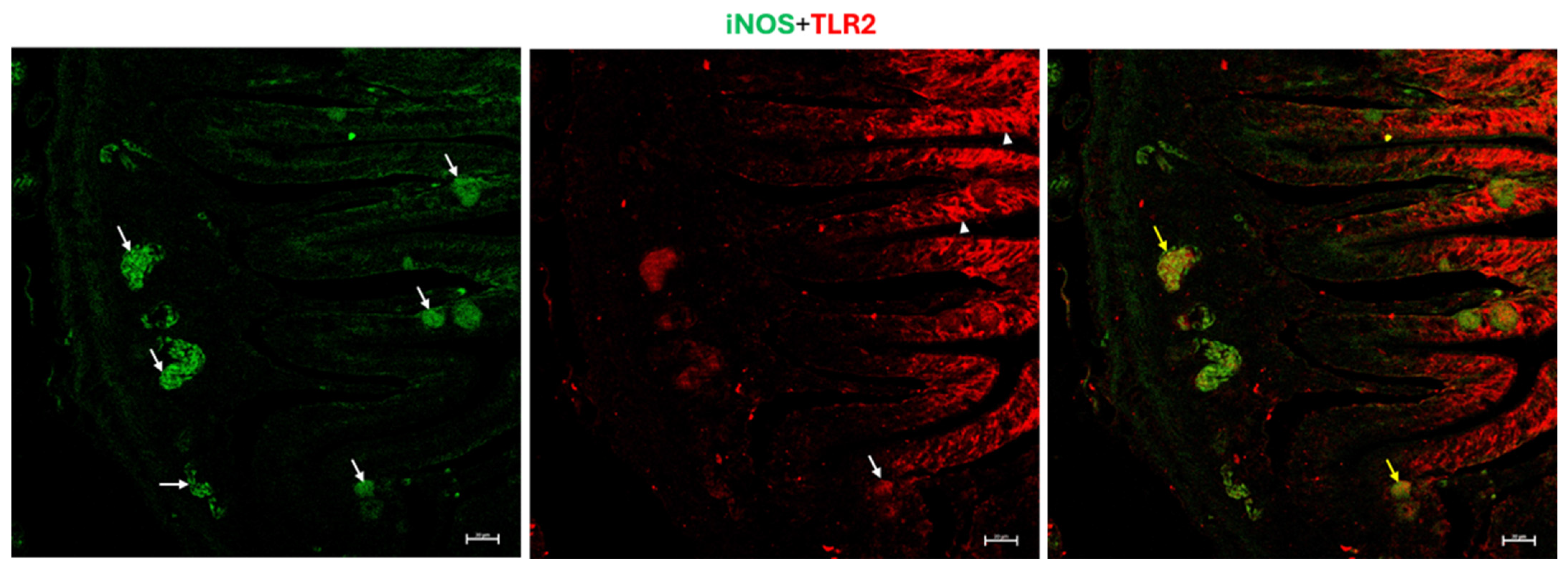
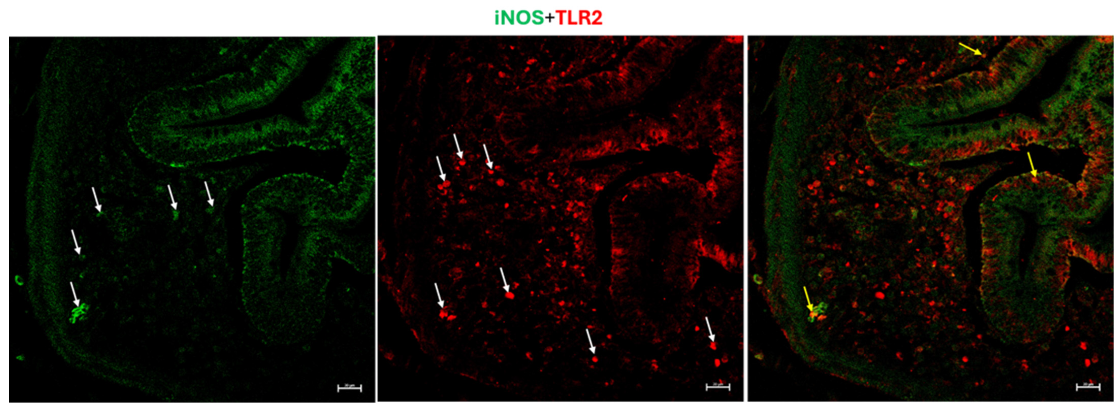
Numerous clusters of immune cells in the submucosa displayed immunopositivity for both iNOS and TLR2, with partial colocalization indicating co-expression in a subset of cells. These immunoreactive cells were predominantly localized in the submucosa, although some were also present in the mucosa. In addition, strong TLR2 immunoreactivity was observed in the intestinal epithelium (Figure 12 and Figure 13).

3.4. Macrophage Density Analysis

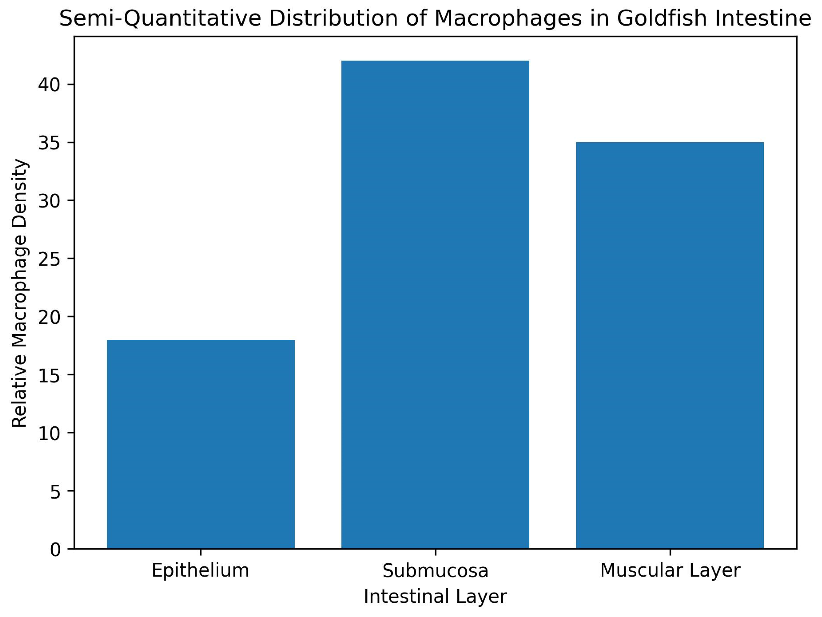
Semi-quantitative analysis demonstrated regional variations in macrophage distribution throughout the intestinal wall. The highest relative density of macrophages was observed in the submucosa, followed by the muscular layer, whereas the epithelium exhibited the lowest macrophage density (Figure 14). These findings are consistent with the histological and ultrastructural observations showing frequent localization of macrophages near enteric neurons and nerve fibers, particularly within the submucosal and muscular regions.

4. Discussion

The present study suggests that the intestine of goldfish possesses a highly organized neuroimmune microenvironment in which immune cells are structurally and functionally integrated with enteric neural elements. The close association observed between macrophages, lymphocytes, dendritic-like cells, and enteric neurons throughout the intestinal wall indicates that neuroimmune communication is a fundamental component of intestinal organization in teleost fish rather than an incidental finding [21]. These observations extend previous reports in fish and provide ultrastructural evidence supporting the concept that the vertebrate gut evolved as a coordinated sensory, immune, and neural interface specialized for continuous interaction with the external environment [22,23].
One of the most striking findings of this study was the intimate association between macrophages and enteric neurons. Macrophages were consistently distributed within the epithelium, submucosa, and muscular layer, where they closely surrounded neuronal cell bodies and nerve fibers. Ultrastructural examination confirmed direct cellular contacts between macrophages and myenteric neural elements. In addition, these macrophages contained lysosomes and phagocytosed material, indicating an activated functional state. Similar neuron-associated macrophage populations have been described in mammals and zebrafish, where they contribute to neuronal maintenance, tissue repair, and regulation of intestinal homeostasis [3,14]. The present findings suggest that comparable neuroprotective and immunoregulatory mechanisms are already established in teleosts.
The extensive development of GALT observed in the posterior intestine further emphasizes the importance of mucosal immunity in goldfish. Both diffuse and aggregated lymphoid structures were identified within the lamina propria and submucosa, reflecting the continuous antigenic stimulation encountered in aquatic environments. The abundance of lymphocytes within the basal epithelium also indicates immune surveillance-related activity at the mucosal surface [24]. Unlike mammals, fish lack organized lymph nodes and therefore rely heavily on mucosal lymphoid tissues as primary defensive sites [7]. The present observations support this concept and suggest that GALT in goldfish is structurally associated with enteric neural components, suggesting coordinated regulation between immune activation and intestinal neural activity.
The immunofluorescence findings provide further evidence for functional specialization of intestinal immune cells. Macrophages expressing both TLR2 and MHC II were frequently observed within lymphoid aggregates, consistent with their role in pathogen recognition and antigen presentation [25]. TLR2 is a major pattern-recognition receptor involved in detecting microbial products and initiating inflammatory signaling pathways [26], whereas MHC II is essential for activation of CD4+ lymphocytes [27]. Their co-expression therefore indicates that these macrophages are actively involved in coordinating innate and adaptive immune responses within the intestinal mucosa.
An important finding was the marked TLR2 and MHC II immunoreactivity detected in enterocytes, particularly along the apical surface. This observation suggests that enterocytes in goldfish are not passive absorptive cells but actively participate in mucosal immune defense. The apical localization of these markers supports their direct involvement in luminal antigen sensing. In fish, this immunoreactivity was detected in Eptatretus cirrhatus (myxines), Scyliorhinus canicula (Chondrichthyes), Polypterus senegalus (Osteichthyes, Brachiopterygii), Lepisosteus oculatus (Osteichthyes, Holostei), and Clarias batrachus (Osteichthyes, Teleostei) [28]. This role may be especially important because of the constant exposure of the intestinal mucosa to waterborne microorganisms and environmental antigens.
The presence of Langerin-positive dendritic-like cells in the submucosa represents additional evidence for specialized antigen-presenting populations within fish GALT. Their co-expression with TLR2 indicates that these cells are capable of microbial recognition and immune activation [29]. Dendritic-like cells have been described previously in teleost mucosal tissues [30,31,32], but information regarding their distribution and functional relationships with other intestinal components remains limited. The clustered distribution observed in the present study suggests localized sites of antigen sampling and immune processing within the intestinal wall.
Another notable finding was the relationship between serotonin signaling and immune cell populations. Aggregates of CD4-positive lymphocytes exhibited partial colocalization with 5-HT immunoreactivity, indicating that serotonin may participate in the regulation of adaptive immune responses in the fish intestine. Serotonin is widely recognized as a neurotransmitter involved in intestinal motility and secretion, but it also exerts important immunomodulatory effects on lymphocytes, macrophages, and other immune cells [33,34]. The present findings support the existence of serotonergic neuroimmune pathways in teleosts [35] and suggest that serotonin-mediated immune organization may be conserved.
Similarly, the colocalization of S100 and 5-HT identified a population of cells likely involved in neuroimmunomodulatory functions. S100 proteins are commonly associated with glial and immune-related cells [36,37], and their coexistence with serotonin suggests participation in local signaling pathways linking neuronal and immune activity. The marked serotonin immunoreactivity observed in goblet cell-like structures and along the microvillar border is also noteworthy. This finding may indicate active serotonin transport or secretion into the intestinal lumen through serotonin transporter-related mechanisms, as previously suggested in inflammatory intestinal conditions [38]. Luminal serotonin release could influence epithelial barrier function, microbial interactions, and mucosal immune responses [39].
The co-expression of iNOS and TLR2 in submucosal immune cells further suggests activation of innate defense pathways. Nitric oxide produced through iNOS activity is an important antimicrobial and immunoregulatory mediator [40]. Its association with TLR2-positive cells suggests that microbial recognition within the intestine triggers localized inflammatory and defensive responses. The predominance of these cells in the submucosa indicates that this region may represent a major site of innate immune activation in the goldfish intestine.
The neuroimmune organization observed in the goldfish intestine reflects an adaptation to the aquatic environment, where fish are continuously exposed to waterborne microorganisms, dietary antigens, and other environmental challenges [6]. The marked expression of TLR2, MHC II, iNOS, and Langerin in immune cells and intestinal tissues suggests the presence of an efficient mucosal surveillance system capable of recognizing, processing, and responding to microbial stimuli. In addition, the coexistence of serotonergic elements and immune cells supports a close functional relationship between the enteric nervous and immune systems, enabling rapid coordination of intestinal defense and physiological activity [41].
The intestinal microenvironment is regulated by a complex interplay among dietary components, resident microbiota, immune cells, and the enteric nervous system [42]. In teleost fish, gut-associated lymphoid tissue is continuously exposed to food-derived antigens and diverse microbial populations, both of which influence the activation and distribution of immune cells [43]. The marked expression of TLR2 observed in enterocytes and immune cells may reflect ongoing recognition of microbial-associated molecular patterns derived from the intestinal microbiota, thereby contributing to mucosal immune surveillance and epithelial barrier maintenance [44,45]. Similarly, MHC II-positive antigen-presenting cells within lymphoid aggregates are likely involved in processing luminal antigens originating from both dietary and microbial sources, supporting adaptive immune responses mediated by CD4+ lymphocytes [46,47].
The observed iNOS immunoreactivity may also be linked to microbial stimulation, since nitric oxide production represents an important antimicrobial mechanism in mucosal tissues [48]. In addition, serotonin is increasingly recognized as a key mediator linking microbial activity, intestinal physiology, and immune regulation [49]. Microbiota-derived signals can influence serotonin synthesis and release, which in turn modulates immune cell function and intestinal homeostasis [50,51]. Consequently, the neuroimmune interactions described in the present study should be viewed within the broader context of a nutrition–microbiota–immune–neural network. Future studies integrating microbiome analysis, dietary manipulation, and molecular profiling would help clarify the relative contribution of these factors to intestinal immune regulation in goldfish and other teleost species.
A notable finding of this study was the high density of macrophages within the submucosa. This strategic localization may provide an effective second line of defense against antigens that cross the epithelial barrier while facilitating communication with nearby enteric neural elements. The close association between macrophages and neurons suggests that the submucosa serves as a major site of neuroimmune interaction, contributing to tissue homeostasis, immune regulation, and protection of intestinal integrity [52,53]. The conservation of these cellular arrangements in goldfish supports the view that neuroimmune cooperation is an evolutionarily conserved mechanism among vertebrates [54]. The teleost intestine exhibits marked regional specialization [55], and the distribution of immune cells may vary among intestinal segments according to local physiological and immunological functions [56,57]. The present study focused exclusively on the posterior intestine, a region known for its prominent immune activity and abundant GALT. Consequently, regional differences in macrophage density along the intestinal tract could not be assessed. Future studies comparing anterior, middle, and posterior intestinal segments would provide a more comprehensive understanding of macrophage distribution and neuroimmune organization throughout the teleost gut.

5. Limitations and Future Directions

The present study was primarily descriptive, combining histological, ultrastructural, and immunohistochemical approaches to characterize neuroimmune organization in the goldfish posterior intestine. Although close associations between neural and immune elements were observed, no functional, molecular, or pharmacological analyses were performed to verify their physiological significance. In addition, the investigation was limited to the posterior intestine, and the semi-quantitative analysis provided only a comparative assessment of cell distribution. Further studies integrating functional assays, molecular techniques, microbiota analysis, and comparisons among intestinal regions are needed to better understand neuroimmune communication in teleost fish.

6. Conclusions

The present findings suggest that the intestine of goldfish contains an integrated neuroimmune system characterized by complex interactions between enteric neurons, epithelial cells, macrophages, lymphocytes, and dendritic-like cells. The close anatomical relationships and overlapping molecular markers observed in this study strongly support the existence of coordinated signaling pathways between neural and immune compartments. These observations provide new insight into the evolution of intestinal neuroimmune regulation in vertebrates and support the use of goldfish intestine as a valuable comparative model for investigating gut neurobiology and mucosal immunity.

Supplementary Materials

The following supporting information can be downloaded at: https://www.mdpi.com/article/10.3390/fishes11060359/s1.

Author Contributions

Conceptualization, methodology, formal analysis, data curation, D.M.M. and G.Z.; resources, N.S.A., M.C.G. and T.A.A.-M.; writing—original draft preparation, H.M.A. and E.R.L.; writing—review and editing, M.A. and A.M.; validation, E.R.L., G.P.L., M.C.G. and A.N.; investigation—supervision, G.Z. and D.M.M. All authors have read and agreed to the published version of the manuscript.

Funding

The authors express their appreciation to the Princess Nourah Bint Abdulrahman University Researchers Supporting Project (PNURSP2026R213), Princess Nourah Bint Abdulrahman University, Riyadh, Saudi Arabia.

Institutional Review Board Statement

The animal study protocol and all the procedures in this study were approved by European Union Directive 63/2010/EU for the use of animals in scientific research and complied with the ARRIVE guidelines. The study protocol received approval from the Ethics Committee of the University of Messina, approved on 7 March 2016.

Informed Consent Statement

Not applicable.

Data Availability Statement

The data presented in this study are available on request from the corresponding author.

Acknowledgments

The authors thank the technical support provided by the department of cell and tissues, Faculty of Veterinary medicine, and Electron Microscopy unit in Assiut University, Egypt.

Conflicts of Interest

The authors declare no conflicts of interest.

References

  1. Zaccone, G.; Mokhtar, D.; Albano, M.; Alesci, A.; Capillo, G.; Tripathy, P.S.; Aragona, M.; Guerrera, M.C.; Icardo, J.M.; Almohaimeed, H.M.; et al. Molecular and Immunohistochemical Characterization of Intestinal Macrophage Subsets in Goldfish. Sci. Rep. 2026, 16, 14397. [Google Scholar] [CrossRef] [Scilit]
  2. Agrawal, H.; Agarwal, N.; Gupta, N. Enteric Nervous System as a Therapeutic Target in Gastrointestinal Disorders. World J. Gastrointest. Pharmacol. Ther. 2025, 16, 110843. [Google Scholar] [CrossRef] [Scilit] [PubMed]
  3. Gabanyi, I.; Muller, P.A.; Feighery, L.; Oliveira, T.Y.; Costa-Pinto, F.A.; Mucida, D. Neuro-Immune Interactions Drive Tissue Programming in Intestinal Macrophages. Cell 2016, 164, 378–391. [Google Scholar] [CrossRef] [Scilit] [PubMed]
  4. Yoo, B.B.; Mazmanian, S.K. The Enteric Network: Interactions between the Immune and Nervous Systems of the Gut. Immunity 2017, 46, 910–926. [Google Scholar] [CrossRef] [Scilit]
  5. Wang, S.; Wang, Y.; Miao, J.; Zheng, X.; Ge, W.; Chen, G.; Yin, Y. Enteric Nervous System and Inflammatory Bowel Disease. Gastroenterol. Rep. 2026, 14, goag005. [Google Scholar] [CrossRef] [Scilit] [PubMed]
  6. Mokhtar, D.M.; Abdelhafez, E.A. An Overview of the Structural and Functional Aspects of Immune Cells in Teleosts. Histol. Histopathol. 2021, 36, 399–414. [Google Scholar] [CrossRef] [Scilit]
  7. Salinas, I. The Mucosal Immune System of Teleost Fish. Biology 2015, 4, 525–539. [Google Scholar] [CrossRef] [Scilit] [PubMed]
  8. Lauriano, E.R.; Alesci, A.; Aragona, M.; Pergolizzi, S.; Miller, A.; Zuwala, K.; Kuciel, M.; Zaccone, G.; Germanà, A.; Guerrera, M.C. Immunohistochemistry of the Gut-Associated Lymphoid Tissue (GALT) in African Bonytongue (Heterotis niloticus, Cuvier 1829). Int. J. Mol. Sci. 2023, 24, 2316. [Google Scholar] [CrossRef] [Scilit]
  9. Ashfaq, H.; Soliman, H.; Saleh, M.; El-Matbouli, M. CD4: A Vital Player in the Teleost Fish Immune System. Vet. Res. 2019, 50, 1. [Google Scholar] [CrossRef] [Scilit]
  10. Drage, M.G.; Pecora, N.D.; Hise, A.G.; Febbraio, M.; Silverstein, R.L.; Golenbock, D.T.; Boom, W.H.; Harding, C.V. TLR2 and Its Co-Receptors Determine Responses of Macrophages and Dendritic Cells to Lipoproteins of Mycobacterium tuberculosis. Cell. Immunol. 2009, 258, 29–37. [Google Scholar] [CrossRef] [Scilit]
  11. Romani, N.; Clausen, B.E.; Stoitzner, P. Langerhans cells and more: Langerin-expressing dendritic cell subsets in the skin. Immunol. Rev. 2010, 234, 120–141. [Google Scholar] [CrossRef] [Scilit]
  12. Koopman, N.; Katsavelis, D.; Hove, A.S.T.; Brul, S.; Jonge, W.J.; Seppen, J. The Multifaceted Role of Serotonin in Intestinal Homeostasis. Int. J. Mol. Sci. 2021, 22, 9487. [Google Scholar] [CrossRef] [Scilit] [PubMed]
  13. Zaccone, G.; Alesci, A.; Mokhtar, D.M.; Aragona, M.; Guerrera, M.C.; Capillo, G.; Albano, M.; de Oliveira Fernandes, J.; Kiron, V.; Sayed, R.K.A.; et al. Localization of Acetylcholine, Alpha 7-NAChR and the Antimicrobial Peptide Piscidin 1 in the Macrophages of Fish Gut: Evidence for a Cholinergic System, Diverse Macrophage Populations and Polarization of Immune Responses. Fishes 2023, 8, 43. [Google Scholar] [CrossRef] [Scilit]
  14. Graves, C.L.; Chen, A.; Kwon, V.; Shiau, C.E. Zebrafish Harbor Diverse Intestinal Macrophage Populations Including a Subset Intimately Associated with Enteric Neural Processes. iScience 2021, 24, 102496. [Google Scholar] [CrossRef] [Scilit]
  15. Arreola, R.; Becerril-Villanueva, E.; Cruz-Fuentes, C.; Velasco-Velázquez, M.A.; Garcés-Alvarez, M.E.; Hurtado-Alvarado, G.; Quintero-Fabian, S.; Pavón, L. Immunomodulatory Effects Mediated by Serotonin. J. Immunol. Res. 2015, 2015, 354957. [Google Scholar] [CrossRef] [Scilit]
  16. Katzenback, B.A.; Katakura, F.; Belosevic, M. Goldfish (Carassius auratus L.) as a model system to study the growth factors, receptors and transcription factors that govern myelopoiesis in fish. Dev. Comp. Immunol. 2016, 58, 68–85. [Google Scholar] [CrossRef] [Scilit] [PubMed]
  17. Filice, M.; Cerra, M.C.; Imbrogno, S. The goldfish Carassius auratus: An emerging animal model for comparative cardiac research. J. Comp. Physiol. B 2022, 192, 27–48. [Google Scholar] [CrossRef] [Scilit]
  18. Blanco, A.M.; Sundarrajan, L.; Bertucci, J.I.; Unniappan, S. Why goldfish? Merits and challenges in employing goldfish as a model organism in comparative endocrinology research. Gen. Comp. Endocrinol. 2018, 257, 13–28. [Google Scholar] [CrossRef] [Scilit]
  19. Karnovsky, M.J. A Formaldehyde-Glutaraldehyde Fixative of High Osmolarity for Use in Electron Microscopy. J. Cell Biol. 1965, 27, 137A. [Google Scholar]
  20. Reynolds, E.S. The Use of Lead Citrates at High pH as an Electron-Opaque Stain in Electron Microscopy. J. Cell Biol. 1963, 17, 208–212. [Google Scholar] [CrossRef] [Scilit]
  21. Zaccone, G. Immunity and Neuroimmune Interactions at the Mucosal Barriers in Fish. Fishes 2022, 7, 381. [Google Scholar] [CrossRef] [Scilit]
  22. Sun, N.; Cao, L.S.; Xia, W.Y.; Wang, J.M.; Wu, Q.F. Gut Sensory Neurons as Regulators of Neuro-Immune-Microbial Interactions: From Molecular Mechanisms to Precision Therapy for IBD/IBS. J. Neuroinflamm. 2025, 22, 172. [Google Scholar] [CrossRef] [Scilit]
  23. Bucking, C.; Terblanche, J.S.; Regan, M.D. The Ins and Outs of Integrative Digestive Biology. J. Exp. Biol. 2025, 228, jeb250770. [Google Scholar] [CrossRef] [Scilit]
  24. Ross, K.F.; Herzberg, M.C. Autonomous Immunity in Mucosal Epithelial Cells: Fortifying the Barrier against Infection. Microbes Infect. 2016, 18, 387–398. [Google Scholar] [CrossRef] [Scilit] [PubMed]
  25. Jablonska, O.; Duda, S.; Gajowniczek, S.; Nitkiewicz, A.; Fopp-Bayat, D. Toll-like Receptor Type 2 and 13 Gene Expression and Immune Cell Profiles in Diploid and Triploid Sterlets (Acipenser ruthenus): Insights into Immune Competence in Polyploid Fish. Int. J. Mol. Sci. 2025, 26, 3986. [Google Scholar] [CrossRef] [Scilit]
  26. Pergolizzi, S.; Fumia, A.; D’Angelo, R.; Mangano, A.; Lombardo, G.P.; Giliberti, A.; Messina, E.; Alesci, A.; Lauriano, E.R. Expression and Function of Toll-like Receptor 2 in Vertebrates. Acta Histochem. 2023, 125, 152028. [Google Scholar] [CrossRef] [Scilit]
  27. Luckheeram, R.V.; Zhou, R.; Verma, A.D.; Xia, B. CD4+ T Cells: Differentiation and Functions. Clin. Dev. Immunol. 2012, 2012, 925135. [Google Scholar] [CrossRef] [Scilit] [PubMed]
  28. Alesci, A.; Capillo, G.; Fumia, A.; Messina, E.; Albano, M.; Aragona, M.; Lo Cascio, P.; Spanò, N.; Pergolizzi, S.; Lauriano, E.R. Confocal Characterization of Intestinal Dendritic Cells from Myxines to Teleosts. Biology 2022, 11, 1045. [Google Scholar] [CrossRef] [Scilit]
  29. Simpson, M.E.; Petri, W.A., Jr. TLR2 as a Therapeutic Target in Bacterial Infection. Trends Mol. Med. 2020, 26, 715–717. [Google Scholar] [CrossRef] [Scilit]
  30. Mokhtar, D.M. Cellular and Stromal Elements Organization in the Liver of Grass Carp, Ctenopharyngodon idella (Cypriniformes: Cyprinidae). Micron 2018, 112, 1–14. [Google Scholar] [CrossRef] [Scilit]
  31. Mokhtar, D.M. The Structural and Ultrastructural Organization of the Cellular Constituents of the Trunk Kidney of Grass Carp (Ctenopharyngodon idella). Microsc. Res. Tech. 2021, 84, 537–547. [Google Scholar] [CrossRef] [Scilit]
  32. Mokhtar, D.M. Characterization of the Fish Ovarian Stroma during the Spawning Season: Cytochemical, Immunohistochemical and Ultrastructural Studies. Fish Shellfish Immunol. 2019, 94, 566–579. [Google Scholar] [CrossRef] [Scilit]
  33. Guzel, T.; Mirowska-Guzel, D. The Role of Serotonin Neurotransmission in Gastrointestinal Tract and Pharmacotherapy. Molecules 2022, 27, 1680. [Google Scholar] [CrossRef] [Scilit] [PubMed]
  34. Wu, H.; Denna, T.H.; Storkersen, J.N.; Gerriets, V.A. Beyond a Neurotransmitter: The Role of Serotonin in Inflammation and Immunity. Pharmacol. Res. 2019, 140, 100–114. [Google Scholar] [CrossRef] [Scilit] [PubMed]
  35. Duffy-Whritenour, J.E.; Zelikoff, J.T. Relationship between Serotonin and the Immune System in a Teleost Model. Brain Behav. Immun. 2008, 22, 257–264. [Google Scholar] [CrossRef] [Scilit] [PubMed]
  36. Singh, P.; Ali, S.A. Multifunctional Role of S100 Protein Family in the Immune System: An Update. Cells 2022, 11, 2274. [Google Scholar] [CrossRef] [Scilit]
  37. García-Domínguez, M. Relationship of S100 Proteins with Neuroinflammation. Biomolecules 2025, 15, 1125. [Google Scholar] [CrossRef] [Scilit]
  38. Pergolizzi, S.; Alesci, A.; Centofanti, A.; Aragona, M.; Pallio, S.; Magaudda, L.; Cutroneo, G.; Lauriano, E.R. Role of Serotonin in the Maintenance of Inflammatory State in Crohn’s Disease. Biomedicines 2022, 10, 765. [Google Scholar] [CrossRef] [Scilit]
  39. González Delgado, S.; Garza-Veloz, I.; Trejo-Vazquez, F.; Martinez-Fierro, M.L. Interplay between Serotonin, Immune Response, and Intestinal Dysbiosis in Inflammatory Bowel Disease. Int. J. Mol. Sci. 2022, 23, 15632. [Google Scholar] [CrossRef] [Scilit]
  40. Webster, C.M.; Shepherd, M. The Nitric Oxide Paradox: Antimicrobial and Inhibitor of Antibiotic Efficacy. Emerg. Top. Life Sci. 2024, 8, 37–43. [Google Scholar] [CrossRef] [Scilit]
  41. Layunta, E.; Buey, B.; Mesonero, J.E.; Latorre, E. Crosstalk Between Intestinal Serotonergic System and Pattern Recognition Receptors on the Microbiota-Gut-Brain Axis. Front. Endocrinol. 2021, 12, 748254. [Google Scholar] [CrossRef] [Scilit]
  42. Wiertsema, S.P.; van Bergenhenegouwen, J.; Garssen, J.; Knippels, L.M.J. The Interplay between the Gut Microbiome and the Immune System in the Context of Infectious Diseases throughout Life and the Role of Nutrition in Optimizing Treatment Strategies. Nutrients 2021, 13, 886. [Google Scholar] [CrossRef] [Scilit]
  43. Kelly, C.; Salinas, I. Under Pressure: Interactions between Commensal Microbiota and the Teleost Immune System. Front. Immunol. 2017, 8, 559. [Google Scholar] [CrossRef] [Scilit]
  44. Lavelle, E.C.; Murphy, C.; O’Neill, L.A.; Creagh, E.M. The role of TLRs, NLRs, and RLRs in mucosal innate immunity and homeostasis. Mucosal Immunol. 2010, 3, 17–28. [Google Scholar] [CrossRef] [Scilit]
  45. Kamdar, K.; Nguyen, V.; DePaolo, R.W. Toll-like receptor signaling and regulation of intestinal immunity. Virulence 2013, 4, 207–212. [Google Scholar] [CrossRef] [Scilit] [PubMed]
  46. Wosen, J.E.; Mukhopadhyay, D.; Macaubas, C.; Mellins, E.D. Epithelial MHC Class II Expression and Its Role in Antigen Presentation in the Gastrointestinal and Respiratory Tracts. Front. Immunol. 2018, 9, 2144. [Google Scholar] [CrossRef] [Scilit]
  47. Heuberger, C.; Janney, A.; Ilott, N. MHC class II antigen presentation by intestinal epithelial cells fine-tunes bacteria-reactive CD4 T-cell responses. Mucosal Immunol. 2023, 17, 416–430. [Google Scholar] [CrossRef] [Scilit] [PubMed]
  48. Roberts, J.M.; Milo, S.; Metcalf, D.G. Harnessing the Power of Our Immune System: The Antimicrobial and Antibiofilm Properties of Nitric Oxide. Microorganisms 2024, 12, 2543. [Google Scholar] [CrossRef] [Scilit]
  49. Grondin, J.A.; Khan, W.I. Emerging Roles of Gut Serotonin in Regulation of Immune Response, Microbiota Composition and Intestinal Inflammation. J. Can. Assoc. Gastroenterol. 2023, 7, 88–96. [Google Scholar] [CrossRef] [Scilit] [PubMed]
  50. Li, G.; Dong, S.; Liu, C.; Yang, J.; Rensen, P.C.N.; Wang, Y. Serotonin signaling to regulate energy metabolism: A gut microbiota perspective. Life Metab. 2024, 4, loae039. [Google Scholar] [CrossRef] [Scilit]
  51. Abubaker, S.; Miri, S.; Hammami, R. Serotonin and the gut microbiome: Pathways, functions, and health implications. Microbe 2026, 10, 100668. [Google Scholar] [CrossRef] [Scilit]
  52. Yip, J.L.K.; Balasuriya, G.K.; Spencer, S.J.; Hill-Yardin, E.L. The Role of Intestinal Macrophages in Gastrointestinal Homeostasis: Heterogeneity and Implications in Disease. Cell. Mol. Gastroenterol. Hepatol. 2021, 12, 1701–1718. [Google Scholar] [CrossRef] [Scilit]
  53. Lou, M.; Heuckeroth, R.O.; Tjaden, N.E.B. Neuroimmune Crossroads: The Interplay of the Enteric Nervous System and Intestinal Macrophages in Gut Homeostasis and Disease. Biomolecules. 2024, 14, 1103. [Google Scholar] [CrossRef] [Scilit] [PubMed]
  54. Litman, G.W.; Rast, J.P.; Fugmann, S.D. The origins of vertebrate adaptive immunity. Nat. Rev. Immunol. 2010, 10, 543–553. [Google Scholar] [CrossRef] [Scilit]
  55. Zhu, K.; Jiang, M.; Yan, M.; Huang, Y.; Yang, T.; Zhu, C. Characteristics and Functions of Different Intestinal Segments in Juvenile Greater Amberjack (Seriola dumerili). Animals 2025, 15, 1672. [Google Scholar] [CrossRef] [Scilit]
  56. Rombout, J.H.; Abelli, L.; Picchietti, S.; Scapigliati, G.; Kiron, V. Teleost intestinal immunology. Fish Shellfish Immunol. 2011, 31, 616–626. [Google Scholar] [CrossRef] [Scilit] [PubMed]
  57. Stosik, M.; Tokarz-Deptuła, B.; Deptuła, W. Immunity of the intestinal mucosa in teleost fish. Fish Shellfish Immunol. 2023, 133, 108572. [Google Scholar] [CrossRef] [Scilit] [PubMed]
Figure 1. Semithin sections of the intestine of goldfish stained with TB. (A) Lamina epithelials form of enterocytes (black arrowhead) and goblet cells (white arrowhead). (B) Many lymphocytes (black arrowheads) are present in the epithelium (EP) and scattered forms (white arrowheads) in lamina propria (LP). (C,D) The posterior intestine showed GALT; massive aggregates of immune cells in form of rounded clusters (arrowheads) were observed in lamina propria-submucosa.
Fishes 11 00359 g001
Figure 2. Semithin sections of the intestine of goldfish stained with TB. (AC) Macrophages (black arrowheads) in the submucosa in close association with enteric neurons (white arrowheads). (D) Macrophages (black arrowhead) in the epithelium are seen in contact with underlying nerve fibers (white arrowhead). (E,F) Macrophages (black arrowheads) in the muscular layer (asterisks) in neighboring to nerve fibers/cells (white arrowheads).
Fishes 11 00359 g002
Figure 3. Digital colored TEM images of neuroimmune communication in the epithelium and submucosa of the intestine of goldfish. (A) Macrophages (blue) in the epithelium are seen in contact with underlying nerve fibers (pink). (B) Macrophages (blue) in the submucosa in close association with enteric neurons (pink). (C,D) Macrophages (blue) in the submucosa in neighboring to nerve fibers (pink). Note the presence of many lymphocytes (L).
Fishes 11 00359 g003
Figure 4. Digital colored TEM images of neuroimmune communication in the muscular layer of the intestine of goldfish. (AD) Macrophages (blue) contained lysosomes, vacuoles, phagocytosed materials seen in direct contact with myenteric nerve fibers (pink).
Fishes 11 00359 g004
Figure 5. Confocal IHC. Section of the C. auratus gut (immunofluorescence 40×, scale bar 20 μm). Numerous TLR2- and MHC II-positive macrophages are clearly observed within the lymphoid tissue aggregates, indicating active immune surveillance in these areas (arrows). Colocalization between the two markers is evident (yellow arrows).
Fishes 11 00359 g005
Figure 6. Confocal IHC. Section of the C. auratus gut (immunofluorescence 40×, scale bar 20 μm). Enterocytes show positivity for TLR2 and MHC II, particularly localized in their apical region (marked by arrows). Colocalization between the two markers is evident (yellow arrows).
Fishes 11 00359 g006
Figure 7. Confocal IHC. Section of C. auratus gut (immunofluorescence 40×, scale bar 20 μm). Immunofluorescence staining shows Langerin and TLR2 positivity in the intestinal epithelium of enterocytes (arrowheads). Clusters of DC-like cells showing immunoreactivity for Langerin and TLR2 are evident in the submucosa (arrows). Colocalization between the two markers is evident (yellow arrows).
Fishes 11 00359 g007
Figure 8. Confocal IHC showing TLR2-positive enterocytes (arrowheads) and clusters of DC-like cells that are immunoreactive for Langerin and TLR2 in the submucosa (white and yellow arrows).
Fishes 11 00359 g008
Figure 9. Confocal IHC. Section of the C. auratus gut (immunofluorescence, 40×, scale bar 20 μm). A marked aggregation of tightly packed lymphocytes in the submucosa is highlighted by CD4 and 5-HT positivity (arrows). Colocalization between the two markers is evident (yellow arrows).
Fishes 11 00359 g009
Figure 10. Confocal IHC. Section of the C. auratus gut (immunofluorescence, 40×, scale bar 20 μm). A marked aggregation of tightly packed lymphocytes in the submucosa is highlighted by CD4 and 5-HT positivity (arrows). Colocalization between the two markers is evident (yellow arrows).
Fishes 11 00359 g010
Figure 11. Confocal IHC. S100-positive immune cells are present in the submucosa and in the lamina propria, with a subset also showing 5-HT positivity (arrows). Colocalization between the two markers is evident (yellow arrows). 5-HT-positive cells morphologically consistent with goblet cells are observed within the epithelium (arrowheads); 5-HT positivity is also visible in microvillus structures (red arrowheads).
Fishes 11 00359 g011
Figure 12. Confocal IHC. Section of the C. auratus gut (immunofluorescence 40×, scale bar 20 μm). Numerous clusters of immune cells (white arrows) in the submucosa show positivity for iNOS and TLR2, with a subset of cells co-expressing both markers, as indicated by colocalization (yellow arrows). These cells are predominantly located in the submucosa, although some are also present within the mucosa. In addition, a strong TLR2 immunoreactivity is observed in the intestinal epithelium (arrowheads).
Fishes 11 00359 g012
Figure 13. Confocal IHC. Section of the C. auratus gut (immunofluorescence 40×, scale bar 20 μm). Numerous clusters of immune cells (white arrows) in the submucosa show positivity for iNOS and TLR2, with a subset of cells co-expressing both markers, as indicated by colocalization (yellow arrows). These cells are predominantly located in the submucosa, although some are also present within the mucosa.
Fishes 11 00359 g013
Figure 14. Semi-quantitative analysis of macrophage distribution in different layers of the goldfish intestine. The graph illustrates the relative density of macrophages in the epithelium, submucosa, and muscular layer. The submucosa exhibited the highest macrophage density, followed by the muscular layer, while the epithelium showed comparatively lower values.
Fishes 11 00359 g014
Table 1. Summary of primary and secondary antibodies.
Primary AntibodiesSupplierCatalog NumberSourceDilution
TLR-2Active Motif40981Rabbit1:125
MHC class II (Y-Ae)Santa Cruz Biotechnologysc-32247Mouse1:250
Langerin/CD207Santa Cruz Biotechnologysc-271272Mouse1:250
Anti-serotonin (5HT)Sigma AldrichS5545Rabbit1:300
S100 (s161)Santa Cruz BiotechnologySc-53438Mouse1:100
CD4 MT310Santa Cruz BiotechnologySc-19641Mouse1:100
iNOSSanta Cruz BiotechnologySc7271Mouse1:200
Secondary AntibodiesSupplierCatalog numberSourceDilution
Alexa Fluor 488 anti-mouse IgG FITC conjugatedInvitrogenA-21202Donkey1:300
Alexa Fluor 594 anti-rabbit IgG TRITC conjugatedInvitrogenA-32754Donkey1:300
Disclaimer/Publisher’s Note: The statements, opinions and data contained in all publications are solely those of the individual author(s) and contributor(s) and not of MDPI and/or the editor(s). MDPI and/or the editor(s) disclaim responsibility for any injury to people or property resulting from any ideas, methods, instructions or products referred to in the content.