Article

Molecular Characterization of DUSP5 in Large Yellow Croaker (Larimichthys crocea) and Its Potential Immunoregulatory Role in Macrophages

1
State Key Laboratory of Mariculture Breeding, Fisheries College, Jimei University, Xiamen 361000, China
2
Roslin Institute, Royal (Dick) School of Veterinary Studies, University of Edinburgh, Edinburgh EH8 9YL, UK
*
Authors to whom correspondence should be addressed.
These authors contributed equally to this work.
Fishes 2026, 11(6), 320; https://doi.org/10.3390/fishes11060320
Submission received: 16 April 2026 / Revised: 18 May 2026 / Accepted: 25 May 2026 / Published: 28 May 2026
(This article belongs to the Special Issue Molecular Research on Immunological Responses in Aquatic Animals)

Abstract

Dual-specificity phosphatase 5 (DUSP5) is a negative regulator of mitogen-activated protein kinase (MAPK) signaling and has been implicated in inflammation and immune regulation. However, its role in teleost fish remains incompletely understood. In this study, DUSP5 from large yellow croaker (Larimichthys crocea), designated as LcDUSP5, was characterized with respect to its molecular features, tissue distribution, subcellular localization, and transcriptomic associations following transient overexpression in macrophages. The full-length open reading frame (ORF) of LcDUSP5 is 1131 bp and encodes a 376-amino-acid protein containing a conserved rhodanese homology domain and a dual-specificity phosphatase catalytic domain. LcDUSP5, was broadly expressed in the examined tissues, with the highest expression in the liver. Subcellular localization analysis showed that LcDUSP5 was predominantly localized in the nucleus. A transient overexpression model was established in the large yellow croaker head kidney-derived macrophage cell line LM10, and samples collected at 24 h post-transfection were subjected to transcriptome sequencing. A total of 365 differentially expressed genes were identified, including 283 up-regulated and 82 down-regulated genes. GO and KEGG enrichment analyses suggested that these genes were associated with pathways related to endocytosis, chemokine signaling, phagosome function, calcium signaling, and lipid metabolism. These results provide the first characterization of DUSP5 in large yellow croaker combined with transcriptomic analysis of macrophage responses and suggest that LcDUSP5 may be associated with immune-related pathways in teleost macrophages.
Key Contribution: This study provides the first characterization of DUSP5 in large yellow croaker combined with transcriptomic analysis of macrophage responses and suggests that LcDUSP5 may be associated with immune-related pathways in teleost macrophages.

1. Introduction

Dual-specificity phosphatases (DUSPs) belong to the superfamily of protein tyrosine phosphatases (PTPs) and are characterized by their ability to dephosphorylate both serine/threonine (Ser/Thr) and tyrosine (Tyr) residues on substrate proteins, thereby enabling precise regulation of protein kinase activity [1]. Members of the DUSP family generally contain a conserved catalytic domain, in which key amino acid residues—including aspartic acid (Asp), cysteine (Cys), and arginine (Arg)—form a catalytic triad that mediates the hydrolysis of phosphoester bonds [2]. Based on the presence or absence of an MAPK-binding domain (MBD) or a kinase-interacting motif (KIM), DUSPs are typically classified into two categories: classical DUSPs and atypical DUSPs. Classical DUSPs, also known as MAPK phosphatases (MKPs), specifically regulate the catalytic activity and subcellular localization of mitogen-activated protein kinases (MAPKs), whereas atypical DUSPs are generally smaller, exhibit greater functional diversity, and possess broader substrate specificity [3]. As key negative regulators of MAPK signaling pathways, DUSPs are widely involved in a variety of biological processes, including immune responses, neural development, metabolic homeostasis, and cell proliferation and differentiation. Dysregulation of DUSPs has been closely associated with pathological conditions such as tumorigenesis, chronic inflammatory diseases, and metabolic syndromes [4,5,6].
DUSP5 (also known as VH3 or hVH3) is an important member of the class III DUSP subfamily and is recognized as a specific negative regulator of the extracellular signal-regulated kinase 1/2 (ERK1/2) signaling pathway. Its molecular mechanism exhibits dual characteristics. On the one hand, DUSP5 can directly bind to activated ERK1/2 and inactivate it through catalytic dephosphorylation, thereby limiting sustained ERK activation within the nucleus and regulating the transcriptional output of downstream target genes. On the other hand, the N-terminal non-catalytic region of DUSP5 contains a nuclear localization signal (NLS), which anchors ERK1/2 in the nucleus and prevents its reactivation in the cytoplasm, thereby forming a finely tuned negative feedback loop within the MAPK signaling pathway [7]. Functional studies have shown that overexpression of DUSP5 suppresses the phosphorylation of signaling molecules in the ERK1/2 pathway and consequently inhibits autophagosome formation. In contrast, RNA interference-mediated knockdown of DUSP5 leads to increased expression of autophagy-related proteins (Atgs) and promotes autophagic flux [8,9]. In the context of inflammation, DUSP5 negatively regulates both ERK and nuclear factor-κB (NF-κB) signaling pathways, thereby effectively suppressing lipopolysaccharide (LPS)-induced production of pro-inflammatory cytokines and exerting anti-inflammatory effects [10]. Immunological studies further reveal that DUSP5-deficient mice exhibit excessive proliferation of CD8+ T cells and enhanced activation-induced cell death (AICD) following viral infection, indicating that DUSP5 is essential for maintaining T-cell homeostasis [11]. In viral infection models, knockdown of DUSP5 in human umbilical vein endothelial cells significantly enhances dengue virus (DENV) replication and exacerbates virus-induced endothelial barrier dysfunction, suggesting a protective antiviral role for DUSP5 [12]. Moreover, the E7 oncoprotein of human papillomavirus type 16 (HPV16) can activate the MAPK/ERK signaling cascade by suppressing DUSP5 transcription and, in cooperation with the mTOR pathway, induce autophagy to promote persistent viral infection [13]. Notably, DUSP5 is also involved in metabolic–immune crosstalk. In macrophages infected with mycobacteria, DUSP5 enhances mitochondrial respiration by promoting fatty acid oxidation (FAO), thereby facilitating the production of pro-inflammatory cytokines such as interleukin-1β (IL-1β), IL-6, and tumor necrosis factor-α (TNF-α), representing a coupling mechanism between metabolic reprogramming and inflammatory responses [14].
In teleost fish, research on DUSP5 remains in its early stages. In Japanese flounder (Paralichthys olivaceus), cloning and expression analysis of DUSP5 (PoDUSP5) demonstrated that the gene is highly expressed in the liver and gill and exhibits distinct temporal expression patterns in response to lipopolysaccharide (LPS) and polyinosinic–polycytidylic acid [poly(I:C)] stimulation, suggesting its potential involvement in responses to both bacterial and viral pathogens [15]. In orange-spotted grouper (Epinephelus coioides), overexpression of DUSP5 (EcDUSP5) significantly suppresses the expression of key viral genes, including major capsid protein (MCP), infected cell polypeptide 18 (ICP18), virion protein 19 (VP19), and lipopolysaccharide-induced TNF factor (LITAF), during Singapore grouper iridovirus (SGIV) infection. Additionally, EcDUSP5 down-regulates the activity of inflammatory transcription factors such as NF-κB and activator protein-1 (AP-1), suggesting a possible negative regulatory role in antiviral innate immunity [16]. Nevertheless, the molecular characteristics of DUSP5 and its downstream transcriptomic associations in large yellow croaker macrophages remain unclear.
The large yellow croaker (Larimichthys crocea) belongs to the order Perciformes and the family Sciaenidae and is widely distributed along the southeastern coast of China. It is one of the most economically important marine aquaculture species in China. According to statistics from the Food and Agriculture Organization (FAO), China is the largest producer of large yellow croaker, accounting for approximately 99% of the global supply [17]. In recent years, advances in genetic breeding programs have contributed to improvements in growth performance and disease resistance, enhancing the sustainability of aquaculture practices [18]. Nevertheless, large yellow croaker still faces a variety of disease challenges, including infections caused by viruses, bacteria, and parasites, particularly in open-water floating or submerged cage culture systems [19]. Among these, infections caused by large yellow croaker iridovirus (LYCIV) are of particular concern [20,21]. In our previous transcriptomic study, LcDUSP5 was found to be significantly up-regulated in resistant individuals after Pseudomonas plecoglossicida infection, suggesting that it may be associated with host immune regulation.
In the present study, we cloned and characterized LcDUSP5, examined its tissue distribution and subcellular localization, and established a transient overexpression model in a large yellow croaker macrophage cell line for transcriptome analysis. We hypothesized that LcDUSP5 may modulate macrophage immune-related pathways through MAPK-associated signaling. This study provides basic information for understanding the potential role of DUSP5 in teleost immunity.

2. Materials and Methods

2.1. Experimental Animals and Cells

A batch of L. crocea was obtained from Ningde Jinling Aquatic Technology Co., Ltd. (Ningde, China). The fish had a body length of 13.0 ± 1.5 cm, a body mass of 28.0 ± 9.0 g, and an estimated age of 9 months. Tissue fragments (muscle, intestine, head kidney, kidney, heart, stomach, gonads, brain, gill, spleen, skin, and liver) were randomly collected from three healthy individuals. All samples were immediately frozen in liquid nitrogen and stored at −80 °C until use.
The large yellow croaker head kidney-derived macrophage cell line (LM10) was kindly provided by Professor Qinghui Ai (Ocean University of China). The HEK293T cell line was maintained in our laboratory.
Fish were maintained in recirculating seawater (salinity 26‰, temperature 17 ± 1 °C, 12 h light/12 h dark cycle) and acclimated for one week prior to sampling. All experimental procedures involving animals were conducted in strict accordance with the guidelines approved by the Animal Ethics Committee of the Fisheries College of Jimei University (protocol code JMU2020012473, approved on 24 January 2020). Before tissue collection, fish were euthanized by immersion in buffered tricaine methanesulfonate (MS-222, Finquel®, 1000 mg/L) for 15 min after cessation of opercular movement; the solution was buffered with sodium bicarbonate (1 g per 1 g MS-222) to pH 7.4. Death was confirmed by the absence of opercular movement, loss of heartbeat (Doppler ultrasound), and lack of response to external stimuli. No adverse events or humane endpoints were encountered.

2.2. RNA Extraction and cDNA Synthesis

Total RNA was extracted from samples using TransZol Up Plus RNA reagent (TransGen Biotech Co., Ltd., Beijing, China). RNA integrity was assessed by 1.2% agarose gel electrophoresis, and purity was evaluated by measuring the A260/A280 ratio (1.8–2.0) using a spectrophotometer.
First-strand cDNA was synthesized using the GoScript™ Reverse Transcription System (Promega Corporation, Madison, WI, USA) according to the manufacturer’s instructions and stored at −20 °C for subsequent experiments.

2.3. Bioinformatic Analysis of LcDUSP5

The physicochemical properties of the LcDUSP5 protein, including molecular weight, theoretical isoelectric point, and amino acid composition, were analyzed using the ExPASy-ProtParam tool (Swiss Institute of Bioinformatics, Lausanne, Switzerland). Protein domains were predicted using the SMART database, and signal peptides were predicted using SignalP 6.0.
The three-dimensional structure of LcDUSP5 was modeled using the SWISS-MODEL (Swiss Institute of Bioinformatics, Lausanne, Switzerland) server and visualized with VMD 1.9.4 software (University of Illinois at Urbana-Champaign, Urbana, IL, USA). Multiple sequence alignment was performed using BLAST (NCBI, Bethesda, MD, USA), GeneDoc (Pittsburgh, PA, USA), and ClustalX (University College Dublin, Dublin, Ireland). A phylogenetic tree was constructed using the neighbor-joining (NJ) method implemented in MEGA 11.0 software.

2.4. Cloning of LcDUSP5 and Construction of Recombinant Plasmids

The open reading frame (ORF) sequence of LcDUSP5 was obtained from transcriptome data generated in our laboratory and identified through BLAST homology analysis. Specific primers (Table 1) were designed using SnapGene software (SnapGene LLC, San Diego, CA, USA) to amplify the ORF.
PCR amplification was carried out in a 20 μL reaction system containing 10 μL of 2× Phanta Max Master Mix (Vazyme, Nanjing, China), 0.4 μL of each forward and reverse primer, 1 μL of cDNA template, and 8.2 μL of nuclease-free water. The PCR conditions were as follows: 95 °C for 3 min; 35 cycles of 95 °C for 30 s, 60 °C for 30 s, and 72 °C for 1 min 30 s; followed by a final extension at 72 °C for 10 min.
PCR products were analyzed by 1% agarose gel electrophoresis, and target bands were excised and purified using a gel extraction kit (Tiangen, Beijing, China). The purified fragments were ligated into pEGFP-N1 and pcDNA3.1 vectors using a one-step cloning method to construct recombinant plasmids.
The recombinant plasmids were transformed into DH competent cells via heat shock and plated on LB agar plates containing ampicillin or kanamycin. After incubation at 37 °C for 12–16 h, single colonies were selected and verified by sequencing.

2.5. Tissue Expression Analysis of LcDUSP5

The expression profile of LcDUSP5 in different tissues was analyzed by qPCR. The reaction system (20 μL) contained 10 μL of 2× ChamQ Universal SYBR qPCR Master Mix, 0.4 μL of each primer, 1 μL of cDNA template, and RNase-free water to volume.
The amplification program was as follows: 95 °C for 30 s; 40 cycles of 95 °C for 10 s and 60 °C for 30 s; followed by melting curve analysis (95 °C for 15 s, 60 °C for 1 min, and 95 °C for 15 s).
Each sample included three biological replicates and three technical replicates. β-actin was used as the internal reference gene. Prior to the formal experiments, we validated the stability of β-actin under our experimental conditions; its Ct values ranged from 25 to 29 with no significant fluctuation across individual samples. Relative expression levels were calculated using the 2−ΔΔCt method.

2.6. Subcellular Localization of LcDUSP5

HEK293T cells were cultured to approximately 80% confluence and transfected with recombinant plasmids encoding the LcDUSP5-EGFP fusion protein using Lipo8000™ transfection reagent (Beyotime Biotechnology, Shanghai, China), following the manufacturer’s instructions. The ratio of plasmid DNA to transfection reagent was 500 ng:0.8 μL. Cells transfected with the empty pEGFP-N1 vector served as the control.
After 24 h of transfection, cells were fixed with 4% paraformaldehyde, permeabilized with Triton X-100 (Sangon Biotech (Shanghai) Co., Ltd., Shanghai, China), and stained with DAPI to label nuclei. Fluorescence signals were observed using a laser scanning confocal microscope (Leica SP8, Leica Microsystems, Wetzlar, Germany).

2.7. Overexpression of LcDUSP5

When LM10 macrophages reached approximately 80% confluence, the LcDUSP5-pcDNA3.1 recombinant plasmid was transfected into the cells via electroporation. For electroporation, a total of 2 × 10⁶ LM10 cells were resuspended in 100 μL of electroporation buffer containing 2 μg of LcDUSP5-pcDNA3.1 plasmid. Electroporation was performed using a Gene Pulser Xcell system (Bio-Rad Laboratories, Hercules, CA, USA) with the following parameters: 250 V, 500 μF, and 4 mm cuvettes. Transfection efficiency, determined by parallel transfection with a GFP-expressing plasmid, was approximately 60%. Cells transfected with empty pcDNA3.1 vector were used as the control group.
After 24 h of transfection, cells were collected for total RNA and protein extraction. qPCR was used to assess mRNA expression levels, and Western blot analysis was performed to verify protein overexpression.
For Western blot analysis, total protein was mixed with loading buffer and denatured at 95 °C for 5 min, followed by SDS-PAGE (12% polyacrylamide gel) and transfer to PVDF membranes (Merck Millipore, Burlington, MA, USA). Membranes were blocked with 5% BSA at room temperature for 2 h and incubated overnight at 4 °C with a rabbit monoclonal anti-Myc antibody (catalog No. 16286, Cell Signaling Technology, 1:1000 dilution). After washing with TBST buffer five times, membranes were incubated with HRP-conjugated goat anti-rabbit IgG (H + L) secondary antibody (catalog No. 7074, Cell Signaling Technology, Danvers, MA, USA 1:5000 dilution) at room temperature for 2 h. Signals were detected using an enhanced chemiluminescence (ECL) substrate (Thermo Fisher Scientific, Waltham, MA, USA). β-Actin (catalog No. 4970, Cell Signaling Technology, 1:2000 dilution) was used as a loading control. Molecular weight markers (PageRuler Prestained Protein Ladder, Thermo Fisher Scientific) were loaded for size determination.

2.8. Transcriptome Sequencing and Bioinformatics Analysis

Total RNA with an RNA integrity number (RIN) ≥ 8.0 was used for library construction. Libraries were prepared using the NEBNext Ultra RNA Library Prep Kit (New England Biolabs, Ipswich, MA, USA) according to the manufacturer’s instructions. A strand-specific library was generated. Paired-end sequencing (150 bp) was performed on the Illumina NovaSeq 6000 platform (Illumina, San Diego, CA, USA), with a target depth of at least 20 million clean reads per sample. Raw reads were processed using fastp. Low-quality reads, adapter-contaminated reads, and reads containing more than 10% ambiguous bases were removed. Clean reads were aligned to the large yellow croaker reference genome (GCA_003845795.1) using HISAT2. Genes with very low expression were filtered out before downstream analysis. Batch effects and sample outliers were evaluated by Pearson correlation analysis and principal component analysis (PCA). Gene expression was quantified as raw read counts, and normalization was performed using DESeq2 size factors.

2.9. qPCR Validation of RNA-Seq Data

To confirm the reliability of the transcriptome results, six differentially expressed genes (map3k15, cxcr4, cd184, ifi44L, dusp1 and hsp40) were selected for qPCR validation using the same RNA samples as those used for RNA-seq. Primers for these genes were designed using Primer Premier 5.0 and are listed in Table 1. The qPCR reaction system and amplification program were the same as described in Section 2.5, with β-actin as the internal reference. Relative expression levels were calculated using the 2−ΔΔCt method. Each sample included three biological replicates and three technical replicates. The qPCR results were compared with the RNA-seq fold changes to assess consistency.

2.10. Statistical Analysis

All statistical analyses were performed with SPSS 26.0 (IBM, Armonk, NY, USA). Quantitative data are presented as mean ± standard error of the mean (SEM). Differences among groups were analyzed by one-way ANOVA followed by Duncan’s multiple range test. Statistical significance was defined as p < 0.05.

3. Results

3.1. Sequence Characteristics of LcDUSP5

In this study, the full-length open reading frame (ORF) of LcDUSP5 was successfully cloned, with a total length of 1131 bp encoding 376 amino acids (Figure 1A). The predicted molecular weight of the LcDUSP5 protein is 42.25 kDa, with a theoretical isoelectric point (pI) of 8.30. A total of 41 potential phosphorylation sites were identified, including 29 serine (Ser), 9 threonine (Thr), and 3 tyrosine (Tyr) phosphorylation sites (Figure 1A).
Prediction using TMHMM 2.0 and SignalP 5.0 indicated that LcDUSP5 lacks both transmembrane domains and a signal peptide. SMART analysis revealed that LcDUSP5 contains a rhodanese homology domain (RHOD), a dual-specificity phosphatase catalytic domain (DSPc), and two low-complexity regions (Figure 1B). The three-dimensional structure of LcDUSP5 was predicted using SWISS-MODEL (Figure 1C).
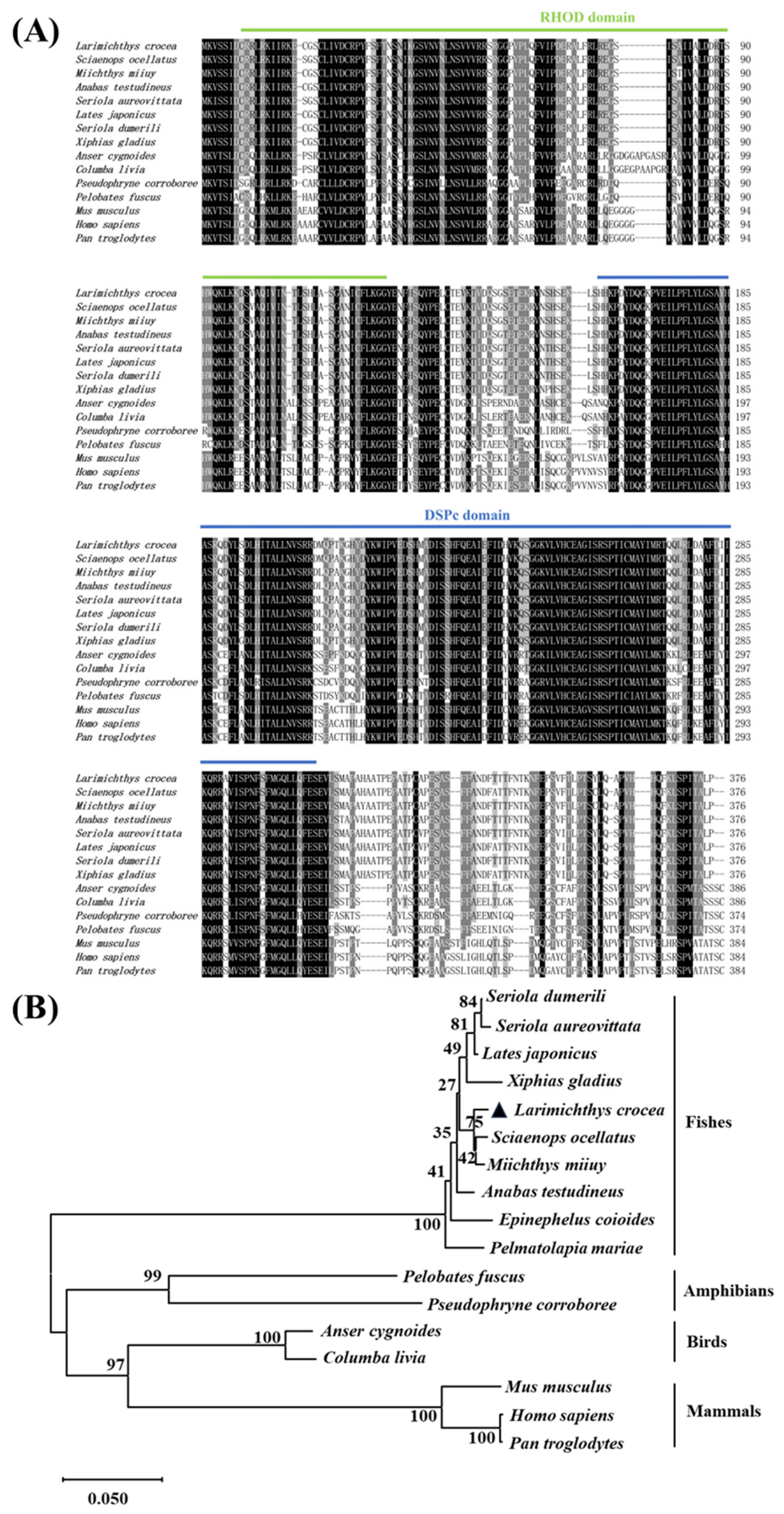
Multiple sequence alignment demonstrated that the RHOD and DSPc domains of LcDUSP5 are highly conserved among vertebrates. The amino acid sequence similarity of LcDUSP5 with those of red drum (Sciaenops ocellatus), brown croaker (Miichthys miiuy), and climbing bass (Anabas testudineus) was 98.67%, 98.40%, and 97.61%, respectively (Figure 2A; Table 2). Phylogenetic analysis showed that LcDUSP5 clusters with other teleost DUSP5 proteins, while those from mammals, amphibians, and birds form a separate clade, consistent with established evolutionary relationships (Figure 2B).

3.2. Tissue Expression Profile and Subcellular Localization of LcDUSP5

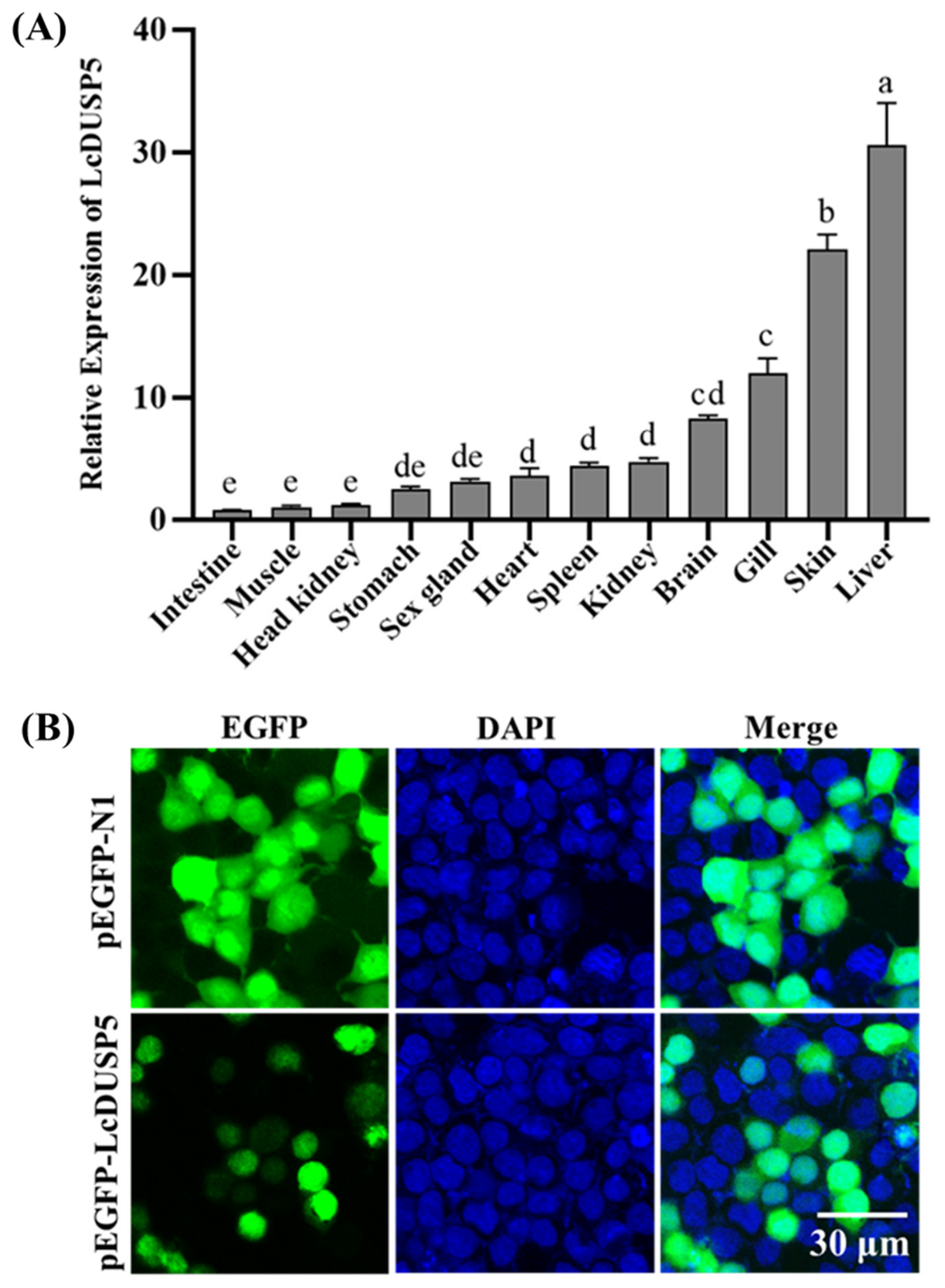
qPCR analysis revealed that LcDUSP5 is widely expressed across multiple tissues of large yellow croaker. Among these tissues, the highest expression level was observed in the liver, followed by the skin, gill, and brain, whereas the lowest expression level was detected in the intestine (Figure 3A).
To determine the intracellular localization of LcDUSP5, a eukaryotic expression vector (pEGFP-N1-LcDUSP5) was constructed and transfected into HEK293T cells, which were used because of their high transfection efficiency in preliminary localization analyses. In contrast to the diffuse cytoplasmic and nuclear distribution observed in the pEGFP-N1 control group, the LcDUSP5-EGFP fusion protein exhibited a predominantly nuclear localization pattern under confocal microscopy (Figure 3B).

3.3. qPCR Analysis of LcDUSP5 Overexpression in Macrophages

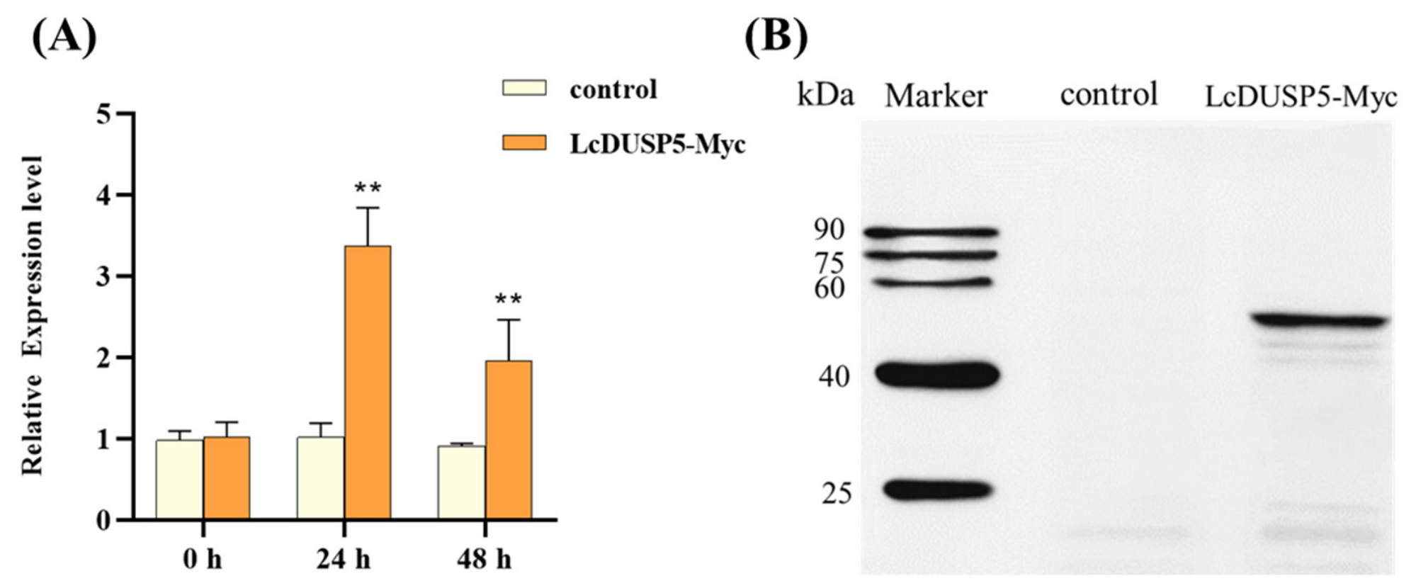
An overexpression plasmid (LcDUSP5-Myc) was constructed and transfected into large yellow croaker macrophages via electroporation. qPCR analysis showed that the mRNA expression level of LcDUSP5 reached its peak at 24 h post-transfection, indicating the highest overexpression efficiency at this time point. At 48 h, the expression level decreased (Figure 4A). Therefore, cells collected at 24 h post-transfection were used for subsequent experiments.

3.4. Western Blot Analysis of LcDUSP5 Overexpression

At 24 h post-transfection with the LcDUSP5-Myc plasmid, cells were harvested for protein extraction, and the expression of the target protein was analyzed by Western blot. As shown in Figure 4B, the LcDUSP5-Myc protein was successfully expressed in large yellow croaker macrophages, indicating that the overexpression system was suitable for subsequent transcriptome analysis.

3.5. Transcriptome Analysis of LcDUSP5 Overexpression

3.5.1. Transcriptome Data Overview

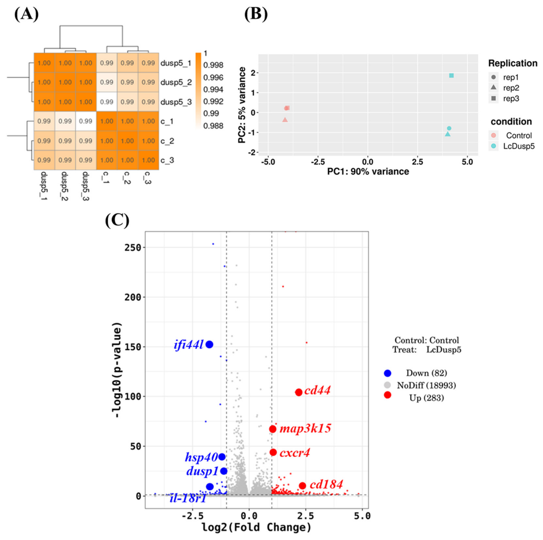
As shown in Table 3, RNA samples from three biological replicates of the control group (pcDNA3.1 empty vector; c1–c3) and the overexpression group (DUSP5 1–3) at 24 h post-transfection were used to construct transcriptome sequencing libraries.
After quality control, an average of 47.63 million clean reads per sample was obtained, with an average Q30 value of 97.07%. These high-quality reads were aligned to the reference genome of large yellow croaker, yielding an average mapping rate of 94.57%.

3.5.2. Sample Correlation Analysis and DEG Identification

The correlation of transcriptomic expression profiles among samples is a key indicator of experimental reliability and grouping validity. Pearson correlation coefficients closer to 1 indicate higher similarity between samples. As shown in Figure 5A, all samples in this study exhibited correlation coefficients greater than 0.98, indicating good reproducibility among biological replicates.
To further assess global expression pattern similarity, principal component analysis (PCA) was performed based on genome-wide expression data. As shown in Figure 5B, samples within the same treatment group clustered closely together, indicating strong intragroup consistency.
Differential expression analysis was conducted using the DESeq2 algorithm. A total of 365 differentially expressed genes (DEGs) were identified, including 283 up-regulated genes and 82 down-regulated genes (Figure 5C).

3.5.3. Functional Enrichment Analysis of DEGs

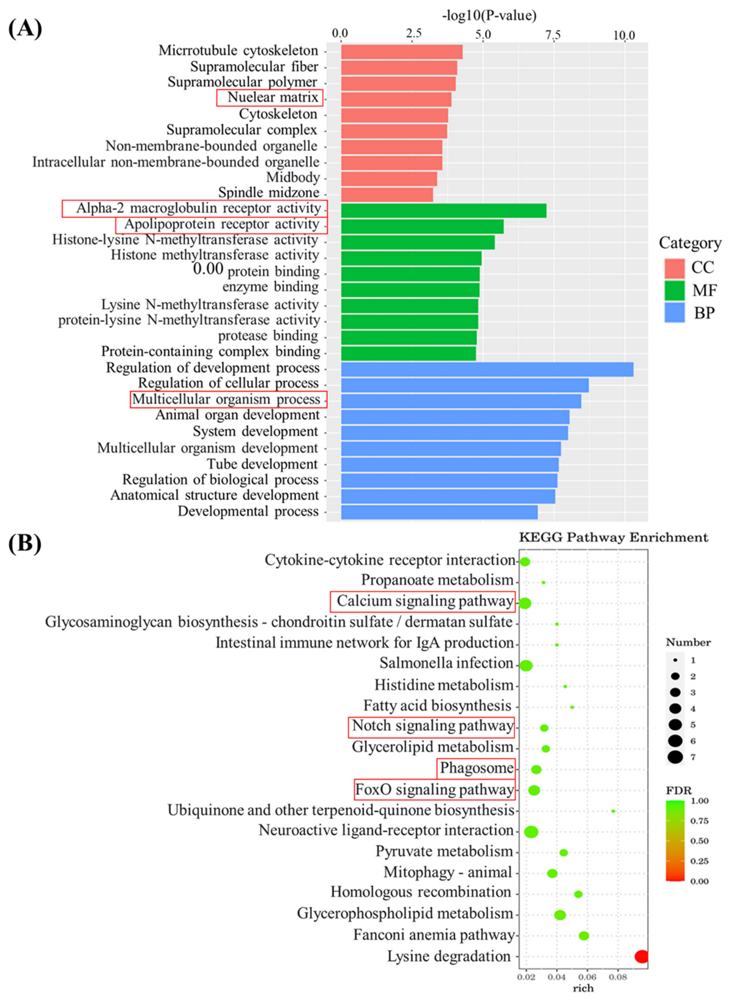
Gene Ontology (GO) enrichment analysis of DEGs revealed that the top 20 significantly enriched terms included α-2-macroglobulin receptor binding, apolipoprotein binding, and regulation of cell development (Figure 6A).
KEGG pathway enrichment analysis showed that DEGs were significantly enriched in several immune-related pathways, including phagosome, Notch signaling pathway, calcium signaling pathway, glycerophospholipid metabolism, fatty acid biosynthesis, and intestinal immune network for IgA production (Figure 6B).

3.5.4. qPCR Validation of Differentially Expressed Genes

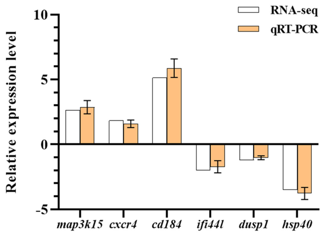
Six representative DEGs (map3k15, cxcr4, cd184, ifi44L, dusp1 and hsp40) were selected for qPCR validation. As shown in Figure 7, the expression changes of all six genes were consistent with the RNA-seq data, confirming the reliability of the transcriptomic analysis.

4. Discussion

In this study, LcDUSP5 from large yellow croaker was cloned and characterized. Domain analysis showed that LcDUSP5 contains the conserved RHOD and DSPc domains typical of DUSP5 family members, indicating substantial structural conservation among vertebrates. Similar domain organization has also been reported in other teleost DUSP5 proteins [16]. In addition, bioinformatic analysis predicted multiple potential phosphorylation sites in LcDUSP5 [7,22], although their functional relevance remains to be experimentally determined.
Tissue expression analysis showed that LcDUSP5 is broadly expressed in multiple tissues of large yellow croaker, with the highest expression in the liver. This pattern is generally consistent with the widespread distribution reported in other teleost species, although the dominant tissue differs among species [15,16]. In teleost fish, the liver not only serves as a central metabolic organ but also plays essential roles in immune surveillance, synthesis of innate immune effectors, and maintenance of inflammatory homeostasis [23]. The high hepatic expression suggests that LcDUSP5 may be associated with immune- or metabolism-related processes in this organ, but further functional evidence is needed.
Subcellular localization analysis indicated that LcDUSP5 is predominantly localized in the nucleus, which is consistent with the known characteristics of DUSP5 family members [4]. However, because this experiment was performed in HEK293T cells rather than fish-derived cells, the result should be interpreted cautiously.
To further elucidate the immunoregulatory mechanisms of LcDUSP5, an overexpression model was established in the large yellow croaker head kidney-derived macrophage cell line (LM10), and transcriptome sequencing was performed to systematically analyze the downstream regulatory network. Transcriptomic analysis revealed that LcDUSP5 overexpression significantly enriched genes associated with the α-2-macroglobulin receptor, namely low-density lipoprotein receptor-related protein 1 (LRP1). LRP1 is a multifunctional endocytic receptor widely involved in ligand clearance, signal transduction, and lipid metabolism regulation in mammals [24,25]. In the context of immunity, LRP1 plays a crucial anti-inflammatory and pro-resolving role in macrophages. For example, macrophages lacking LRP1 exhibit excessive activation of LPS-induced NF-κB signaling and increased secretion of pro-inflammatory cytokines such as TNF-α and IL-6, indicating that LRP1 acts as a key negative regulator of inflammatory responses [26]. Furthermore, LRP1 can recognize and bind apoptotic cell markers such as phosphatidylserine, thereby mediating efferocytosis and promoting inflammation resolution and tissue homeostasis [27]. In addition, LRP1 has been identified as a co-receptor or entry factor for multiple viruses, including Sindbis virus and Rift Valley fever virus [28]. The significant enrichment of α-2-macroglobulin receptor-related pathways in this study suggests that LcDUSP5 overexpression leads to altered expression of LRP1-associated genes, which may indicate a potential role in macrophage functions such as pathogen clearance and inflammatory responses. However, direct evidence is needed to confirm this hypothesis. However, these associations were inferred from transcriptomic enrichment and were not directly validated at the protein or functional level.
Another key finding of this study is the significant enrichment of apolipoprotein-related pathways. Traditionally, apolipoproteins are considered essential regulators of lipid metabolism, responsible for lipoprotein assembly, secretion, and receptor recognition. However, increasing evidence indicates that apolipoproteins also play important roles in innate immunity and host defense [29]. For instance, apolipoprotein E (ApoE) can inhibit influenza virus entry by maintaining membrane cholesterol homeostasis, thereby preventing the interaction between viral hemagglutinin and host receptors [30]. In teleost fish, apolipoprotein A-I (ApoA-I) from orange-spotted grouper exhibits direct antibacterial activity against Aeromonas hydrophila and can inhibit SGIV replication by upregulating antiviral genes such as interferon-stimulated gene 15 (ISG15) and Mx-I [31]. In terms of inflammation, ApoE promotes macrophage polarization toward an alternatively activated (M2) phenotype, facilitating tissue repair, whereas ApoA-IV suppresses NF-κB nuclear translocation by inhibiting IκB kinase activity, thereby attenuating LPS-induced systemic inflammation [32,33].
The enrichment of apolipoprotein-related genes observed in this study suggests that LcDUSP5 overexpression may affect macrophage function through modulation of lipid metabolism–immune crosstalk. Combined with previous findings that DUSP5 participates in fatty acid oxidation and metabolic reprogramming in mammals [14], it is plausible that the LcDUSP5–ERK axis integrates metabolic signals with immune responses, thereby optimizing the antimicrobial and antiviral functions of macrophages through regulation of intracellular lipid environments.
The up-regulation of the chemokine receptor CXCR4 represents another notable finding in this study. CXCR4 is the specific receptor for the chemokine CXCL12 (stromal cell-derived factor-1, SDF-1) and plays a central role in immune cell development, homing, migration, and effector function [34,35]. In teleost fish, the functional conservation of the CXCR4/CXCL12 axis has been well demonstrated. For example, CXCR4 in Nile tilapia mediates B-cell chemotaxis and phagocytosis, and inhibition of this signaling pathway significantly increases bacterial burden and reduces survival following infection [36]. CXCR4 is also involved in T-cell development, hematopoietic stem cell homing, and leukocyte recruitment to inflammatory sites [37].
In this study, LcDUSP5 overexpression significantly up-regulated CXCR4 expression. Together with the enrichment of cytokine–cytokine receptor interaction pathways in KEGG analysis, this finding suggests that LcDUSP5 may enhance CXCR4-mediated chemotactic signaling, thereby promoting directed migration of immune cells to sites of infection or tissue injury. Notably, studies in mammals have shown that CXCR4 signaling can crosstalk with MAPK and NF-κB pathways [38]. Therefore, regulation of CXCR4 by LcDUSP5 may represent an additional mechanism by which it modulates inflammatory responses, acting synergistically with its direct ERK dephosphorylation function.
Beyond these key findings, KEGG pathway analysis further revealed that LcDUSP5 is involved in multiple immunometabolism-related processes. The enrichment of the Notch signaling pathway suggests that LcDUSP5 may influence macrophage fate determination and functional polarization, as Notch signaling is a critical regulator of M1/M2 macrophage differentiation [39]. Enrichment of the calcium signaling pathway is consistent with the role of DUSP5 in regulating intracellular calcium homeostasis and immune cell activation [40]. Additionally, significant alterations in lipid metabolism pathways, including glycerophospholipid metabolism and fatty acid biosynthesis, further support the hypothesis that LcDUSP5 modulates immune function through metabolic reprogramming.
Overall, the transcriptomic results provide an initial framework for understanding how LcDUSP5 overexpression is associated with immune-related gene networks in teleost macrophages. However, the present data do not establish direct regulation of macrophage polarization, antiviral defense, cytokine production, or MAPK pathway suppression.
Several limitations of this study should be acknowledged. First, our conclusions rely primarily on transcriptomic correlations following transient LcDUSP5 overexpression, which may produce non-physiological protein levels and do not directly demonstrate endogenous function. Complementary loss-of-function experiments (e.g., siRNA knockdown or CRISPR knockout) are needed to confirm the immunoregulatory role of LcDUSP5. Second, although DUSP5 is known as a nuclear MAPK phosphatase, we did not directly measure ERK phosphorylation, dephosphorylation, or MAPK pathway activity; therefore, the mechanistic link between LcDUSP5 and MAPK signaling remains speculative. Third, the RNA-seq findings were not followed by functional assays such as phagocytosis, migration, cytokine detection, or pathogen challenge. Finally, in vivo validation in fish was not performed. Future studies combining knockdown approaches, direct ERK phosphorylation measurements, and in vivo infection models will be important for clarifying the physiological role of LcDUSP5.

5. Conclusions

This study cloned and characterized LcDUSP5 from large yellow croaker. LcDUSP5 is predominantly localized in the nucleus and is highly expressed in the liver. Transcriptomic analysis showed that transient overexpression of LcDUSP5 altered the expression of genes associated with several immune-related and metabolism-related pathways, suggesting that LcDUSP5 may participate in macrophage immune regulation. However, direct functional validation, loss-of-function analysis, and in vivo infection studies are still required to confirm its physiological role.

Author Contributions

Conceptualization, Z.Z., T.H. and H.W.; methodology, Z.Z., T.H. and H.W.; software, H.W. and D.Y.; validation, Z.Z., T.H. and H.W.; formal analysis, T.H.; investigation, Z.Z. and H.W.; data curation, Z.Z., H.W.,T.H. and D.Y.; writing—original draft preparation, Z.Z. and H.W.; writing—review and editing, K.Y. and F.H.; visualization, K.Y.; supervision, Z.Z., T.H. and H.W.; project administration, F.H. and K.Y.; funding acquisition, F.H. and K.Y. All authors have read and agreed to the published version of the manuscript.

Funding

This research was funded by the Natural Science Foundation of Fujian Province, grant number 2023J01768; the National Research and Development Program of China Grant No. 2022YFD2401002; the College Students’ Innovation and Entrepreneurship Training Program of Jimei University; the National Key R&D Program of China, grant number 2018YFD0900301; and the China Agriculture Research System, grant number CARS-47-G04.

Institutional Review Board Statement

The animal study protocol was approved by the Animal Ethics Committee of Jimei University (Approval code JMU2020012473, approved on 24 January 2020).

Informed Consent Statement

Not applicable.

Data Availability Statement

The data supporting the findings of this study are available within the article. Additional datasets generated or analyzed during the current study are available from the corresponding author upon reasonable request. The RNA sequencing data have been submitted to the NCBI Sequence Read Archive (SRA), and the accession number is PRJNA1455091.

Conflicts of Interest

The authors declare no conflicts of interest.

References

  1. Pulido, R.; Stoker, A.W.; Hendriks, W.J.A.J. PTPs emerge as PIPs: Protein tyrosine phosphatases with lipid-phosphatase activities in human disease. Hum. Mol. Genet. 2013, 22, R66–R76. [Google Scholar] [CrossRef]
  2. Lokareddy, R.K.; Bhardwaj, A.; Cingolani, G. Atomic structure of dual-specificity phosphatase 26, a novel p53 phosphatase. Biochemistry 2013, 52, 938–948. [Google Scholar] [CrossRef] [PubMed]
  3. Pulido, R.; Lang, R. Dual specificity phosphatases: From molecular mechanisms to biological function. Int. J. Mol. Sci. 2019, 20, 4372. [Google Scholar] [CrossRef]
  4. Mandl, M.; Slack, D.N.; Keyse, S.M. Specific inactivation and nuclear anchoring of extracellular signal-regulated kinase 2 by the inducible dual-specificity protein phosphatase DUSP5. Mol. Cell. Biol. 2005, 25, 1830–1845. [Google Scholar] [CrossRef]
  5. Kidger, A.M.; Keyse, S.M. The regulation of oncogenic Ras/ERK signalling by dual-specificity mitogen activated protein kinase phosphatases (MKPs). Semin. Cell Dev. Biol. 2016, 50, 125–132. [Google Scholar] [CrossRef] [PubMed]
  6. Lang, R.; Hammer, M.; Mages, J. DUSP meet immunology: Dual specificity MAPK phosphatases in control of the inflammatory response. J. Immunol. 2006, 177, 7497–7504. [Google Scholar] [CrossRef]
  7. Kucharska, A.; Rushworth, L.K.; Staples, C.; Morrice, N.A.; Keyse, S.M. Regulation of the inducible nuclear dual-specificity phosphatase DUSP5 by ERK MAPK. Cell. Signal. 2009, 21, 1794–1805. [Google Scholar] [CrossRef]
  8. Rushworth, L.K.; Kidger, A.M.; Delavaine, L.; Stewart, G.; van Schelven, S.; Davidson, J.; Bryant, C.J.; Caddye, E.; East, P.; Caunt, C.J.; et al. Dual-specificity phosphatase 5 regulates nuclear ERK activity and suppresses skin cancer by inhibiting mutant harvey-ras (HRasQ61L)-driven serpinb2 expression. Proc. Natl. Acad. Sci. USA 2014, 111, 18267–18272. [Google Scholar] [CrossRef]
  9. Luo, J.; Xue, D.; Song, F.; Liu, X.; Li, W.; Wang, Y. DUSP5 (dual-specificity protein phosphatase 5) suppresses BCG-induced autophagy via ERK 1/2 signaling pathway. Mol. Immunol. 2020, 126, 101–109. [Google Scholar] [CrossRef] [PubMed]
  10. Seo, H.; Cho, Y.-C.; Ju, A.; Lee, S.; Park, B.C.; Park, S.G.; Kim, J.-H.; Kim, K.; Cho, S. Dual-specificity phosphatase 5 acts as an anti-inflammatory regulator by inhibiting the ERK and NF-κB signaling pathways. Sci. Rep. 2017, 7, 17348. [Google Scholar] [CrossRef]
  11. Kutty, R.G.; Xin, G.; Schauder, D.M.; Cossette, S.M.; Bordas, M.; Cui, W.; Ramchandran, R. Dual specificity phosphatase 5 is essential for T cell survival. PLoS ONE 2016, 11, e0167246. [Google Scholar] [CrossRef] [PubMed]
  12. Liang, M.; Li, Y.; Zhang, K.; Zhu, Y.; Liang, J.; Liu, M.; Zhang, S.; Chen, D.; Liang, H.; Liang, L.; et al. Host factor DUSP5 potently inhibits dengue virus infection by modulating cytoskeleton rearrangement. Antivir. Res. 2023, 215, 105622. [Google Scholar] [CrossRef]
  13. Hua, C.; Zheng, Q.; Zhu, J.; Chen, S.; Song, Y.; van der Veen, S.; Cheng, H. Human papillomavirus type 16 early protein E7 activates autophagy through inhibition of dual-specificity phosphatase 5. Oxidative Med. Cell. Longev. 2022, 2022, 1863098. [Google Scholar] [CrossRef] [PubMed]
  14. Luo, J.; Tian, Z.; Song, F.; Ren, C.; Liu, W. Dual-specificity phosphatase 5-mediated fatty acid oxidation promotes mycobacterium bovis BCG-induced inflammatory responses. Exp. Cell Res. 2024, 434, 113869. [Google Scholar] [CrossRef]
  15. Li, S.; Hao, G.; Li, J.; Peng, W.; Geng, X.; Sun, J. Comparative analysis of dual specificity protein phosphatase genes 1, 2 and 5 in response to immune challenges in Japanese flounder Paralichthys olivaceus. Fish Shellfish Immunol. 2017, 68, 368–376. [Google Scholar] [CrossRef]
  16. He, J.; Cai, Y.; Huang, W.; Lin, Y.; Lei, Y.; Huang, C.; Cui, Z.; Qin, Q.; Sun, H. The role of Epinephelus coioides DUSP5 in regulating Singapore grouper iridovirus infection. Viruses 2023, 15, 1807. [Google Scholar] [CrossRef]
  17. Zhou, T.; Chen, B.; Ke, Q.; Zhao, J.; Pu, F.; Wu, Y.; Chen, L.; Zhou, Z.; Bai, Y.; Pan, Y.; et al. Development and evaluation of a high-throughput single-nucleotide polymorphism array for large yellow croaker (Larimichthys crocea). Front. Genet. 2020, 11, 571751. [Google Scholar] [CrossRef] [PubMed]
  18. Han, X.; Zhang, S.; Wang, Y.; Fang, H.; Peng, S.; Yang, S.; Wu, Z. Seedling selection of the large yellow croaker (Larimichthys crocea) for sustainable aquaculture: A review. Appl. Sci. 2025, 15, 7307. [Google Scholar] [CrossRef]
  19. Zhang, H.; Wang, J.; Jing, Y. Larimichthys crocea (large yellow croaker): A bibliometric study. Heliyon 2024, 10, e37393. [Google Scholar] [CrossRef]
  20. Wang, G.; Luan, Y.; Wei, J.; Li, Y.; Shi, H.; Cheng, H.; Bai, A.; Xie, J.; Xu, W.; Qin, P. Genetic and pathogenic characterization of a new iridovirus isolated from cage-cultured large yellow croaker (Larimichthys crocea) in China. Viruses 2022, 14, 208. [Google Scholar] [CrossRef]
  21. Chen, X.H.; Lin, K.B.; Wang, X.W. Outbreaks of an iridovirus disease in maricultured large yellow croaker, Larimichthys crocea (Richardson), in China. J. Fish Dis. 2003, 26, 615–619. [Google Scholar] [CrossRef] [PubMed]
  22. Chen, H.F.; Chuang, H.C.A.; Tan, T.H. Regulation of dual-specificity phosphatase (DUSP) ubiquitination and protein stability. Int. J. Mol. Sci. 2019, 20, 2668. [Google Scholar] [CrossRef]
  23. Wu, N.; Song, Y.-L.; Wang, B.; Zhang, X.-Y.; Zhang, X.-J.; Wang, Y.-L.; Cheng, Y.-Y.; Chen, D.-D.; Xia, X.-Q.; Lu, Y.-S. Fish gut-liver immunity during homeostasis or inflammation revealed by integrative transcriptome and proteome studies. Sci. Rep. 2016, 6, 36048. [Google Scholar] [CrossRef] [PubMed]
  24. Lillis, A.P.; Van Duyn, L.B.; Murphy-Ullrich, J.E.; Strickland, D.K. LDL receptor-related protein 1: Unique tissue-specific functions revealed by selective gene knockout studies. Physiol. Rev. 2008, 88, 887–918. [Google Scholar] [CrossRef]
  25. Gonias, S.L.; Campana, W.M. LDL receptor-related protein-1 a regulator of inflammation in atherosclerosis, cancer, and injury to the nervous system. Am. J. Pathol. 2014, 184, 18–27. [Google Scholar] [CrossRef]
  26. May, P.; Bock, H.H.; Nofer, J.R. Low density receptor-related protein 1 (LRP1) promotes anti-inflammatory phenotype in murine macrophages. Cell Tissue Res. 2013, 354, 887–889. [Google Scholar] [CrossRef]
  27. Yancey, P.G.; Blakemore, J.; Ding, L.; Fan, D.; Overton, C.D.; Zhang, Y.; Linton, M.F.; Fazio, S. Macrophage LRP-1 controls plaque cellularity by regulating efferocytosis and akt activation. Arterioscler. Thromb. Vasc. Biol. 2010, 30, 787–795. [Google Scholar] [CrossRef]
  28. Ganaie, S.S.; Schwarz, M.M.; Mcmillen, C.M.; Price, D.A.; Feng, A.X.; Albe, J.R.; Wang, W.; Miersch, S.; Orvedahl, A.; Cole, A.R.; et al. LRP-1 is a host entry factor for Rift Valley fever virus. Cell 2021, 184, 5163–5176.e14. [Google Scholar] [CrossRef]
  29. Concha, M.I.; Smith, V.J.; Castro, K.; Bastías, A.; Romero, A.; Amthauer, R.J. Apolipoproteins A-I and A-II are potentially important effectors of innate immunity in the teleost fish Cyprinus carpio. Eur. J. Biochem. 2004, 271, 2984–2990. [Google Scholar] [CrossRef] [PubMed]
  30. Gao, P.; Ji, M.; Liu, X.; Chen, X.; Liu, H.; Li, S.; Jia, B.; Li, C.; Ren, L.; Zhao, X.; et al. Apolipoprotein E mediates cell resistance to influenza virus infection. Sci. Adv. 2022, 8, eabm6668. [Google Scholar] [CrossRef]
  31. Wei, J.; Gao, P.; Zhang, P.; Guo, M.; Xu, M.; Wei, S.; Yan, Y.; Qin, Q. Isolation and function analysis of apolipoprotein A-I gene response to virus infection in grouper. Fish Shellfish Immunol. 2015, 43, 396–404. [Google Scholar] [CrossRef]
  32. Baitsch, D.; Bock, H.H.; Engel, T.; Telgmann, R.; Müller-Tidow, C.; Varga, G.; Bot, M.; Herz, J.; Robenek, H.; von Eckardstein, A.; et al. Apolipoprotein E induces antiinflammatory phenotype in macrophages. Arterioscler. Thromb. Vasc. Biol. 2011, 31, 1160–1168. [Google Scholar] [CrossRef]
  33. Shearston, K.; Tan, J.T.M.; Cochran, B.J.; Rye, K.-A. Inhibition of vascular inflammation by Apolipoprotein A-IV. Front. Cardiovasc. Med. 2022, 9, 901408. [Google Scholar] [CrossRef]
  34. Caspar, B.; Cocchiara, P.; Melet, A.; Van Emelen, K.; Van der Aa, A.; Milligan, G.; Herbeuval, J.-P. CXCR4 as a novel target in immunology: Moving away from typical antagonists. Future Drug Discov. 2022, 4, FDD77. [Google Scholar] [CrossRef]
  35. Guyon, A. CXCL12 chemokine and its receptors as major players in the interactions between immune and nervous systems. Front. Cell. Neurosci. 2014, 8, 65. [Google Scholar] [CrossRef]
  36. Gao, A.; Lin, Y.; Chai, Y.; Han, J.; Wu, L.; Ye, J. CXCL12/CXCR4 axis promotes the chemotaxis and phagocytosis of B cells through the PI3K-AKT signaling pathway in an early vertebrate. J. Immunol. 2024, 213, 1676–1690. [Google Scholar] [CrossRef] [PubMed]
  37. Yellowley, C. CXCL12/CXCR4 signaling and other recruitment and homing pathways in fracture repair. BoneKEy Rep. 2013, 2, 300. [Google Scholar] [CrossRef] [PubMed]
  38. Tian, X.; Xie, G.; Xiao, H.; Ding, F.; Bao, W.; Zhang, M. CXCR4 knockdown prevents inflammatory cytokine expression in macrophages by suppressing activation of MAPK and NF-κB signaling pathways. Cell Biosci. 2019, 9, 55. [Google Scholar] [CrossRef] [PubMed]
  39. Xu, H.; Zhu, J.; Smith, S.; Foldi, J.; Zhao, B.; Chung, A.Y.; Outtz, H.; Kitajewski, J.; Shi, C.; Weber, S.; et al. Notch-RBP-J signaling regulates the transcription factor IRF8 to promote inflammatory macrophage polarization. Nat. Immunol. 2012, 13, 642–650. [Google Scholar] [CrossRef]
  40. Feske, S.; Wulff, H.; Skolnik, E.Y. Ion channels in innate and adaptive immunity. Annu. Rev. Immunol. 2015, 33, 291–353. [Google Scholar] [CrossRef]
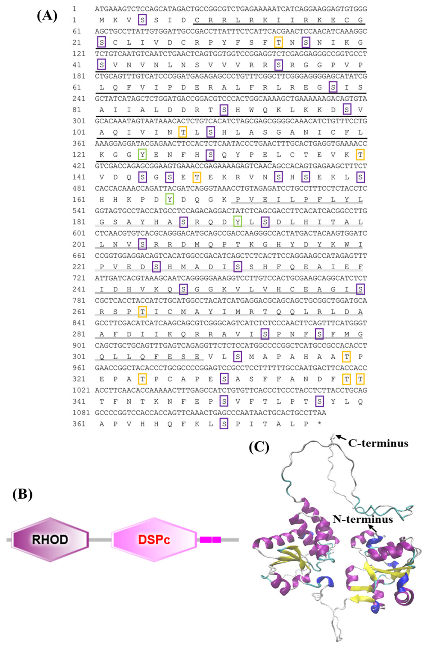
Figure 1. Bioinformatic analysis of the LcDUSP5 ORF. Notes: (A) Nucleotide and corresponding amino acid sequences of the LcDUSP5 ORF. The stop codon is indicated by an asterisk (*). The RHOD is underlined in black, and the DSPc domain is double underlined. Phosphorylation sites for serine (Ser), threonine (Thr), and tyrosine (Tyr) are highlighted with purple, green, and orange boxes, respectively. (B) Domain architecture of the LcDUSP5 protein. The RHOD is represented by a purple hexagon, and the DSPc domain is represented by a pink hexagon. (C) Tertiary structure of the LcDUSP5 protein. α-Helices are colored purple, and β-sheets are colored yellow.
Fishes 11 00320 g001
Figure 2. Amino acid sequence alignment and phylogenetic tree of DUSP5 from different species. Notes: (A) Sequence alignment of DUSP5 among different species. Black shading indicates identical amino acids, gray shading indicates low homology, the green underline marks the RHOD, and the blue underline marks the DSPc domain. (B) Phylogenetic tree constructed from LcDUSP5 and DUSP5 sequences from other species. The black triangle indicates large yellow croaker; node numbers represent bootstrap support values; the scale bar of 0.05 indicates genetic distance; GenBank accession numbers for the sequences are listed in Table 2.
Fishes 11 00320 g002
Figure 3. Tissue expression profile and subcellular localization of LcDUSP5. Notes: (A) Expression of LcDUSP5 in various tissues of large yellow croaker, with β-actin as the internal reference. Data are presented as mean ± SEM (n = 3 biological replicates). Different letters indicate significant differences (p < 0.05). (B) Immunofluorescence staining showing DUSP5 signal in green and nuclei stained with DAPI in blue. The merged image indicates predominant nuclear localization of LcDUSP5. The scale bar shown in the bottom-right panel applies to all panels in this figure.
Fishes 11 00320 g003
Figure 4. qPCR (A) and Western blot (B) analyses of LcDUSP5 overexpression in large yellow croaker macrophages. Data are presented as mean ± SEM (n = 3). ** indicates statistically significant difference compared with the control group (p < 0.01).
Fishes 11 00320 g004
Figure 5. Transcriptome data analysis following LcDUSP5 overexpression. Notes: (A) Correlation coefficient analysis between samples. (B) Principal component analysis (PCA). (C) Volcano plot of differentially expressed genes (DEGs).
Fishes 11 00320 g005
Figure 6. Enrichment analysis of DEGs following LcDUSP5 overexpression. Notes: (A) Gene Ontology (GO) enrichment analysis. (B) Kyoto Encyclopedia of Genes and Genomes (KEGG) pathway enrichment analysis.
Fishes 11 00320 g006
Figure 7. qPCR validation of differentially expressed genes. Six representative DEGs (map3k15, cxcr4, cd184, ifi44L, dusp1 and hsp40) were selected for validation. The expression changes in all six genes were consistent with the RNA-seq data. Data are presented as mean ± SEM (n = 3 biological replicates).
Fishes 11 00320 g007
Table 1. Primers used in this study.
PrimersSequence (5-3′)Purpose and Application
LcDUSP5-FgccctctagactcgaGCGGCCGCATGAAAGTCTCCAGCATAGACTGOverexpression
LcDUSP5-RtagtccagtgtggtgGAATTCAGGCAGTGCAGTTATTGGGC
qLcDUSP5-FGAGTCAACAGCCACAGTGAGAAGqPCR
qLcDUSP5-RTGTGAAGGTCGCTGAGATAGTCC
β-actin-FTTATGAAGGCTATGCCCTGCC
β-actin-RTGAAGGAGTAGCCACGCTCTGT
map3k15-FGCTGTGCTCATCCGTTACCT
map3k15-RGGTGAGGTAGGCGTTGTCAT
cxcr4-FAGCCTGTTGGCTACTTCAAG
cxcr4-RGATGATGGAGAGCACCAGGT
cd184-FTGGTCAACGTGGTCTACCTC
cd184-RAGCACACTGACCTTCCAGTT
ifi44L-FGAGACCTACGAGCTGTGCTC
ifi44L-RCTTGGCCTTGACAGTGGTCT
dusp1-FCCACTACGCCATCCTCAACT
dusp1-RCCTGCACAGAGAGAGTTGGA
hsp40-FGCTGAAGAGGTTCGACGAGA
hsp40-RCTTGTCCCCGAACTGCTTCT
sLcDUSP5-FtgaaccgtcagatccGCGGCCGCATGAAAGTCTCCAGCATAGACTGSubcellular localization
sLcDUSP5-RgtaccgtcgactgcaGAATTCCAGGCAGTGCAGTTATTGGGC
Table 2. Comparative analysis of amino acid sequence similarity of DUSP5 in large yellow croaker and other species.
SpeciesForeign NameGenBank
GenBank Accession Number
Identity (%)
Larimichthys croceaLarge yellow croakerXP_010733180.3100
Sciaenops ocellatusRed drumXAV45655.198.67
Miichthys miiuyBrown CroakerQCL11413.198.40
Anabas testudineusClimbing bassXP_026216642.197.61
Seriola dumeriliAllied kingfishXP_022611123.197.34
Lates japonicusAsian seabassGAA6229669.197.34
Seriola aureovittataYellowtail amberjackXP_056227911.196.81
Xiphias gladiusSwordfishXP_040002958.196.81
Epinephelus coioidesOrange-spotted grouperWJJ08744.196.01
Pelmatolapia mariaeLargescale shoveljaw fishXP_063332679.195.21
Anser cygnoidesSwan gooseXP_066856770.161.03
Columba liviaPigeonXP_064923110.160.51
Pelobates fuscusFrogXP_063290669.159.10
Mus musculusHouse mouseNP_001078859.158.24
Pseudophryne corroboreeCorroboree frogXP_063818706.157.26
Homo sapiensHumanNP_004410.353.77
Pan troglodytesChimpanzeeXP_016774803.353.26
Table 3. Summary of transcriptome sequencing data statistics.
SampleClean Reads (M)Clean Bases (Gb)Q30 (%)Total Mapping Genome RatioUniquely Mapping Gene Ratio
c-140.786.1496.9889.5594.67
c-250.557.6097.0589.5794.59
c-342.966.4697.1289.6494.60
DUSP5-142.086.3297.2289.5194.61
DUSP5-246.136.9397.0789.5994.52
DUSP5-363.289.5297.0089.5994.42
Disclaimer/Publisher’s Note: The statements, opinions and data contained in all publications are solely those of the individual author(s) and contributor(s) and not of MDPI and/or the editor(s). MDPI and/or the editor(s) disclaim responsibility for any injury to people or property resulting from any ideas, methods, instructions or products referred to in the content.