Article

The lncRNA011760/miR-Novel-91/NIPA2 ceRNA Network Regulates Salinity Stress Response in Sea Cucumber (Apostichopus japonicus)


1
Key Laboratory of Mariculture & Stock Enhancement in North China’s Sea, Ministry of Agriculture, Dalian Ocean University, Heishijiao Street, No. 52, Dalian 116023, China
2
College of Marine Science and Environment, Dalian Ocean University, Dalian 116023, China
*
Author to whom correspondence should be addressed.
Fishes 2026, 11(5), 275; https://doi.org/10.3390/fishes11050275
Submission received: 2 April 2026 / Revised: 2 May 2026 / Accepted: 5 May 2026 / Published: 8 May 2026

Abstract

Low-salinity stress poses a critical constraint on commercial aquaculture and the survival of the sea cucumber (Apostichopus japonicus). This study investigated the regulatory network involving lncRNA011760, miR-novel-91, and their target gene NIPA2 in response to salinity fluctuations. Using integrated in vivo and in vitro functional assays, we demonstrate that lncRNA011760 acts as a competitive endogenous RNA (ceRNA) to sponge miR-novel-91, thereby alleviating the post-transcriptional repression of NIPA2. Based on these molecular dynamics, we propose a novel inhibition-adaptation-survival three-stage model. Initially (0–3 h), acute NIPA2 upregulation enhances Mg2+ transport efficiency to mitigate osmotic shock. During the mid-stage (3–24 h), miR-novel-91-mediated NIPA2 suppression creates a transient biosynthetic window, facilitating a shift from passive tolerance to active metabolic adaptation. Ultimately (24–48 h), lncRNA-driven NIPA2 restoration sustains Mg2+ homeostasis, allowing the organism to enter a low-metabolism survival mode. These stage-specific shifts reflect the inherent physiological strategies of sea cucumbers as osmoconformers.
Key Contribution: This study identifies the lncRNA011760/miR-novel-91/NIPA2 ceRNA axis as a novel post-transcriptional regulator of Mg2+ transport in the sea cucumber under salinity stress, expanding the known repertoire of ncRNA-mediated osmoregulation to divalent cation homeostasis.

1. Introduction

The sea cucumber (Apostichopus japonicus) is an important marine aquaculture species in East Asia. The growing market demand has made the sustainable farming of sea cucumbers particularly important. However, intensifying global climate change and human activities have led to significant fluctuations in coastal salinity, posing a serious threat to the sustainable production of sea cucumbers [1,2]. As stenohaline invertebrates, sea cucumbers lack specialized osmoregulatory organs and are strictly confined to a narrow salinity range and are highly sensitive to changes in the salinity of their surrounding environment [3,4,5]. Consequently, hypoosmotic shock leads to a rapid disruption of intracellular osmotic equilibrium, triggering physiological dysfunctions, suppressed metabolic activity, and high mortality rates in aquaculture systems [6,7,8].
As an osmo-conforming marine invertebrate, sea cucumbers are considered to be strictly marine animals confined to a narrow salinity range [9] because sea cucumbers lack specialized osmoregulatory organs, rendering them particularly susceptible to salinity changes [10,11]. To overcome these challenges, sea cucumbers and other echinoderms have evolved a range of strategies to enhance their adaptability. According to Binyon (1972) and Diehl (1986), sea cucumbers exhibit hypo-osmotic regulatory mechanisms at low salinity and hyperosmotic regulatory strategies at high salinity [10,12]. The gray sea cucumber (Holothuria grisea) showed obvious protective behaviors: temporally regulating the osmotic pressure of its coelomic fluids by possibly contracting ambulacral feet and oral tentacles to reduce body wall permeability and slow coelomic fluid flow [13,14]. In extreme cases, sea cucumbers may discharge their intestines to adapt to salinity changes in the environment [15]. Adult Echinus esculentus can acclimate to decreased salinity, thereby increasing their resistance to subsequent hypo-osmotic stress, yet their capacity for acute tolerance is restricted [16]. Physiologically, they rely on cellular-level adjustments, primarily the active regulation of osmotic pressure through ion transport mechanisms [14,17,18]. Hypo-salinity stress induced an increase in the activity of Na+/K+-ATPase and antioxidant enzymes to mitigate oxidative damage from osmotic stress [19]. Inorganic ions, amino acids, nucleotides, and their derivatives play crucial osmoregulatory roles in sea cucumbers under hypo-osmotic conditions [20]. A sea star and two sea urchin species (Asterias rubens, Psammechinus milliaris, and Strongylocentrotus droebachiensis) could utilize various combinations of free amino acids (FAAs) to maintain osmotic equilibrium [21]. While these physiological and metabolic responses are documented, the upstream molecular networks—specifically the epigenetic mechanisms orchestrating these complex processes—remain largely elusive.
Recent research has highlighted non-coding RNAs (ncRNAs), such as microRNAs (miRNAs) and long non-coding RNAs (lncRNAs), as master regulators of gene expression under environmental stress [22,23]. Specifically, lncRNAs can function as competitive endogenous RNAs (ceRNAs), acting as molecular sponges to sequester miRNAs and thereby alleviating the post-transcriptional repression of target mRNAs [24]. Transcriptomic studies have found that lncRNA001074 acts as a ceRNA and can regulate the expression of the Na+/K+-ATPaseαsubunit (NKAα) under low-salt stress through let-7. Let-7 is a conserved microRNA family that was originally identified as a developmental timing regulator and later as a tumor suppressor in mammals. However, studies in marine organisms have revealed an additional function of let-7 in osmotic regulation. Let-7 directly targeted the Na+/K+-ATPase α subunit gene (NKAα) through imperfect base-pairing, post-transcriptionally repressing NKAα expression [25]. Under low-salinity stress, lncRNA001074 acts as a ceRNA to competitively bind let-7, thereby relieving its repression on NKAα and allowing increased NKA enzyme activity to compensate for the osmotic challenge. This regulatory interaction represents a rapid post-transcriptional mechanism for adjusting ion transport capacity in response to salinity fluctuations, distinct from let-7’s canonical roles in development and tumor suppression. Research indicated that the let-7 miRNAs participate in low-salinity tolerance in the swimming crab [26,27]. In zebrafish, the miR-8 family (a member of the let-7 family) enabled precise control of ion transport by modulating the expression of Nherf1, a regulator of apical trafficking of transmembrane ion transporters. Disruption of miR-8 family member function leads to an inability to respond to osmotic stress and blocks the ability to properly traffic and/or cluster transmembrane glycoproteins at the apical surface of ionocytes [28]. The over-expression of miR-novel-3 suppressed SLC17a9, whereas lncRNA015168 acted as a competitive endogenous RNA (ceRNA) to sequester miR-novel-3 to deal with salinity stress [29]. These findings emphasized the critical role of ncRNA networks in maintaining ionic gradients. However, the specific ceRNA axes governing small-molecule metabolic pathways and cation transport during salinity adaptation have not been fully characterized.
Among the various cations, Mg2+ is the second most abundant cation in cells, present in high concentrations and tightly regulated within cells, playing an important role in biochemical functions, particularly those involving ATP hydrolysis [30,31]. In vertebrates, Mg2+ homeostasis is tightly controlled by a diverse array of transporters, including MAGT1, MRS2, and the SLC41 family [32]. Magnesium transporter protein 1 (MAGT1) is upregulated by low Mg2+ concentrations and is an essential protein in Mg2+ uptake into cells [30,31]. The SLC41A1 and SLC41A2 encode magnesium transport proteins 1 and 2 respectively (MagT1 and MagT2), which both mediate the uptake of Mg2+ into cells [30,33]. MAGT1 and SLC41 families mediate generalized cellular uptake. The magnesium transporter MRS2 homolog, mitochondrial (MRS2), mediates the influx of Mg2+ into the mitochondrial matrix [34]. TRPM6, along with its close relative TRPM7, may play an important role in regulating Mg2+ homeostasis in vertebrates [35]. Membrane magnesium transporter 1 (MMGT1) mediates the uptake of Mg2+ across the Golgi membrane [36]. NIPA2 (non-imprinted in Prader–Willi syndrome region gene 2) has emerged as a highly selective Mg2+ transporter localized in the plasma membrane and early endosomes [37]. In contrast to the generalized uptake mediated by MAGT1/SLC41 family members or the organelle-specific transport by MRS2 and MMGT1, NIPA2 combines high substrate selectivity with stress-responsive plasma membrane localization—it is specifically recruited to the cell surface under low extracellular Mg2+ conditions. Given that seawater Mg2+ concentrations correlate linearly with salinity, dynamic changes in environmental osmotic pressure directly impact the intracellular demand for Mg2+ to sustain metabolic flux. However, whether NIPA2 is integrated into a ceRNA regulatory network to facilitate active salinity adaptation of echinoderms remains entirely unknown.
Based on previous high-throughput transcriptome screening under simulated low-salinity stress (18 psu), we identified a set of differentially expressed lncRNAs and miRNAs (data unpublished). Among them, a novel microRNA, temporarily designated miR-novel-91 (mature sequence length: 21 nt), was predicted by miRanda v3.3a and TargetScan v7.2. against a local transcriptome database. A novel lncRNA, lncRNA011760, was identified from the same transcriptome dataset and characterized as an intergenic lncRNA. Target prediction supported that NIPA2 harbors a potential miR-novel-91 binding site within its 3′-UTR, with a minimum free energy of −24.0 kcal/mol and an alignment score of 154. Previous studies have established that ceRNA networks play pivotal roles in the salinity adaptation of A. japonicus. For example, lncRNA001074 acts as a ceRNA to sponge let-7, thereby relieving post-transcriptional repression of the Na+/K+-ATPase α subunit (NKAα) and maintaining Na+/K+ ionic homeostasis under low-salinity stress [25]. More recently, a miR-novel-3-LNC_015168-SLC17A9 axis was reported to mediate salinity adaptation [29]. These findings demonstrate that lncRNA–miRNA–mRNA regulatory networks are fundamental to the epigenetic control of osmotic homeostasis in echinoderms. However, existing studies have primarily focused on Na+/K+ balance, while the regulatory mechanisms governing Mg2+ homeostasis during salinity stress remain entirely unexplored. Notably, NIPA2 is recruited to the plasma membrane specifically in response to low extracellular magnesium, a condition mimicking the ionic fluctuations during hypoosmotic stress when water influx globally dilutes divalent cation concentrations [37]. Because seawater Mg2+ concentrations correlate linearly with salinity, acute salinity decline directly challenges the intracellular Mg2+ supply required to sustain hundreds of Mg2+-dependent enzymatic reactions and metabolic flux. The unique combination of high substrate selectivity, stress-responsive localization, and plasma membrane accessibility positions NIPA2 as an ideal candidate ion transporter whose regulation via a ceRNA network would enable rapid post-transcriptional tuning of Mg2+ transport capacity during osmotic challenge.
We therefore hypothesized that lncRNA011760 functions as a ceRNA that competitively binds miR-novel-91, thereby relieving the miRNA-mediated post-transcriptional repression of NIPA2 and enhancing Mg2+ transport capacity to counteract hypoosmotic stress. This hypothesis is supported by the coordinated identification of all three components from a single low-salinity transcriptome, the bioinformatic prediction of high-confidence binding between miR-novel-91 and NIPA2, the established precedent of ceRNA-mediated ion transporter regulation in A. japonicus, and the unique physiological properties of NIPA2 as a low-Mg2+-responsive transporter. In the present study, we combined expression profiling, in vivo and in vitro functional interference, and physiological assessments to test this proposed lncRNA011760/miR-novel-91/NIPA2 regulatory interaction and to evaluate its contribution to low-salinity stress adaptation in the sea cucumber.

2. Materials and Methods

2.1. Tissue Sample Collection and Low-Salt Stress

The experiment was conducted at the Key Laboratory of Marine Aquaculture and Stock Enhancement in the Northern Seas of the Ministry of Agriculture and Rural Affairs, Dalian Ocean University. The animal research was approved by the Institutional Animal Care and Use Committee (IACUC) of Dalian Ocean University (Approval No.: DOU-IACUC-2023-0901). All experiments were conducted in accordance with the Guidelines for the Management and Use of Laboratory Animals of the Chinese Academy of Fishery Sciences and local regulations. Healthy sea cucumbers (average weight 20.2 g ± 1.1 g) were collected from a commercial farm in Liaoning Province, China. Before the experiment, the sea cucumbers were acclimated for 14 days under controlled conditions in 1000-LL fiberglass tanks (30 individuals per tank): the temperature was 16–18 °C, the pH was 7.8–8.0, and the dissolved oxygen was 5.0–6.0 mg/L (measured using the YSI ProODO® multiparameter meter, Yellow Springs Instruments, Yellow Springs, OH, USA). Sediment (sieved to remove debris) was replaced every two weeks, and a customized diet plan (60% Sargassum thunbergii, 40% sea mud) was provided daily at 3–5% of body weight.
After acclimation, sea cucumbers were randomly distributed to the control and experimental groups, each in three replicates (n = 3 tanks/group, with 30 sea cucumbers per tank). Each replicate tank was equipped with an independent, self-contained water-recirculation system, ensuring no water exchange between tanks. For the salinity challenge, the seawater salinity was gradually reduced from 30 PSU (control) to 18 PSU within 24 h using deionized water to minimize osmotic shock. Control groups were maintained at 30 PSU throughout the experiment. Five experimental subgroups were established to verify the function under 18 PSU stress: negative control (NC) group (transfected with non-targeting oligonucleotides); miR-novel-91 agomir-transfected group; miR-novel-91 mimics-transfected group; si-NIPA2-transfected group; and lncRNA011760 mimics-transfected group. Transfection was carried out 24 h before salinity stress by intraperitoneal injection (50 µL per individual). Sea cucumber samples were randomly collected at six time points after salinity stress (1.5, 3, 6, 12, 24, and 48 h), with 3 individuals per group at each time point. Coelomic fluid was extracted by puncturing the coelom with a syringe, centrifuged at 3000× g for 5 min at 4 °C, and then the coelomocyte pellet obtained by centrifugation of coelomic fluid was immediately frozen in liquid nitrogen and stored at −80 °C for subsequent RNA extraction and sequencing. The entire experimental design used in this study is shown in Figure 1.

2.2. RNA Extraction, cDNA Synthesis, mRNA, miRNA, and LncRNA Expression

miRNA target genes among the DEGs were predicted using miRanda v3.3a and TargetScan v7.2. For miRanda, target sites were filtered with a pairing score ≥150 and ΔG ≤ −20 kcal/mol; for TargetScan, only sites with a cumulative weighted context++ score <−0.2 were retained.
Total RNA was extracted from coelomocytes using TRIzol® Reagent (Invitrogen, Carlsbad, CA, USA) according to the manufacturer’s instructions [38]. The purity and integrity of the RNA were assessed using a NanoDrop 2000 spectrophotometer (Thermo Fisher Scientific, Waltham, MA, USA) and 1.5% agarose gel electrophoresis, respectively. miRNA-specific cDNA was synthesized using the miRcute Plus miRNA First-Strand Kit (Sangon Biotech, Shanghai, China). The 20 μL reaction system contained 100 ng of total RNA, 10 μL of 2× miRNA P-RT Solution mix, 2 μL of miRNA P-RT Enzyme mix, and nuclease-free water to reach the final volume. The reaction was carried out at 37 °C for 60 min, followed by incubation at 85 °C for 5 min. The first-strand synthesis of lncRNA cDNA was performed using the lnRcute lncRNA cDNA First-Strand Synthesis Kit (Degenmol, Tiangen Company, Shanghai, China). The preparation and handling of the reaction system were as follows: 100 ng of RNA, 2 μL of 5× gDNA buffer, and RNase-free water were added in a 10 μL volume. The mixture is combined and incubated at 42 °C for 3 min, then placed on ice. Into the mixture, 2 μL of 10× lnR RT Buffer, 1 μL of lnR RT Enzyme mix, 2 μL of lnR-RT Primer mix, and RNase-free water were added to reach a final volume of 20 μL. The reaction mixture is incubated at 42 °C for 15 min, then at 95 °C for 3 min, and stored at 80 °C in preparation for subsequent experiments.
The qRT-PCR was performed using the LightCycler® 96 (Roche Diagnostics, Basel, Switzerland) with 2× SG Fast qPCR Master Mix (Sangon Biotech, Shanghai, China). The 20 µL reaction system consisted of 10 µL of 2× SG Fast qPCR Master Mix, 1.0 µL of cDNA template, 0.4 µL of each primer (10 mmol/L), and nuclease-free water to a final volume. The reaction program was as follows: pre-denaturation at 95 °C for 30 s; 35 cycles of denaturation at 95 °C for 3 s and annealing at 60 °C for 30 s. All reactions were performed with three technical replicates and three biological replicates, with Cytb and RNU6B serving as reference genes for mRNA and miRNA/lncRNA, respectively [25,39]. All primers were designed using Primer 5.0 software and synthesized by Sangon Biotech (Shanghai, China) (Table 1). Each sample was analyzed in three technical replicates and three biological replicates. Relative expression levels were calculated using the 2−ΔΔCt method.

2.3. Culture of Sea Cucumber Coelomocytes

We collected 1 mL of the coelomocyte pellet obtained by centrifugation of coelomic fluid from healthy sea cucumbers and mixed it 1:1 with ice-cold anticoagulant solution, centrifuged at 1000× g for 5 min at 4 °C (Eppendorf 5810R centrifuge, Hamburg, Germany), then washed twice with isotonic buffer. The isotonic buffer was prepared using Leibovitz’s L-15 medium (Gibco; Thermo Fisher Scientific, Waltham, MA, USA), supplemented with 10% fetal bovine serum (Merck, Darmstadt, Germany), 100 U/mL penicillin, and 100 μg/mL streptomycin (Fisher BioReagents, Pittsburgh, PA, USA). We resuspended the coelomic cells in Leibovitz’s L-15 medium, adjusting the cell density to 6 × 10⁵ cells/mL. Next, we dispensed 500 μL of the cell suspension (6 × 10⁵ cells per well) into 24-well culture plates, which were then cultured in the dark under humid conditions at 16 °C with 5% CO2. After 24 h of culture, the adherent coelomic cells were washed twice with ice-cold PBS (pH 7.4) and collected by centrifugation at 1000× g for 5 min for transfection.

2.4. miRNAs, lncRNA, and Targeted Gene Overexpression and Knockdown Experiments in Sea Cucumber Coelomocytes

We selected miR-novel-91, lncRNA011760, and their target gene NIPA2 for functional validation through overexpression (agomir) and inhibition (antagomir) to elucidate their roles in salinity stress response. We used Lipofectamine 2000 (Invitrogen, Carlsbad, CA, USA) according to the manufacturer’s instructions, and then we transfected 30 pmol miR-novel-91 agomir/mimic, si-NIPA2, lncRNA011760 mimics, or the corresponding negative controls into adherent co-cultured cells. In short, we diluted the oligonucleotides and Lipofectamine 2000 in 50 μL of serum-free L-15 medium, mixed them, and incubated them at room temperature for 20 min. Then we added the transfection mixture to each culture well and continued incubating the cells for 24 h. After transfection, the cells were exposed to low-salinity stress (18 PSU) by diluting seawater with deionized water, and we collected samples at 6, 24, and 48 h post-stress for RNA extraction and qRT-PCR analysis.

2.5. Statistical Analysis

All data are presented as mean ± standard deviation (SD). The statistical differences in the temperature and spatial expression of miR-novel-91, NIPA2, and lncRNA011760 were analyzed using one-way ANOVA (post-test using LSD), tested in SPSS software (version 22.0). A significance level of p < 0.05 was set. Graphs were generated using GraphPad Prism 8.0 software (GraphPad Software, San Diego, CA, USA).

3. Results

3.1. Identification of miR-Novel-91 and Target Gene NIPA2

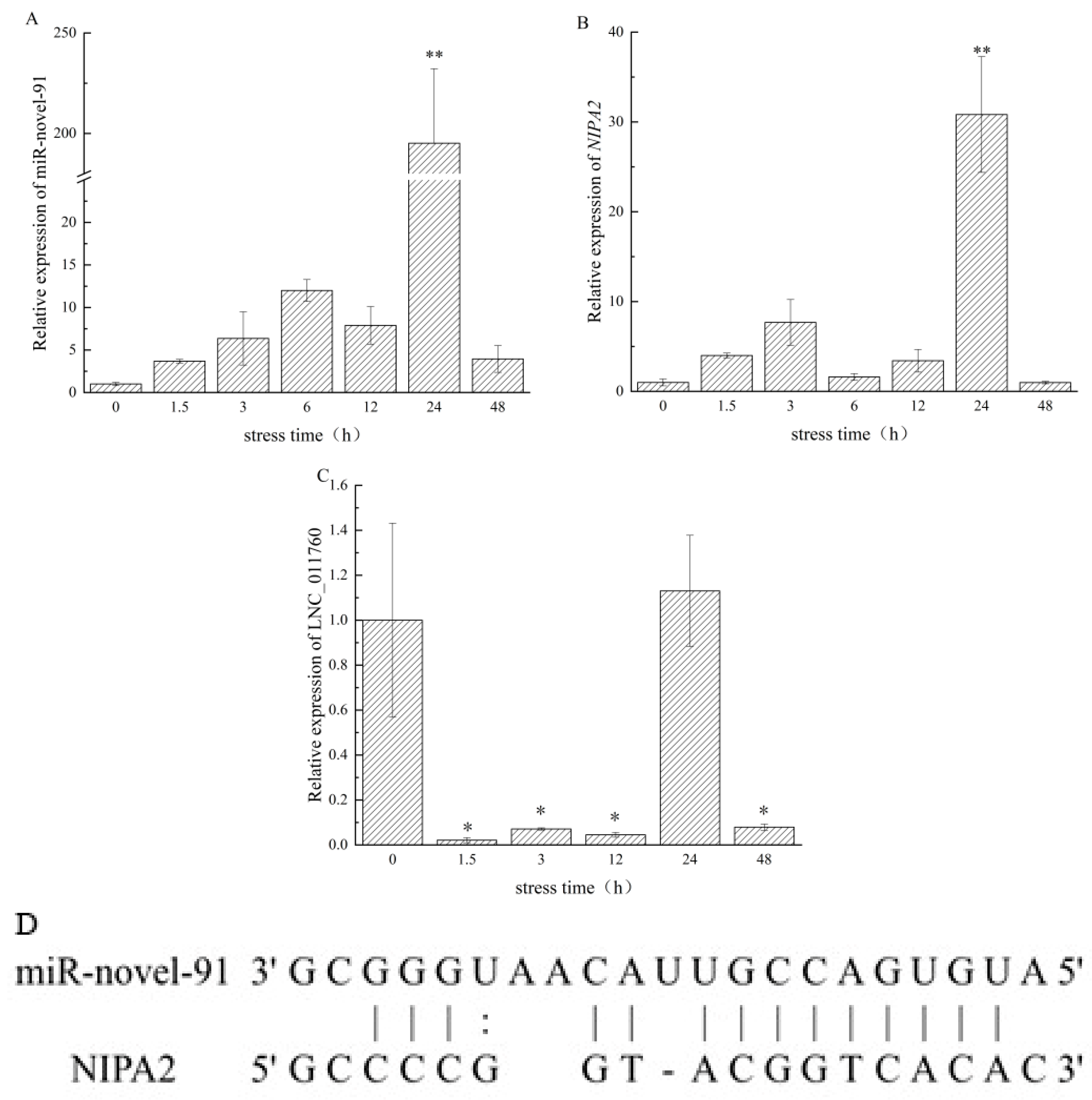
The corresponding target genes of miR-novel-91 were predicted, and the miR-novel-91 and NIPA2 corresponding binding sites are shown in Figure 2D. The length of the mature region of miR-novel-91 was 21, the length of the target gene NIPA2 was 1000, the sequence alignment score was 154, the minimum free energy was −24.0 kcal/mol, the p < 0.05 was correlated, and the 2–20 on the mature region of miR-novel-91 was combined with the 433–450 locus on the 3′-UTR of NIPA2.

3.2. Expression Profiles of miR-Novel-91, lncRNA011760 and NIPA2 Under Salinity Stress

The temporal expression patterns of miR-novel-91, lncRNA011760, and target gene NIPA2 during salinity stress response were determined by real-time quantitative PCR (Figure 2). The expression level of miR-novel-91 gradually increased with the duration of stress, peaking at 24 h with a significantly higher expression relative to the control group (Figure 2A). Exhibiting a similarity to that of miR-novel-91, NIPA2 showed a gradual upregulation from 0 to 3 h and peaked significantly at 24 h, before returning to the basal, unstressed level by 48 h (Figure 2B). The relative expression of lncRNA011760 displayed significant fluctuations across the time course. Notably, its expression dropped below the detection limit at 6 h—a result supported by repeated assays—yet rebounded at 24 h to a level showing no significant difference from the unstressed control group (Figure 2C). These dynamic patterns can be hypothesized to indicate that the 24 h time point represents an important temporal window for the involvement of miR-novel-91 and its target NIPA2 in the salinity adaptation process of the sea cucumber.

3.3. Validation of the miR-Novel-91/NIPA2 Regulatory Interaction In Vivo

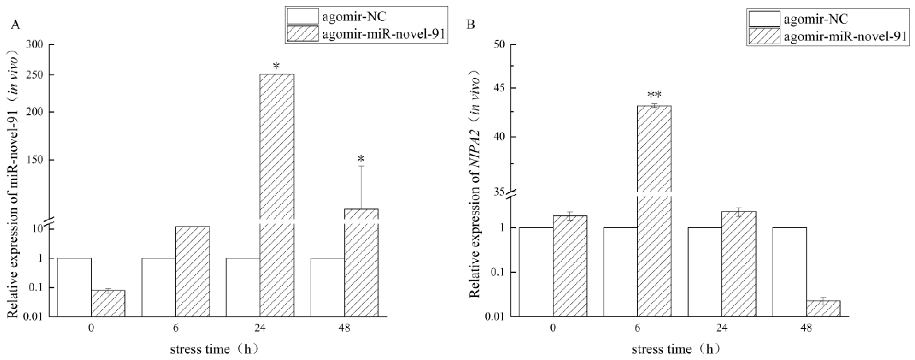
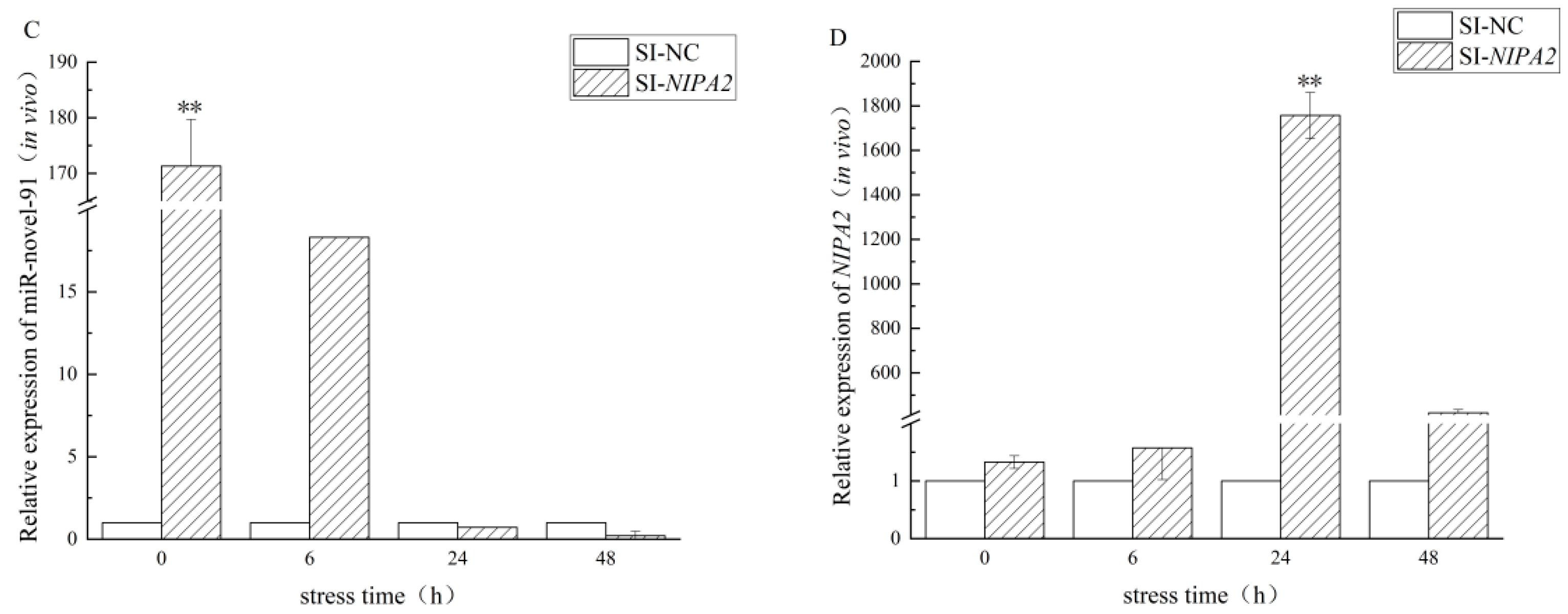
To verify the regulatory relationship between miR-novel-91 and NIPA2, in vivo functional assays were conducted under low-salinity stress using miR-novel-91 agomir and small interfering RNA targeting NIPA2 (si-NIPA2) (Figure 3). The transcript levels of both genes exhibited significant dynamic alterations depending on the delivery conditions. In the miR-novel-91 agomir-treated group, expression of miR-novel-91 was initially lower than that of the control group prior to stress induction; however, it increased progressively with the duration of low-salinity stress. This upregulation reached a highly significant level at 24 h (p < 0.01) and remained significantly elevated at 48 h (p < 0.05) (Figure 3A). Concurrently, the expression of NIPA2 displayed a highly significant variation at 6 h and was notably repressed compared to the control group by 48 h (Figure 3B), displaying an expression pattern inversely related to that of the microRNA. Conversely, in the si-NIPA2-treated group, the miR-novel-91 expression exhibited a highly significant difference prior to stress and was subsequently downregulated as the stress duration extended, falling below the control group level after 24 h (Figure 3C). Meanwhile, NIPA2 expression was upregulated over the stress time course, peaking with a highly significant difference at 24 h (Figure 3D). Taken together, these temporal expression dynamics support a potential negative regulatory interaction between miR-novel-91 and NIPA2 in A. japonicus during low-salinity adaptation.

3.4. Validation of the miR-Novel-91/NIPA2 Regulatory Interaction In Vitro

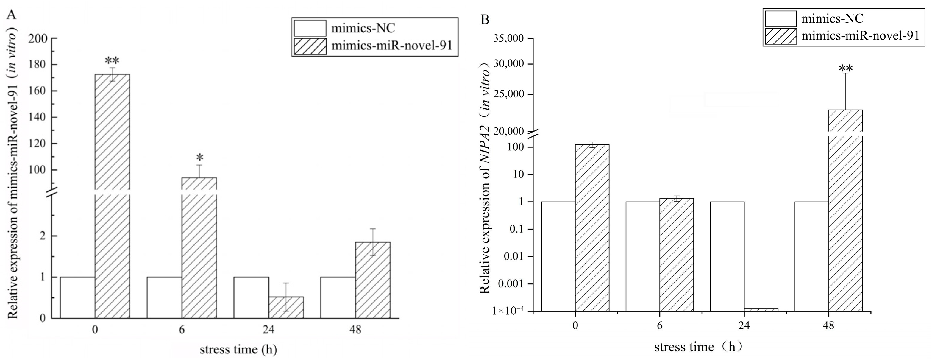
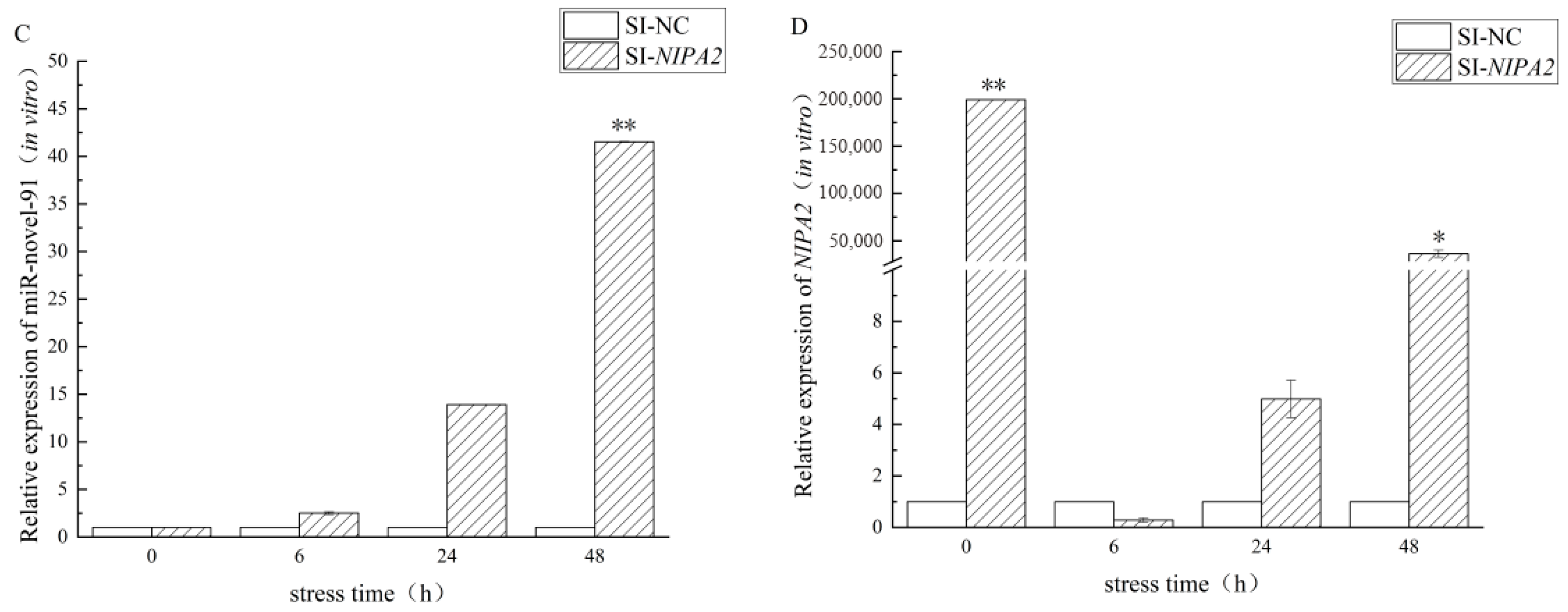
The regulatory interaction was further validated in vitro using cultured A. japonicus coelomocytes transfected with miR-novel-91 mimics or si-NIPA2 (Figure 4). In the miR-novel-91 mimics-transfected group, the expression of miR-novel-91 peaked post-transfection (0 h) immediately and maintained a highly significant elevation at 6 h (p < 0.01). Subsequently, with prolonged stress exposure, its expression was downregulated, dropping below that of the control group at 24 h (Figure 4A). The expression of the target gene NIPA2 was significantly repressed at 24 h compared to the control but later exhibited a highly significant rebound at 48 h (Figure 4B). In the experimental group transfected with si-NIPA2, the expression of miR-novel-91 was progressively upregulated throughout the stress period, reaching a highly significant peak at 48 h (Figure 4C). Meanwhile, the expression of NIPA2 displayed a highly significant peak at 0 h, sharply declined below the control level at 6 h, and subsequently rebounded, showing a significant upregulation again by 48 h (Figure 4D). Collectively, these in vitro temporal dynamics are consistent with a regulatory interaction in which miR-novel-91 influences NIPA2 expression, further suggesting a possible role of this microRNA-target interaction in the cellular response to salinity stress.

3.5. Proposed Mechanism of Salinity Response via the lncRNA011760/miR-Novel-91/NIPA2 Axis and Small Molecule Pathway

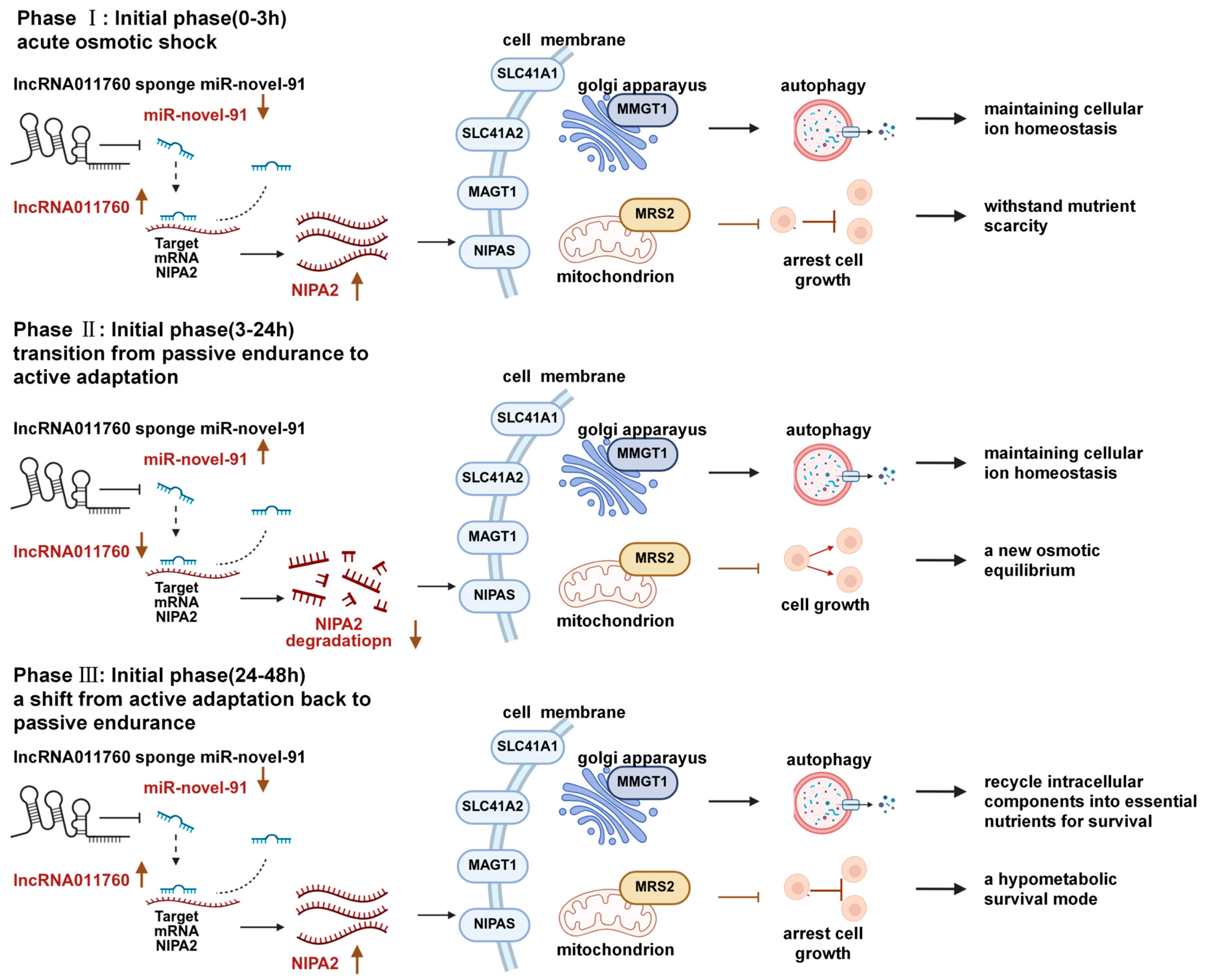
Based on the observed expression profiles and functional validation results, a three-stage adaptive model for sea cucumber salinity adaptation was proposed (Figure 5). This model operates through a ceRNA regulatory network, in which the lncRNA011760-miR-novel-91 axis tightly regulates NIPA2 expression, thereby fine-tuning Mg2+ transport and associated small-molecule metabolic pathways. The chronological cascade of this osmotic stress response is categorized into the following three distinct physiological phases. The initial stage (0–3 h) is acute osmotic shock and passive tolerance. Upon exposure to acute salinity fluctuations, the intracellular ion balance of the sea cucumber is rapidly disrupted, challenging the homeostasis of inorganic cations such as Mg2+. At 0 h, lncRNA011760 is significantly upregulated to act as a ceRNA sponge, sequestering miR-novel-91. This event effectively relieves the post-transcriptional repression of NIPA2, driving a marked increase in its expression (Figure 2). As an essential cation transporter, the enriched NIPA2 facilitates the transmembrane flux of Mg2+ and other inorganic cations. This immediate molecular response stabilizes fundamental intracellular cation homeostasis and mitigates the structural and metabolic damage induced by the early osmotic shock.
Mid-stage (3–24 h) was an active physiological adaptation period. As the stress duration extends, the sea cucumbers gradually transition from passive tolerance to an active osmotic remodeling state. During this critical window, lncRNA011760 expression is markedly downregulated, which releases the sequestered miR-novel-91, leading to its significant upregulation and subsequent direct targeting of NIPA2 (Figure 5). The targeted suppression of NIPA2 strategically modulates the transport efficiency of inorganic cations. This regulatory bottleneck recalibrates the metabolic rhythm of small-molecule pathways, accommodating the dynamic energetic and ionic demands required for the cells to establish a newly adapted intracellular ionic equilibrium.
Late stage (24–48 h) is low-metabolism survival and potential autolysis. Under prolonged osmotic stress, the intracellular energy and nutrient reserves approach critical depletion. By 48 h, the expression of lncRNA011760 significantly rebounds, re-engaging its sponge effect on miR-novel-91 and consequently restoring NIPA2 expression levels. The recovered NIPA2 sustains the basal transport of Mg2+ and maintains the essential functions of small-molecule pathways. This shift ensures the homeostasis of critical intracellular cations while minimizing global energy expenditure, effectively transitioning the organism into a low-metabolism survival mode to endure extreme stress. However, if long-term salt stress persists beyond physiological limits, the prolonged energetic deficit may trigger excessive autophagy and an irreversible arrest of anabolic metabolism, ultimately culminating in autolysis and visceral damage.

4. Discussion

4.1. The miR-Novel-91/NIPA2 Regulatory Interaction Under Salinity Stress

MicroRNAs (miRNAs) are key post-transcriptional regulatory factors that trigger mRNA degradation or translational repression by binding to the 3′ untranslated region (UTR) of target mRNAs [22,40]. In this study, we identified a negative regulatory relationship between miR-novel-91 and its target gene NIPA2 in sea cucumbers under salinity stress.
Under low-salinity conditions, miR-novel-91 and NIPA2 exhibited coordinated temporal dynamics, both reaching peak significance at 24 h. The apparent co-upregulation at this time point seems to contradict the canonical repressive function of miRNAs. However, miRNA-mediated silencing often operates at the translational level without immediate mRNA decay, meaning that mRNA abundance may not reflect protein output. It is therefore plausible that the surge in NIPA2 transcription during acute stress is simultaneously counterbalanced by elevated miR-novel-91 to fine-tune Mg2+ uptake and prevent detrimental overload. Protein-level measurements will be required to resolve this dynamic fully. Interestingly, this temporal inflection at 24 h is not unique to our axis. The previously characterized lncRNA001074/let-7/NKAα axis and the lncRNA015168/ miR-novel-3/SLC17A9 axis in A. japonicus also display critical regulatory transitions around 24 h post-stress [19,29]. The convergence of multiple ceRNA networks within this time window suggests a coordinated epigenetic program that synchronizes the remodeling of both monovalent (Na+/K+) and divalent (Mg2+) ion transporters during the early phase of salinity acclimation. Our findings extend this paradigm by revealing that Mg2+ homeostasis—a previously unexplored dimension in echinoderm osmoregulation—is similarly governed by a ceRNA network, thereby expanding the known repertoire of stress-responsive ion-regulatory axes.
In vivo experiments further uncovered a complex regulatory interplay. The miR-novel-91 overexpression elicited a biphasic NIPA2 response, while NIPA2 knockdown triggered a compensatory upregulation of miR-novel-91. This reciprocal regulation is consistent with a negative feedback loop, wherein the cell adjusts miRNA levels to transiently relieve NIPA2 inhibition, facilitating rapid Mg2+ uptake upon osmotic challenge. This feedback loop suggests that A. japonicus may modulate miR-novel-91 levels to transiently alleviate NIPA2 inhibition, thereby facilitating a rapid response to osmotic fluctuations. Such a feedback design would allow sea cucumbers to quickly tune Mg2+ transport capacity without requiring de novo protein synthesis, representing an energetically efficient strategy to cope with fluctuating salinity.

4.2. Physiological Implications of Metabolic Modulation in Salinity Adaptation

NIPA2 is a plasma membrane- and early endosome-localized Mg2+ transporter that is selectively upregulated under conditions of low extracellular Mg2+ [37]. Because seawater Mg2+ concentration declines linearly with salinity, hypoosmotic shock rapidly dilutes both environmental and coelomic fluid Mg2+ levels, creating a cellular Mg2+ deficiency signal. Our data show that NIPA2 is acutely induced as early as 3 h after low-salinity exposure, which likely represents a compensatory mechanism to enhance Mg2+ import.
The functional significance of this early NIPA2 induction lies in the central role of Mg2+ as an indispensable cofactor for ATP and a vast array of enzymatic reactions [41]. As a key mediator linking Mg2+ transport with the regulation of small molecule pathways, the loss or downregulation of NIPA2 can have a significant impact on small molecule pathways. Mg2+ is an indispensable cofactor for ATP-dependent enzymatic reactions, linking its homeostasis intrinsically to energy metabolism [42,43]. At the early stage of acute low-salinity stress (0–3 h), the rapid decline in environmental and coelomic fluid Mg2+ concentration acts as an intracellular starvation signal, triggering the immediate upregulation of NIPA2 (Figure 5). Previous physiological studies on intertidal holothurians demonstrate that while sodium and chloride levels fluctuate passively with external salinity, Mg2+ exhibits a distinct pattern of active homeostatic control [43]. The acute upregulation of NIPA2 enhances the efficiency of Mg2+ transmembrane transport and uptake, thereby reshaping intracellular Mg2+ homeostasis [44], systemically regulating small molecule pathways and inorganic cation balance, and alleviating damage caused by initial osmotic stress.
As stress progresses to the mid-stage (3–24 h), A. japonicus undergoes a strategic shift from passive endurance to active physiological adaptation. This transition involves a complex synergy of ion transport—particularly the fluctuant activation of Na+/K+-ATPase and the mobilization of intracellular osmolytes [19,20,45]. NIPA1 expression increases as Mg2+ concentration decreases, enhancing Mg2+ uptake to maintain homeostasis [46]. Behaviorally, adaptive strategies are manifested through defensive mechanisms, such as reducing body wall permeability. This was observed in the gray sea cucumber (Holothuria grisea), which lowers body wall permeability by contracting its tube feet and oral tentacles, thereby reducing water influx and alleviating osmotic stress [43]. The miR-novel-91 is significantly upregulated, inhibiting NIPA2 and reducing the transport efficiency of inorganic cations such as Mg2+ (Figure 2A,B). The significant upregulation of miR-novel-91 during this phase suppresses NIPA2, effectively recalibrating the transport efficiency of Mg2+. This regulatory bottleneck adjusts the metabolic process of small-molecule pathways, allowing cells to establish a newly adapted ionic equilibrium and providing support for adapting to the osmotic environment [46,47].
At the later stage (48 h), the rebound of lncRNA011760 adds a third layer of control. Acting as a ceRNA sponge for miR-novel-91, lncRNA011760 restores NIPA2 expression, thereby maintaining a basal level of Mg2+ transport necessary for survival under prolonged low-salinity conditions. As an organism that lacks specialized organs for regulating osmotic pressure, sea cucumbers can only live within a certain salinity range and are very sensitive [17]. Prolonged salinity stress eventually leads to the depletion of cellular energy reserves, such as glucose and branched-chain amino acids [47]. As cellular energy and amino acid reserves are depleted, they shift from active adaptation to passive tolerance. At 48 h, the rebound of lncRNA011760 acts as a ceRNA sponge to sequester miR-novel-91, effectively restoring NIPA2 levels and maintaining basal Mg2+ influx (Figure 4). This shift ensures the persistence of essential metabolic functions while transitioning the organism into a low-metabolism survival mode [44].
Mg2+ homeostasis is intimately linked to mitochondrial function and cellular energy status. In vertebrate cells, intracellular Mg2+ deficiency triggers mitophagy via the PINK1/Parkin pathway to clear damaged mitochondria [48,49]. It is plausible that in sea cucumbers, the miR-novel-91-mediated suppression of NIPA2 during the mid-phase of stress leads to a controlled reduction in Mg2+ influx, which could serve as a signal to initiate mitochondrial quality control and autophagy. Such a mechanism would facilitate the recycling of damaged components and conserve energy, aligning with the observed shift from active adaptation to a low-metabolism survival mode in stenohaline osmoconformers. However, direct measurements of Mg2+ fluxes, ATP levels, and autophagic markers are needed to test this hypothesis. We postulate a similar mechanism in A. japonicus: to minimize energy expenditure under prolonged stress, the sea cucumber halts energy-intensive protein synthesis and fully activates systemic autophagy to recycle intracellular components. Unlike the early protective autophagy, the late-stage autophagy mainly recycles intracellular components into essential nutrients needed to maintain energy [47]. This shift is a direct result of the sea cucumber’s limited salinity tolerance—because it cannot sustain active osmoregulation, it prioritizes saving energy to prolong survival. However, this survival strategy has limitations: with prolonged salinity stress, the continued activation of autophagy and complete cessation of anabolic metabolism may exceed physiological thresholds, and the sea cucumber may adopt its ultimate survival strategy—evisceration [15].

4.3. Limitations and Future Perspectives

This study demonstrates the pivotal role of the lncRNA011760/miR-novel-91/NIPA2 axis in the salinity adaptation of Apostichopus japonicus, specifically by modulating metabolic pathways and cation homeostasis. Our findings offer a novel framework for understanding how stenohaline echinoderms’ multi-stage adaptive strategies operate at the epigenetic level. Nevertheless, several limitations warrant further consideration. While our study establishes the molecular framework of the lncRNA011760/miR-novel-91 /NIPA2 ceRNA axis, several limitations must be explicitly acknowledged. First, the absence of direct binding assays and NIPA2 protein-level data means that the proposed physical interactions and post-transcriptional repression remain to be experimentally supported. Second, the subcellular localization of lncRNA011760 has not been determined, which is essential for validating its proposed cytoplasmic sponge function. Third, rescue experiments in which both the miRNA and the lncRNA are simultaneously manipulated were not performed; consequently, the linear causality of the ceRNA axis is inferred from single-gene perturbations and should be further verified. Fourth, whole-animal physiological endpoints—including survival rate, coelomic fluid Mg2+ concentration, osmotic pressure, and metabolic indicators—were not assessed, and therefore the organism-level adaptive significance of this axis remains to be directly demonstrated. We have accordingly interpreted our findings as providing a novel candidate regulatory mechanism for Mg2+ homeostasis rather than definitive proof of its role in salinity adaptation. Future studies that integrate dual-luciferase reporter assays, subcellular fractionation, genetic rescue experiments, and physiological phenotyping will be necessary to fully substantiate the proposed model. Experiments to achieve these validations—including dual-luciferase reporter assays, subcellular localization, rescue experiments, and physiological phenotyping—are currently underway in our laboratory.

5. Conclusions

This study identifies the lncRNA011760/miR-novel-91/NIPA2 axis as a novel ceRNA regulatory mechanism governing Mg2+ transport in A. japonicus under low-salinity stress. Through integrated in vivo and in vitro functional assays, we provide evidence consistent with a model in which lncRNA011760 acts as a molecular sponge for miR-novel-91, thereby alleviating the post-transcriptional repression of the magnesium transporter NIPA2. The temporal dynamics suggested a stage-specific adaptive strategy, transitioning from acute ion compensation to metabolic reorganization and ultimately to a low-energy survival mode. This work expands the paradigm of ncRNA-mediated osmoregulation in echinoderms, revealing a previously unrecognized layer of epigenetic control over ion transport during salinity adaptation.

Author Contributions

Y.T., J.C., Y.Z., J.Z. and H.X., project administration, visualization, writing. X.W. and Q.G., data curation, supervision, review and editing. All authors have read and agreed to the published version of the manuscript.

Funding

This work was supported by the National Natural Science Foundation of China (approval code No. 32273117, and approval date 1 July 2023) and the National Key R&D Program of China (2018YFC0310702).

Institutional Review Board Statement

Animal study was reviewed and approved by the Institutional Animal Care and Use Committee of Dalian Ocean University (approval No. DOUIACUC-2023-0901 and approval date 1 September 2023).

Data Availability Statement

The original contributions presented in this study are included in the article. Further inquiries can be directed to the corresponding author.

Conflicts of Interest

The authors declare no conflicts of interest.

References

  1. Allan, R.P.; Barlow, M.; Byrne, M.P.; Cherchi, A.; Douville, H.; Fowler, H.J.; Gan, T.Y.; Pendergrass, A.G.; Rosenfeld, D.; Swann, A.L.S.; et al. Advances in Understanding Large-Scale Responses of the Water Cycle to Climate Change. Ann. N. Y. Acad. Sci. 2020, 1472, 49–75. [Google Scholar] [CrossRef] [Scilit]
  2. Röthig, T.; Trevathan-Tackett, S.M.; Voolstra, C.R.; Ross, C.; Chaffron, S.; Durack, P.J.; Warmuth, L.M.; Sweet, M. Human-Induced Salinity Changes Impact Marine Organisms and Ecosystems. Glob. Change Biol. 2023, 29, 4731–4749. [Google Scholar] [CrossRef] [Scilit]
  3. Hu, M.; Li, Q.; Li, L. Effect of Salinity and Temperature on Salinity Tolerance of the Sea Cucumber Apostichopus japonicus. Fish. Sci. 2010, 76, 267–273. [Google Scholar] [CrossRef] [Scilit]
  4. Meng, X.; Dong, Y.; Dong, S.; Yu, S.; Zhou, X. Mortality of the Sea Cucumber, Apostichopus japonicus Selenka, Exposed to Acute Salinity Decrease and Related Physiological Responses: Osmoregulation and Heat Shock Protein Expression. Aquaculture 2011, 316, 88–92. [Google Scholar] [CrossRef] [Scilit]
  5. Li, L.; Li, Q.; Sun, X.; Kong, L. Effects of Temperature and Salinity on Larval Growth, Survival, and Development of the Sea Cucumber Apostichopus japonicus. N. Am. J. Aquac. 2011, 73, 296–303. [Google Scholar] [CrossRef] [Scilit]
  6. Wang, F.; Yang, H.; Gao, F.; Liu, G. Effects of Acute Temperature or Salinity Stress on the Immune Response in Sea Cucumber, Apostichopus japonicus. Comp. Biochem. Physiol. Part A Mol. Integr. Physiol. 2008, 151, 491–498. [Google Scholar] [CrossRef] [Scilit]
  7. Yuan, X.; Yang, H.; Wang, L.; Zhou, Y.; Gabr, H.R. Effects of Salinity on Energy Budget in Pond-Cultured Sea Cucumber Apostichopus japonicus (Selenka) (Echinodermata: Holothuroidea). Aquaculture 2010, 306, 348–351. [Google Scholar] [CrossRef] [Scilit]
  8. Mei, Y.; Xu, Y.; Gao, Q.; Li, Z.; Dong, S. Effects of Temperature and Salinity on CO2 Fluxes Dynamics and Respiration Metabolism in the Sea Cucumber Apostichopus japonicus (Selenka). Mar. Pollut. Bull. 2025, 215, 117928. [Google Scholar] [CrossRef] [Scilit]
  9. Brusca, R.C.; Brusca, G.J. Invertebrates; Sinauer Associates: Sunderland, MA, USA, 2004; Volume 53, ISBN 0-87893-097-3. [Google Scholar]
  10. Binyon, J. Physiology of Echinoderms; Elsevier: Amsterdam, The Netherlands, 1972; ISBN 978-0-08-016991-0. [Google Scholar]
  11. Willmer, P.; Stone, G.; Johnston, I.A. Environmental Physiology of Animals, 2nd ed.; Blackwell Pub: Malden, MA, USA, 2004; ISBN 978-1-4051-0724-2. [Google Scholar]
  12. Diehl, W.J. Osmoregulation in Echinoderms. Comp. Biochem. Physiol. Part A Physiol. 1986, 84, 199–205. [Google Scholar] [CrossRef] [Scilit]
  13. Vidolin, D.; Gouvea, I.A.S.; Freire, C.A. Estabilidade Osmótica Dos Fluídos Celômicos de Um Pepino Do Mar (Holothuria grisea) e de Uma Estrela-Do-Mar (Asterina stellifera) (Echinodermata) Expostos Ao Ar Durante a Maré Baixa: Um Estudo de Campo. Acta Biol. Par. 2002, 31, 113–121. [Google Scholar] [CrossRef] [Scilit]
  14. Castellano, G.C.; Souza, M.M.; Freire, C.A. Volume Regulation of Intestinal Cells of Echinoderms: Putative Role of Ion Transporters (Na+/K+-ATPase and NKCC). Comp. Biochem. Physiol. A Mol. Integr. Physiol. 2016, 201, 124–131. [Google Scholar] [CrossRef] [Scilit] [PubMed]
  15. Fankboner, P.V. Seasonal Visceral Atrophy and Response to Salinity by Parastichopus californicus (Stimpson): Osmoregulation? SPC Beche-De-Mer Inf. Bull. 2002, 17, 22–26. [Google Scholar]
  16. Barrett, N.J.; Harper, E.M.; Last, K.S.; Reinardy, H.C.; Peck, L.S. Behavioural and Physiological Impacts of Low Salinity on the Sea Urchin Echinus esculentus. J. Exp. Biol. 2024, 227, jeb246707. [Google Scholar] [CrossRef] [Scilit]
  17. Dong, Y.; Dong, S.; Meng, X. Effects of Thermal and Osmotic Stress on Growth, Osmoregulation and Hsp70 in Sea Cucumber (Apostichopus Japonicus Selenka). Aquaculture 2008, 276, 179–186. [Google Scholar] [CrossRef] [Scilit]
  18. Huang, L.; Zhong, S.; Pan, C.; Mo, H.; Pan, Z.; Huang, G.; Mi, S.; Gao, C.; Liu, Y. Effects of Acute Salinity Stress on Physiology and Immunoenzymatic Activity in Juvenile Sea Cucumber, Stichopus Monotuberculatus. Aquaculture 2024, 578, 740094. [Google Scholar] [CrossRef] [Scilit]
  19. Russell, M.P. Echinoderm Responses to Variation in Salinity. Adv. Mar. Biol. 2013, 66, 171–212. [Google Scholar] [CrossRef] [Scilit]
  20. Jiang, J.; Tang, Y.; Cao, Z.; Zhou, C.; Yu, Z. Effects of Hypo-Osmotic Stress on Osmoregulation, Antioxidant Response, and Energy Metabolism in Sea Cucumber Holothuria Moebii under Desalination Environment. Environ. Res. 2024, 252, 118800. [Google Scholar] [CrossRef] [Scilit]
  21. Podbielski, I.; Hiebenthal, C.; Hajati, M.-C.; Bock, C.; Bleich, M.; Melzner, F. Capacity for Cellular Osmoregulation Defines Critical Salinity of Marine Invertebrates at Low Salinity. Front. Mar. Sci. 2022, 9, 898364. [Google Scholar] [CrossRef] [Scilit]
  22. Esteller, M. Non-Coding RNAs in Human Disease. Nat. Rev. Genet. 2011, 12, 861–874. [Google Scholar] [CrossRef] [Scilit]
  23. Zheng, J.; Zhang, G.; Ren, L. Advances in miRNA research: Unraveling the complexities of gene regulation. Anim. Models Exp. Med. 2025, 8, 1741–1759. [Google Scholar] [CrossRef] [Scilit]
  24. Zhang, S.; Shao, Y.; Li, C. Characterization of Host lncRNAs in Response to Vibrio Splendidus Infection and Function as Efficient miRNA Sponges in Sea Cucumber. Front. Immunol. 2021, 12, 792040. [Google Scholar] [CrossRef] [Scilit]
  25. Shang, Y.; Tian, Y.; Wang, Y.; Guo, R. Novel lncRNA lncRNA001074 Participates in the Low Salinity-Induced Response in the Sea Cucumber Apostichopus japonicus by Targeting the Let-7/NKAα Axis. Cell Stress Chaperones 2021, 26, 785–798. [Google Scholar] [CrossRef] [Scilit]
  26. Chen, X.; Chen, J.; Shen, Y.; Bi, Y.; Hou, W.; Pan, G.; Wu, X. Transcriptional Responses to Low-Salinity Stress in the Gills of Adult Female Portunus trituberculatus. Comp. Biochem. Physiol. Part D Genom. Proteom. 2019, 29, 86–94. [Google Scholar] [CrossRef] [Scilit] [PubMed]
  27. Roush, S.; Slack, F.J. The Let-7 Family of microRNAs. Trends Cell Biol. 2008, 18, 505–516. [Google Scholar] [CrossRef] [Scilit]
  28. Flynt, A.S.; Thatcher, E.J.; Burkewitz, K.; Li, N.; Liu, Y.; Patton, J.G. miR-8 microRNAs Regulate the Response to Osmotic Stress in Zebrafish Embryos. J. Cell Biol. 2009, 185, 115–127. [Google Scholar] [CrossRef] [Scilit]
  29. Wei, X.; Chen, J.; Zhao, Y.; Li, N.; Wang, N.; Xu, T.; Yang, Y.; Chang, Y.; Tian, Y. A ceRNA Network Mediates Salinity Adaptation Via miR-Novel-3-LNC_015168-SLC17A9 Axis in Sea Cucumber. Mar. Biotechnol. 2026, 28, 21. [Google Scholar] [CrossRef] [Scilit]
  30. Goytain, A.; Quamme, G.A. Identification and Characterization of a Novel Mammalian Mg2+ Transporter with Channel-like Properties. BMC Genom. 2005, 6, 48. [Google Scholar] [CrossRef] [Scilit]
  31. Zhou, H.; Clapham, D.E. Mammalian MagT1 and TUSC3 Are Required for Cellular Magnesium Uptake and Vertebrate Embryonic Development. Proc. Natl. Acad. Sci. USA 2009, 106, 15750–15755. [Google Scholar] [CrossRef] [Scilit]
  32. Brandao, K.; Deason-Towne, F.; Perraud, A.-L.; Schmitz, C. The Role of Mg2+ in Immune Cells. Immunol. Res. 2013, 55, 261–269. [Google Scholar] [CrossRef] [Scilit] [PubMed]
  33. Sahni, J.; Nelson, B.; Scharenberg, A.M. SLC41A2 Encodes a Plasma-Membrane Mg2+ Transporter. Biochem. J. 2007, 401, 505–513. [Google Scholar] [CrossRef] [Scilit] [PubMed]
  34. Zsurka, G.; Gregán, J.; Schweyen, R.J. The Human Mitochondrial Mrs2 Protein Functionally Substitutes for Its Yeast Homologue, a Candidate Magnesium Transporter. Genomics 2001, 72, 158–168. [Google Scholar] [CrossRef] [Scilit] [PubMed]
  35. Schmitz, C.; Deason, F.; Perraud, A.-L. Molecular Components of Vertebrate Mg2+-Homeostasis Regulation. Magnes. Res. 2007, 20, 6–18. [Google Scholar]
  36. Goytain, A.; Quamme, G.A. Identification and Characterization of a Novel Family of Membrane Magnesium Transporters, MMgT1 and MMgT2. Am. J. Physiol.—Cell Physiol. 2008, 294, C495–C502. [Google Scholar] [CrossRef] [Scilit]
  37. Goytain, A.; Hines, R.M.; Quamme, G.A. Functional Characterization of NIPA2, a Selective Mg2+ Transporter. Am. J. Physiol.—Cell Physiol. 2008, 295, C944–C953. [Google Scholar] [CrossRef] [Scilit]
  38. Simms, D.; Chomczynski, P. TRIzolTM: A New Reagent for Optimal Single-Step Isolation of RNA. Focus 1993, 15, 532–535. [Google Scholar]
  39. Tian, Y.; Shang, Y.; Guo, R.; Chang, Y.; Jiang, Y. Salinity Stress-Induced Differentially Expressed miRNAs and Target Genes in Sea Cucumbers Apostichopus japonicus. Cell Stress Chaperones 2019, 24, 719–733. [Google Scholar] [CrossRef] [Scilit]
  40. Ali, S.A.; Peffers, M.J.; Ormseth, M.J.; Jurisica, I.; Kapoor, M. The Non-Coding RNA Interactome in Joint Health and Disease. Nat. Rev. Rheumatol. 2021, 17, 692–705. [Google Scholar] [CrossRef] [Scilit]
  41. Camilli, A.; Bassler, B.L. Bacterial Small-Molecule Signaling Pathways. Science 2006, 311, 1113–1116. [Google Scholar] [CrossRef] [Scilit] [PubMed]
  42. Madrid, E.; Zanders, I.P.; Herrera, F.C. Changes in Coelomic Fluid and Intracellular Ionic Composition in Holothurians Exposed to Diverse Sea Water Concentrations. Comp. Biochem. Physiol. Part A Physiol. 1976, 54, 167–174. [Google Scholar] [CrossRef] [Scilit]
  43. Castellano, G.C.; Santos, I.A.; Freire, C.A. Maintenance of Ionic Gradients and Tissue Hydration in the Intertidal Sea Cucumber Holothuria Grisea under Hypo- and Hyper-Salinity Challenges. J. Mar. Biol. Assoc. UK 2018, 98, 325–332. [Google Scholar] [CrossRef] [Scilit]
  44. Quamme, G.A. Molecular Identification of Ancient and Modern Mammalian Magnesium Transporters. Am. J. Physiol.—Cell Physiol. 2010, 298, C407–C429. [Google Scholar] [CrossRef] [Scilit] [PubMed]
  45. Geng, C.; Tian, Y.; Shang, Y.; Wang, L.; Jiang, Y.; Chang, Y. Effect of Acute Salinity Stress on Ion Homeostasis, Na+/K+-ATPase and Histological Structure in Sea Cucumber Apostichopus Japonicus. SpringerPlus 2016, 5, 1977. [Google Scholar] [CrossRef] [Scilit] [PubMed]
  46. Goytain, A.; Hines, R.M.; El-Husseini, A.; Quamme, G.A. NIPA1(SPG6), the Basis for Autosomal Dominant Form of Hereditary Spastic Paraplegia, Encodes a Functional Mg2+ Transporter. J. Biol. Chem. 2007, 282, 8060–8068. [Google Scholar] [CrossRef] [Scilit]
  47. Zhao, W.; Zhang, W.-L.; Yang, B.; Sun, J.; Yang, M.-W. NIPA2 Regulates Osteoblast Function via Its Effect on Apoptosis Pathways in Type 2 Diabetes Osteoporosis. Biochem. Biophys. Res. Commun. 2019, 513, 883–890. [Google Scholar] [CrossRef] [Scilit]
  48. Rabanal-Ruiz, Y.; Otten, E.G.; Korolchuk, V.I. mTORC1 as the Main Gateway to Autophagy. Essays Biochem. 2017, 61, 565–584. [Google Scholar] [CrossRef] [Scilit] [PubMed]
  49. Carroll, B.; Korolchuk, V.I.; Sarkar, S. Amino Acids and Autophagy: Cross-Talk and Co-Operation to Control Cellular Homeostasis. Amino Acids 2015, 47, 2065–2088. [Google Scholar] [CrossRef] [Scilit]
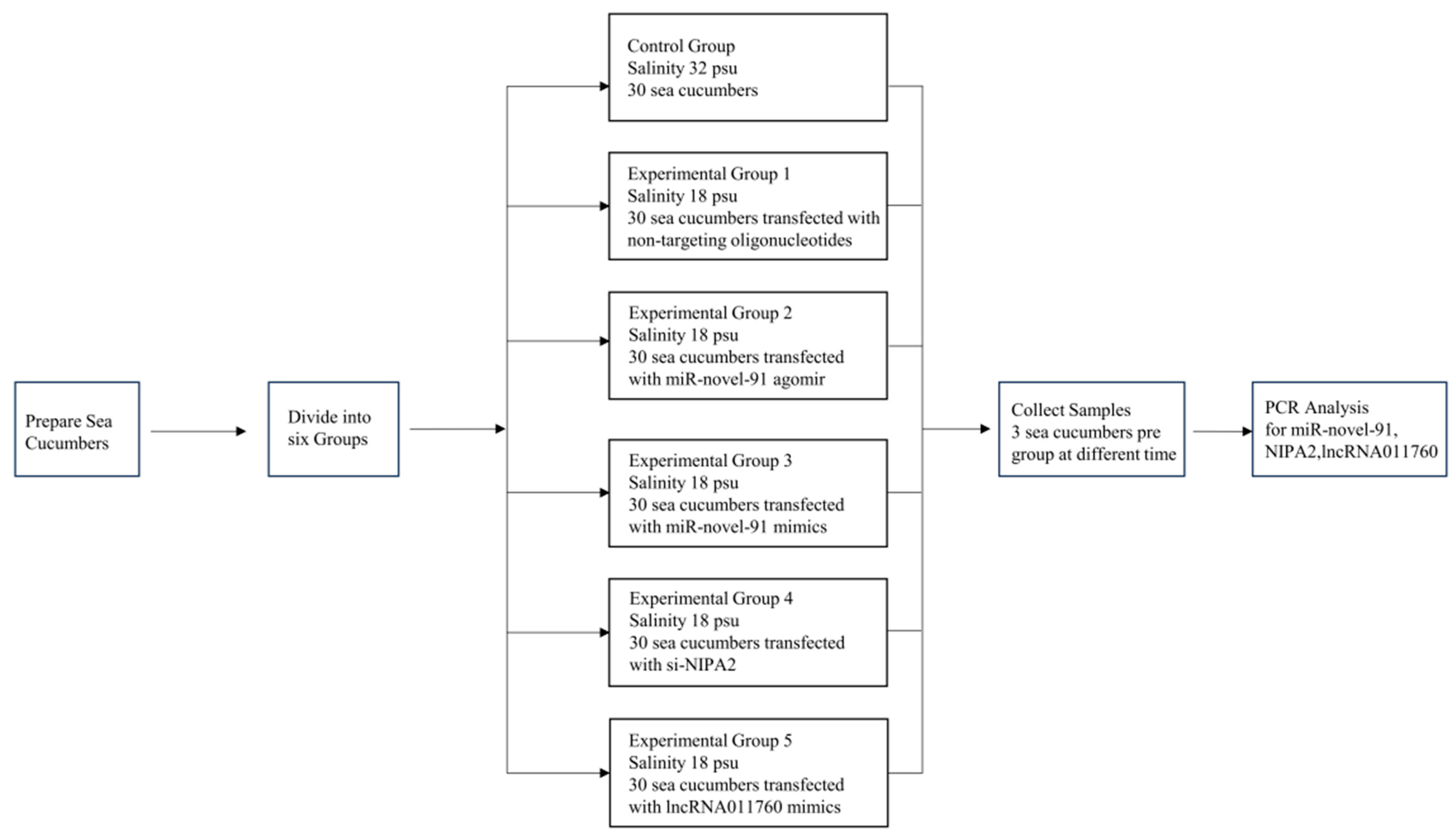
Figure 1. The entire experimental procedure used in this study.
Fishes 11 00275 g001
Figure 2. Expression profiles of miR-novel-91 and NIPA2 during low-salinity adaptation in A. japonicus. (A) Relative expression of miR-novel-91. (B) Relative expression of NIPA2. (C) Relative expression of lncRNA011760. Data are presented as mean ± standard deviation of the mean (SD) (n = 3 biological replicates). Note: * indicates a significant difference compared to the control group (p < 0.05); ** indicates a highly significant difference compared to the control group (p < 0.01). (D) Binding site maps of miR-novel-91 and its corresponding target genes.
Fishes 11 00275 g002
Figure 3. Expression profiles of miR-novel-91 and NIPA2 in A. japonicus under low-salinity stress following in vivo delivery of miR-novel-91 agomir or si-NIPA2. (A) Expression of miR-novel-91 after injection of miR-novel-91 agomir. (B) Expression of NIPA2 after injection of miR-novel-91 agomir. (C) Expression of miR-novel-91 after transfection with si-NIPA2. (D) Expression of NIPA2 after transfection with si-NIPA2. Data are presented as mean ± SD (n = 3 biological replicates). Note: * indicates a significant difference compared with the control group (p < 0.05); ** indicates a highly significant difference compared with the control group (p < 0.01).
Fishes 11 00275 g003aFishes 11 00275 g003b
Figure 4. Expression profiles of miR-novel-91 and NIPA2 in A. japonicus coelomocytes (in vitro) under low-salinity stress following the transfection with miR-novel-91 mimics or si-NIPA2. (A) Expression of miR-novel-91 after transfection with miR-novel-91 mimics. (B) Expression of NIPA2 after transfection with miR-novel-91 mimics. (C) Expression of miR-novel-91 after transfection with si-NIPA2. (D) Expression of NIPA2 after transfection with si-NIPA2. Note: * indicates a significant difference compared with the control group (p < 0.05); ** indicates a highly significant difference compared with the control group (p < 0.01).
Fishes 11 00275 g004aFishes 11 00275 g004b
Figure 5. A three-stage adaptive model for salinity acclimation in sea cucumbers based on the results and small-molecule pathway. Created in https://BioRender.com. A standard arrow (→) indicates that one entity promotes, activates, or positively regulates another. A perpendicular blunt line (—|) indicates that one entity inhibits, blocks, or negatively regulates another. Different colors are used to distinguish experimental groups, cellular components, or molecular labels.
Fishes 11 00275 g005
Table 1. miR-novel-91, NIPA2, and lncRNA011760 homologous gene primers used for expression.
CategroySequences NameSequence (5′-3′)
qRT-PCR PrimersNIPA2-FCTGACCTGCCTTCCGTGAGTAAG
NIPA2-RTGGCTGCTCTCCTCCTCTGAC
miR-novel-91CATGTGACCGTTACAATGGGCG
lncRNA011760-FGGAGAGCCTAGATTATGATACCGTTAC
lncRNA011760-RTTCACCACTAATCGTTCCACCAAG
Cytb-FTGAGCCGCAACAGTAATC
Cytb-RAAGGGAAAAGGAAGTGAAAG
U6ACGCAAATTCGTGAAGCGTT
Functional oligonucleotidesNegative controlUUGUACUACACAAAAGUACUG
GUACUUUUGUGUAGUACAAUU
miR-novel-91 agomir/mimicsAUGUGACCGUUACAAUGGGCG
CCCAUUGUAACGGUCACAUUU
si-NIPA2GCGAUAUCUACAAGAAACUTT
AGUUUCUUGUAGAUAUCGCTT
Disclaimer/Publisher’s Note: The statements, opinions and data contained in all publications are solely those of the individual author(s) and contributor(s) and not of MDPI and/or the editor(s). MDPI and/or the editor(s) disclaim responsibility for any injury to people or property resulting from any ideas, methods, instructions or products referred to in the content.